HAL Id: hal-01898264
https://hal.archives-ouvertes.fr/hal-01898264
Submitted on 18 Apr 2019
HAL is a multi-disciplinary open access
archive for the deposit and dissemination of
sci-entific research documents, whether they are
pub-lished or not. The documents may come from
teaching and research institutions in France or
abroad, or from public or private research centers.
L’archive ouverte pluridisciplinaire HAL, est
destinée au dépôt et à la diffusion de documents
scientifiques de niveau recherche, publiés ou non,
émanant des établissements d’enseignement et de
recherche français ou étrangers, des laboratoires
publics ou privés.
Design and properties of a novel radiopaque injectable
apatitic calcium phosphate cement, suitable for
image-guided implantation
Myriam Le Ferrec, Charlotte Mellier, Florian Boukhechba, Thomas Le
Corroller, Daphne Guenoun, Franck Fayon, Valérie Montouillout, Christelle
Despas, Alain Walcarius, Dominique Massiot, et al.
To cite this version:
Myriam Le Ferrec, Charlotte Mellier, Florian Boukhechba, Thomas Le Corroller, Daphne Guenoun,
et al.. Design and properties of a novel radiopaque injectable apatitic calcium phosphate cement,
suitable for image-guided implantation. Journal of Biomedical Materials Research Part B: Applied
Biomaterials, Wiley, 2018, 106, pp.2786-2795. �10.1002/jbm.b.34059�. �hal-01898264�
Design and properties of a novel radiopaque injectable apatitic calcium
phosphate cement, suitable for image-guided implantation
Myriam Le Ferrec,1 Charlotte Mellier,1 Florian Boukhechba,1 Thomas Le Corroller,2
Daphne, Guenoun,2 Franck Fayon,3 Vale,rie Montouillout,3 Christelle Despas,4 Alain Walcarius,4
Dominique Massiot,3 Franc¸ois-Xavier Lefe` vre,5 Caroline Robic,6 Jean-Claude Scimeca,7
Jean-Michel Bouler,5 Bruno Bujoli5
1Graftys SA, Eiffel Park, Ba^timent D, Po^le d’activite,s d’Aix en Provence, 13854 Aix en Provence CEDEX 3, France
2Ho^pitaux Sud – Ho^pital Sainte-Marguerite, CHU APHM, 13274, Marseille Cedex 9, France
3CNRS, UPR 3079, CEMHTI, 45071 Orle,ans Cedex 02, France
4Universite, de Lorraine, CNRS, UMR 7564, LCPME, 54600 Villers-le` s-Nancy, France
5Universite, de Nantes, CNRS, UMR 6230, CEISAM, UFR Sciences et Techniques, BP 92208, 44322 NANTES Cedex 3, France
6Guerbet, Ba^timent Rimbaud, 93420 Villepinte, France
7Universite, Co^te d’Azur, CNRS, Inserm, iBV, UMR 7277, Tour Pasteur, UFR Me,decine, 06107 Nice Cedex 02, France
Received 12 September 2017; revised 9 November 2017; accepted 18 November 2017
Published online 00 Month 2017 in Wiley Online Library (wileyonlinelibrary.com). DOI: 10.1002/jbm.b.34059 Abstract: An injectable purely apatitic calcium phosphate
cement (CPC) was successfully combined to a water-soluble
radiopaque agent (i.e., XenetixVR
), to result in an optimized composition that was found to be as satisfactory as poly(- methyl methacrylate) (PMMA) formulations used for verte- broplasty, in terms of radiopacity, texture and injectability. For that purpose, the Xenetix dosage in the cement paste was optimized by injection of the radiopaque CPC in human cadaveric vertebrae under classical PMMA vertebroplasty conditions, performed by interventional radiologists familiar with this surgical procedure. When present in the cement
paste up to 70 mg I mL21, Xenetix did not influence the
injectability, cohesion, and setting time of the resulting com- posite. After hardening of the material, the same observation
was made regarding the microstructure, mechanical strength and alpha-tricalcium phosphate to calcium deficient apatite transformation rate. Upon implantation in bone in a small animal model (rat), the biocompatibility of the Xenetix–con- taining CPC was evidenced. Moreover, an almost quantitative release of the contrast agent was found to occur rapidly, on the basis of in vitro static and dynamic quantitative studies
simulating in vivo implantation. VC 2017 Wiley Periodicals, Inc. J
Biomed Mater Res Part B: Appl Biomater 00B: 000–000, 2017.
Key Words: calcium phosphate, radiopaque agent, injectable cement, image-guided implantation, bone reconstruction
How to cite this article: Le Ferrec M, Mellier C, Boukhechba F, Le Corroller T, Guenoun D, Fayon F, Montouillout V, Despas C, Walcarius A, Massiot D, Lefe` vre F-X, Robic C, Scimeca J-C, Bouler J-M, Bujoli B. 2017. Design and properties of a novel radiopaque injectable apatitic calcium phosphate cement, suitable for image-guided implantation. J Biomed Mater Res Part B 2017:00B:000–000.
INTRODUCTION
Calcium phosphate cements (CPCs)1–7 are now widely used
for bone void filling and have obtained regulatory approval in many countries in Europe, America, and Asia. In particu- lar, injectable CPCs give access to implantations under mini- mally invasive surgery conditions with high benefits (small incision, rapidity, and low complication rates), due to their injectable character, and act as a mechanically resistant sac- rificial calcium phosphate source for bone reconstruction. For example, we have codeveloped two injectable formula- tions of these bioactive bone substitutes which are on the
market since 2008.8 These CPCs benefit from >8 years of
clinical experience and have proven efficiency in
traumatol-ogy.9–11 Histological and radiographic data of clinical cases
performed by clinicians have indeed shown bone recon- struction and quasitotal resorption of the CPCs within 12– 18 months, and analysis of the newly formed bone gave evidence of its excellent quality.
However, substantial improvements are still needed to extend their application in spine (e.g., filling of cages for intervertebral fusion, vertebral body augmentation), in particular the inclusion of an adapted contrast agent (i.e.,
Additional Supporting Information may be found in the online version of this article. Correspondence to: B. Bujoli; e-mail: bruno.bujoli@univ-nantes.fr
Contract grant sponsor: OSEO; contract grant number: F1210010 Contract grant sponsor: Graftys Company
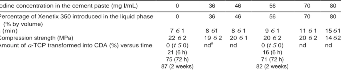
TABLE I. Characteristic Parameters of CPC/Xenetix Combinations as a Function of the Contrast Agent Loading (mg I mL21 of
Cement Paste): Initial Setting Time Monitored at 378C Using Gillmore Needles, Compression Strength Measured after 72 h,
Amount of a-TCP Transformed into CDA Versus Time Measured From 31P MAS NMR Spectra
Iodine concentration in the cement paste (mg I/mL) 0 36 46 56 70 80
Percentage of Xenetix 350 introduced in the liquid phase 0 36 46 56 70 80
(% by volume)
ti (min) 7 6 1 8 61 8 6 1 9 6 1 11 6 1 15 6 1
Compression strength (MPa) 22 6 2 19 6 2 20 6 1 20 6 2 20 6 2 14 6 2
Amount of a-TCP transformed into CDA (%) versus time 0 (t 5 0) nda nd 0 (t 5 0) nd nd
21 (6 h) 16 (6 h)
75 (72 h) 71 (72 h)
87 (2 weeks) 82 (2 weeks)
a
nd, not determined.
compatible with the bioresorption of the associated cement) to assist their implantation under image-guided procedures, since the opacity of CPCs is close to that of bone tissues. Filling this gap is highly strategic, since the fast improve- ment of imaging technologies should result in their increas- ing use in operating rooms. Indeed, interventional radiology has contributed to some of the most significant medical
improvements in recent years, including overall survival,12
quality of life improvement for patients13 and reduced
healthcare costs. The current state of the art in the field mainly concerns image-guided bone augmentation with injectable poly(methyl methacrylate) (PMMA) resins that are used for over two decades to treat fractures related to osteoporosis in different locations. In particular, a steady increase in the use of vertebroplasty is observed, consisting in the augmentation of vertebral compression fractures by image-guided intracorporeal injection of PMMA, in order to
palliate pain after failure of noninvasive therapies.14,15 How-
ever these resins are not biodegradable and contain barium sulfate particles for their visualization under fluoroscopy. From the best of our knowledge, only one biodegradable alternative has been developed recently by Bone Support
AB (CeramentTM), which consists of a mixture of 40%
hydroxyapatite and 60% of a-calcium sulfate hemihydrate. When mixed with an aqueous solution of a nonionic iodin- ated aromatic polyol (iohexol), an injectable paste is obtained that hardens in about 15 min and can be moni- tored by fluoroscopy. Clinical cases were thus reported for
different indications including, vertebroplasty16–19 and treat-
ment of aneurysmal bone cyst20 or benign bone tumours,21
but no detailed physical–chemical characterization of the product was however given. In the present article, an inject- able purely apatitic CPC was combined with a water-soluble contrast agent commonly used in angiography (iobitridol,
available as XenetixVR
at the Guerbet company), in order to develop a radiopaque composition suitable for X-ray fluoros- copy-guided implantation in bone. The influence of Xenetix on the setting time, mechanical properties, chemical trans- formation and microstructure of the resulting composite was investigated, as well as the kinetics of the contrast agent release from the CPC, under conditions simulating in vivo implantation. A small animal model (rat) was used to implant the composite in bone defects to evaluate its local
bone tolerance. Finally, CPC formulations loaded with vari- ous amounts of Xenetix were implanted in human cadaveric vertebrae, under conditions similar to image-guided verte- broplasty, to determine the optimal loading of radiopaque agent to reach performances as good as the PMMA gold standard, as regards the injectability, texture, and radiopac- ity of the CPC.
MATERIALS AND METHODS
Calcium phosphate cement formulations
The apatitic calcium phosphate cement (CPC) used in this study, abbreviated as MIADROS, was obtained from Graftys SA (Aix-en-Provence, France). MIADROS is mainly composed of a mixture of 78 wt % alpha-tricalcium phosphate (a-TCP)
(Ca3(PO4)2), 5 wt % dicalcium phosphate dihydrate (DCPD)
(CaHPO4-2H2O), 5 wt % monocalcium phosphate monohy-
drate (MCPM) (Ca(H2PO4)2-H2O), 10 wt % calcium deficient
apatite (CDA) (Ca102x[]x(HPO4)y(PO4)62y(OH)22z[]z), 2 wt %
hydroxypropyl methyl cellulose. The liquid phase consists of
a 5 wt % Na2HPO4 aqueous solution (liquid/powder
ratio 5 0.4 mL g21). Xenetix 350 was obtained from Guerbet
(Villepinte, France) as 20 mL sealed glass vials with a
350 mg mL21 iodine content. The MIADROS cement paste
samples were prepared by mixing the powder mixture with the liquid phase until reaching homogeneity of the obtained paste (i.e., 2 min). The same conditions were applied for the preparation of the CPC loaded with the radiopaque agent, except that the liquid phase was partially replaced by Xene- tix 350 (see Table I).
Methods
The high frequency impedance measurements were recorded, between 0.4 and 100 MHz, with a HP 4194A impedance/gain-phase analyzer (Hewlett-Packard), using an experimental setup allowing to concomitantly perform com- plex impedance and Gillmore needles measurements at
378C, as reported previously.22
The experimental device was completed by a computer allowing automatic data acquisition and real-time calcula- tion of the complex impedance, Z* from which the dielectric
permittivity, E0 (related to dipole variation), and dielectric
losses, E00 (related to the motion of free charges), were
The initial setting time (ti) is defined as the time elapsed
until the small Gillmore needle24 (diameter 2.12 mm, weight
113.4 g) fails to indent the surface of the sample, while the
final setting time (tf) is the corresponding value when using
the large Gillmore needle (diameter 1.06 mm, weight 453.6 g)
All NMR experiments were performed after quenching the cement hydration process by grounding the samples in
acetone. The 31P magic angle spinning (MAS) NMR spectra
were recorded on a Bruker Advance I spectrometer operat-
ing at 7.0 T (1H and 31P Larmor frequencies of 300 and
121.5 MHz, respectively). 13C MAS NMR experiments were
conducted on a Bruker Advance III spectrometer operating
at 9.4 T (1H and 13C Larmor frequencies of 400 and 100.6
MHz). All experiments were performed with a 4 mm double-resonance MAS probe head with a spinning fre- quency of 12
kHz and 1H SPINAL-6425 decoupling with a nutation
frequency of 70 kHz was applied during signal acquisition.
The 31P quantitative MAS spectra were recorded using a flip
angle of 248 (pulse length of 1 ms) and 128 scans were coadded with a recycle delay of 60 s to ensure complete
recovering of the longitudinal magnetization. The 13C-{1H}
CP-MAS spectra were acquired using a contact time of 1 ms,
with a linear amplitude ramp26 on the 1H channel and a
recycle delay of 1 s. Approximately 63,000 transients were coadded for pure iobitridol in its solid form and the CPC
loaded with Xenetix (56 mg I mL21) (experiment dura- tion of
about 18 h), while 235,000 scans were recorded for the MIADROS CPC reference (experiment duration of about
65 h). 31P and 13C chemical shifts were referenced relative
to H3PO4 85% and TMS, respectively.
In order to follow ex situ the progressive transformation
of a-TCP to CDA, the quantitative 31P MAS spectra were
reconstructed with the Dmfit program27 using four indepen-
dent contributions, three of them corresponding to the 31P
experimental spectra of a-TCP, DCPA, and DCPD, and the remaining one being a Lorentzian line centered at approxi- mately 2.9 ppm characteristic of the CDA phase. The relative amount (in wt %) of each phase as a function of the setting time was determined assuming that the composition of the
formed CDA phase was close to Ca9(HPO4)(PO4)5OH.
Scanning electron microscope (SEM) observation of the cement samples was performed using a field emission gun scanning electron microscope (Jeol 7600F). Images were acquired on back scattered electron mode with a 9 pA
beam current and a 10 kV accelerated voltage. A 1 mm2 pol-
ished cross-section of the cement samples was obtained using a JEOL cross section polisher SM09010, by applying an argon ion beam accelerated by a voltage up to 6 kV per- pendicular to the surface of each specimen for 6 h.
Compressive strength measurements and texture analyses versus time were performed using a AMETEK LS5 texture analyzer.
In vitro monitoring of the iobitridol release from CPCs Hydrodynamic conditions. A glass column (6.6 mm in diameter) fitted with a PE filter was filled with a weighed quantity (ca. 5 g) of cement paste loaded with Xenetix, with
an iodine concentration of 56 mg I mL21 in the sample. The
CPC was allowed to set for 7 days at 378C in a humid cham- ber, by immersing the entire column in a 0.9 wt % sodium chloride aqueous solution (NaCl), which was replaced every 2–3 days. Percolation of a 0.9 wt % sodium chloride aque- ous solution through the column was then performed using a high-pressure pump (Gilson 307), to control the flow rate. The eluted solution was collected in tared vials at the col- umn outlet and weighed. The amount of iobitridol released as a function of the percolated volume was measured by UV-visible, using a Shimadzu UV-2501PC spectrometer. Quantification was made at the wavelength of maximum
absorption of iobitridol (kmax 5 243 nm). A calibration curve
in the 0–0.035 mg I mL21 range was recorded prior to the
experiments by dilution of a galenic Xenetix solution
(350 mg I mL21 iodine content) in pure water. Two col-
umns were prepared under similar conditions, to ensure of the repeatability of the method.
Batch conditions. Cement samples loaded with Xenetix
(56 mg I mL21), prepared as described above, were directly
introduced into PTFE molds (inner diameter: 6 mm; height 12 mm), that were weighed and placed after 30 min in a known volume of 0.9 wt % sodium chloride aqueous solu- tion for 24 h. The amount of iobitridol desorbed during this step was measured by UV-vis spectroscopy. Then the obtained hardened CPC blocks were taken out of the mold, weighed and then placed in individual beakers that were hermetically sealed to prevent evaporation. A 0.9 wt % sodium chloride aqueous solution was used as desorption medium. Different liquid to solid ratios were investigated, with regular renewal of the solution (for details, see Figure 3) and the determination of the iobitridol content in the suc- cessive baths was again carried out by UV-vis spectroscopy. In vivo evaluation of the local bone tolerance of
iobitridol-loaded CPCs
All animal handling and surgical procedures were conducted according to European Community guidelines for the care and use of laboratory animals (DE 86/609/CEE). The study was approved by the local ethical committee for animal experimentation.
The animals (14 male Lewis rats aged 14 weeks, and weighing approximately 350 g) were placed in quarantine for at least 10 days prior to surgery. General anesthesia was performed using an intramuscular injection of ketamine and xylazine. The animals were surgically prepared by shaving both lower limbs, skin disinfection with iodine solution and sterile draping. A longitudinal skin incision (1.5 cm) was made to expose the distal lateral femoral condyle. A large size defect of 2.7 mm in diameter and 3 mm in depth was created at the epiphyseal-metaphyseal junction by using a motor-driven driller. The drilling process was achieved in three successive steps using burs of 1.5, 2, and 2.7 mm in diameter. During the drilling process, the defect site was continuously irrigated using a syringe of sterile saline solu- tion. Bone chips and particles were removed from the defects by irrigation with saline solution. The defects were
cement paste under fluoroscopy (Philips ALLURA FD20). The injections were performed under blind conditions (i.e., the two operators did not know the Xenetix content in the injected formulations), and for each vertebra, a rating of the radiopacity, injectability and texture of the CPC was given by the operating interventional radiologist, in comparison with standard PMMA used for vertebroplasty (see below).
FIGURE 1. Chemical formula of iobitridol. Rating Radiopacity Injectability Texture
1 Insufficient Difficult Too liquid/thick
then packed with sterile swabs and ribbon gauzes until 2 Limited Uncomfortable Fair
bleeding had subsided. The femoral cavities were then filled 3 Satisfactory Satisfactory Satisfactory
with about 0.6–0.7 g of the selected cement composition, 4 Optimal Comfortable Optimal
that is, loaded with various amounts of Xenetix (0, 37, 56,
and 80 mg I mL21 of cement paste, respectively). The sub-
cutaneous tissues and skin were closed in different layers using degradable sutures. The surgical site was finally cov- ered with an adhesive bandage. Postoperative radiographic monitoring of the animals (Faxitron, 9.5 s, 35 kV) was per- formed at day 0, 7, 14, and 28, and the rats were sacrificed
at day 28 by CO2 inhalation for histological analysis of the
implanted area.
For that purpose, bone explants were fixed by immer- sion into 10% formol for 2 days, then decalcified in a 10 wt % ethylenediaminetetraacetic acid solution for 3 weeks, and finally embedded into paraffin. The resulting samples were cut into 4 mm thin slices from which paraffin was removed using xylene, before hematoxylin-eosin-safran staining. His- tological analysis was carried out using an Axioskop Zeiss microscope equipped with a digital camera, and the image processing was run using the Axio Vision (Zeiss) software. Image-guided implantation of iobitridol-loaded CPCs in human cadaveric vertebra
This study was performed in the Radiology and Medical Imaging Department of the Sainte Marguerite Hospital in Marseille. Injection of the CPC formulations was performed under classical PMMA vertebroplasty conditions, by two interventional radiologists with, respectively, 10 and 5 years of experience in this procedure.
For the first part of the experiment, four humid human cadaveric vertebrae blocks from two donors were used and placed in a plastic dummy stuffed with polyurethane to mimic soft tissues. For each injected vertebra, one individual syringe of CPC was prepared and the operators were asked to inject the necessary volume to fill the vertebra. The amount of radiopaque agent introduced in the CPC formula- tion was adjusted through partial replacement of the liquid
phase (5 wt % Na2HPO4) by the appropriate volume of sterile
Xenetix 350, while keeping all other parameters fixed. After preparation of each dose of cement (6 g of powder 1 2.4 mL of liquid, leading to a cement paste containing 0, 37, 47, 56,
or 70 mg I mL21, respectively), the resulting paste was imme-
diately transferred into an injector (SomatexVR
4 mL—ref
180710) that was then connected to a trocart (OptimedVR
10 gauge, 0.7 mL—ref 1394-1010), before injection of the
In accordance with local ethics and safety regulations, three female human cadavers with distinct body mass index (BMI) were selected for the second part or the experiment: specimen 1 (83 years old, average BMI ;:: 23), specimen 2 (92 years old, high BMI ;:: 30), and specimen 3 (84 years old, low BMI ;:: 18). A strictly similar protocol was used by the same two operators, assisted by a fellow in interven- tional radiology as third operator, for the image-guided injection of the CPC (one unique loading of radiopaque agent, corresponding to a cement paste containing 56 mg I
mL21), except that a less performing fluoroscope was
employed (STENOSCOP2 General Electric CGR).
RESULTS
In the present study, we have introduced a water-soluble iodinated contrast agent into the composition of an inject- able purely apatitic cement currently developed by Graftys for the prevention of the secondary fracture of the proximal femur in osteoporotic patients [abbreviated as MIADROS]. Iobitridol (Xenetix—Figure 1), a non-ionic low-osmolality iodinated contrast agent, was selected because it is one of the gold standards for radiographical examination with >20 years of experience since it was first launched in 1994. To facilitate the preparation of sterile samples, the liquid phase
(5 wt % Na2HPO4) was partially replaced by the sterile
galenic form (350 mg iodine per mL) of the radiopaque agent, while keeping all other parameters fixed. Various amounts of Xenetix were loaded in the CPC formulation, in order to (i) investigate whether the presence of the contrast agent might influence the general properties of the CPC and (ii) determine the minimal dose for appropriate visualiza- tion of the product under X-ray imaging conditions.
Influence of the contrast agent on the setting time and mechanical properties
The Gillmore needles standard test method was used to investigate the influence of the addition of increasing amounts of Xenetix 350 (up to 80 mg of iodine per mL of cement paste) on the CPC setting reaction at body tempera- ture. Based on a change in the material’s penetration resis-
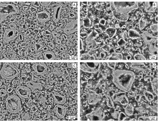
FIGURE 2. SEM observations of a polished cross-section of a radiopaque agent free MIADROS sample (bottom views; magnification: 32500 (C)
and 35000 (D)) and its analogue loaded with Xenetix (56 mg I mL21—top views; magnification: 32500 (A) and 35000 (B)), after a setting time of
72 h.
very similar results, except for the higher dose for which a significant augmentation of the setting time was observed (Table I). This is very likely related to a decrease of the phosphate concentration in the liquid phase upon partial replacement of the phosphate buffer by Xenetix, which is known to slow down the CPC setting reaction. We have
recently reported22 that high frequency impedance meas-
urements can be used to monitor the a-TCP to calcium- deficient hydroxyapatite (CDA) transformation occurring during the setting process of CPCs, in particular in the pres- ence of additives which can influence this reaction. When conversion of a-TCP into CDA starts, an increase in the
dielectric permittivity (E0) is thus observed, in relation with
the accumulation of mobile nonbonded charged species on the surface of the solid reactants and the formation of a supersaturated medium. At the same time, a sharp decrease
of the dielectric losses (E00) takes place as a result of the
precipitation of CDA crystals in the intergranular space as well as on the surface of a-TCP particles. For increasing amounts of Xenetix in the CPC composition up to 70 mg I
mL21, experimental data (see Figure S1 in Supporting Infor-
mation) show that evolution of the E0 and E00 parameters
has already started (i.e., 6 min are necessary to prepare the cement paste and load it in the dielectric cell before record- ing the first measurable dielectric values). In addition, the radiopaque agent does not inhibit or delay the setting reac- tion. Then, the hydrolysis of a-TCP into CDA propagates from the surface to the inner part of the particles and an
attenuation of the variation of the E0 and E00 parameters is
therefore observed, before slow stabilization after about 25– 30 min for the Xenetix–containing formulations, as com- pared to about 15 min for the CPC reference (Figure S1). Ex situ 31P MAS NMR analyses of the amount of a-TCP trans-
formed into CDA after an ageing period of 6 h, 3 days, and 2 weeks, respectively, showed no significant difference between the radiopaque agent free CPC and its analog
loaded with Xenetix (56 mg I mL21), thus confirming that
the setting reaction is not significantly influenced by the presence of the radiopaque agent in the CPC (Table I; see also Figure S2).
Moreover, the introduction of Xenetix into the CPC com- position did not result in significant changes in the mechan- ical properties of the resulting composite (see Table I), since the compressive strength after a setting time of 72 h was in the similar range in the presence (22 6 2 MPa) or absence (19–20 6 2 MPa) of Xenetix, except for the higher dose for which the cement paste was not cohesive anymore, leading to a decrease of the mechanical properties. Influence of the contrast agent on the microstructure of hardened cements
In order to investigate whether the presence of the radi- opaque agent in the CPC formulation might influence the microstructure of the hardened cement, SEM observations were performed on polished cross-sections of the radi- opaque agent free CPC and its analogue loaded with a
FIGURE 3. Amount of iobitridol desorbed from cement blocks loaded with Xenetix (56 mg I mL21), as a function of time. Desorption solu-
tion: 0.9 wt % sodium chloride aqueous solution at 378C. Blue curve:
S/L 5 0.06 g mL21 (eight renewals of the desorption solution); red
curve: S/L 5 0.12 g mL21 (eight renewals of the desorption solution);
green curve: S/L 5 0.25 g mL21 (nine renewals of the desorption solu-
tion); yellow curve: S/L 5 0.25 g mL21 (two renewals of the desorption
solution). Note that for S/L 5 0.12 g mL21, with no renewal of the
desorption medium, the iobitridol release after 2 months was 88(66)%.
In the case of the MIADROS composition, a large amount of geode-like particles can be observed, showing a dense shell lined in its inner part with entangled needle-shaped crystals, consistent with a complete hydrolysis of a-TCP into CDA (bottom right view in Figure 2). However, hydrolysis is only partial for larger a-TCP particles (bottom left view in Figure 2), while a few DCPA particles are also present that are dense along their whole cross-section. Finally, the area in between all these particles is mostly occupied by a rather porous network of platelet-like CDA crystals. When com- bined with Xenetix, the microstructure of the cement looks fully similar (top views in Figure 2).
Release of the contrast agent under in vitro conditions In order to model the release of the contrast agent from the cement paste under bone implantation conditions, molded blocks (6 mm in diameter and 12 mm in height) of the CPC
loaded with a median dose of Xenetix (i.e., 56 mg I mL21)
were immerged in a 0.9 wt % sodium chloride solution at 378C, under static conditions. The iobitridol concentration in the desorption solution was then measured by UV-vis spec- troscopy. Different options were investigated (i.e., solid to
liquid ratio [S/L 5 0.06, 0.12, 0.25 g mL21, respectively],
number of renewals of the sodium chloride solution [2 vs. 8–9]) and the results are summarized in Figure 3. After 2 weeks, about 85(66)% of iobitridol was released from the CPC, with very little influence of the solid to liquid ratio.
The release of iobitridol was then investigated under hydrodynamic conditions, using a glass column filled with a
cement paste loaded with Xenetix (56 mg I mL21) and
allowed to set for 7 days at 378C under humid conditions (see Materials and Methods). Percolation of a 0.9 wt % sodium chloride aqueous solution was then performed under controlled pressure (around 1–2 bars) resulting in an
outlet flow rate close to 10 lL min21. In addition to the
amount of radiopaque agent released during the equilibra- tion time of the column, the iobitridol concentration at the column outlet was then measured, as a function of the per- colated volume (Figure 4). The release of iobitridol is very rapid, since about 97(67)% is washed out of the column after a percolated volume of about 75 mL which corre- sponds to a time period of about 3 days under these experi- mental conditions.
In vivo evaluation of the bone local tolerance of CPCs loaded with Xenetix
The radiopaque CPC formulations were implanted in large size cavitary defects created in the distal lateral femoral condyle of rats, to evaluate their local bone tolerance. Each of the 14 animals were implanted on both sides and showed a good recovery after surgery, with comparable weight gain. Radiographic studies gave evidence of a correct filling of the defects with no cement leakage. No fibrous tissue or delete- rious effect on surrounding bone marrow was observed upon histological examination of bone explants and the for- mation of new trabecular bone was present in all condi- tions. In summary, there was no significant difference in the biological response of iobitridol-loaded CPC by comparison with the radiopaque agent free CPC control. Moreover, the characteristic process involved in the CPC remodeling simi- larly occurred, whatever the amount of Xenetix present in
the cement paste (0, 37, 56, and 80 mg I mL21—see Figure
S4).
Assessment of the potential of radiopaque CPCs for their image-guided implantation
A first experiment was designed to assess the safety of a vertebroplasty procedure using the CPCs loaded with vari-
ous amount of Xenetix (0, 37, 47, 56, and 70 mg I mL21 of
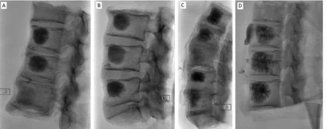
cement paste). For that purpose, the different CPC formula- tions were injected under fluoroscopy guidance, in humid human cadaveric vertebrae blocks placed in a plastic dummy stuffed with polyurethane to mimic soft tissues (Fig- ure 5). The procedure was performed under blind condi- tions by two interventional radiologists familiar with PMMA
FIGURE 4. Amount of iobitridol desorbed under hydrodynamic condi- tions from a hardened cement column loaded with Xenetix (56 mg I mL21), as a function of the percolated volume. Desorption solution:
0.9 wt % sodium chloride aqueous solution at 378C. The blue and red curves correspond to two different columns prepared under similar conditions, to ensure of the repeatability of the experiment.
FIGURE 5. Fluoroscopy images of human cadaveric vertebrae blocks after injection of CPCs loaded with various amounts of Xenetix (inje cted volume: ca. 3–4 mL, injection time: ca. 4 min): (A) Block 1–1 (from top to bottom): 70, 47, 0 mg I mL21; (B) Block 1–2 (from top to bottom): 56,
37, 70 mg I mL21; (C) Block 2–1 (from top to bottom): 56, 37, 0, 70, 47 mg I mL21; (D) Block 2–2 (from top to bottom): 56, 56, 70 mg I mL21.
vertebroplasty, with a rating of the radiopacity, texture and injectability of the five investigated compositions by com- parison to PMMA, on a scale ranging from 1 (unsatisfactory) to 4 (excellent), reported in Table II.
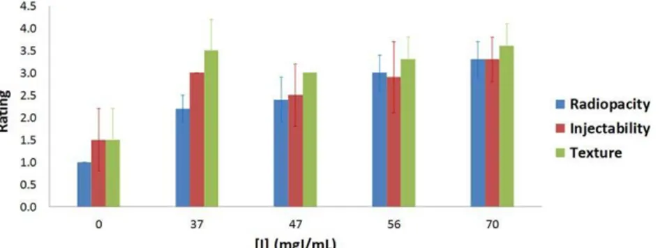
The global assessment of the five formulations for the selected criteria (Figure 6) showed that for a loading of
Xenetix greater or equal to 56 mg I mL21 of cement paste,
acceptable performances were achieved.
In order to confirm that the 56 mg I mL21 dosage of the
CPC was sufficient for a comfortable visualization of the cement under the typical conditions of vertebroplasty, a sec- ond experiment was performed on three female human cadavers of different body mass, using an old generation fluoroscopy equipment available in the surgical room (see Figure S5). The injection was performed by three interven- tional radiologists, and again the radiopacity, texture, and
injectability of the CPC composition were rated on a scale ranging from 1 to 4, and the results are reported in Table III. For the three operators the radiopacity of the CPC was considered as suitable for the easy monitoring of the injec- tion under fluoroscopy guidance, whatever the body mass of the specimen. However, a leakage out of the vertebrae was systematically observed for specimen 1 and 2, resulting in a low rating of the texture (2 or 3). Interestingly, when the time elapsed between the preparation of the cement paste and the start of the injection was longer than 11 min (Table III, specimen 3), no more leakage occurred, with no detrimental effect on the injectability.
DISCUSSION
PMMA is currently the gold standard material for the vertebroplasty/kyphoplasty-mediated management of acute
TABLE II. Assessment of the Radiopacity, Texture, and Injectability of CPCs Loaded with Various Amounts of Xenetix (0, 37,
47, 56, and 70 mg I mL21 of Cement Paste) and Injected in Humid Human Cadaveric Vertebrae Blocksa
Xenetix Radiopacity Rating Injectability Rating Texture Rating
Specimen
Vertebra
Block Vertebra
Loading
(mg I/mL) Operator A Operator B Operator A Operator B Operator A Operator B
1 (operator 1-1 T12 70 3 4 nab 4 na 4 B) L1 47 2 3 na 3 na 3 L2 0 1 1 na 1 na 1 1–2 L3 56 3 31 na 4 na 4 L4 37 2 3- na 3 na 4 L5 70 3 31 na 3 na 31 2 (operator 2-1 L1 56 21 3- 2 na 3 na A) L2 37 2 2 3 na 3 na L3 0 1 1 2 na 2 na L4 70 3 4 3 na 3 na L5 47 2 3- 2 na 3 na 2-2 T10 56 3 31 3 na 3 na T11 56 3 31 3- na 3 na T12 70 3 31 3 na 4 na a
Experiments were run under blind conditions and the rating of the selected criteria was made on a scale ranging from 1 (unsatisfactory) to 4 (excellent). For each vertebra, one individual syringe of CPC was prepared. Note that the radiopacity assessment could be mad e by the two operators who were both present during the whole experiment.
b
FIGURE 6. Global assessment of the CPCs loaded with various amounts of Xenetix (0, 37, 47, 56, and 70 mg I mL21 of cement paste—x-axis), on the
basis of radiopacity, texture and injectability criteria. The rating (y-axis) was made on a scale ranging from 1 (unsatisfactory) to 4 (excellent).
vertebral compression fractures, for the treatment of pain. Since the vertebral augmentation procedure is conducted
under image-guided conditions,28–42 the injectable PMMA
resin contains barium sulfate particles to enhance its radio- pacity and allow appropriate visualization of the product during its injection into the fractured vertebral body. How- ever, barium sulfate which is hardly soluble in aqueous media, cannot be used for the preparation of radiopaque CPCs, since the gradual resorption of the CPC would result in the release of solid particles in the blood stream. There- fore, an aromatic iodinated water soluble contrast agent was selected for this study, in an attempt to prepare inject- able radiopaque apatitic CPC formulations. Replacement of part of the liquid phase of the CPC by Xenetix (iobitridol, Figure 1), a liquid contrast agent commonly used for angi- ography, led to CPC formulations compatible with their sur- gical use. Indeed, for an iodine content in the cement paste
up to 70 mg I mL21, no significant difference could be
observed with respect to the undoped CPC analogue, in terms of setting time, compression strength, or transforma- tion kinetics of a-TCP into CDA (Table I). In addition, the final microstructure of the CPC once hardened, assessed by SEM observation of polished cross-sections of the samples, was found to be similar in the absence or presence of
Xenetix. On the other hand, the release of iobitridol from hardened cement blocks was monitored under static and hydrodynamic conditions in a 0.9 wt % sodium chloride solution at 378C, to investigate whether the contrast agent is irreversibly trapped or not during the setting process. In both conditions, the most part of the radiopaque agent was released quite rapidly, giving evidence that (i) the interac- tion of iobitridol with the inorganic apatitic network is very limited, (ii) the permeability of the CPC once hardened is high enough to allow iobitridol to diffuse out of the bone substitute quite rapidly. As a consequence, the quick release of the contrast agent makes long-term follow-up of the cement impossible. However, this potential drawback might be compensated by the fact that the risk of chronic adverse effects upon implantation of the developed radiopaque CPC should be limited. Furthermore, histological analysis of bone explants clearly evidenced a similar biological response for iobitridol-loaded CPCs and the unloaded CPC control, with no incidence of the radiopaque agent on the CPC remodel- ing process.
To evaluate the potential of Xenetix-containing CPCs for vertebroplasty procedures, CPC loaded with different doses of radiopaque agent have been implanted in humid human cadaveric vertebrae blocks, by interventional radiologists
TABLE III. Assessment of the Radiopacity, Texture, and Injectability of a CPC Loaded with Xenetix (56 mg I mL21 of Cement
Paste) and Injected in Vertebrae of Human Cadavers (Injected Volume: ca. 3–5 mL)a
Specimen Operator Vertebra t0 [t1] Radiopacity Rating Injectability Rating Texture Rating
1 A L5 3.5 [6.5] 3 3 2 T10 3.5 [5.5] 3 3 2 T12 4 [5.8] 3 3 2 L2 4 [5.9] 3 3 2 L4 4.6 [5.2] 3 3 2 2 B T9 5 [6] 4 4 3 T11 6 [7] 4 4 3 L1 6.5 [7.5] 4 4 3 L3 7 [8.3] 4 4 3 3 C L4 9 [11.1] 4 4 3 L3 11 [12.5] 4 4 4 T12 11 [11.7] 4 4 4 T11 11 [11.7] 4 4 4 a
The rating of the selected criteria was made on a scale ranging from 1 (unsatisfactory) to 4 (excellent). t0 (in minutes) corresponds to the
time elapsed between the preparation of the cement paste and the start of the injection; t1 (in minutes) corresponds to the time elapsed between
following an image-guided procedure performed thanks to classical fluoroscopy equipments used in osteoarticular radi- ology, having a resolution (pixel size) in the 0.1–0.2 mm range. This investigation was conducted under blind condi- tions, to determine whether some of the investigated com- positions gave results comparable to the PMMA standard, in terms of injectability, texture, and radiopacity. This was effectively the case when the cement paste contained at
least 56 mg I mL21. A second similar experiment was then
performed to confirm that the 56 mg I mL21 minimal dose
was appropriate, except that the surgery was directly per- formed on three female human cadavers. While the radio- pacity was again found to be suitable for a comfortable visualization of the product, we have found that the texture of the composite is advantageously improved when a wait- ing time of about 10 min before injection is applied right after the preparation of the cement paste. Such conditions allowed to prevent any leakage phenomena, while still retaining good injectability of the product.
CONCLUSION
In this article, an injectable purely apatitic calcium phos- phate cement was successfully combined to Xenetix, a non- ionic water-soluble iodinated contrast agent. For a final iodine concentration in the cement paste up to 70 mg I
mL21, the main general properties of the resulting compos-
ite were retained, as regards the injectability, cohesion, and setting time. Moreover, when hardened, similar microstruc- ture, mechanical strength and a-TCP to CDA transformation were observed, regardless of whether or not Xenetix was present in the CPC formulation. In vivo implantation of Xenetix–containing CPC in bone large size defects gave evi- dence that the radiopaque cement was fully tolerated, with no adverse effect resulting from the presence of the contrast agent. Moreover, all Xenetix–containing CPCs exhibited a rapid and nearly quantitative release of the contrast agent. All together, these data demonstrate that Xenetix–containing CPCs are compatible for a surgical use. Moreover, their implantation in human cadaveric vertebrae was performed under image-guided procedure, to determine the minimal Xenetix dosage in the cement paste to allow comfortable visualization of the material during surgery. Hence, a 56 mg
I mL21 content in the cement paste was found to be appro-
priate, and suitable conditions for the preparation of the cement paste before injection were found, leading to a behavior during the implantation procedure that was con- sidered by the interventional radiologists as satisfactory as with PMMA formulations used for vertebroplasty. Comple- mentary studies are underway to investigate the potential of the radiopaque CPC developed in this work for indica- tions for which minimally invasive image-guided implanta- tion of injectable CPCs might represent an added value.
ACKNOWLEDGMENTS
The authors greatly acknowledge the Scanning Electron Microscopy facility at the Institut des Mat,eriaux Jean Rouxel laboratory, and particularly Nicolas Stephant for his technical support.
DISCLOSURE
Some authors of this publication have research support from Graftys SA. The terms of this arrangement have been reviewed and approved by both CNRS and the University of Nantes in accordance with their policy on objectivity in research.
REFERENCES
1. Ambrosio L, Guarino V, Sanginario V, Torricelli P, Fini M, Ginebra MP, Planell JA, Giardino R. Injectable calcium-phosphate-based composites for skeletal bone treatments. Biomed Mater 2012;7: 024113.
2. Bohner M, Gbureck U, Barralet JE. Technological issues for the development of more efficient calcium phosphate bone cements: A critical assessment. Biomaterials 2005;26:6423–6429.
3. Brown WE, Chow LC. A new calcium phosphate setting cement. J Dent Res 1983;62:672–679.
4. Dorozhkin SV. Calcium orthophosphate cements for biomedical application. J Mater Sci 2008;43:3028–3057.
5. Dorozhkin SV. Calcium orthophosphate cements and concretes. Materials 2009;2:221–291.
6. Dorozhkin SV. Self-setting calcium orthophosphate formulations. J Funct Biomater 2013;4:209–311.
7. LeGeros R, Chohayeb A, Shulman A. Apatitic calcium phosphates: Possible restorative materials. J Dent Res 1982;61 (Special Issue): 343.
8. Graftys. https://clinicaltrialsgov/ct2/show/study/NCT02575352. 9. Ollivier M, Gay AM, Cerlier A, Lunebourg A, Argenson JN,
Parratte S. Can we achieve bone healing using the diamond con- cept without bone grafting for recalcitrant tibial nonunions? Injury 2015;46:1383–1388.
10. Ollivier M, Turati M, Munier M, Lunebourg A, Argenson J-N, Parratte S. Balloon tibioplasty for reduction of depressed tibial plateau fractures: Preliminary radiographic and clinical results. Int Orthop 2016;40:1961–1966.
11. Young AA, Neyton L, Molony DC, Boileau P, Walch G. Glenoid tri- cortical iliac crest structural bone graft enhanced with resorbable cement for the treatment of aseptic glenoid loosening. Tech Shoulder Elb Surg 2011;12:12–17.
12. Charalel RA, McGinty G, Brant-Zawadzki M, Goodwin SC, Khilnani NM, Matsumoto AH, Min RJ, Soares GM, Cook PS. Inter- ventional radiology delivers high-value health care and is an imaging 3.0 vanguard. J Am Coll Radiol 2015;12:501–506. 13. Monsky WL, Khorsand D, Nolan T, Douglas D, Khanna P. Quality
of life assessment in interventional radiology. Acad Radiol 2014; 21:407–414.
14. Beall DP, Datir A, D’Souza SL, D’Souza LS, Gunda D, Morelli J, Johnson MB, Nabavizadeh N. Percutaneous treatment of insuffi- ciency fractures. Skeletal Radiol 2010;39:117–130.
15. Hurley MC, Kaakaji R, Dabus G, Shaibani A, Walker MT, Fessler RG, Bendok BR. Percutaneous vertebroplasty. Neurosurg Clin North Am 2009;20:341–359.
16. Marcia S, Boi C, Dragani M, Marini S, Marras M, Piras E, Anselmetti GC, Masala S. Effectiveness of a bone substitute (CERAMENTTM) as an alternative to PMMA in percutaneous verte- broplasty: 1-year follow-up on clinical outcome. Eur Spine J 2012; 21:112–118.
17. Masala S, Nano G, Marcia S, Muto M, Fucci FPM, Simonetti G. Osteoporotic vertebral compression fractures augmentation by injectable partly resorbable ceramic bone substitute (Cerament (TM)|SPINE SUPPORT): A prospective nonrandomized study. Neu- roradiology 2012;54:589–596.
18. Rauschmann M, Vogl T, Verheyden A, Pflugmacher R, Werba T, Schmidt S, Hierholzer J. Bioceramic vertebral augmentation with a calcium sulphate/hydroxyapatite composite (Cerament (TM) SpineSupport) in vertebral compression fractures due to osteopo- rosis. Eur Spine J 2010;19:887–892.
19. Siemund R, Nilsson LT, Cronqvist M, Stro€mqvist B. Initial clinical experience with a new biointegrative cement for vertebroplasty in osteoporotic vertebral fractures. Interv Neuroradiol 2009;15:335– 340.
20. Guarnieri G, Vassallo P, Muto M, Muto M. Percutaneous treat- ment of symptomatic aneurysmal bone cyst of L5 by percutane- ous injection of osteoconductive material (Cerament). J Neurointerv Surg 2014;6:e43.
21. Kaczmarczyk J, Sowinski P, Goch M, Katulska K. Complete twelve month bone remodeling with a bi-phasic injectable bone substi- tute in benign bone tumors: A prospective pilot study. BMC Mus- culoskelet Disord 2015;16: 369.
22. Despas C, Schnitzler V, Janvier P, Fayon F, Massiot D, Bouler J- M, Bujoli B, Walcarius A. High-frequency impedance measure- ment as a relevant tool for monitoring the apatitic cement setting reaction. Acta Biomater 2014;10:940–950.
23. Thiebaut JM, Roussy G, Chlihi K, Bessiere J. Dielectric study of the activation of blende with cupric ions. J Electroanal Chem 1989;262:131–144.
24. Standard Test Method for Time of Setting of Hydraulic Cement Paste by Gillmore Needles. ASTM C266-89 and ASTM C266-08. Philadelphia: Cement, Lime, Gypsum. American Society for Test- ing and Materials; 1993.
25. Fung BM, Khitrin AK, Ermolaev K. An improved broadband decoupling sequence for liquid crystals and solids. J Magn Reson 2000;142:97–101.
26. Metz G, Wu XL, Smith SO. Ramped-amplitude cross-polarization in magic-angle-spinning NMR. J Magn Reson Ser A 1994;110:219–127. 27. Massiot D, Fayon F, Capron M, King I, Le Calve, S, Alonso B, Durand J-O, Bujoli B, Gan Z, Hoatson G. Modelling one- and two- dimensional solid-state NMR spectra. Magn Reson Chem 2002;40: 70–76.
28. Alvarez L, Alcaraz M, Perez-Higueras A, Percutaneous vertebro- plasty—Functional improvement in patients with osteoporotic compression fractures. Spine 2006;31:1113–1118.
29. Anselmetti GC, Corrao G, Monica PD, Tartaglia V, Manca A, Eminefendic H, Russo F, Tosetti I, Regge D. Pain relief following percutaneous vertebroplasty: Results of a series of 283 consecu- tive patients treated in a single institution. Cardiovasc Interv Radiol 2007;30:441–447.
30. Buchbinder R, Osborne RH, Ebeling PR, Wark JD, Mitchell P, Wriedt C, Graves S, Staples MP, Murphy B. A randomized trial of vertebroplasty for painful osteoporotic vertebral fractures. N Engl J Med 2009;361:557–568.
31. Clark W, Lyon S, Burnes J. Trials of vertebroplasty for vertebral fractures. N Engl J Med 2009;361:2097–2098.
32. Eck JC, Nachtigall D, Humphreys SC, Hodges SD. Comparison of vertebroplasty and balloon kyphoplasty for treatment of vertebral compression fractures: A meta-analysis of the literature. Spine J 2008;8:488–497.
33. Evans AJ, Jensen ME, Kip KE, DeNardo AJ, Lawler GJ, Negin GA, Remley KB, Boutin SM, Dunnagan SA. Vertebral compression
fractures: Pain reduction and improvement in functional mobility after percutaneous polymethylmethacrylate vertebroplasty- retrospective report of 245 cases. Radiology 2003;226:366–372. 34. Hulme PA, Krebs J, Ferguson SJ, Berlemann U. Vertebroplasty
and kyphoplasty: A systematic review of 69 clinical studies. Spine 2006;31:1983–2001.
35. Kallmes DF, Comstock BA, Heagerty PJ, Turner JA, Wilson DJ, Diamond TH, Edwards R, Gray LA, Stout L, Owen S, Hollingworth W, Ghdoke B, Annesley-Williams DJ, Ralston SH, Jarvik JG. A randomized trial of vertebroplasty for osteoporotic spinal frac - tures. N Engl J Med 2009;361:569–579.
36. Klazen CAH, Lohle PNM, de Vries J, Jansen FH, Tielbeek AV, Blonk MC, Venmans A, van Rooij WJJ, Schoemaker MC, Juttmann JR, Lo TH, Verhaar HJJ, van der Graaf Y, van Everdingen KJ, Muller AF, Elgersma OEH, Halkema DR, Fransen H, Janssens X, Buskens E, Mali WPThM. Vertebroplasty versus conservative treatment in acute osteoporotic vertebral compres- sion fractures (Vertos II): An open-label randomised trial. Lancet 2010;376:1085–1092.
37. Klazen CAH, Verhaar HJJ, Lampmann LEH, Juttmann JR, Blonk MC, Jansen FH, Tielbeek AV, Schoemaker MC, Buskens E, van der Graaf Y, Janssens X, Fransen H, van Everdingen KJ, Muller AF, Mali WPThM, Lohle PNM. VERTOS II: Percutaneous vertebro- plasty versus conservative therapy in patients with painful osteo- porotic vertebral compression fractures; rationale, objectives and design of a multicenter randomized controlled trial. Trials 2007;8: 33.
38. Legroux-Ge,rot I, Lormeau C, Boutry N, Cotten A, Duquesnoy B, Cortet B. Long-term follow-up of vertebral osteoporotic fractures treated by percutaneous vertebroplasty. Clin Rheumatol 2004;23: 310–317.
39. McGraw JK, Lippert JA, Minkus KD, Rami PM, Davis TM, Budzik RF. Prospective evaluation of pain relief in 100 patients undergo - ing percutaneous vertebroplasty: Results and follow-up. J Vasc Interv Radiol 2002;13:883–886.
40. Pe,rez-Higueras A, Alvarez L, Rossi RE, Quin~ones D, Al-Assir I. Per-cutaneous vertebroplasty: Long-term clinical and radiological out- come. Neuroradiology 2002;44:950–954.
41. Voormolen MHJ, Lohle PN,Lampmann LE, van den Wildenberg W, Juttmann JR, Diekerhof CH, de Waal Malefijt J. Prospective clinical follow-up after percutaneous vertebroplasty in patients with painful osteoporotic vertebral compression fractures. J Vasc Interv Radiol 2006;17:1313–1320.
42. Zoarski GH, Snow P,Olan WJ, Stallmeyer MJB, Dick BW, Hebel JR, De Deyne M. Percutaneous vertebroplasty for osteoporotic compression fractures: Quantitative prospective evaluation of long-term outcomes. J Vasc Interv Radiol 2002;13:139–148.