Extensive aortic prosthesis dilatation
Bernhard Winkler
a,*, Jens Bremerich
b, Friedrich Eckstein
aand Peter Matt
aa
Division of Cardiac Surgery, University Hospital Basel, Basel, Switzerland
b Division of Radiology, University Hospital Basel, Basel, Switzerland
* Corresponding author. Division of Cardiac Surgery, University Hospital Basel, Spitalstrasse 21, CH-4031 Basel, Switzerland. Tel: +41-61-2652525; fax: +41-61-2657324; e-mail: [email protected] (B. Winkler).
Received 5 June 2012; received in revised form 18 October 2012; accepted 4 November 2012
Keywords:
Aneurysm
• Thoracic aorta • Prosthesis • Follow-up
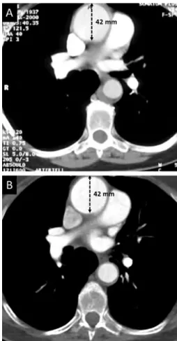
A 75-year old woman underwent successful ascending aortic
repair for type A aortic dissection 14 years ago (Figs
1
and
2
). A
recent computed tomographic (CT) scan showed a Gelweave
™
prosthesis (Vascutek, UK) that was originally 30 mm, with an
extensive dilatation of 42 mm in diameter. This dilatation had
already occurred within the
first years and has remained stable
since then.
Figure 1:A CT scan 2 years (A) and 14 years (B) after the initial aortic repair.
It shows the enlarged prosthesis diameter of 42 mm (black dotted arrows), which has remained stable over time. Compared with the original dimension, the cross-sectional area of the prosthesis increased by 96%, and the prosthesis wall tension by 40%.
Figure 2: Reconstructed three-dimensional angiography demonstrating the
enlarged aortic prosthesis compared with the aortic root and the proximal aortic arch. White arrows mark the proximal and distal anastomosis site.
© The Author 2012. Published by Oxford University Press on behalf of the European Association for Cardio-Thoracic Surgery. All rights reserved.
IMAGES IN C A RDIO-THO R A C IC SU RG E R Y
European Journal of Cardio-Thoracic Surgery 43 (2013) e93