Actinomyces neuii Isolated From a 20-Month-Old
Girl With Cervical Lymphadenitis
Kathi Walther,1Elisabeth Bruder,2Daniel Goldenberger,3Johannes Mayr,4Urs Beat Schaad,1and Nicole Ritz1,5 1Paediatric Infectious Diseases Unit, Departments of2Pathology,3Clinical Microbiology, University Hospital Basel;4Department of Paediatric Surgery, University Children’s Hospital Basel; and5Paediatric Infectious Diseases Unit, Royal Children’s Hospital Melbourne, Australia
Corresponding Author: Dr. Nicole Ritz, MD, PhD, University Children’s Hospital Basel, Spitalstrasse 33, CH-4056 Basel, Switzerland. E-mail: nicole.ritz@ukbb.ch.
Received April 8, 2014; accepted August 18, 2014; electronically published October 14, 2014.
Actinomycetes are Gram-positive bacteria that can be part of the normal human flora of the gastrointestinal, pulmonary, and genital tract. Infections are rare, slowly progressing and most commonly affect the cervicofacial region. Actinomyces israelii is the most frequently isolated species but a number of other species may cause infection. We report thefirst postnatally acquired case of an actinomycosis caused by A. neuii in a child. We also provide a systematic review of all published cases of A. neuii infections. In children, there is one case report of a premature infant with perinatally acquired A. neuii sepsis. In adults 21 cases have currently been reported and A. neuii infection was associated with endophthalmitis after eye surgery, foreign material-associated infection and abscess formation in the inguinal, axillary, and mammary area. Our case highlights that a A. neuii infection is also a potential differential diagnosis in children with chronic lymphadenitis.
Key words. Actinomyces neuii; cervical lymphadenitis; children; MALDI-TOF; Prevotella.
Actinomycosis is a rare infection in children younger than 10 years of age [1]. It mainly affects the cervicofacial re-gion, but many other sites of infection have been described [2]. Actinomycosis is usually caused by Actinomyces isra-elii, a Gram-positive bacterium that colonizes the oral cav-ity. With the development of molecular techniques in recent years, it was recognized that some Actinomyces spe-cies were misclassified (eg, as Actinobaculum species), and a number of new Actinomyces species have been identified [3,4]. Atypical coryneform bacteria (initially designated as Centers for Disease Control and Prevention [CDC] fermen-tative coryneform group 1 [5]) were isolated for thefirst time in the 1980s from patients with endophthalmitis, and the organism was subsequently named A. neuii in 1994 [6]. Interestingly current adult literature suggests that A. neuii has a different spectrum of disease and most frequently presents with skin and soft tissue infection or abscesse. In children, there is currently only 1 published case report of A. neuii sepsis in a premature infant born to a mother with pelvic infection and chorioamnionitis [7]. We report here the first, to our knowledge, case of a postnatally acquired A. neuii infection in a child presenting with cervical lymphadenitis.
CASE
A 20-month-old previously healthy girl presented to our emergency department with a 3-week history of subman-dibular swelling. Ultrasonographic examination per-formed 1 week before presentation showed a multilobar calcified structure measuring 1.5 cm in diameter. After a rapid increase of the swelling within 24 hours, the child presented to our hospital. The parents reported that since birth, the child was known to have a small cervical sinus tract that intermittently drained foul-smellingfluid. The child had not had any contact with sick individuals, and her family history was unremarkable.
On physical examination, the girl was afebrile and had a submandibular,firm, nonfluctuant, nontender swelling with a sinus tract. She also had an itching macular rash over the cervical area. Results of the remaining examination were normal. In particular, there were no other enlarged lymph nodes. Her dentition was normal, and there were no lesions in her mouth. A full blood count revealed a hemoglobin value of 117 g/L, a white blood cell count of 13.7 × 10⁹/L (62% neutrophils, 31% lymphocytes, 6% monocytes, 1% eo-sinophils), a platelet count of 491 × 109/L, and a C-reactive protein level of 9 mg/L. Repeated ultrasonography of the
Case Report
Journal of the Pediatric Infectious Diseases Society, Vol. 4, No. 3, pp. e32–e37, 2015. DOI:10.1093/jpids/piu096 © The Author 2014. Published by Oxford University Press on behalf of the Pediatric Infectious Diseases Society. All rights reserved. For Permissions, please e-mail: journals.permissions@oup.com.
neck confirmed the multilobar structure located at the an-terior border of the sternocleidomastoid muscle with a diameter of 2 cm and afistula adjacent to the multilobular structure extending to the skin.
The following day, the mass andfistula were excised. Histopathological examination revealed purulent in flam-mation and small fragments of squamous epithelium
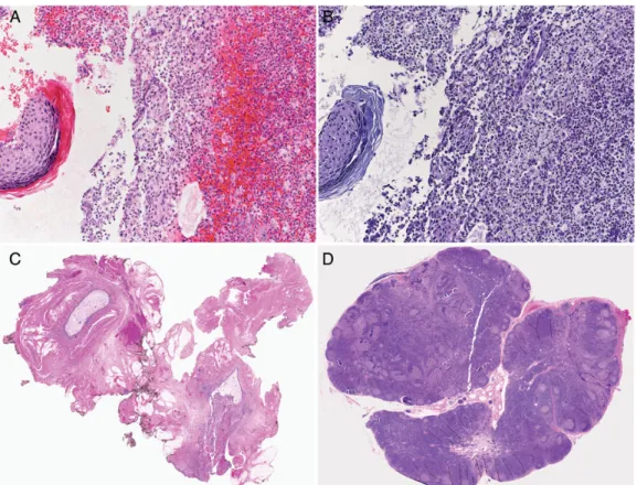
(Figure1A and B). Sulfur granules were not identified. A Gram stain from several deep cervical swabs showed Gram-positive rods (Figure2), Gram-positive cocci, and Gram-negative pleomorphic rods. Culture resulted in poly-microbial growth of Prevotella timonensis, viridans strep-tococci, anaerobic Gram-positive cocci, and A. neuii. Identification of A. neuii was achieved from pure culture on Columbia agar supplemented with 5% sheep blood (BD Diagnostic Systems, Allschwil, Switzerland). The cat-alase reaction was positive, and the Gram stain revealed coryneform, short rods. In addition, matrix-assisted laser desorption ionization time-of-flight mass spectrometry (MALDI-TOF MS) (Microflex LT, Bruker Daltonics) was performed using a short extraction protocol with 1 µL of 70% formic acid added to the smears followed by applica-tion of the matrix soluapplica-tion. Analysis of the raw spectral data was performed with MALDI Biotyper software 3.0 (Bruker Daltonics) with reference database version 3.1.2.0 (3995 database entries) and identified A. neuii with a score of 2.108. Identification of P. timonensis was done using a 16S rRNA gene sequence analysis. The anaer-obe cocci and the viridans streptococci identified with culture and Gram staining were not further identified to the species level. Staining for acid-fast bacilli and Figure 1. Hematoxylin and eosin (H&E) (A) and periodic acid–Schiff (PAS) (B) staining of histological sections of excised cervical lymph node showing purulent inflammation and small fragments of squamous epithelium (×200 magnification). H&E (C) and PAS (D) staining of histological sections from the second operation showing the remaining sinus tract lined with squamous epithelium cells adjacent to elastic cartilage (C) and follicular hyperplasia of the lymph node (D) (×10 magnification).
Figure 2. Gram stain of the A neuii isolate showing coryneform nonbranching Gram-positive rods (×400 magnification).
Table 1. Details of All Currently Reported Cases of Actinomyces neuii Infection Highlighting the Variance of Infection Locations and Treatment
Ref. Age Gender
Type of Infection/Underlying Condition
Sample(s) Positive for A neuii
Antibiotic
susceptibility Surgical Treatment
Antibiotic Treatment (Dose per d) Duration of Treatment (d) Outcome 7 1 d F Sepsis/maternal chorioamnionitis
Blood culture, culture of gastric aspirate and residual amniotic fluid in external ear canal
Pen, Cefo, Vanco, Imi, Ery
None Amp (100 mg/kg), Gent
(3 mg/kg) iv
14 Cured
Pen G (30 mg/kg) oral 28 Our case 1.6 y F Infection of lateral cervical cyst Culture of intraoperative
sample NS Drainage with excochleation, secondary selective neck dissection Amox-Clav (180 mg/kg) iv 4 Cured Amox-Clav (80 mg/kg) oral 178
18 28 y M Infection of pilonidal cyst Culture of purulent fluid NS None Pen V NS NS
20 39 y F Chronic pericarditis PCR of pericardial fluid NS Pericardial fluid
drainage
NS NS NS
25 46 y F Breast abscess Culture of fine-needle
aspirate
NS Surgical debridement Amox (2–3 g) oral 28 Cured
19 48 y F Breast abscess Culture of fine-needle
aspirate
NS None Amox 21 Cured
18 48 y F Breast abscess Culture of intraoperative
sample
NS Surgical debridement Pen V NS NS
12 58 y M Endophthalmitis/
phacoemulsification with posterior chamber intraocular lens implant
Culture of anterior chamber and vitreous body taps
Pen, Amox/Clav, Cefa, Cefu, Ceftr, Vanco, Imi, Oxa, Levo
None Intravitreous: Vanco (2 mg),
Ami (400μg) NS Poor visual acuity (20/40),complicated by central vein occlusion Peribulbar: Vanco (25 mg) NS
Ocular: Tobra NS
Cephalexin (2 g) NS 17 64 y F Mammary prosthesis infection Culture of swab from
mammary prosthesis
Ery, Pen, Tetra, Vanco Removal of mammary prosthesis
Amox-Clav (4.4 g) Preop period Cured Amox-Clav (2.4 g) Postop period
27 64 y F VP-shunt infection Culture of CSF Pen, Ceftr, Clinda,
Vanco
Removal of VP shunt Vanco, Cefepime, Amp, Metro
18 Cured
Pen G (24 Mio IU) iv 42
Pen oral 180
21 66 y M Prosthetic valve endocarditis Blood culture Pen None Pen G (20 Mio IU), Metro
(2 g), Ery (4 g)
21 Cured
Pen G (20 Mio IU) iv 25
Amox (2 g) oral 330
23 67 y M Perirenal abscess Blood culture NS Drainage Amp, followed by Pen and
Cipro
37 Cured
22 68 y M Endocarditis/aortic paravalvular abscess
Blood culture Pen, Amp, Ceftr, Vanco, Genta
Open heart surgery Amp (9 g), Gent (24 mg), Ceftr (2 g) iv 4 NS Amp (9 g), Gent (24 mg) iv 5 Amp iv 21 Ceftr (2 g) iv 63 Doxycycline oral 252 28 68 y F Toe ulcer/type 2 diabetes Cultures of intraoperative
samples
Pen G, Cefa, Cefo, Ery, Clinda, Vanco, Teico Surgical debridement, amputation of toe Metro (1500 mg), Cipro (200 mg) iv 3 Cured Clinda (600 mg) iv 2 Clinda (600 mg), Teico (400 mg) iv 15 Teico (800 mg) im 10 29 69 y F Bilateral endophthalmitis/ immunosuppression not further specified
Culture of anterior chamber fluid
NS None Intravitreous: Vanco, Cefta 1 Limited improvement of
visual acuity in right (6/20) and left (6/120) eyes
Pen G (4 Mio IU) iv, Sulf ocular
21
14 73 y M Chronic endophthalmitis/ phacoemulsification with intraocular lens implantation
Culture of anterior chamber fluid
Ery, Pen, Tetra, Gent, Cefu
Pars plana vitrectomy Neomycin ocular 21 Satisfactory with visual acuity (6/18) Levo (1 g) NS Azit (500 mg) NS Chloramphenicol ocular NS e34 W alther et al
mg/0.1 mL
Oflox, Cefa 600 mg/12 mL ocular
21
Cipro (1 g) 14
24 76 y M Chronic osteomyelitis of the calcaneum with fistulation
Culture of bone from curettage
NS Surgical curettage Cefa (2 g) 77 Cured
30 78 y F Periprosthetic infection/total hip arthroplasty
Culture from joint fluid, intraoperative periprosthetic tissue
Pen, Amp, Clinda, Levo, Vanco, Rif
Surgery (removal of prosthesis, Girdlestone arthroplasty)
Cefa (6 g), Rif (900 mg) iv Pen G (20 Mio IU) iv
7 2 wk after reimplantation, no signs of local infection, no further follow up Antibiotic-loaded bone cement (Vanco 2 g, Clinda 1 g, Gent 1 g per 40 g polymethyl methacrylate)
14
Amox (3 g) oral 28
31 79 y M Infection of IPP reservoir Culture of purulent fluid collection around the prosthesis tubing
Amp Surgery (removal of IPP) Vanco, Piperacillin/ Tazobactam iv Kan/Cefa, Vanco/Genta, Baci (wound irrigation)
Preop Cured
Antibiotic treatment with Vanco iv, cephalexin, Amox-Clav oral, and Amox oral
365
23 91 y M Urosepsis/chronic nephropathy Blood culture NS None Cefu and mecillinam 9 Cured
32 NS NS 2 patients with endophthalmitis/ implantation of anterior chamber lenses
Culture of vitreous fluid Pen, Cefu, Gent None None NS NS
Abbreviations: iv, intravenous; im, intramuscular; CSF, cerebral spinal fluid; Ami, amikacin; Amox, amoxicillin; Amp, ampicillin; Azit, azithromycin; Baci, bacitracin; Cefa, cefazolin; Cefta, ceftazidime; Ceftr, ceftriaxone; Cefo, cefotaxime; Cefu, cefuroxime; Cipro, ciprofloxacin; Clav, clavulanate; Clinda, clindamycin; Ery, erythromycin; Gent, gentamicin; Imi, imipenem; Kan, kanamycin; Levo, levofloxacin; Metro, metronidazole; Oflox, ofloxacin; Oxa, Oxacillin; Pen, penicillin; Pred, prednisolone; Rif, rifampicin; Sulf, sulfacetamide; Tetra, tetracycline; Teico, teicoplanin; Tobra, tobramycin; Vanco, vancomycin; NS, not stated; VP, ventriculoperitoneal; IPP, inflatable penile prosthesis.
Actinomyc es neuii Infection in Childhood e35
Mycobacterium tuberculosis complex polymerase chain re-action (PCR) remained negative.
Intravenous amoxicillin-clavulanate (180 mg/kg/d) was started. When the swelling and redness subsided, antibiotic treatment was changed to oral amoxicillin-clavulanate (80 mg/kg/d), and the patient was discharged 4 days after sur-gery. At follow-up 2 weeks later, we noted persistent dis-charge from the wound that continued during the following 2 months despite local antiseptic and oral antibi-otic treatment. A remaining sinus tract was seen, and there-fore excision of the remainingfistula and adjacent lymph nodes was performed 3 months after the initial surgery. Pathology examination confirmed a remaining sinus tract lined with squamous epithelium cells adjacent to elastic cartilage (Figure1C). The resected lymph node was charac-terized by distinct follicular hyperplasia (Figure1D). Gram staining did not reveal any bacteria, and culture remained negative. At the next follow-up 2 weeks after the second surgery, the wound had healed and left a small scar (1 cm long). Treatment with amoxicillin-clavulanate was stopped after a total of 6 months, at which time complete resolution of the swelling was documented.
DISCUSSION
A. neuii is a coryneform, nonbranching, aerobically grow-ing, Gram-positive rod that was named in honor of Harold Neu in 1994 [6]. A positive catalase reaction and a positive CAMP test result are keyfindings in the biochemical iden-tification of this species today. Although the gold-standard method for identification of A. neuii is 16S rRNA gene sequencing, recent reports showed that identification with MALDI-TOF MS is excellent even to the species level [8,9]. Therefore, it has been suggested that for Gram-positive rods, including those of A. neuii, a species identification can be accepted without 16S rRNA sequencing analysis if the MALDI-TOF MS cutoff value is higher than 2.0 [8]. Actinomyces spp. are believed to be part of the endoge-nousflora of mucous membranes in the gastrointestinal, pulmonary, and genital tracts [10]. Recent studies have shown that by the age of 2 years, the oral cavity of every child is colonized with Actinomyces spp. [11]. Actinomyces odontolyticus and Actinomyces naeslundii are the most com-monly found species [11]. In contrast, A. neuii has not been identified thus far as part of the normal oral flora in the first 2 years of life [11].
After colonization, disruption of the mucosa leading to a microaerophilic environment is thought to promote inva-sive infection. In adults, a total of 21 cases of A. neuii in-fection have been described in the literature (Table1). A. neuii has been reported most frequently to cause
endophthalmitis after eye surgery [12–14], abscess forma-tion, superinfections of ulcers predominantly located in the inguinal, axillary, and mammary areas, and foreign mate-rial–associated infections [3,15–19]. In addition further re-ports include 3 cases of cardiac infections [20–22], 2 cases of A. neuii bacteremia as a result of a urinary tract infection and a perianal abscess [23] and 1 case of chronic osteomy-elitis [23]. Additional details of all previously reported A. neuii infections in children and adults are summarized in Table1.
To our knowledge, only 1 pediatric case of A. neuii infec-tion (in a neonate whose infecinfec-tion was caused by maternal bacteremia and subsequent chorioamnionitis) has been re-ported [7]. Our case represents thefirst, to our knowledge, postnatally acquired A. neuii infection in a child. On the basis of the clinical presentation and the age of the child, infection with atypical mycobacteria was initially suspect-ed, and excision of the enlarged lymph node was per-formed. The results of culture and PCR remained negative for atypical mycobacteria but showed polymicro-bial growth, including growth of A. neuii. A. neuii is com-monly isolated together with other bacterial species, mainly anaerobes. We considered A. neuii to be the most important pathogen with potential contribution of the other isolated bacteria. The subspecies of A. neuii was not determined. Because the child was afebrile, we did not perform a blood culture; culture results have been shown to be positive in up to 10% of adult patients with A. neuii infection [6,15]. Interestingly, histopathological examination did not reveal any sulfur granules, which are usually a hallmark of actinomycosis. However, the absence of sulfur granules has been reported, particularly in A. neuii infections [25]. On the basis of reports on adults, antibiotic treatment with amoxicillin-clavulanate was started. In addition, amoxicillin-clavulanic acid was also considered to be active against the other isolated bacteria. Antimicrobial susceptibility testing for Actinomyces spp. is not routinely performed at our microbiology laboratory, because internal data have shown that all Actinomyces species are susceptible to amoxicillin-clavulanic acid. Other potential treatment options reported in the literature are ampicillin, penicillin, and cephalosporins [26]. On the basis of experience with infections with other Actinomyces spp., we opted for a 6-month antibiotic treatment course with regular follow-ups. Three months after starting treat-ment, persistent drainage from the lymph node was noted to be a result of a remaining sinus tract rather than treat-ment failure, because cultures from the second sample re-mained sterile.
In conclusion, infection with A. neuii is a potential dif-ferential diagnosis for children with chronic lymphadenitis e36 Walther et al
and particularly those with presumed atypical mycobacte-rial infection with negative mycobactemycobacte-rial culture and PCR results from lymph nodes.
Acknowledgments
We thank Kathrin Kalinowski for technical assistance with microsco-py. We cordially thank Dr Reno Frei for the helpful discussion and comments on the manuscript.
Financial support. N.R was supported by a grant from the University of Basel and by the Rozalia Foundation.
Potential conflicts of interest. All authors: No reported conflicts. All authors have submitted the ICMJE Form for Disclosure of Potential Conflicts of Interest. Conflicts that the editors consider rele-vant to the content of the manuscript have been disclosed.
References
1. Pulverer G, Schutt-Gerowitt H, Schaal KP. Human cervicofacial actinomycoses: microbiological data for 1997 cases. Clin Infect Dis 2003; 37:490–7.
2. Bennhoff DF. Actinomycosis: diagnostic and therapeutic consid-erations and a review of 32 cases. Laryngoscope 1984; 94: 1198–1217.
3. Hall V. Actinomyces—gathering evidence of human colonization and infection. Anaerobe 2008; 14:1–7.
4. Zimmermann P, Berlinger L, Liniger B, et al. Actinobaculum schaalii an emerging pediatric pathogen? BMC Infect Dis 2012; 12:201.
5. Na’Was TE, Hollis DG, Moss CW, Weaver RE. Comparison of biochemical, morphologic, and chemical characteristics of Centers for Disease Control fermentative coryneform groups 1, 2, and A-4. J Clin Microbiol 1987; 25:1354–8.
6. Funke G, Stubbs S, von Graevenitz A, Collins MD. Assignment of human-derived CDC group 1 coryneform bacteria and CDC group 1-like coryneform bacteria to the genus Actinomyces as Actinomyces neuii subsp. neuii sp. nov., subsp. nov., and Actinomyces neuii subsp. anitratus subsp. nov. Int J Syst Bacteriol 1994; 44:167–71.
7. Mann C, Dertinger S, Hartmann G, et al. Actinomyces neuii and neonatal sepsis. Infection 2002; 30:178–80.
8. Schulthess B, Bloemberg GV, Zbinden R, et al. Evaluation of the Bruker MALDI Biotyper for identification of Gram-positive rods: development of a diagnostic algorithm for the clinical laboratory. J Clin Microbiol 2014; 52:1089–97.
9. De Vreese K, Verhaegen J. Identification of coryneform Actinomyces neuii by MALDI-TOF MS: 5 case reports and re-view of literature. Acta Clin Belg 2013; 68:210–4.
10. Smego RA Jr, Foglia G. Actinomycosis. Clin Infect Dis 1998; 26: 1255–61; quiz 1262–53.
11. Sarkonen N, Kononen E, Summanen P, et al. Oral colonization with Actinomyces species in infants by two years of age. J Dent Res 2000; 79:864–7.
12. Garelick JM, Khodabakhsh AJ, Josephberg RG. Acute postoper-ative endophthalmitis caused by Actinomyces neuii. Am J Ophthalmol 2002; 133:145–7.
13. Perez-Santonja JJ, Campos-Mollo E, Fuentes-Campos E, et al. Actinomyces neuii subspecies anitratus chronic endophthalmitis after cataract surgery. Eur J Ophthalmol 2007; 17:445–7. 14. Raman VS, Evans N, Shreshta B, Cunningham R. Chronic
postoperative endophthalmitis caused by Actinomyces neuii. J Cataract Refract Surg 2004; 30:2641–3.
15. Funke G, von Graevenitz A. Infections due to Actinomyces neuii (former“CDC coryneform group 1” bacteria). Infection 1995; 23:73–5.
16. Clarridge JE 3rd, Zhang Q. Genotypic diversity of clinical Actinomyces species: phenotype, source, and disease correlation among genospecies. J Clin Microbiol 2002; 40:3442–8. 17. Brunner S, Graf S, Riegel P, Altwegg M. Catalase-negative
Actinomyces neuii subsp. neuii isolated from an infected mam-mary prosthesis. Int J Med Microbiol 2000; 290:285–7. 18. Gomez-Garces JL, Burillo A, Gil Y, Saez-Nieto JA. Soft tissue
in-fections caused by Actinomyces neuii, a rare pathogen. J Clin Microbiol 2010; 48:1508–9.
19. Lacoste C, Escande MC, Jammet P, Nos C. Breast Actinomyces neuii abscess simulating primary malignancy: a case diagnosed byfine-needle aspiration. Diagn Cytopathol 2009; 37:311–2. 20. Levy PY, Fournier PE, Charrel R, et al. Molecular analysis of
pericardialfluid: a 7-year experience. Eur Heart J 2006; 27: 1942–6.
21. Grundmann S, Huebner J, Stuplich J, et al. Prosthetic valve endo-carditis due to Actinomyces neuii successfully treated with antibi-otic therapy. J Clin Microbiol 2010; 48:1008–11.
22. Cohen E, Bishara J, Medalion B, et al. Infective endocarditis due to Actinomyces neuii. Scand J Infect Dis 2007; 39:180–3. 23. Hansen JM, Fjeldsoe-Nielsen H, Sulim S, et al. Actinomyces
spe-cies: a danish survey on human infections and microbiological characteristics. Open Microbiol J 2009; 3:113–20.
24. Van Bosterhaut B, Boucquey P, Janssens M, et al. Chronic osteo-myelitis due to Actinomyces neuii subspecies neuii and Dermabacter hominis. Eur J Clin Microbiol Infect Dis 2002; 21:486–7.
25. Roustan A, Al Nakib M, Boubli L. Primary actinomycosis of the breast due to Actinomyces neuii [in French]. J Gynecol Obstet Biol Reprod (Paris) 2010; 39:64–7.
26. von Graevenitz A. Actinomyces neuii: review of an unusual infec-tious agent. Infection 2011; 39:97–100.
27. Watkins RR, Anthony K, Schroder S, Hall GS. Ventriculoperitoneal shunt infection caused by Actinomyces neuii subsp. neuii. J Clin Microbiol 2008; 46:1888–9.
28. Papaefstathiou K, Sonikian M, Zoumberi M, et al. Actinomyces neuii isolation from foot necrotic ulcer in an immunocompro-mised patient. Clin Microbiol Infect 2004;10 Suppl 3:404–5. 29. Graffi S, Peretz A, Naftali M. Endogenous endophthalmitis with
an unusual infective agent: Actinomyces neuii. Eur J Ophthalmol 2012; 22:834–5.
30. Rieber H, Schwarz R, Kramer O, et al. Actinomyces neuii subsp. neuii associated with periprosthetic infection in total hip arthroplasty as causative agent. J Clin Microbiol 2009; 47:4183–4.
31. Hsi RS, Hotaling JM, Spencer ES, et al. Isolated infection of a de-commissioned penile prosthesis reservoir with Actinomyces neuii. J Sex Med 2011; 8:923–6.
32. Coudron PE, Harris RC, Vaughan MG, Dalton HP. Two similar but atypical strains of coryneform group A-4 isolated from pa-tients with endophthalmitis. J Clin Microbiol 1985; 22:475–7.