Glycine-rich cell wall proteins act as specific antigen targets in autoimmune and food allergic disorders
11
0
0
Texte intégral
(2) 648 Glycine-rich cell wall protein and autoimmunity Table 1. Homology of GRP peptide with antigens GRP peptide GRP 1.8 (436–454). GGYGDGGAHGGGYGG *************** SGGGYGDGGAHGGGYGGGA. Procollagen α1 (V) (327–345). GGYGDGGAHGGGYGG :: :**::* ** * ANYDTYGGGRRGKGYKGRA. Fibrillar collagen (247–265). GGYGDGGAHGGGYGG ****::*::* RRGGYGRKGRAGAVGDVGD. EBNA-I (NP) (425–443). GGYGDGGAHGGGYGG :******::::* * GRRGYGDGRRKKGGWGKH. hnRNP A2 (266–284). GGYGDGGAHGGGYGG ******: :*::** GRGGYGGGPGYGNQGGGY. Epidermal keratin (568–582). GGYGDGGAHGGGYGG **** ** ** * GGYGSGGGSGGRYGS. to screen Ig fractions obtained from patients with multiple sclerosis (8), chronic immune thrombocytopenic purpura (9), type 1 diabetes mellitus (10), systemic lupus erythematosus (SLE) (11) and rheumatoid arthritis (RA) (12–14). One of the selected peptides identified by Ig from RA patients (12) showed homology with cereal glycine-rich cell wall proteins (GRP) and with the Epstein–Barr virus nuclear (EBV) antigenI (EBNA-I). Analysis of GRP 1.8 (15), a ubiquitous protein found in French bean and related species, shows the presence of Gly–Ala repeated sequences homologous to EBNA-I and cytokeratins. Many studies have shown the ability of antiEBNA-I antibodies to cross-react with different autoantigens such as heterogeneous nuclear ribonucleoprotein (hnRNP) (16), cytokeratin, collagen and actin in RA (17,18); the crossreactions can be inhibited with synthetic peptides containing the Gly–Ala repeat sequence, implying that the cross-reactivity is due to anti-Gly–Ala antibodies that recognize both the host proteins and the EBNA-I repeat. A 15 amino acid synthetic peptide corresponding to amino acids 436–454 of GRP 1.8 and sharing homology with EBNA-I, fibrillar collagen and procollagen (Table 1) has been shown to induce humoral (14) and T cell responses in RA (19). Taken together, these data suggest that the autoimmune response in RA but also in other autoimmune diseases may not be as heterogeneous as originally thought. A possible consequence of such hypothesis is that the identification of widespread (auto)antigens may allow their use in the suppression of autoimmunity by oral administration. The suppression of autoimmunity by oral antigen administration may be a feasible therapeutic option (reviewed in 20) and the induction of oral tolerance can be a therapeutic strategy also in food allergy (reviewed in 21). Based on the observation that Gly–Ala repeated sequences are present in food proteins as well as in viruses and selfproteins, we decided to investigate the presence of antiGRP antibodies in a large panel of sera from patients with. autoimmune disorders. The 15 amino acid peptide derived from GRP was used to analyze human sera in a direct and competitive ELISA assay. Serum IgG antibodies directed against such peptide were detected in different percentages of several autoimmune disorders and in food allergy. The GRP peptide was able to elicit a specific T cell response: peripheral mononuclear cells (PBMC) derived from patients with different diseases proliferated to the peptide and antigenspecific T cell clones could be generated from such patients. These data suggest that (auto)immune responses can be triggered by protein epitopes with crucial amino acids homologous to self proteins.. Methods Patients The patients enrolled in the study attended the outpatient clinic of the Department of Clinical and Experimental Medicine, University of Verona, Italy. The patients affected by SLE and RA fulfilled the diagnostic criteria of the American College of Rheumatology (22,23). Psoriatic arthritis (PsA) was diagnosed by the presence of psoriasis and a seronegative peripheral arthritis, with or without axial skeletal involvement. Chronic idiopatic urticaria (CIU) was characterized by recurrent episodes of diffuse erythematous wheals with severe pruritus lasting for ⬎6 weeks. Food allergy was identified by the presence of specific IgE and positive double-blind placebocontrolled challenge test; the offending foods were cereals, fruits and vegetables in 20 of 29 patients, fish in five of 29 and diary products in four of 29 patients. Aeroallergy was assessed by a history of seasonal or perennial rhinitis and/or asthma and positive skin test for pollens or house dust mite. The patients with chronic parvovirus B19 infection (CPI) are described in details elsewhere (24). The number of the patients studied is reported in Table 2. The HLA status of patients with CPI has already been reported (24). In patients with PsA HLA DR4, DR1 and DR7 were found in three patients each; DR3, which is associated with a more aggressive form of the disease and the presence of erosive arthritis, was present in two patients. We did not find particular HLA class II antigens more frequently present in patients with SLE, CIU and food allergy, whereas in RA there was an increased prevalence of DR4 (30% of patients versus 15% of controls; P ⫽ 0.008). When the DR4 and DR1 where analysed at molecular level no difference was found in DR4 suballeles (*0401, *0402, *0403, *0404, *0405, *0408) between patients and controls; as far as DR1 suballeles were concerned (*0101, *0102, *0103) there was a statistical difference in the frequency of *0102 between the two groups (46% of patients versus 19% of controls; P ⫽ 0.008). IgM rheumatoid factor was present in 25 of 32 RA patients and its level varied between 105 and 770 IU/ml (n.v. ⬍ 60 IU/ml). All the patients’ sera were tested for the presence of the following autoantibodies: anti-ssDNA antibodies, anti-keratin antibodies, anti-collagen II antibodies, anti-RNP antibodies. Anti-ssDNA antibodies were present in 37 of 37 SLE patients and in two of 32 RA patients, anti-keratin antibodies in seven of 32 RA patients and two in 12 of PsA patients, anti-collagen.
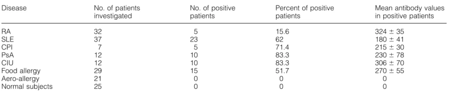
(3) Glycine-rich cell wall protein and autoimmunity 649 Table 2. Patient’s sera with IgG antibodies against GRP peptide Disease. No. of patients investigated. No. of positive patients. Percent of positive patients. Mean antibody values in positive patients. RA SLE CPI PsA CIU Food allergy Aero-allergy Normal subjects. 32 37 7 12 12 29 21 25. 5 23 5 10 10 15 0 0. 15.6 62 71.4 83.3 83.3 51.7 0 0. 324 ⫾ 35 180 ⫾ 41 215 ⫾ 30 230 ⫾ 78 306 ⫾ 70 270 ⫾ 55 0 0. antibodies in eight of 32 RA patients and in one of 12 PsA patients, and anti-hnRNP antibodies in eight of 37 SLE patients. The sera of patients affected by CIU, food allergy and aero-allergy were negative for the autoantibody specificities studied. Peptide synthesis and recombinant GRP proteins The synthetic peptides (GRP peptide: GGYGDGGAHGGGYGG and the irrelevant control peptide: ALYPSSVGQPFQGAP) were obtained by solid-phase synthesis using Fmocprotected amino acids according to the method of Merrifield as modified by Atherton (25), and were purified by gel filtration. Recombinant GRP proteins with or without the sequence corresponding to the GRP peptide were expressed in Escherichia coli and purified according to Ryser et al. (15). mAb and reagents The purified mAb anti-CD56 (Leu19, IgG1), anti-CD8 (Leu2a, IgG1), anti-CD4 (Leu3a, IgG1), anti-CD3 (Leu4, IgG1) and anti-CD19 (Leu12, IgG1) were from Becton Dickinson (San Jose, CA). The purified mAb anti-CD16 (KD1, IgG2a) was a kind gift of Professor L. Moretta (Genoa, Italy). The class II specific mAb DC1-12 was a kind gift of Professor R. Accolla (Genoa, Italy). Phycoerythrin (PE)- or FITC-conjugated goat anti-mouse antisera were from Southern Biotechnology Associates (Birmingham, AL). Human keratin, collagen type II and actin were purchased from Sigma (St Louis, MO). Affinity purification of IgG anti-peptide and anti-keratin antibodies Serum IgG was obtained by affinity purification using a Protein A–Sepharose column (Pharmacia, Uppsala, Sweden). The synthetic peptides or human keratin (5 mg antigen/g dried Sepharose powder) were coupled to Sepharose CL4B (Pharmacia), according to the manufacturer’s instructions. Affinitypurified IgG samples diluted in PBS were applied to the column. Bound IgG was eluted with 0.1 M glycine (pH 2.5) and dialyzed against PBS. ELISA assay The synthetic peptides and the recombinant proteins were used at a concentration of 20 µg/ml in PBS to coat polystyrene plates (Nunc, Roskilde, Denmark). After blocking with 5% dry non-fat milk in PBS, the antibodies diluted in 2.5% dry nonfat milk and 0.05% Tween in PBS were added and incubated. for 4 h. The plates were then washed and alkaline phosphatase-conjugated goat anti-human IgG (F(ab⬘)2 fragment) or anti-mouse IgG or IgM (Sigma) were added and incubated overnight at 4°C. After washings, the bound enzymatic activity was measured with p-nitrophenylphosphate (Sigma). Each antibody preparation was tested on a control plate not coated with the antigen. This non-specific binding never exceeded 10% of the specific binding (e.g. to the antigen coated plate). For competitive assays the amount of antibody that gave 50% of the maximum binding to the antigen on the solid phase was preincubated with different amounts of competitors or buffer for 1 h at 37°C and then transferred to the antigencoated plates. The assay was then carried on as the direct binding assay. In the ELISA assay for the detection of serum antibodies directed against the peptide or the recombinant GRP protein containing the peptide sequence of interest, 25 sera diluted 1: 100 from normal age- and sex-matched subjects were used as control group. Optical density values higher than the mean ⫹ 3 SD of each serum dilution of the control group (OD ⬎ 70 for the GRP peptide and OD ⬎ 85 for the recombinant protein) were considered positive. The direct and competitive ELISA for human keratin, collagen type II and actin has been described (24). The ELISA assay for EBNA-I was performed using a commercially available kit (Sigma). Immunization of BALB/c mice The synthetic peptide was coupled to the carrier protein keyhole limpet hemocyanin (KLH) and emulsified in Freund’s adjuvant. Mice were injected 5 times (the first time in complete Freund’s adjuvant, the other times in incomplete adjuvant) at the base of the tail at 15 day intervals. The animals were bled 7 days after the last injection and the sera tested in ELISA. Control animals were injected with adjuvant alone, keyhole limpet hemocyanin (KLH) alone or coupled with the irrelevant peptide. Generation of CD4⫹-specific T cells clones and flow cytofluorimetric analysis PBMC derived from patients were isolated on Ficoll-Hypaque gradient and cells were then incubated with peptide at 20 µg/ml in 96 U-bottomed microplates in complete medium. After 10 days of culture in absence of rIL-2 the cells were incubated with a mixture of anti-CD16 (KD1), anti-CD56 (Leu19) and anti-CD8 (Leu2a) mAb and purified by immunodepletion using goat anti-mouse Ig coated with magnetic.
(4) 650 Glycine-rich cell wall protein and autoimmunity beads (Unipath, Milan, Italy) (26). Viable cells were cloned under limiting dilution in the presence of irradiated peripheral blood lymphocytes as feeder cells in complete medium and of exogenous rIL-2 (Cetus, Emeryville, CA) as described for T cell cloning (27). Cells were stained with the appropriate mAb followed by fluoresceinated goat anti-mouse Ig (28); control aliquots were stained with the fluoresceinated reagent alone. All samples were analyzed on a flow cytometer FACSort (Becton Dickinson) gated to exclude non-viable T cells. Human mAb Epstein–Barr virus-transformed cell lines were derived from selected individuals according to standard procedures (29) and were cloned in soft agar; the resulting Ig-producing clones were tested for antigen-binding activity by ELISA assay. Proliferation assay PBMC or highly purified CD4⫹ clones were cultured for 3 days in complete medium in 96-well U-bottom microplates (5⫻105 cells/well) with irradiated autologous PBMC or B-EBVtransformed cell lines (5⫻105 cells/well). The following stimuli were used: GRP peptide (20 µg/ml), an irrelevant peptide (20 µg/ml) and rIL-2 (20 U/ml) as positive control. Cells were then pulsed with 20 µCi [3H]thymidine and incubated for an additional 18 h at 37°C. Results are expressed in c.p.m.⫻10–3 of the mean ⫾ SD of triplicate samples of two different tests for each patient. Identification of the phenotype of individual clones. The Th subsets of the clones obtained was analyzed by flow cytometric assessment of intracytoplasmic cytokine content (reviewed in 30). Cells were stimulated with 25 ng/ml phorbol 12-myristate 13-acetate (Sigma) plus 1 µg/ml ionomycin (Sigma) for 4 h in the presence of 10 µg/ml Brefeldin A (Sigma) (31) and were subsequently fixed with PBS containing 4% (v/v) paraformaldehyde and permeabilized in PBS/saponin buffer (Sigma). Directly conjugated monoclonal anti-cytokine antibodies specific for IL-2, IFN-γ, 1L-4, IL-5 and IL-10 were used to identify the cytokines produced [FITC-conjugated antiIFN-γ and R-PE-conjugated anti-IL-2 and -IL-4, from Becton Dickinson; R-PE-anti-IL-5 and -IL-10, from PharMingen (San Diego, CA)]. Specificity controls were performed using isotypical mAb (IgG2a–FITC and IgG1–R-PE, both from Becton Dickinson). At least 30,000 events were acquired by FACScan flow cytometer equipped with an argon ion laser (488 nm) and CellQuest software (Becton Dickinson).. Table 2 shows the results of the ELISA assay performed in the patients studied: the frequency of patients’ sera able to recognize the GRP peptide was particularly high in certain diseases such as PsA, CIU and SLE. A large number (15 of 29) of sera from patients with food allergy also reacted with the peptide; on the contrary, none of the 21 patients with aeroallergy and of normal donors recognized the peptide. The results were confirmed in each patient’s serum by competitive immunoassays where the peptide in liquid-phase competition experiments was shown to be able to displace the binding of serum IgG from the peptide on the solid phase (Fig. 1A). Anti-GRP IgA antibodies were detected in two of 32 RA patients, in four of 37 SLE patients, in none of seven CPI patients, in one of 12 PsA patients, in three of 12 CIU patients and in 11 of 29 food allergy patients. Anti-GRP IgE antibodies were not detected in any patients’ group. These data suggest that anti-GRP peptide IgG antibodies are widely present in the serum of patients with autoimmune disorders and with food allergy. Serum anti-peptide antibodies specifically recognize the native recombinant protein Prokaryotic expression plasmids encoding truncated forms of the GRP were expressed in E. coli (15). SDS–PAGE analysis of lysates of E. coli expressing these truncated forms demonstrated that each lysate contained an additional protein of the expected molecular mass. The different recombinant proteins were then purified and used in an ELISA assay to confirm the reactivity of the sera with the peptide. Fifteen patients’ sera containing serum antibodies against the GRP peptide as well as normal controls’ sera were tested on the various forms of recombinant proteins. All the 15 sera reacted with the recombinant protein (GST1fGRPC) containing the insert corresponding to the GRP peptide sequence, but not with the other truncated versions of the molecule. Similarly sera which did not recognize the GRP peptide did not react with the recombinant protein. Normal human sera did not recognize the recombinant protein (data not shown). In a competitive immunoassay the liquid-phase GRP peptide could displace the binding of serum Ig from the recombinant protein on the solid phase (Fig. 1B), further confirming the specificity of such antibody interaction. The data obtained indicate that serum antibodies are able to recognize the GRP peptide sequence even in the context of a larger molecule such as the GRP recombinant protein. This observation is important because it may constitute the basis for a wide crossreactivity of antibodies directed against the GRP peptide.. Results. Serum anti-peptide antibodies cross-react with other autoantigens. Patients’ sera recognize the synthetic peptide.. To further characterize the fine specificity of the binding of serum anti-GRP peptide antibodies we isolated the IgG antipeptide component from the sera of five different patients by affinity chromatography using a peptide–Sepharose column. Anti-GRP peptide antibodies affinity purified from five normal donors were used as control. Anti-GRP peptide antibodies isolated from the patients’ sera recognized the peptide in both direct and competitive ELISA, and they also reacted with the recombinant protein (data not shown).. A peptide immunoselected from a phage display library using a pool of Ig from RA patients showed interesting homologies with a food-derived antigen, viral-encoded proteins and autoantigens (Table 1) (19). Based on the high homology between the peptide derived from GRP and common autoantigen targets we synthesized and used the GRP peptide to screen a large number of patients’ sera affected by either autoimmune or allergic diseases as well as normal donors..
(5) Glycine-rich cell wall protein and autoimmunity 651. Fig. 1. (A) Inhibition of binding of serum IgG antibodies to solid phase GRP peptide. The serum (from patient LM) was preincubated with different amounts of antigen (GRP peptide and control irrelevant peptide, µg/ml in diluting buffer) for 1 h at 37 °C and then transferred to a GRP peptide-coated plate. Bound antibodies were detected by an alkaline phosphatase-labeled anti-human IgG antiserum. Results are expressed as percent inhibition. Inhibition experiments were performed in all the patients included in the study. The mean of the amount of liquid-phase inhibitor required to obtain 50% inhibition of the binding of serum Ig to the solid-phase antigen in the competitive assay is 4 ⫾ 1.2. (B) Inhibition of binding of serum IgG antibodies to solid-phase GRP recombinant protein. The serum (from patient VM) was preincubated with different amount of antigens [GRP recombinant protein (GST1fGRPC), GRP peptide and control irrelevant peptide, µg/ml in diluting buffer] for 1 h at 37°C and then transferred to a plate coated with GRP recombinant protein. Bound antibodies were detected by an alkaline phosphatase-labeled anti-human IgG antiserum. Results are expressed as percent inhibition. The other 14 patients’ sera had a similar behavior. The means of liquid-phase inhibitors required to achieve a 50% inhibition of the binding of serum Ig to the solid-phase GRP recombinant protein are: (i) GRP peptide: 10 ⫾ 1.2 and (ii) GRP recombinant protein: 8 ⫾ 1.1.. Table 3. Autoantigen binding properties of anti-GRP peptide antibodies Patients. Keratin. Collagen II. Actin. EBNA-1. Normal Iga GC CA PA SP PG. –b 500c/5d 600/5 700/2.5 300/10 410/5. –b 400/10 520/5 350/10 650/2.5 700/2.5. –b 400/10 300/10 250/10 480/5 450/5. –b 550/NPe 480/NPe 620/NPe 420/NPe 200/NPe. aNormal serum Ig isolated from five different normal donors. GC, patient with RA; CA, patient with food allergy; PA, patient with CIU; SP, patient with PsA; PG, patient with SLE. bNo direct binding. cA 405 for 5 µg/ml affinity-purified anti-GRP antibodies in direct binding assay. dMicrograms of liquid-phase inhibitor required for 50% inhibition in competitive homologous assay. eCompetitive assay was not performed.. These antibody preparations were then tested for their ability to recognize other autoantigens such as human keratin, collagen type II, actin, EBNA-I. As shown in Table 3, the antiGRP peptide reacted with different autoantigens and the affinity of the interaction varied within the different serum samples. On the contrary anti-GRP peptide antibodies purified from the serum of normal subjects showed a low-affinity interaction with the GRP peptide and did not cross-react with any of the autoantigens tested. Moreover 20 times more serum was needed to purify the same amount of anti-peptide Ig when compared to the patients’ sera, indicating that such. anti-peptide antibody population was much less represented in the sera of the healthy donors as compared to the patients studied. Figure 2(A and B) shows a cross-inhibition experiment in which the binding of two anti-GRP peptide antibody preparations to solid-phase keratin is cross-inhibited by the GRP peptide but not by an irrelevant control peptide. To further assess the specificity of the autoantigen binding activity of the anti-GRP antibodies isolated from the patients’ sera the irrelevant control peptide was used to affinity purify anti-peptide antibodies from the same five patients. Such anti-control peptide antibodies were then tested for their ability to cross-react with autoantigens (collagen II, keratin, actin and EBNA-I). None of the five anti-peptide antibody preparations reacted with the autoantigens studied (data not shown). These results indicate that anti-GRP peptide antibodies isolated from the sera of autoimmune patients are able to cross-react with autoantigens including keratin, collagen and EBNA-I. This cross-reactivity is not shown by anti-control peptide antibodies obtained from the same subjects. In a separate set of experiments anti-keratin antibodies were affinity purified from the serum of patients GC and SP using a keratin–Sepharose column. The purified antibody preparations recognized human keratin in direct and competitive ELISA (data not shown). Such anti-keratin antibody populations were used to perform a cross-inhibition experiments in which the binding of these antibodies to solid-phase keratin is competed by keratin and GRP peptide. As shown in Fig. 2(C and D), the binding of these antibodies to keratin is completely inhibited by keratin and only partially by GRP peptide. These data indicate that anti-GRP antibodies are.
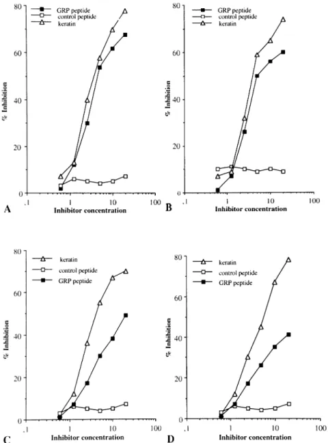
(6) 652 Glycine-rich cell wall protein and autoimmunity. Fig. 2. Inhibition of binding of affinity-purified anti-GRP peptide antibodies to keratin (A and B). Affinity-purified anti-GRP peptide antibodies [from patient GC (A) and patient CA (B)] were preincubated with different amount of antigens (keratin, GRP peptide and control irrelevant peptide, µg/ml in diluting buffer) for 1 h at 37°C and then transferred to a plate coated with keratin. Bound antibodies were detected by an alkaline phosphatase-labeled anti-human IgG antiserum. Results are expressed as percent inhibition. Inhibition of binding of affinity-purified anti-keratin antibodies to solid-phase keratin (C and D). Affinity-purified anti-keratin antibodies [from patient GC (C) and patient SP (D)] were preincubated with different amount of antigens (keratin, GRP peptide and control irrelevant peptide, µg/ml in diluting buffer) for 1 h at 37°C and then transferred to a keratin-coated plate. Bound antibodies were detected by an alkaline phosphatase-labeled anti-human IgG antiserum. Results are expressed as percent inhibition.. able to cross-react with autoantigens (keratin, actin, EBNAI), whereas not all the antibodies directed against such autoantigens are able to recognize the GRP sequence. These results indicate that the peptide studied identifies a shared epitope widely expressed in different diseases and that antibodies against such peptide recognize different autoantigen targets. EBV-transformed cell clones producing anti-GRP antibodies Six EBV-transformed cell clones obtained from patients PG, GC and SP produced anti-GRP peptide antibodies able to. recognize keratin, actin and EBNA-I. An example of this behavior is given in Fig. 3. These results confirm at the clonal level our previous observations that anti-GRP antibodies produced in subjects with autoimmune disorders are able to cross-react with several autoantigens and this cross-reactivity may be relevant in the pathogenesis of the disease. Normal mice immunized with the GRP peptide develop an autoimmune response To further substantiate the role of the GRP peptide in inducing an autoimmune response, 10 BALB/c mice were immunized.
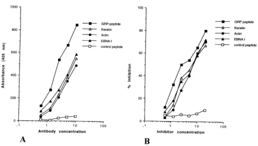
(7) Glycine-rich cell wall protein and autoimmunity 653. Fig. 3. Autoantigen binding activity of a human monoclonal anti-GRP peptide antibody. (A) Direct binding of mAb 1K1 to different antigens: keratin, actin, EBNA-I, GRP peptide and control peptide. Bound antibodies were detected by an alkaline phosphatase-labeled anti-human antiserum. Results are expressed as absorbance at 405 nm, 30 minutes after substrate addition. (B) Inhibition of binding of human mAb 1K1 to solid-phase GRP peptide. The amount of purified Ig which gave 50% of the maximal binding to the antigen in direct ELISA was preincubated for 1 h at 37° C with different amount of antigens (keratin, actin, EBNA-I, GRP peptide and control peptide) and then transferred to the GRP peptide-coated plate. Bound antibodies were detected by an alkaline phosphatase-labeled anti-human antiserum. Results are expressed as percent inhibition.. Table 4. Autoantibody response in mice injected with GRP peptide Mice injected with. Mice with IgG anti-GRP peptide antibodies. Mice with IgG anti-keratin antibodies. GRP peptide–KLH Control peptide–KLH KLH PBS. 8/10a 600 ⫾ 30b 0/10 ⬍80c 0/10 ⬍80 0/10 ⬍80. 8/10 0/10 0/10 0/10. 450 ⫾ 50b ⬍75c ⬍75 ⬍75. Mice with IgG anti-collagen II 8/10 0/10 0/10 0/10. 400 ⫾ 20b ⬍50c ⬍50 ⬍50. Mice with IgG anti-EBNA I 8/10 0/10 0/10 0/10. 350 ⫾ 25b ⬍50c ⬍50 ⬍50. aNumber of animals considered in the study. bMean IgG Ab values in mice immunized with cMean. the GRP peptide which developed an autoantibody in response (1:200 serum dilution). IgG antibody values ⫹3 SD in the control groups (1:200 serum dilution).. with the GRP peptide coupled to the carrier protein KLH. Eight mice developed an anti-GRP peptide IgG and IgM response (Table 4). Autoantibody activity against keratin, collagen II and EBNA-I was detected in all the mice which developed a strong anti-GRP response, and anti-peptide antibodies affinity purified from the pooled sera of five individual mice recognized the autoantigens as well (data not shown). On the contrary, autoantibody activity could not be detected in the sera of the animals injected with an irrelevant peptide coupled to KLH, KLH alone or PBS alone. These data further indicate that the GRP peptide is able to induce an autoimmune response through the production of anti-peptide antibodies cross-reacting with autoantigens. Generation of peptide-specific T cell clones To evaluate the T cell response to the GRP peptide, 11 patients affected by different diseases were selected on the basis of a high anti-peptide serum IgG antibody titer. The 11 patients selected for the preparation of T cell clones were affected by the following diseases: food allergy (patient CA),. SLE (patients LoM, PG, ZP), PsA (patients LM and SP), RA (patients VM and GC) and CPI (patients BS, CoL, SE). PBMC isolated from these patients were able to proliferate to the GRP peptide, but not to a control irrelevant peptide, whereas normal subjects did not show any proliferative response to the GRP peptide (data not shown). One hundred and fifty antigen-specific T cell clones were derived from the PBMC of the 11 patients studied. The clonal efficiency of the clonal procedures was 4.82% (SD ⫾ 1.1). We then chose to characterize 38 clones on the basis of their growth capability; all the clones proved to be CD4⫹ by FACS analysis. The antigen specificity of the 38 clones was then evaluated using the GRP peptide and EBV-transformed autologous B lymphocytes or autologous PBMC as antigenpresenting cells. Twenty-five T cell clones showed a specific proliferative response to the GRP peptide, but not to an irrelevant control peptide. Of the 25 peptide-specific T cell clones, six were derived from patient SP, four from patient CA, two from patients SE, CG, VM, LM, PG, LoM, one from each of ZP, CoL, BS. The specificity of the proliferative.
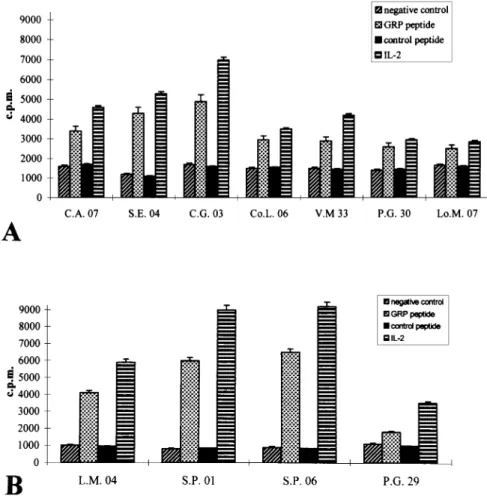
(8) 654 Glycine-rich cell wall protein and autoimmunity. Fig. 4. Proliferative responses of GRP peptide-specific T cell clones. Proliferation of antigen-specific T-cell clones derived from different individual patients. CD4⫹ clones derived were cultured in complete medium in 96-well U-bottom microplates (5⫻105 cells/well) with irradiated autologous PBMC or autologous B-EBV-transformed cell lines derived from patients (5⫻105 cells/well). The following stimuli were used: GRP peptide (20 µg/ml), an irrelevant peptide (20 µg/ml) and rIL-2 (20 U/ml) as positive control. After 3 days of culture cells were pulsed with 20 µCi [3H]thymidine and incubated for an additional 18 h at 37°C. Results are expressed in c.p.m.⫻10–3 of the mean ⫾ SD of triplicate samples of two different tests for each patient’s clone. The antigen-presenting cells used are: autologous PBMC (A) and autologous B EBVtransformed cell lines (B). Statistical analysis of the data was performed using the paired t-test; the differences between the proliferative responses of T cell clones to the GRP peptide and to the control peptide were statistically significant (P ⬍ 0.001).. response of the CD4⫹ T cell clones obtained from the patients studied was confirmed by the following lines of evidence: (i) all the anti-GRP T cell clones did not proliferate in the presence of the GRP peptide when heterologous irradiated EBV cells or heterologous PBMC were used as antigen-presenting cells and (ii) preincubation of the CD4⫹ anti-GRP peptide clones with the class II-specific mAb D1-12 abolished the proliferative response of these clones to the GRP peptide (data not shown). Figure 4 shows the proliferative response of representative clones derived from different patients. PBMC obtained from two normal subjects were stimulated with the GRP peptide and subsequently cloned with the previously described procedure: differently from what was observed with the clones derived from the patients’ PBMC, the CD4⫹ T cell clones obtained from the two normal subjects failed to proliferate to the GRP peptide (data not shown), further confirming the specificity of the anti-GRP T cell response observed in the group of patients examined. Taken together, these results suggest that a specific T cell. response against the GRP peptide can be elicited in patients affected by different autoimmune diseases. Intracytoplasmic cytokine profile of the T cell clones studied In order to evaluate the Th subset of the peptide-specific T cell clones, we performed intracellular cytokine staining by FACS analysis. The 18 clones analyzed did not show a Th1 or Th2 cytokine profile, since IFN-γ, IL-2 and IL-4 were simultaneously present (data not shown). These data suggest that GRP peptide-specific T cell clones belong to the Th0 subset. Discussion This study shows that a peptide from a food-derived glycinerich cell wall protein is able to elicit a B and T cell immune response in several autoimmune diseases. Such peptide has a high homology with EBNA-I, cytokeratins, and other self antigens such as hnRNP, fibrillar collagen and procollagen.
(9) Glycine-rich cell wall protein and autoimmunity 655 (19). Interestingly the reactivity to this peptide is not limited to a particular disease condition since anti-peptide IgG antibodies and peptide-specific T cell clones can be found in several autoimmune disorders and in food allergy, whereas normal subjects do not have detectable serum antibodies directed against the peptide and their PBMC fail to proliferate to the peptide. The anti-GRP peptide antibodies purified from the sera of the patients studied were able to specifically recognize EBNA-I and different autoantigen targets (keratin, collagen, actin), whereas such autoantigen binding activity was not displayed by the anti-control peptide antibodies affinity purified from the same patients’ sera and by the antiGRP peptide antibodies eluted from the sera of healthy donors. In the latter case the eluted antibodies showed a lowaffinity interaction with the peptide and did not recognize any of the autoantigens tested. The autoantigen binding profile of the anti-GRP peptide antibodies is similar to the one observed with serum antibodies directed against P62, a synthetic peptide corresponding to the Gly–Ala repeat sequence of EBNA-I (18). In both cases affinity purified anti-peptide antibodies cross-reacted with the same autoantigens: keratin, actin and collagen. The similar behavior of the two antipeptide antibody populations (anti-P62 and anti-GRP peptide) can be ascribed to the presence of the common sequence Gly–Gly–Ala, which suggests that anti-Gly–Ala antibodies may be responsible for the cross-reactions observed. An increased level of anti-Gly–Ala antibodies may account for the crossreaction with structural endogenous proteins such as cytokeratins and collagen. In this regard, recent data obtained in our laboratory using recombinant peptide libraries show that the Gly–Gly–Ala motif is recognized at high frequency in randomly generated dodecamer peptides by autoantibodies belonging to different autoimmune diseases. This finding further demonstrates an important role for this amino acid sequence as an antigen target in different diseases (Lunardi et al., manuscript in preparation). The finding that anti-GRP IgG antibodies are present at high frequency in the sera of autoimmune and food allergic individuals was further confirmed by the generation of human mAb from EBV-transformed lymphocytes belonging to representative patients. The anti-GRP peptide reactivity was present at high frequency in a panel of randomly selected EBV clones. Similarly to the serum counterpart, these antibodies were able to cross-react with several autoantigens. These results again support an important role for the GRP sequence in priming an autoimmune response. This hypothesis was further substantiated by experiments in which the 15 amino acid GRP peptide was coupled to a carrier protein and its ability to elicit an autoimmune response in normal animals (BALB/c mice) was analyzed. Interestingly mice displaying a strong anti-peptide response produced antibodies able to bind the GRP peptide, and to cross-react with several autoantigens including keratin, collagen II and EBNA-I. In accordance to the humoral response, T cell responses can be elicited by the GRP peptide in different patients and in several disease conditions. A T cell response against the GRP peptide has been already described in the synovial fluid lymphocytes of patients with juvenile RA and GRP peptide specific CD4⫹ T cell clones have been obtained from the synovial fluid of these patients (19). Interestingly the GRP. peptide is able to induce proliferation of PBMC isolated from patients with different diseases, whereas normal subjects do not proliferate to the peptide. To further analyze the T cell response to the peptide, a panel of CD4⫹ peptide-specific T cell clones was derived from PBMC of patients with different disease conditions. CD4⫹ T cell clones specific for the GRP peptide could not be derived from the PBMC of normal healthy donors. Peptidespecific CD4⫹ T cell clones may either be the consequence of a previous activation and expansion by exogenous antigens, e.g. in the gut mucosa or more likely derive from normally present autoreactive T cell subsets (3). Independently from the mechanism involved in the generation of such CD4⫹ T cell clones, these cells may be able to provide a T cell help to B cells for the generation of an IgG response towards the peptide, which will result in the production of large amounts of cross-reacting anti-peptide antibodies. The presence of the same structural motif in the different autoantigens may account for the promiscuity of such autoantibody response which seems to be a common feature of the immune response in different autoimmune disorders. These results suggest that the immune response in autoimmune diseases may not be as heterogeneous as originally thought. In this regard it is possible to view the autoimmune response as an oligoclonal expansion of a rather limited number of T cell subsets which share the capability of responding to a particular amino acid sequence. The process may result in the stimulation of cells able to deliver T cell help to a great number of B cell clones. Thus, a large number of antibodies specific for different autoantigenic targets may arise as a response to a relatively small number of amino acid sequences, probably due to a mechanism of molecular mimicry. Interestingly, a B and T cell response to the GRP peptide can also be found in patients with food allergy. The absence of anti-GRP-specific IgE antibodies in patients with food allergy suggests the possibility of a non-IgE-mediated immune response to particular food-derived antigens which can be able either to block or to mediate histamine release from basophils and mast cells. The type of immune response elicited may be determined by the genetic background: the TCR–peptide–MHC interaction can control the direction of the functional immune response, and MHC linkage to polarized Th1-type and Th2type immune responses has now been reported for several antigens and peptides (32–38). The analysis of some of our peptide-specific T cell clones derived from peripheral blood of patients with different diseases revealed a predominant Th0 phenotype. We can hypothesize either that the GRP peptide is unable to induce a dominant Th1 or Th2 cytokine response, or that this peptide-specific CD4⫹ subset found in the peripheral blood may switch to a Th1 or Th2 cytokine response at sites of autoimmune inflammation. Several lines of evidence suggest that the induction of oral tolerance by orally administered antigens has potential therapeutic applications for the treatment of autoimmmune and food-allergic diseases (20,21). In this regard the finding that the same peptide epitope can induce an immune response in different diseases may have important practical implications. The identification of widespread peptide antigens may suggest their potential utilization in the suppression.
(10) 656 Glycine-rich cell wall protein and autoimmunity of the immune response by oral administration. This therapeutic strategy has so far been hampered mainly by the limited knowledge of disease relevant antigens. Random peptide library technology is a powerful tool for identifying potentially pathologically relevant peptide antigens in different disease conditions. Using this approach it is possible to dissect common features at the amino acid level in individuals affected by the same or by different diseases. This could allow the identification of peptide sequences recognized at high frequency in certain conditions: the GRP peptide represents an example of an antigenic peptide sequence able to prime a B and T cell immune response in different and apparently unrelated diseases. Finally, the finding of a common peptide epitope able to elicit an immune response in patients with food allergy and different autoimmune disorders give rise to the question of a possible link between food antigens, gut mucosa and systemic immune response. In the past few years several groups have studied the role of dietary manipulation in RA (39–41), juvenile RA (42) and vasculitis (43), reaching conflicting results. T cell clones specific for particular food antigen epitopes may arise in the gut mucosa and be recruited to particular sites, such as joints, where they proliferate in response to homologous peptides derived from synovial proteins, following local inflammation and up-regulation of MHC molecules. The release of additional self-antigens and/or epitope spreading can lead to a chronic self-perpetuating process of organ inflammation and destruction. In conclusion, our data suggest that phylogenetically highly conserved epitopes in plants, viruses and humans may be responsible for an autoimmune response in susceptible individuals. Acknowledgements This work was partially supported by AIRC (Italian Association for Cancer Research; to A. P.), by the Italian Ministry of Scientific Research (MURST; to A. P.), by the Italian National Research Council (CNR; to G. De S.) and by a grant from Regione Veneto, Ricerca Sanitaria Finalizzata Venezia-Italia (to G. De S.). The authors are indebted to Dr Alessandro Moretta for his invaluable suggestions and help in the revision of the manuscript.. Abbreviations CIU CPI EBNA-I EBV GRP hnRNP KLH PBMC PE PsA RA SLE. chronic idiopathic urticaria chronic parvovirus infection Epstein–Barr nuclear antigen I Epstein–Barr virus glycine-rich cell wall protein heterogeneous nuclear ribonucleoprotein keyhole limpet hemocyanin peripheral blood mononuclear cells phycoerythrin psoriatic arthritis rheumatoid arthritis systemic lupus erythematosus. References 1 Hemmer, B., Fleckenstein, B. T., Vergelli, M., Jung, G., McFarland, H., Martin, R. and Wiesmuller, K. H. 1997. Identification of high potency microbial and self ligands for a human autoreactive class II-restricted T cell clone. J. Exp. Med. 185:1651.. 2 Lehman, P. V., Sercarz, E. E, Forsthuber, T., Dayan, C. M. and Gammon, G. 1993. Determinant spreading and the dynamics of the autoimmune T-cell repertoire. Immunol. Today 14:203. 3 Hemmer, B., Vergelli, M., Pinilla, C., Houghten, R. and Martin, R. 1998. Probing degeneracy in T-cell recognition using peptide combinatorial libraries. Immunol. Today 19:163. 4 Smith, G. 1985. Filamentous fusion phage: novel expression vectors that display cloned antigens on the virion surface. Science 228:1315. 5 Scott, J. K. and Smith, J. P. 1990. Searching for peptide ligands with an epitope library. Science 249:386. 6 Devlin, J. J, Panganiban, L. C. and Devlin, P. E. 1990. Random peptide libraries: a source of specific protein binding molecules. Science 249:404. 7 Cwirla, S. E., Peters, E. A., Barrett, R. W. and Dower, W. J. 1990. Peptides on phage: a vast library of peptides for identifying ligands. Proc. Natl. Acad. Sci. USA 87:6378. 8 Cortese, I., Tafi, R., Grimaldi, L. M., Martino, G., Nicosia, A. and Cortese, R. 1996. Identification of peptides specific for cerebrospinal fluid antibodies in multiple sclerosis by using phage libraries. Proc. Natl. Acad. Sci. USA 93:11063. 9 Bowditch, R. D., Tani, P., Fong, K. C. and McMillan, R. 1996. Characterization of autoantigenic epitopes on platelet glycoprotein IIb/IIIa using random peptide libraries. Blood 12:4579. 10 Mennuni, C., Santini, C., Lazzaro, D., Dotta, F., Arilla, L., Fierabracci, A., Bottazzo, G. F., Di Mario, U., Cortese, R. and Luzzago, A. 1997. Identification of a novel type 1 diabetesspecific epitope by screening phage libraries with sera from prediabetic patients. J. Mol. Biol. 268:599. 11 Sibille, P., Ternynck, T., Nato, F., Buttin, G., Strosberg, D. and Avrameas, A. 1997. Mimotopes of polyreactive anti-DNA antibodies identified using phage-display peptide libraries. Eur. J. Immunol. 27:1221. 12 Dybwad, A., Forre, O., Kjeldsen-Kragh, J., Natvig, J. B. and Sioud, M. 1993. Identification of new B cell epitopes in the sera of rheumatoid arthritis patients using a random nanopeptide phage library. Eur. J. Immunol. 23:3189. 13 Dybwad, A., Forre, O., Natvig, J. B. and Sioud, M. 1995. Structural characterization of peptides that bind synovial fluid antibodies from RA patients: a novel strategy for identification of diseaserelated epitopes using a random peptide library. Clin. Immunol. Immunopathol. 75:45. 14 Dybwab, A., Forre, O. and Sioud, M. 1996. Increased serum and synovial fluid antibodies to immunoselected peptides in patients with rheumatoid arthritis. Ann. Rheum. Dis. 55:437. 15 Ryser, U., Schorderet, M., Zhao, G. F., Studer, D., Ruel, K., Hauf, G. and Keller, B. 1997. Structural cell-wall proteins in protoxylem development: evidence for a repair process mediated by a glycine-rich protein. Plant J. 12:97. 16 Rhodes, G. H., Valbracht, J. R., Nguyen, M. D. and Vaughan, J. H. 1997. The p542 gene encodes an autoantigen that crossreacts with EBNA-I of the Epstein–Barr virus and which may be a heterogeneous nuclear ribonucleoprotein. J. Autoimmun. 10:447. 17 Baboonian, C., Halliday, D., Venables, P. J. W., Pawlowski, T., Millman, G. and Maini, R. N. 1989. Antibodies in rheumatoid arthritis react specifically with the glycine alanine repeat sequence of Epstein–Barr nuclear antigen-1. Rheumatol. Int. 9:161. 18 Baboonian, C., Venables, P. J. W., Williams, D. G., Williams, R. O. and Maini, R. N. 1991. Cross reaction of antibodies to glycine/ alanine repeat sequence of Epstein–Barr virus nuclear antigen-1 with collagen, cytokeratin, and actin. Ann. Rheum. Dis. 50:772. 19 Ostenstad, B., Dybwad, A., Lea, T., Forre, O., Vinje, O. and Sioud, M. 1995. Evidence for monoclonal expansion of synovial T cells bearing V alpha 2.1/V beta 5.5 gene segments and recognizing a synthetic peptide that shares homology with a number of putative autoantigens. Immunology 86:168. 20 Weiner, H. L. 1997. Oral tolerance: immune mechanisms and treatment of autoimmune diseases. Immunol. Today 18:335. 21 Strobel, S. and McI. Mowat, A. 1998. Immune responses to dietary antigens: oral tolerance. Immunol. Today. 19:173. 22 Tan, E. M., Cohen, A. S., Fries, J. F., Masi, A. T., McSahne, D. J., Rothfield, N. F., et al. 1982. The 1982 revised criteria for the.
(11) Glycine-rich cell wall protein and autoimmunity 657. 23. 24. 25 26. 27. 28. 29. 30 31. 32. 33. classification of systemic lupus erythematosus. Arthritis Rheum. 25:1271. Arnett, F. C., Edworthy, S. M., Bloch, D. A., McShane, D. J., Fries, J. F., Cooper, N. S., et al. 1988. The American Rheumatism Association 1987 revised criteria for the classification of rheumatoid arthritis. Arthritis Rheum. 31:315. Lunardi, C., Tiso, M., Borgato, L., Nanni, L., Millo, R., De, Sandre, G., Severi, A. B. and Puccetti A. 1998. Chronic parvovirus B19 infection induces the production of anti-virus antibodies with autoantigen binding properties. Eur. J. Immunol. 28:936. Atherton, E., Logan, C. J. and Sheppard, R. C. 1979. Peptide synthesis. Part 2. Bioorg. Chem. 8:351. Poggi, A., Biassoni, R., Pella, N., Paolier, F., Bellomo, R., Bertolini, A., Moretta, L. and Mingari, M. C. 1990. In vitro expansion of CD3/TCR-human thymocyte populations that selectively lack CD3 delta gene expression: a phenotypic and functional analysis. J. Exp. Med. 172:1409. Moretta, A., Pantaleo, G., Moretta, L., Cerottini, J. C. and Mingari M. C. 1983. Direct demonstration of the clonogenic potential of every human peripheral blood T cell. J. Exp. Med. 157:743. Poggi, A., Tomasello, E., Revelli, V., Nanni, L., Costa, P. and Moretta, L. 1997. p40 molecule regulates NK cell activation mediated by NK receptors for HLA class I antigens and TCRmediated triggering of T lymphocytes. Int. Immunol. 9:1271. Lefkovits, I. 1987. Generation of EBV-transformed continuous B Lymphoblastoid cell lines (BLCL). In Lefkovits, I., ed., Immunology Methods Manual, p. 1978. Academic Press, San Diego, CA. Carter, L. L. and Swain, S. L. 1997. Single cell analyses of cytokine production. Curr. Opin. Immunol. 9:177. Jung, T., Schauer, U., Heusser, C., Neumann, C. and Rieger, C. 1993. Detection of intracellular cytokines by flow cytometry. J. Immunol. Methods 159:197. Murray, J. S., Madri, J., Tite, J., Carding, S. R. and Bottomly, K. 1989. MHC control of CD4⫹ T cell subset activation. J. Exp. Med. 170:2135. Brunner, M., Larsen, S., Sette, A. and Mitchison, A. 1995. Altered Th1/Th2 balance associated with the immunosuppressive/. 34. 35. 36 37. 38 39 40 41. 42 43. protective effect of the H-2Ab allele on the response to allo-4hydroxyphenylpyruvate dioxygenase. Eur. J. Immunol. 25:3285. Milich, D. R., Peterson, D. L., Schodel, F., Jones, J. E. and Hughes, J. L. 1995. Preferential recognition of hepatitis B nucleocapsid antigens by Th1 or Th2 cells is epitope and major histocompatibility complex dependent. J. Virol. 69:2776. Harrison, L. C., Honeyman, M. C., De Aizpurua, H. J., Schmidli, R. S., Colman, P. G., Tait, B. D. and Cram, D. S. 1993. Inverse relation between humoral and cellular immunity to glutamic acid decarboxylase in subjects at risk of insulin-dependent diabetes. Lancet. 341:1365. Mutis, T., Cornelisse, Y. E., Datema, G., Van den Elsen, P. J, Ottenhoff, T. H. and de Vries, R. R. P. 1994. Definition of a human suppressor T-cell epitope. Proc. Natl Acad. Sci. USA 91:9456. Roep, B. O., Duinkerken, G., Schreuder, G. M. T., Kolbm, H., de Vries, R. R. P. and Martin, S. 1996. HLA-associated inverse correlation between T cell and antibody responsiveness to islet autoantigen in recent-onset insulin-dependent diabetes mellitus. Eur. J. Immunol. 26:1285. Murray, J. S. 1998. How the MHC selects Th1/Th2 immunity. Immunol. Today. 19:157. Darlington, L. G., Ramsey, N. W. and Mansfield, J. R. 1986. Placebo-controlled, blind study of dietary manipulation therapy in rheumatoid arthritis. Lancet i:236. Panush, R. S., Carter, R. L., Katz, P., Kowsari, B., Longley, S. and Finnie, S. 1983. Diet therapy for rheumatoid arthritis. Arthritis Rheum. 26:462. Kavanaghi, R., Workman, E., Nash, P., Smith, M., Hazleman, B. L. and Hunter, J. O. 1995. The effects of elemental diet and subsequent food reintroduction on rheumatoid arthritis. Br. J. Rheumatol. 34:270. Schrander, J. J., Mracelis, C., de Vries, M. P. and van SantenHoeufft, H. M. 1997. Does food intolerance play a role in juvenile chronic arthritis? Br. J. Rheumatol. 36:905. Lunardi, C., Bambara, L. M., Biasi, D., Zagni, P., Caramaschi, P. and Pacor, M. L. 1992. Elimination diet in the treatment of selected patients with hypersensitivity vasculitis. Clin. Exp. Rheumatol. 10:131..
(12)
Figure


+3
Documents relatifs