Protein Engineering vol.8 no.6 pp.535-542, 1995
The predicted secondary structure of the G-type glutamine
amidotransferase is compatible with TEVf-barrel topology
Thomas Niermann1>2 and Kasper Kirschner1"3
'Department of Biophysical Chemistry, Biozentrum der Universitat Basel, Klingelbergstrasse 70, CH-4056 Basel, Switzerland
2Present address: Ciba-Geigy AG, Biotechnologie, CH 4002 Basel,
Switzerland
'To whom correspondence should be addressed
Glutamine amidotransferase (GAT) subunits or domains catalyze an important partial reaction in many complex biosynthetic reactions. The structure of one member of the F-type GATs is known, but the structure of the unrelated G-type is still unknown. Because many protein sequences are available for anthranilate synthase component II (product of the trpG gene), we have predicted its average secondary structure by a joint prediction method [Niermann and Kirschner (1991a) Protein Engng, 4, 359-370]. The predicted eight p-strands and seven a-helices follow an 8-fold cyclic repetition of a (3-strand-4oop-a-helix-loop module with helix a7 missing. This pattern of
secondary structure suggests that the G-type GAT domain has an 8-fold Pa-barrel topology, as found first in triose phosphate isomerase (TIM-barrel)- This model is supported by the location of known catalytically essential residues in loops between p-strands and a-helices. Evidence from published sequencing and mutational studies on selected members of the GAT superfamily (carbamoyl phosphate, imidazoleglycerol phosphate, GMP and CTP synthases) support both the secondary structure prediction and the TIM-barrel topology.
Key words: glutamine amidotransferase/prediction of secondary structure/sequence alignments/TIM-barrel proteins/tryptophan biosynthesis
Introduction
More than a dozen biosynthetic enzymes possess both an ammonia (NH3)-dependent synthase and a glutamine
amido-transferase (GAT) activity. A recent review is given by Zalkin (1993). In general, the synthase incorporates either NH3 or
preferentially the amino group released from glutamine into a variety of intermediates in the biosynthesis pathways of amino acids, nucleotides and coenzymes. The two different active sites of these complex enzymes are provided for by different domains (or subunits), and are thought to interact intimately in terms of both structure and function. A striking consequence of this interdomain interaction is the failure of 'nascent NH3',
which is released from glutamine at the GAT active site, to equilibrate with the bulk solvent.
There are two apparently unrelated families of GAT domains, designated F- and G-type, for historical reasons. The structure of the first complex enzyme containing an F-type GAT domain has been determined recently for glutamine 5'-phosphoribosyl-1-pyrophosphate amidotransferase, the product of the purF gene from Bacillus subtilis (Smith et al., 1994). The F-type
GAT domain consists of two antiparallel f}-sheets positioned face to face, and covered by a-helices on both outer surfaces. In contrast, the structure of the G-type GAT domain is still unknown. However, several predictions have tentatively assigned an entirely different protein fold, namely that of the familiar 8-fold parallel Pa (or TIM)-barrel (Farber, 1993), to these domains (Wilmanns and Eisenberg, 1993; T.Niermann and K.Kirschner, personal communication to Wilmanns and Eisenberg, 1993). A preliminary report on the X-ray crystallo-graphic structure analysis of GMP synthetase from Escherichia coli that contains a G-type GAT domain has been published recently (Tesmer et al., 1994). The anticipated first determina-tion of the structure of a G-type GAT domain provides for a test of the combination of methods suitable for the improved prediction of the secondary structure of a/p" proteins (Niermann and Kirschner, 1991b), particularly that of TIM-barrel proteins (Crawford et al, 1987; Niermann and Kirschner, 1991a).
Here we report the results of applying the methods to 26 sequences of the G-type GAT domains of both anthranilate synthase and the closely related aminodeoxychorismate syn-thase. The predicted average secondary structure consists of a sequence of eight pa modules, with helix a7 predicted as a
loop. The cysteine, histidine and glutamate residues known to be catalytically essential are located in loops following the C-termini of predicted strands p4 and Pg that would be spatially
adjacent in a TIM-barrel fold. Data from aligned sequences of the superfamily of synthetases with G-type GAT domains narrow down the number of invariant residues at the carboxyl ends of p-strands still further, and support the TIM-barrel fold.
Materials and methods
Alignment of amino acid sequences
We consider here 22 trpG sequences (Sma, Serratia marces-cens; Eco, Escherichia coli; Lbi, Leptospira biflexa; Aca, Acinetobacter calcoaceticus; Pal, Pa2, Pseudomonas aerugi-nosa (two genes; Essar et al., 1990); Sty, Salmonella typhimur-ium; Bla, Brevibacterium lactofermentum; Tth, Thermus thermophilus; Ncr, Neurospora crassa; See, Saccharomyces cerevisiae; And, Aspergillus nidulans; Ang, Aspergillus niger, Pch, Penicillium chrysogenum; Sdy, Shigella dysenteria; Vpa, Vibrio parahaemolyticus; Bsu, Bacillus subtilis; Ppu, Pseudomonas putida; Rme, Rhizobium meliloti; Pbl, Phyco-myces blakesleeanus; Pep, Phanerochaete chrysosporium; Hpo, Hansenula polymorpha; and four pab A. sequences [E.coli, S.typhimurium, Kae (Klebsiella aerogenes) and S.marcescens]. All sequences were obtained from the MIPS data bank (Mewes, 1990). The sequences were first aligned in pairs, using FASTA (Pearson and Lipman, 1988; GCG Sequence Analysis Software Package, 1991). The computer programs PROFILE and PRO-FILEGAP (Gribskov et al., 1987; GCG Sequence Analysis Software Package, 1991) were used for multisequence align-ment. Minor rearrangements in the sequence alignments were greatly aided by the clustering of both identical residues and
T.Niermann and K.Kirschner 04 «i 05 l l l l l l l l l l l l l l l l l l l l l l l l l l i w i 1 1 1 1 1 1 1 1 1 1 1 1 1 0i «i 02 10 20 30 40 50 60 70 80 90 100 110
I I I I I I I I I I I
cons. DN DSFt N g RN SPGP P G g P G C G Q GgSma MADILLLDNVDSFTYNLVDQLRASGHQ WIYRNQIGAEVIIERLQHMEQPVLMLSPGPGTP . . . SEAGCMPELLQRLRGQLPI IGICLGHQAI VEAYGGQVGQAGEI Eco MADILLLDNIDSFTYNLADQLRSNGHN W I YRNHIPAQTLIERLATMSNPVLMLSPGPGVP. . . SEAGCMPELLTRLRGKLPIIGICLGHOAIVEAYGGYVGQAGEI Lbi .MKVLILDNYDSFTFNLYQIVGEILEEREKPFQLDVIRND. . . EKPFEWIKSANYDKIIISPGPGHPADPAYFGVSADI LKELGKTPPVLGICLGMQGMATVFGGEWRANIA Aca . .MLLMIDNYDSFTYNIVQYFGELNQD V K W R N D . . . QVTLEDIERWQPKYLWGPGPCSP. . . TEAGISIPAIHHFAGRIPLLGVCLGHQAIGQAFGGNIIRAKTV Sty MADILLLDNIDSFTWNLADQLRTNGHN W I YRNHIPAQTLIDRLATMKNPVLMLSPGPGVP . . . SEAGCMPELLTRLRGKLPIIGICLGHQAIVEAYGGYVGQAGE I Bla MTHWLIDNHDSFVYNLVDAFAVAGYK CTVFRNT . . . . VPVETILAAJJPDLICLSPGPGYP
Tth VMRVLWDNYDSFTYNLVQYLGELGAE PIVWRND. . .RFRLEEVEALDPDRILISPGPCTP Sdy MADILLLDNIDSFTYNLADQLRSNGHN WIYRNHIPAQTLIERLATMSNPVLMLSPGPGVP Vpa MANIVFIDNFDSFTYNLVDQFRSLGHS VKIYRNHIPAETIEQAINELENPWLLSPGPGAP Bsu . .MILMIDNYDSFTYNLVQYLGELGEE L W K R N D . . . SITIDEIEELSPDFLMISPGPCSP
.AITVPEIAALNPDTLLISPGPGHP See NKHWLIDNYDSFTWNVYEYLCQEGAK VSVYRND .
Hpo SKNWMIDNYDSFTWNLYEYLCQEGAN VEVFRND . And ASNVILIDNYDSFTWNVYQYLVLEGAT VTVIRND. Ang ASNVILIDNYDSFTWNVYQYLVLEGAT VNVFRND .
Pch ASNVILIDNYDSFTWNIYQYLVLEGAT VTVYRND . . .EVTVEDLVAKKPTQLVISPGPGHP Ncr ASNLILIDNYDSFTWNVYQYLVLEGAK VTVFRND . . . QITIDELIAKNPTQLVISPGPGHP Pbl .MATLLIDNYDSFTYNVYQYLCSQGAD W V Y R N D . . . KITVDEIVKLNPVNIVISPGPGHP Pep PIDILMIDNFDSFTWNLYQSLCLLGAD VTVIRND. . . AIPRSAI PQLRIKRLIVSPGPGHP Rme GVSILLVDHEDSFVHTLANYFRQTGAS VTTVRTP . . . . VAEEI FDRVKPDLWLSPGPGTP Pal . . MLLMIDNYDSFTYNLVQYFGELXAE V K W R N D . . . ELSVEQIEALAPERIVLSPGPCTP Pa2 . .MLLMIDNYDSFTYNWQYLGELGAE VKVIRND. . . EMTIAQIEALNPERIWSPGPCTP Ppu .MRITLLDNFDSFTYNLVEQFCLLGAE VRVMRNDTPLPTIQAALLADGCELLVLSPGPGRP ECO . .MILLIDNYDSFTWNLYQYFCELGAD VLVKRND . . . ALTLADIDALKPQKIVISPGPCTP Sty . .MILLIDNYDSFTWNLYQYFCELGAE VQVRRND . . . ALTLAHIDALNPQKIVISPGPCTP .MILLIDNYDSFTWNLYQYFCELGAE VLVRRND. . . ELTLADIISLAPAKIVISPGPCTP Kae
Sma
.ADAGNMMALIERTLGQIPLLGICLGYQALIEYHGGKVEPCG.P .FEAGLSVPLVQRYAPRYPILGVCLGHQAIGAAFGGKWPAPVL .SEAGCMPELLTRLSGKLPI IGICLGHQAI VEAYGGYVGQAGEI .SEAGSMPELIQRMKGKVPMIGICLGHQAIVEAYGGTVAGAGEI .DEAGISLEAIKHFAGKIPIFGVCLGHQSIAQVFGGDWRAERL KTDSGISRDCIRYFTGKIPVFGICMGQQCMFDVFGGEVAYAGEI QITIPEIEQLKPDVWISPGPGHP. . RTDSGISRDVISHFKGKI PVFGVCMGQQCIFEEFGGDVEYAGEI EISLEELIAKKPTQLWSPGPGHP. . KSDAGISNAAIQYFAGKI PIFGVCMGQQCIHHSFGGKVDVTGEI QITLEELIAKKPTQLVISPGPGHP. . ETDAGISSAAIQYFSGKI PIFGVCMGQQCIITCFGGKVDVTGEI DTDAGISNAVIKHFSGKVPIFGVCMGQQCMITSFGGKVDVTGEI GTDSGISRDAIRHFAGKIPIFGVCMGQQCIFDVYGGDVCFAGEI SHDAGVSRDVISYFAGKLPILGICMGEQCIFEVFGGTVSYAGDI QTDSGISREAIKYFAGKVPIMGVCMGLECWDVFGGQIAYAGEI . KDFDCKATIKKARARDLPIFGVCLGLQALAEAYGGDLRQLAI P . NEAGVSLAVI ERFAGKLPLLGVCLGHQSIGQAFGGEWRARQV . SEAGVSIE AILHFAGKLPILGVCLGHQSIGQAFGGDWRARQV . EDAGCMLELLAWARGRLPVLGVCLGHQALALAAGGAVGEARKP . DEAGISLD VIRHYAGRLPILGVCLGHQAMAQAFGGKWRAAXV .NDAGISLAVIRHYAGRIPMLGVCLGHQAMAQAFGASWRAAXV .DESGISLAAIRHFSGQTPILGVCLGHQAIAQVFGAAIVRAAKV .MLLLIDNYDSFTYNLYQYFCELGAE V W K R N D . . . ELQLTDIERLAPQHLVISPGPCTP . . . NEAGISVAAIRHFAGKLPILGVCLGHQALGQAFGAEWRARAV
var. 122 111 2 4 2 121212 3113 34 33 1 311 3111 1 221 3 22 111
gaps throughout the sequences, which provided the necessary internal register for confident alignment.
Averaging of secondary structure propensities
The secondary structure 'GOR" prediction algorithm [Gamier et al. (1978), with decision constants as suggested by Gibrat el al. (1987)] was first used to calculate the propensity of each residue for the a-helix. P-strand and coil state along each continuous amino acid sequence. Then the propensities corres-ponding to each residue were located to that residues position in the alignment. The average state propensities at each position
were obtained by summing up and dividing the sum by the number of amino acids in that column (Gamier et al.. 1978). The state profiles' were finally smoothed with a three-residue span. The state with the highest average propensity defines the predicted secondary structure at that position.
Averaging of residue properties
The chain flexibility profiles of individual sequences were calculated according to Karplus and Schul/ < 1485). Hydropathy profiles were calculated using the scale of Kyte and Doolittle (1982). over a five-residue span setting, and were linally
Secondary structure of glutamine amidotransferase 120 130 140 150 160 170 190 200 210 220
I
230I
180I I I I I I I I I
HGk s h RYHSL P M h g QFHPES LHGKASAI . . . .AHDGEGMFAGMANP LPVARYHSLVGS . . NIPADLTVNARSG EMVMAVRDDRRRVCGFQFHPESILT LHGKASSI. . . .EHDGQAMFAGLTNP LPVARYHSLVGS. . NIPAGLTINAHFN GMVMAVRHDADRVCGFQFHPESILT MHGKLSPI. . . . EHDGKGVFSGLTQG IEIMRYHSLVAKEISLPNDLEITARVSAGEGK . .GEIMGLRHKSLKIEGVQFHPESFGS MHGRLSDM. . . . YHTDKGIFSNLPSP FSATRYHSLVIEQESLPECLEVTCWTNQNDGS . IEEIMGVKHKTLPVEGVQFHPESILS LHGKASSI . . . .EHDGQAMFAGLANP LPVARYHSLVGS. .NVPAGLTINAHFN GMVMAVRHDADRVCGFQFHPESILT VHGTTDNMILTDAGVQSPVFAGLATDVEPDHPEVPGRKVPIGRYHSLGCV. . VAPDGIESLGTCSSEI . . .GDVIMAARTTDGKAIGLQFHPESVLS MHGKVSPI. . . . HHDGTGVFRGLDSP FPATRYHSLAW. . EVPEALWNAWAEEA . . . GGRTVMGFRHRDYPTHGVQFHPESYLTLHGKASSI . . . .EHDGQAMFAGLTNP LPVARYHSLVGS. .NIPAGLTINAHFN GMVMAVRHDADRICGFQFHPESILT . . .TQGARLLEQTLAWAQRK IHGKVSMM. . . .EHQDHAIYQNLPSP LAIARYHSLVAT . . KVPDSLTITAEVD NLVMSWHEQDKVCGFQFHPESIMT . . .TYGATLLGNAIEWALEK MHGKTSDI. . . .EHDGKTIFEGLKNP LVATRYHSLIVKPETLPSCFTVTAQTKE GEIMAIRHNDLPIEGVQFHPESIMT . . . SFGKEMLRNFIETYRKE VHGKTSPI . . . .SHDNCGIFRNVPQG IAVTRYHSLAGTESSLPSCLKVTASTE NGIIMGVRHKKYTVEGVQFHPESILT . . . EEGHLMIRNILNVSGGT
. THGARLLEQTLAWALAK . TQGARLLEQTLAWAQHK .EEGKCLLRNFINS.... .QHGHQIFKNFLEIYA.. .TQGARLLEQTLAWAQQK .PTGPIILSRCVEQLLAN .EAGKLILKNFLEDPWTR EEGHLMIQNILNVSGGY EHGQTMFRNFLKLTAGT EYGRIMFRN FLK LTAGT QYGRKMFRNFLELTAGT AEGRGMFRN FLHMQGGT EHGHTMISNFLSLRGGN ESGDDLLRHFMKLKGGT VHGKTSTV. . . . KHDNKGMFKNVPQD VAVTRYHSLAGTLKSLPDCLEITARTDN GIIMGVRHKKYTIEGVQFHPESILT LHGKTSVL. . . . KHDGRGAYEGLPPS VIITRYHSLAGTHSTIPECLEVSSFAQLGEDADKTVIMGVRHKQFAVEGVQFHPESILT LHGKTSPL. . . .KHDGKGAYEGLPGS LAVTRYHSLAGTHATIPDCLEVSSSVQLADDSNKDVIMGVRHKKLAVEGVQFHPESILT LHGKTSEL. . . . KHDSKGVYQGLPTS LEVTRYHSLAGTHSTIPDCLEVTSRVELGDASGKNIIMGVRHKEFAVEGVQFHPESILT LHGKTSPL. . . . RHDGKGAYAGLSQD LPVTRYHSLAGTHVTLPECLEVTSWIAKEDG . SKGVIMGVRHKEYTIEGVQFHPESILS LHGKTSTI . . . . KHDNRGLFKNVPQD NQVTRYHSLAGMPSTLPEVLEVTATTDD GVIMGVRHKKYTVEGVQFHPESILC MHGKVSGI. . . .RHDARGCFKDLPQG IQSTRYHSLSAGVKTLPDELAVTAVTEHE RVIMGIRHRKYTVEAVQYHPESILS
MHGKPSRI. . .RVLEPGIVFSGLGKE VTVGRYHSI FADPSNLPREFVITAESED GTIMGIEHSKEPVAAVQFHPESIMTLGGDAGMRMIENWAHLAKR MHGKTSPI. . . . HHKDLGVFAGLANP LTVTRYHSLWKRESLPECLEVTAWTQHADGS . LDEIMGVRHKTLNVEGVQFHPESVLT . . . EQGHELLANFLRQQGGV MHGKTSPV. . . . HHRDLGVFTGLNNP LTVTRYHSLWKRETLPDCLEVTAWTAHEDGS . VDEIMGLRHKTLNIEGVQFHPESILT . . . EQGHELFANFLKQTGGR LHGKSTSL. . . RFDQRHPLFDGIAD L R V A R Y H S L W S . . RLPEGFDCLAESD GEIMAMADPRNRQLGLQFHPESILT. . . THGQRLLENALLWCGAL MHGKTSPI. . . .THNGEGVFRGLANP LTVTRYHSLWEPDSLPACFDVTAWSET REIMGIRHRQWDLEGVQFHPESILS. . . EQGHQLLANFLHR . . . . MHGKTSPV. . . .THNGQGVFRGLPSP LTVTRYHSLIVDPATLPECFEITAWSET QEIMGIRHREWDLEGVQFHPSSILS. . . EQGHALLKNFLRR . . . . MHGKTSPV. . . . SHTGQGVFLGLNNP LTVTRYHSLLIDPRTLPECFEVTARSEE GEIMGIRHRVFDLEGVQFHPESILS . . . EQGHQLLANFLNR . . . . MHGKTSAI . . . .RHLGVGVFRGLSDP LTVTRYHSLVLKADTLPDCFEVTAWSERDGVR . . DEIMGIRHRALALEGVQFHPESVLS . . . EQGHQLLDNFLNR . . . .
1 2 4 114 1 3 22 3 113221 11 1 22271 1111 1 13331 12 24 2 1 344112 911303971113356517423451391111111111118366111125404976173416659330577605563141639936292412111154111733118656505582667
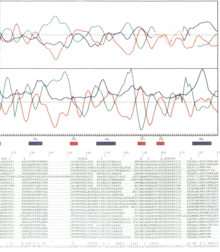
Fig. 1. Joint prediction of average secondary structure of the G-Iype GAT. Section of aligned sequences. Top row 'cons': upper case, invariant residues,
allowing for one possible sequencing error lower case, highly conserved residues, allowing for three differences. There was a total of 22 sequences of anthranilate synthase component II (Smu lo I'pu: for definition of acronyms of microorganisms see Materials and methods) and four sequences of aminodeoxychorismate synthase {Ecu lo Snuu. The single-letter amino acid codes have been used. Numbering begins with Ml of GAT from S.manescens. and is continuous throughout the longest sequences across the gaps. Bottom row 'var': sequence variability index Isee Materials and methods). Upper row. tens: lower row. ones. (Ai Profiles of averaged secondary structure propensity Color code: blue, a-helix; red. (3-strand; green, coil. (B) Profiles of averaged property parameters. Color code: blue, amphipathic moment; red. hydropathy: green, chain flexibility. Joint prediction of secondary structural elements: blue bars (helices (/.,) and red bars (strands (5,1 separated by coil segments. See text for details.
smoothed with a three-residue span. The scale of Kyte and Doolittle (1982) (Cornette et ai. 1987) was also used for the calculation of the helical amphipathic moment at 100
(Eisenberg el ai. 1984). with a span setting of seven residues and a final smoothing with a three-residue span. The chosen span setting of seven residues for the amphipathic moment
T.Niermann and K.KJrschner
profile is shorter than the value we originally preferred (Nierm-ann and Kirschner, 1991a). The justification here is that the centra] amphipathic regions of some predicted helices seem to be rather short, therefore a longer span setting would yield less pronounced maxima. The average 'property profiles' of the aligned sequences were obtained as described above for the state profiles. Peaks of the property profiles were used in the joint prediction procedure (Niermann and Kirschner, 1991b) to enhance the cognate propensity profile, where appropriate.
The variability index 'var' (Wu and Kabat, 1970) is a measure of sequence variability, var = mlf where m is the number of different residues, and / (0 < / < 1) the fraction of the most frequent residue at that position. It follows that 1
< var < n2, where n is the number of aligned sequences.
Results and discussion
Information from aligned sequences
The bottom of Figure 1 presents 26 aligned sequences of G-type GAT domains. The first 22 sequences represent component II of anthranilate synthase (i.e. products of the trpG gene), and the last four sequences correspond to the closely related 4-amino-4-deoxychorismate synthase (pabA gene). The pre-sented alignment spans the entire sequence of the monomeric GAT subunit from S.marcescens (Tso and Zalkin, 1980) to eliminate irrelevant extensions at the N- and C-termini. The sequences are numbered continuously across the gaps for later comparison with the sequences of the superfamily of GAT domains. Similar alignments, albeit with fewer sequences, have been published previously (Kaplan et al., 1985; Essar et al., 1990) as evidence for the evolutionary relationship between the products of the trpG and pabA genes. Here the emphasis is on the information content of aligned sequences of a G-type GAT domain regarding the probable occurrence of secondary structural elements along the sequence.
There are eight aligned gaps, indicating that the protein fold tolerates insertions and deletions at these sites. It is generally accepted that the residues that border the gaps reside in loops on the protein surface (Zvelebil et al., 1987). This assignment is reinforced by the preferred occurrence of Pro, Gly and charged residues in the segments that span the gaps. The gaps divide the set of aligned sequences into nine blocks that probably contain the uninterrupted secondary structural ele-ments of the protein core (Niermann and Kirschner, I991a,b; Benner, 1992).
The row labeled 'cons' above the block of aligned sequences denotes the 36 out of 230 (16%) conserved residues. The eight clusters of conserved residues reported previously by Kaplan et al. (1985) and Essar et al. (1990) are preserved despite the larger number of sequences. However, the number of conserved residues has decreased, focusing on those that are important for folding and catalysis. The clusters are numbered as follows: (I) 8 DNxDSFt 14, (II) 38 RN 39, (III) 61 SPGxxP 67, (TV) 91 GxCxGxQ 97, (V) 104 Gg 106, (VI) 115 HGk 117, (VII) 156 RYHSL 160, (VIII) 202 QFHPES 207, where x is a variable residue position.
The two rows below the block of aligned sequences give the sequence variability at each position, as defined by Wu and Kabat (1970). These data confirm the earlier work of Crawford (1989), showing that regions of high and low variability alternate.
Prediction of secondary structure
Figure 1A displays the three profiles of averaged secondary structural propensities based on the GOR method (Gibrat et al.,
1987; red, P-strand; blue, a-helix; green, coil). We have shown previously (Niermann and Kirschner, 1991a,b) that averaging of propensities is preferable to consensus prediction, and that the accuracy of prediction increases with sequence variability. It is seen that the G-type GAT domain belongs to the a/p class of proteins (Levitt and Chothia, 1976) because p-strands tend to alternate with a-helices. Figure IB displays the profiles of averaged residue properties that correlate with P-strands (red, hydrophobicity; Kyte and Doolittle, 1982), a-helices (blue, amphipathic moment; Eisenberg et al., 1984) and coils (green, chain flexibility; Karplus and Schulz, 1985). In general, the cognate state propensity and property profiles vary syn-chronously across the entire alignment, and are therefore mutually supportive.
We have used the qualitative approach as described by Niermann and Kirschner (1991b) to obtain a joint prediction of average secondary structural elements along the protein sequence. The state propensity profiles represent the primary information. Where the amplitudes of the helix and strand propensities are weak and of identical sign and magnitude, the prediction is guided qualitatively by the stronger of the correlated property profiles. The joint prediction is presented below Figure IB as a sequence of blue (a-helix) and red (p-strand) bars, with the intervening spaces assigned to surface loops.
Ignoring the profiles across the aligned gaps, the alternating patterns of hydrophobicity and chain flexibility in Figure IB strongly support the prediction in Figure 1A of five P-strands (Pi, P2, P3, p4 and P7). Similarly, the maxima of amphipathic
moment reinforce the prediction of five a-helices (a:, a2, a3,
Of, and Og). Strands p6 and pg are so predicted because
the maxima of strand propensity and hydropathy are more pronounced than those of helix propensity and amphipathic moment. Strand PJ should be assigned to an a-helix by these criteria, but its P-strand character is justified by applying the template criterion as discussed later on. Similarly, the assignment of Oj is based on the strong peak of amphipathic moment. In summary, up to this point only the assignments of P5 and 04 remain ambiguous; a-] is missing, and there is the indication of an additional P-strand overlapping with the C-terminus of o^.
Evidence in favor of a TIM-barrel fold
The pattern of strongly predicted P-strands and a-helices suggests that the G-type GAT domain has the TTM-barrel topology. This fold consists of a cyclic 8-fold repeat of the P-strand-loop-a-helix-loop module. The P-strands assemble to a central eight-stranded parallel P-barrel connected on the outside by a-helices. All TIM-barrels with a known enzymatic function have their active sites at the C-terminal face of the central p-barrel (Farber, 1993), with catalytic residues located in the C-terminus of several P-strands and in the loops that connect these P-strands to the next a-helix. The overall length of the polypeptide chain (190 < n < 241 residues), as well as the average lengths of the predicted P-strands (five ± one residues) and a-helices (10 ± two residues), are consistent with the TIM-barrel topology (Lesk et al., 1989; Lasters et al.,
1990; Murzin et al., 1994a,b).
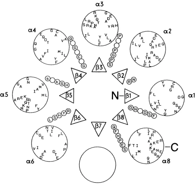
Figure 2 depicts the predicted secondary structural elements of Figure 1 arranged into an idealized TTM-barrel. The triangles represent P-strands rising towards the observer. They are connected to the next a-helix (large circles labeled a,) by strings of small circles representing residues of the connecting loops. The amino acids (single-letter code) contained in the
Secondary structure of glutamine amidotransfera.se
a3
a4
a2
a5
a1
a6
Fig. 2. Arrangement of predicted secondary structural elements and loops into an idealized TIM-barrel. Projection down the central ^-barrel axis. N,
N-terminus; C, C-terminus. (A) Strands p1,, labeled as in Figure 1, C-termini facing upwards. (.) Individual residues of loops connecting C-termini of fJ-strands
with N-termini of a-helices. Single-letter amino acid code indicates conserved residues from Figure I. (O) Helices a( labeled as in Figure 1. Single-letter
amino acid code indicates predominant residues of the central helix region, presented as a conical helical wheel to illustrate the amphipathic character of the predicted a-helices.
small circles correspond to the invariant residues given in Figure 1. Note that all invariant residues that are candidates for a catalytic function (e.g. general acid/base and nucleophilic catalysis) are located in loops at the C-terminal end of the central p-barrel.
The letters within the large circles correspond to the pre-dominant residues of the predicted a-helices, and are distrib-uted at 100° intervals in a right-handed helical fashion on the surface of a cone descending away from the observer. The seven predicted a-helices decorated in this manner as helical wheels are clearly amphipathic, as generally observed in TTM-barrels. In Figure 2 they are arranged arbitrarily, with their hydrophobic surfaces facing the central P-barrel.
Further support for secondary structural elements that are ambiguously predicted in Figure 1 is derived from the distribu-tion of clusters of conserved residues. The predicdistribu-tion method used here was optimized using multiple sequences from seven different TIM-barrel proteins (Niermann and Kirschner, 1991 a). It was found that a template of supersecondary structure occurred frequently along each sequence, and it helped to correct most of the mis- or unpredicted secondary structural
elements. The template consists of a hydrophobic P-strand followed by a loop and an amphipathic a-helix. Importantly, conserved residues are frequently clustered at the C-terminus of the P-strand, and in the adjacent loop segment.
Figure 1 shows that the strongly predicted Pa units 1, 2, 3, 4 and 6 fit to this template. Moreover, it supports the otherwise ambiguous assignment of p5. The occurrence of the conserved
cluster 104 Gg 105 at the C-terminus of the ambiguously predicted helix 04 does not necessarily invalidate the assign-ment, because tight loops between a-helices and P-strands in TTM-barrels frequently belong to the a p , or aP3 type, which includes a conserved glycine residue (Scheerlinck et ai, 1992). Conserved glycines are also found at the C-termini of a, (G25) and a3 (G85). It is also predicted from the previous
analysis of TIM-barrel proteins (Niermann and Kirschner, 1991a) that insertions are not tolerated between the predicted P-strands and the overlapping conserved residues, but rather between these conserved residues and the subsequent a-helices. Figure 1 shows that where aligned gaps do occur between P-strands and a-helices (e.g. p*2a2, p3a3, Psa^, p6««, PgOs), they
are always located after the cluster of conserved residues that 539
T.Niermann and ICKlrschner 10 20
I
HHHHHHHHH 40 50 60 80 90I
HHHHHHHHH SSSS HHHHHHHHH SSSS HHHHHHHHH SSSS HHHHHHHHH SSSS D G p G C G DYDWANESINDYDGVFISNGPGDPSLC. . . . GKAIENIRKVLALPVAKAVFGVCMG DYDFTKEDY. . . . DGLFYSNGPGDPSV LDDLSQRLSNVLEAKKTPVFGICLG DYRIQDVASEF. . DGIFLSNGPGNPELCQA. .TISNVRELLNNPVYDCIPIFGICLG QTSAEDVLKMNP.DGIFLSNGPGDPA PCDYAITAIQKFLETDIPVFGICLG MADILLLDNIDSFTYNLADQLRSNGHNWIYRNHIPAQTLIERLATMSN.PVLHLSPGPGVP SEAGCMPELLTRLRGKLPIIGICLG MADIIXLDNIDSFTWNLADQLRTNGHNWIYRNHIPAQTLIDRLATMKN.PVLMLSPGPGVP SEAGCMPELLTRLRGKLPIIGICLG ..EKRKNKKVIVLDCGIKN ..PDGKVLRILAIDVGMKY ..PGKAKANVALIDCGVKE . . EDELPFHWAYDFGAKR . NQIRCLLNRGVDLKWPW. .NQIRCFIKRGVRIESCSM. .NIIRCLVKRGANVTVFPY. .NILRMLVDRGCRLTIVPA.ALTLADIDALKP. QKIVISPGPCTP DEAGISLDVIRHYAGRLPILGVCLG ALTLAHIDALNP. QKIVISPGPCTP NDAGISLAVIRHYAGRIPMLGVCLG DVTEAQIRDFNP.SGIILSGGPEST TEENSPRAPQYVFEAGVPVFGVCYG QREAEIVLRA. . . DKLFL. PGVGTAQAA. . .MDQVRERELIDLIKACTQPVLGICLG KVSRDPDWLLA.DKLFL.PGVGTAQAA. . .MDQVRERELFDLIKACTQPVLGICLG EITRDYDKAMNA. DGLLV. PGVGAFAACMEGLKAARGDWIVDRRLSGGRPVMGICVG YIELPDAYKSVIEALKHGGLKNRVSVNIKLIDSQDVETRGLEILKGL.DAILV.PGGFGYRGV EGMITTARFARENNIPYLGICLG
I I
I
I
I I I
10 20 40 50 60 80 90 .MILLIDNYDSFTWNLYQYFCELGADVLVKRND .MILLIDNYDSFTWNLYQYFCELGAEVQVRRND MTENIHKHRILILDFGSQYTQLVARRVRELGVYCELWAW. MNWILDTCA NLSSVKSA.GAPRLHPGG. MNWILDTGCA. . .NLNSVKSAI. .ARHGYEP.VWFDYGFG. . -NVRSAERAL. .ARAGADV.
100 110 120 130 160 170
HHHHHHH SSSS HHHHHHHH SSSS Q h
NQ. .LLGLAAGA. . .QTHKMAFGNRGLN QPCVDQISGRC HITSQNHGFVIDSN. HQ. .LIARAAVQ. . .STLKLKFGNRGHN IPCTSTISGRC YITSQNHGFAVD. . . HQ. .LLALASGA. . .STHKLKYGNRAHN IPAHDLTTGQC HITSQNHGYAVDP. . HQ. .LLALASGA KTVKMKFGHHGGN HPVKDVEKNW MITAQNHGFAVDE. . HQ. .AIVEAYGGYVGQAGEILHGKASSI EHDGQAMFAGLTNP LPVARYHSLVGS. . . HQ. .AIVEAYGGYVGQAGEILHGKASSI EHDGQAHFAGLANP LPVARYHSLVGS. . . HQ. .AMAQAFGGKWRAAKVMHGKTSPI THNGEGVFRGLANP LTVTRYHSLWEP. . HQ. .AMAQAPGASWRAAKVMHGKTSPV THNGQGVFRGLPSP LTVTRYHSLIVDP. . MQCTCMAMQLGGHVEASNEREFGYAQVE WNDSALVRGIEDXLTADGKPLLDVWMSHGDKVT. . . MQ. .LLGRRSEE.TRGVDLLNIIEQDVP. .KMTDFGLPLPHMGWNRVYPQRGNRLFQGIEDG AYFYFVHSYA MQ. .IXGRRSEE.SNGVDLLGIIDEDVP. .KMTDFGLPLPHMGWNRVYPQAGNRLFQGIEDG AYFYFVHSYA MQ. .ILFSRGIE. . HDVEAEG. LDE. WPGTVGPLEADVVPHMGWNTVEAPADSQLFAGLDAD ARFYFVHSYAVHEW. MQ.. 100 110 120 190 200 210 130 220 160 HHHHHHHHHH SLPAGSGWKTYFINA VDTLTSGWKPLFVNA ETLPKDQWKPYFVNL ATLPANLRVTHKSLF .NIPAGLTINAHFN. .NVPAGLTINAHFN. DSLPACFDVTAWSET ATLPECFEITAWSET .AIPSDFITVASTES ..MPVNPWTIAQCNY ..MPVNPWTIAQCNY TQESHNPLIAEPRVT 170 230 SSSS SSSS HHHHHHHHHHH QfHPE
ND ASNEGIYHESKPWFSVQFHPEAMAG. . . PTDTEYLFDNFVDNVCGE Ddi pyrl ND DSNERFYHSELPYFSVQFHPESTPG. . .PEIQNSCLTFIQAVKEFK See Ura2 NU KSNEGMIHLQRPIFSTQFHPEAKGGPLDTAILFDKFFDNIEKYQLQ See Cpal D GTLQGIHRTDKPAFSFQGHPEASPGPHDAAPLFDHFIELIEQYRKT Eco carA
GMVMAVRHDADRVCGFQFHPESILT. . . . TQGARLLEQTLAWAQHK Eco trpG GMVMAVRHDADRVCGFQFHPESILT. . . .TQGARLLEQTLAWAQQK Sty trpG REIMGIRHRQWDLEGVQFHPESILS EQGHQLLANFLHR Eco pabA QEIMGIRHREWDLEGVQFHPESILS .... EQGHALLKNFLRR Sty pabA CPFAIMANEEKRFYGVQFHPEVTHT RQGMRMLERFVRDICQC Eco guaA GEPFTAAVQKDNFFGVQFHPERSGAA GAQLLKNFLEM Eco hisH GEPFTAAVQKDNTYGVQFHPERSGAA GAKLLKNFLEM. . . . Sty hisH WSTH GKPFVAAVENGALWATQFHPEKSGDA GAQLLTNWIETL. . . SCO hisH QIEDAGLRVRARSGDDQLVEIIEVPNHPWFVACQFHPEFTSTPRD. . .GHPLFAGFVKAASEF Eco pyrG
I I I I I
190 200 210 220 230Fig. 3. Illustrative alignment of the superfamily of G-type GATs. Numbering is as in Figure 1. (S) Predicted (J-strand position. (H) Predicted ct-helix position. The single-letter amino acid code has been used. Top row: conserved residues; upper case, invariant; lower case, allowing for one difference. Section of aligned sequences: Ddi pyr\ to Eco carA, carbamoyl phosphate synthetases; Eco trpG and Sty trpG, anthranilate synthase components II; Eco pobA and Sty
pabA, aminodeoxychorismate synthases; Eco guaA, GMP synthetase; Eco hisH to Sco hisH, imidazoleglycerol phosphate synthase; Eco pyrG, CTP
synthetase. For definitions of acronyms of microorganisms see the text and Materials and methods.
follow the (i-strand. Interestingly, the genomic DNA of the fungus P.chrysosporium carries an intron at a position corres-ponding to the peptide bond between A108 and Y109, i.e. between 04 and p5 (cf. Figure 1; Schrank et al, 1991).
Helices (X4, ctj and 07 are the only underpredicted secondary structural elements expected for a basic TIM-barrel protein. The region between residues 95 and 106 that is a candidate for 04 is characterized by small amplitudes of the three propensity profiles (Figure 1 A), with a preference for p*-strand. In contrast, amphipathy and low chain flexibility are the dominant average properties in Figure IB in this segment.
Conversely, the region between residues 107 and 116 that is a candidate for P5 is predicted ambiguously as either p-strand
or a-helix from the propensity profiles in Figure 1A, but again amphipathy and low chain flexibility are the dominant properties. Several members of the TTM-barrel family deviate from the standard fold shown in Figure 2. Thus, the region of residues 95-120 may resemble the unorthodox topology of enolase (Lebioda et al., 1989) in having the secondary structure P4P5<X(a5, rather than p4<X4P5a5 as given in Figure 1. Similarly,
the missing a-helices in the TIM-barrels of mandelate racemase and muconate lactonizing enzyme (Neidhart et al., 1990),
Secondary structure of glutamine amldotransferase
phosphoribosyl anthranilate isomerase (Wilmanns et al., 1992) and A/-acetylglucosaminidase (Van Roey et ai, 1994) are precedents for the underpredicted helix a7 of the GAT domain.
Information from the G-type GAT superfamily
Chemical labeling and mutational studies have provided direct evidence that the invariant residues cysteine C93, histidine H204 and glutamate E206 (numbering as in Figure 1) are catalytically essential for the G-type GAT domains of both anthranilate (trpG) and aminodeoxy chorismate (pabA) syn-thases (Roux and Walsh, 1992, 1993; Zalkin, 1993). It has been suggested that these three residues cooperate as a 'catalytic triad' during the hydrolysis of the carboxamide bond of glutamine. In any case, all three residues would be located at the C-terminal face of the proposed TIM-barrel of the G-type GAT domain (Figure 2).
To gain further insight into the subset of conserved residues that affect the intrinsic activity of the G-type GAT domain, we compared selected sequences of the corresponding superfamily (Zalkin et ai, 1985; Zalkin, 1993) with the sequences and predicted secondary structural elements of the trpG/pabA subfamily given in Figure 1. For the sake of comparison, the arbitrary numbering of Figure 1 is also used in Figure 3. The alignment contains four sequences of carbamyl phosphate synthetase (Ddi pyrl from Dictyostelium discoideum; Faure et ai, 1989; URA2, CPA1 and car A; Zalkin, 1993), two representative sequences each of anthranilate synthase (trpG) and aminodeoxychorismate (pabA) synthases from Figure 1, a single sequence of GMP synthase (guaA from E.coli; Mantsala and Zalkin, 1992) out of four, three sequences of the GAT domain of imidazolglycerol phosphate synthase (hisli; Kuenzler et al., 1993), and a single sequence of CTP synthetase (pyrG from E.coli; Zimmer and Hundeshagen, 1994) out of four. The two known sequences of the GAT domain of 5'-phosphoribosylformyl-glycinamide amidotransferase [product of the bifunctional purL(Q) gene of E.coli and the monofunc-tional purQ gene of B.subtilis] showed significant sequence identities in only two regions (residues 91-97 and 202-207 in Figure 3; Sampei and Mizobuchi, 1989; Schendel etal., 1989), and were therefore not included in the alignment.
On one hand, several of the aligned gaps in Figure 1 have disappeared (e.g. around position 30), because only four out of the 26 sequences of trpG and pabA from Figure 1 were used for the comparison. On the other hand, the alignment in Figure 3 required the introduction of five new aligned gaps around positions 15, 55, 61, 97 and 105. Although the alignments are equivocal in certain regions, it is reassuring that all the newly introduced gaps lie between and not within predicted secondary structural elements.
Five out of seven groups of invariant residues in Figure 1 are retained in Figure 3: (I) D8; (III) G63, (IV) 91 GxCxGxQ 97; (VII) H158 and (VIII) 202 QxHPE 206. Based on the joint prediction of Figure 1, all five groups are located a few residues beyond the C-termini of the predicted strands P,, p3,
P4, p6 and Pg. Weng and Zalkin (1987) have studied the role
of G63 in the G-type GAT domain of CTP synthetase from E.coli (pyrG). The corresponding mutant G351A appeared to be unstable. The histidine residue in the GAT domain of carbamoyl phosphate synthetase from E.coli (carA) that corresponds to HI58 was mutated by Miran et al. (1991) to asparagine (H312N). Because k^ was unaffected but the KM value for glutamine was increased 100-fold, it was suggested that H312 is involved in the binding of the substrate. Similarly, Mareya and Raushel (1994) showed by mutagenesis of C248
in the same enzyme that bulky replacements increase the unproductive glutaminase activity but abolish the transfer of the glutamine amino group to carbamyl phosphate. C248 corresponds to the variable position 72 in Figures 1 and 3, which is located in the loop between P3 and a3. Clearly this
loop must be involved both in the intrinsic hydrolytic activity of the GAT domain and in its interaction with the synthase domain of carbamoyl phosphate synthase.
To our knowledge, the invariant residues D8, G91, G94, Q96, Q202 and P205 in the G-type GAT domain have not been challenged as yet by mutagenesis. Because El70 in the GAT domain of aminodeoxychorismate synthase (which corresponds to E206 in Figures 1 and 3) can be replaced by aspartate with the retention of 25% catalytic efficiency (Roux and Walsh, 1993), it remains to be seen whether D8 is an alternative general base in the catalytic triad. If the G-type GAT domain possesses the TIM-barrel fold, strand P, followed by D8 would be in close proximity to strand Pg followed by 602 QxHPE 206 (see Figure 2). In summary, several residues that affect indirectly the function of the various GAT domains of the superfamily are also located in P,-a,- loops of the postulated TIM-barrel fold.
The a/p hydrolase fold described recently comprises eight P-strands and six cc-helices (Ollis etal., 1992). This superfamily of enzymes utilizes a catalytic triad of Asp/Glu-His-Ser/Cys to catalyze the hydrolysis of a great variety of compounds, including peptides and esters. Moreover, the C-terminal half of the fold comprises four parallel pa modules that carry the residues of the catalytic triad. Nevertheless, the a/p hydrolase fold is not a reasonable alternative topology for GAT because the four N-terminal Pa motifs of GAT are strongly predicted (see Figure 1), whereas the a/p hydrolase fold has the four consecutive Pa motifs at its C-terminus [secondary sequence PP(Pa)6]. The catalytic triad of the a/p hydrolase fold is
located at the ends of the following P-strands: P5 (Cys/Ser),
P7 (Asp/Glu) and fa (His).
Results from alternative prediction methods
The earlier mean secondary structure prediction by Zalkin et al. (1985), which was based on the alignment of single sequences each of anthranilate synthase (trpG), aminodeoxy-chorismate synthase (pabA) and GMP synthetase (guaA), differs from our joint prediction in Pa units 1, 3, 5, 6, 7 and 8, but does predict a7 in the segment 193-198. Wilmanns and
Eisenberg (1993) used the 3-D profile method on a large database of protein sequences of unknown structure. The GAT sequence was found to be compatible with the profiles of the TIM-barrel proteins triose phosphate isomerase and indole-glycerol phosphate synthase. Pickett et al. (1992) tested the sequence template method for discriminating the TIM-barrel fold on a limited database that unfortunately did not contain a GAT sequence, perhaps because some sequences are <200 residues in length (Figure 1).
Conclusion
The predicted secondary structural elements of the G-type GAT domains of the anthranilate (trpG) and aminodeoxychorismate (pabA) synthases fall into the pattern of eight consecutive P-strand-loop-a-helix-loop modules that are typical for TIM-barrel topology. This working model is supported further by the location of (i) aligned gaps and clusters of invariant residues and (ii) the known catalytically essential residues Cys93, His204 and Glu206 in loops at the C-terminal face of the 8-fold parallel P-barrel. Because the sequences of four
T.Niermann and K.KJrschner
members of the GAT domain superfamily can be aligned to the predicted secondary structural elements, several additional residues involved indirectly in substrate binding and the transfer of 'nascent NH3' are predicted to reside in the
neighborhood of the catalytic triad. The forthcoming elucida-tion of the structure of GMP synthase, which contains a G-type GAT domain (Tesmer et al, 1994), will decide the issue.
Acknowledgements
We thank EJohner for assembling the manuscript. The work was supported by the Swiss National Science Foundation grant no. 31-25711.88.
References
Benner.S.A. (1992) Curr. Opin. Struct. Bioi, 2, 402^412.
CometteJ.L., Cease.K.B., Margalit.H., SpougeJ.L., BerzofskyJ.A. and DeLisi.C. (1987) J. Mol. Bioi, 195, 659-685.
Crawford.I.P. (1989) Annu. Rev. Micmbiol., 43, 567-600.
Crawford.I.P., Niermann.T. and Kirschner.K. (1987) Proteins, 2, 118-129. Eisenberg.D., Weiss.R.M. and Terwilliger.T.C. (1984) Proc. Nail Acad. Set.
USA, 81, 140-144.
Essar.D.W., Eberly.L., Han,D.-Y. and Crawford.I.P. (1990) /. Bacterial, 111, 853-866.
Farber.G.K. (1993) Curr. Opin. Struct. Bioi, 3, 409-412.
Faurejvl., CamonisJ.H. and Jacquet,M. (1989) Eur. J. Biochem., 179,345-358. GamierJ., Osguthorpe.DJ. and Robson.B. (1978) J. Mol. Bioi, 120, 97-120. GCG (1991) Program Manual for the Wisconsin Package. Version 7 April 1991.
Genetics Computer Group, 575 Science Drive, Madison, Wl 53711, USA. GibraU-F., GarnierJ. and Robson.B. (1987) J. Mol. Bioi, 198, 425-443. Gribskovjvl., McLachlan.A.D. and Eisenberg.D. (1987) Proc. Natl Acad. Sa.
USA, 84, 4355-4358.
KaplanJ.B., Merkel.W.K. and Nichols.B.P. (1985) J. Mol. Bioi, 183, 327-340. Karplus.P.A. and Schulz.G.E. (1985) Naturwissenschaften, 72, 212-213. Kuenzler.M., Balmelli.T., Egli.C.M., Paravicini.G. and Braus.G. (1993)
J. Bacterioi, 175, 5548-5558.
KyteJ. and Doolittle.R.F. (1982) J. Mol. Bioi, 157, 105-132.
Lasters.I., Wodak,S.J. and Pio.F. (1990) Proteins: Struct. Funct. Genet., 1, 249-256.
LebiodaX., Stec.B. and BrewerJ.M. (1989) J. Bioi Chem., 264, 3685-3693. Lesk^A.M., Brflnd£n,C.-I. and Chothia,C. (1989) Proteins: Struct. Funct.
Genet., 5, 139-148.
LevhxM. and Chothia,C. (1976) Nature, 261, 552-557. MantsSla,P. and Zalkin.H. (1992) J. Bacterioi, 174, 1883-1890. Mareya,S.M. and Raushel.F.M. (1994) Biochemistry, 33, 2945-2950. Mewes.H.W. (1990) Institute protein sequences.
Max-Planck-Institute fllr Biochemie, Munich, Germany.
Miran.S.G., Chang.S.H. and Raushel.F.M. (1991) Biochemistry, 30, 7901-7907.
Murzin.A.G., LesMi.M. and Chothia,C. (1994a) J. Mol. Bioi, 236, 1369-1381. Murzin.A.G., Lesk^VM. and Chothia,C. (1994b)/ Moi Bioi, 236,1382-1400. Neidhart.DJ., Kenyon.G.L., GerltJA. and Petsko.G.A. (1990) Nature, 347,
692-694.
Niermann.T. and Kirschner.K. (1991a) Protein Engng, 4, 359-370. Niermann.T. and Kirschner.K. (1991b) Methods Enzymoi, 202, 45-59. Ollis.D.L. et al. (1992) Protein Engng, 5, 197-211.
Pearson.W.R. and Lipman.DJ. (1988) Proc. Natl Acad. Set. USA, 85, 2444-2448.
Pickett,S.D., Saqi.M.A.S. and Stemberg.M.J.E. (1992) J. Mol. Bioi, 228,
170-187.
Roux3- and Wa)sh,C.T. (1992) Biochemistry, 31, 6904-6910. Roux.B. and Wa]sh,C.T. (1993) Biochemistry, 32, 3763-3768. Sampei.G. and Mizobuchi.K. (1989) J. Bioi Chem., 264, 21230-21238. ScheerlinckJ.-P.Y, Lasters.I., Claessens>1., De Mayerjvl., PioJ7., Delhaise.P.
and Wodak,SJ. (1992) Proteins: Struct. Funct. Genet., 12, 299-313. Schendel.FJ., Mueller.E., StubbeJ., Shiau^A. and Smith J.M. (1989)
Biochemistry, 28, 2459-2471.
Schrank.A., Tempelaars.C, Sims.P.F.G., Oliver.S.G. and Broda,P. (1991) Mol.
MicrobioL, 5, 467^*75.
SmithJ.L., Zaluzec.EJ., WeryJ.-P., Niu.L., Switzer.R.L., Zalkin.H. and Satow.Y. (1994) Science, 264, 1427-1433.
TesmerJJ.G., Stemmler.T.L., Penner-HahnJ.E., Davisson.V.J. and SmithJ.L. (1994) Proteins: Struct. Funct. Genet., 18, 394-403.
TsoJ. and Zalkin.H. (1980) / Bioi Chem., 255, 1451-1457.
Van Roey.P., Rao.V., Plummer.T.H.Jr and Tarentino.A.L. (1994) Biochemistry,
33, 13989-13996.
Weng,M. and Zalkin.H. (1987) J. Bacterioi, 169, 3023-3028.
Wilmanns.M. and Eisenberg.D. (1993) Proc. Nail Acad. Sci. USA, 90, 1379-1383.
Wilmanns.M., PriestleJ.P., Niermann.T. and JansoniusJ.N. (1992) / Mol.
Bioi, 223, 477-507.
Wu.T.T. and Kabat,E.A. (1970) / Exp. Med., 132, 211.
Zalkin.H. (1993) Adv. Enzymoi. Relat. Areas Mol. Bioi, 66, 203-310. Zalkin.H., Argos.P., Naranyana,S.V.L., TiedemanAA. and SmithJ.M. (1985)
J. Bioi Chem., 260, 3350-3354.
Zimmer.W. and Hundeshagen.B. (1994) FEMS Micmbiol. Lett., 115, 273-278. Zvelebil.MJ., Barton.GJ., Taylor.W.R. and StembergJVi.J.E. (1987) J. Mol.
Bioi, 195, 957-961.
Received January 4, 1995: revised March 27. 1995: accepted April 26, 1995 Note added in proof
The G-type glutamine amidotransferase domain of GMP synthase does not have the TIM barrel topology. The predicted assignments of the first four (J-strands and a-helices (positions 1-104 in Figure 2) and of the last two [J-strands and the last a-helix (positions 188-227) are essentially correct. However, the assignments of the intervening region (positions 105-187) are largely incorrect (Dr Janet Smith, personal communication).