www.elsevier.com / locate / cardiores www.elsevier.nl / locate / cardiores
Angiotensin II receptor blockade attenuates the deleterious effects of
exercise training on post-MI ventricular remodelling in rats
a a a b a c ,
*
Mohit Jain , Ronglih Liao , Soeun Ngoy , Peter Whittaker , Carl S. Apstein , Franz R. Eberli
a
The Cardiac Muscle Research Laboratory, Whitaker Cardiovascular Institute, Boston University School of Medicine, Boston, MA, USA
b
The Heart Institute, Good Samaritan Hospital, University of Southern California, Los Angeles, CA, USA
c
Swiss Cardiovascular Center Bern, University Hospital, 3010 Bern, Switzerland Received 6 September 1999; accepted 3 December 1999
Abstract
Objectives: The effects of exercise training on LV remodelling following large anterior myocardial infarction (MI) remains
controversial. Blockade of the renin–angiotensin system has been shown to prevent ventricular dilation and deleterious remodeling. We therefore tested, in a rat model of chronic MI, whether any potentially deleterious effects of exercise on post-MI remodelling could be ameliorated by angiotensin II receptor blockade. Methods: Male Wistar rats underwent coronary ligation or sham operation. Treatment with losartan (10 mg / kg / day) began 1 week post-MI and moderate treadmill exercise (25 m / min, 60 min / day, 5 days / week) was initiated 2 weeks post-MI. Systolic and diastolic pressure–volume relationships were measured in isolated, red-cell perfused, isovolumically beating hearts 8 weeks post-MI. Morphometric measurements were performed in trichrome stained cross sections of the heart. Five groups of animals were compared: sham (n513), control MI (MI; n511), MI plus losartan (MI–Los; n513), MI plus exercise (MI–Ex; n510) and MI plus exercise and losartan (MI–Ex–Los; n512). Results: Infarct size (% of left ventricle, LV) was similar among the infarcted groups [MI54364%, MI–Los54962%, MI–Ex54561%, MI–Ex–Los54862% (NS)]. Exercise, losartan and exercise1losartan treatments all attenuated LV dilation post-MI to a similar degree. Exercise training increased LV developed pressure in both untreated and losartan treated hearts (P,0.05 vs. other MI groups). In addition, exercise resulted in additional scar thinning in untreated hearts, while no additional scar thinning was seen in post-infarct hearts receiving both losartan and exercise. Conclusions: Following large anterior MI, losartan attenuated LV dilation and scar thinning. In untreated animals, exercise decreased dilation, but also contributed to scar thinning. Therefore, exercise concurrent with blockade of the renin–angiotensin system may provide optimal therapeutic benefit following large anterior MI. 2000 Elsevier Science B.V. All rights reserved.
Keywords: Infarction; Remodelling; Renin angiotensin system; Fibrosis; Ventricular function
1. Introduction sial in clinical studies. In an early study of patients with
extended anterior myocardial infarction, Jugdutt et al. [4]
Myocardial infarction (MI) causes acute and chronic showed an increase in LV dilation and a decrease in
transformation of the necrotic infarct zone and subsequent regional and global cardiac function following exercise. In
compensatory hypertrophy of the non-infarct tissue, lead- contrast, other studies have shown no detrimental effect of
ing to global alterations and cavity dilation that have exercise post-MI [5,6]. The ELVD trial, a more extensive,
collectively been termed ‘ventricular remodelling’ [1,2]. recent investigation of patients with large anterior MI and
Current treatment of patients post-MI often includes reduced ejection fractions, reported that exercise training
exercise training as an element of cardiac rehabilitation [3]. attenuated LV dilation and increased systolic function [7].
The effects of exercise training on left ventricle (LV) The medical treatment of patients enrolled in these studies
remodelling post-MI, however, have remained controver- was similar, with one remarkable exception — level of
treatment with angiotensin-converting enzyme inhibitors (ACE inhibitors). Earlier studies, which showed deleterious
*Corresponding author. Tel.: 3062; fax: 141-31-632-4770.
E-mail address: [email protected] (F.R. Eberli) Time for primary review 17 days.
0008-6363 / 00 / $ – see front matter 2000 Elsevier Science B.V. All rights reserved. P I I : S 0 0 0 8 - 6 3 6 3 ( 9 9 ) 0 0 4 2 9 - 0
effects of post-MI exercise training, used minimal ACE infarction without treatment (MI, n510), infarction with
inhibitor therapy [4], while in more recent studies [6,7], exercise treatment (MI–Ex, n510), infarction with
losar-extensive ACE inhibitor therapy was utilized, in up to tan treatment (MI–Los, n513), and infarction with both
100% of patients. Post-MI exercise training in experimen- exercise and losartan treatment (MI–Ex–Los, n513). Four
tal studies, in which no ACE inhibition therapy was used, rats died before the end of the protocol (MI 1; MI–Ex 1;
have repeatedly shown deleterious results on LV remodel- MI–Los 0; MI–Ex–Los 2).
ling, including an increase in cellular hypertrophy, left
ventricular dilation, further scar thinning, and ultimately, a 2.3. Drug and exercise protocol
reduction in survival [8–11], with the exception of one
study [12]. ACE inhibition might confound the results of Losartan treatment (10 mg losartan / kg body weight /
these exercise trials, since post-MI, it improves cardiac day) was initiated, 1 week post-infarction, similarly to
systolic and diastolic performance, reduces hypertrophy previous studies performed with ACE-inhibitors [20]. The
and ventricular dilation, and prolongs survival in both drug was added to the drinking water, with careful
experimental and clinical studies [1,13–17]. Similar effects monitoring of water consumption and body weight to
have also been shown with angiotensin II receptor blockers ensure proper drug dosage. The exercise protocol was
in both experimental [18–20] and clinical studies [21]. initiated 2 weeks after infarction. The rats were initially
We therefore hypothesized that post-MI treadmill exer- exercised on a rodent treadmill at 0.5 km / h for 35 min.
cise would exacerbate deleterious remodelling, and the The speed and duration of running were increased in 0.20
addition of angiotensin type-1 (AT ) receptor blocker,1 km / day and 5 min / day increments until animals were
losartan, in conjunction with exercise training would result exercising at 1.5 km / h for 1 h. The rats were then
in an attenuation of this unfavorable exercise effect. We exercised 5 days / week for 5 subsequent weeks.
utilized a rat model of post-infarct rehabilitation that
closely mimics current clinical procedures of treadmill 2.4. Whole heart perfusion protocol
exercise and AII intervention therapy, to examine
left-ventricular dilation and function, as well as morphometric Hemodynamic studies were performed in an isolated
elements of ventricular remodelling. erythrocyte perfused heart, as described by Eberli et al.
[22] Briefly, rats were anesthetized with 35 mg / kg sodium pentobarbital (i.p.) and the hearts excised, weighed,
se-2. Methods cured on an aortic perfusion cannula, and retrogradely
perfused.
2.1. Animals and experimental myocardial infarction The perfusate consisted of cow erythrocytes resuspended
in calcium free Krebs–Henseleit buffer (Krebs–Henseleit
Male Wistar rats 200–250 g (Charles River Laborator- buffer contained NaCl 118 mM, KCl 4.7 mM, KH PO2 4
ies), housed one per cage under a 12 h light–dark cycle, 1.2 mM, MgSO 1.2 mM, NaHCO 25 mM, glucose 5.54 3
received a constant diet of laboratory chow (Purina) and mM, lactate 1 mM, palmitic acid 0.4 mM, Gentamycin 0.2
water. Rats were anesthetized with sodium pentobarbital mg / dl, and 4 g% bovine serum albumin at a final
(35 mg / kg, i.p.), intubated, and mechanically ventilated hematocrit of 40%. CaCl was added to the perfusate to a2
using a Harvard apparatus rodent ventilator. Following left final ionized calcium concentration of 1.2 mM. Accurate
thoracotamy, the left large marginal coronary artery was final ionic concentrations were ensured using a Nova 6
ligated approximately 2 mm below the left atrium with a electrolyte analyzer (Nova Biomedical). The erythrocyte
5-0 Ethilon silk suture. Successful ligation was confirmed perfusate was pumped (Digi Staltic pump, Masterflex)
by observation of pallor of the left ventricular free wall through capillary tubing into an enclosed cylinder with
and bulging of the left atrium. Sham-operated animals 77% N2, 20% O , and 3% CO . A final P2 2 O2 of 140–160
underwent an identical procedure without tying the suture. mmHg and a pH of 7.35 to 7.4 were attained and
All animal handling and protocols were approved by the confirmed using a blood gas analyzer (BG3,
Instrumen-Institutional Animals Care and Use Committee at Boston tation Laboratory).
University School of Medicine and strictly adhere to the The heart’s coronary perfusion was maintained at a
regulations of the National Society for Medical Research. constant pressure of 80 mmHg. Coronary perfusion
pres-sure was recorded by a prespres-sure transducer
(Gould-2.2. Experimental groups and mortality Statham P23dB, Gould Oxnard, CA, USA) fastened to the
aortic cannula via a sidearm. The left atria was incised and
A total of 66 rats were used, 13 animals as sham a small plastic drain was inserted through the apex of the
operated controls and 53 rats underwent coronary ligation. left ventricle for venting of Thebesian drainage. A second
Seven animals died within 48 h of occlusion, yielding a drain was inserted into the right ventricle, via the
pulmon-peri-infarct mortality rate of 13%. The remaining 46 rats ary artery, for collection of coronary venous effluent.
(model 59 stimulator, Grass Instrument, Quincy, MA, red stained sections using a published image-analysis
USA) were secured to the sides of the left ventricle and method [24].
hearts were paced at 5 Hz. A collapsed balloon
custom-made from non-compliant, flexible polyvinyl chloride film 2.7. Assessment of pulmonary congestion
connected to a short polyethylene tube was inserted in the
left ventricle via the left atrium. The balloon was con- At sacrifice, lungs were extracted, weighed, and placed
nected to a pressure transducer (Gould-Statham P23dB, in an oven at 558C. After 72 h, the lungs were again
Gould Oxnard) for constant monitoring of left ventricular weighted and the lung wet / dry ratio calculated, as an
pressure. indirect assessment of pulmonary congestion.
2.8. Statistics 2.5. Pressure–volume analysis
Statistical analysis of pressure–volume relationships and After an equilibration period of 30 min, the LV balloon
wall stress curves was conducted using a repeated mea-was inflated to an end-diastolic pressure of approximately
sures two-factor analysis of variance (ANOVA). If an 40 mmHg and emptied to ensure proper adhesion of the
overall ANOVA indicated a significant difference, indi-balloon in the ventricular cavity. Active pressure–volume
vidual pairs were compared using the least significant relationships were then generated. From a balloon volume
difference method. Animal characteristics were analyzed of zero, the balloon was filled in increments of 0.05 ml and
with a one-factor ANOVA. All data are presented as subsequent pressures recorded.
mean6S.E.M. Diastolic pressure–volume curves were generated using
a model derived by Fletcher et al. [23] End-diastolic pressures, at incremental volumes were plotted and a
kV
best-fit exponential curve (P 5 b ? e ) generated for each 3. Results
rat (DELTAGRAPH PRO 3). The volumes at a given pressure
were averaged for animals in each group, and a final 3.1. Animal characteristics
pressure–volume exponential relationship obtained.
Contractile function was assessed by developed pres- Body mass increased in all groups during the protocol,
sure–volume analysis, wherein developed pressure was though infarcted animals that underwent exercise training
plotted versus LV end-diastolic volume. The developed gained the least weight. MI caused a significant increase in
pressures at given diastolic volumes were averaged for heart weight / body weight ratio in all MI groups,
sug-hearts within each group, and a final contractile function gesting similar levels of overall myocardial compensatory
relationship was determined. hypertrophy. In addition, infarction resulted in increased
lung wet / dry ratios; however, the increase was only significant in MI–Ex rats (Table 1).
2.6. Histology and infarct size measurement
3.2. Diastolic pressure–volume relationships After the pressure–volume experiments, the heart was
arrested in diastole by an infusion of 1 ml of high
Fig. 1 shows the left ventricular diastolic pressure– concentration potassium chloride with the LV balloon in
volume curves, normalized for body weight. All rats that place and filled to a final distending pressure of 5 mmHg.
underwent MI (MI, MI–Ex, MI–Los, MI–Ex–Los) ex-The hearts were then flushed with 2 ml of saline and
hibited ventricular dilation and a rightward shift of the perfusion fixed with 200 ml of 10% buffered formalin
diastolic pressure–volume curve, placing the sham curve acetate (Fisher Scientific). The tissue was processed for
significantly (P ,0.05) leftward of all other groups. In paraffin embedding and sections (6-mm thick) from each of
addition, the untreated MI group experienced the greatest six equally spaced levels (base through apex) were stained
ventricular enlargement, and shifted significantly (P , with trichrome and picosirius red.
0.05) rightward of all other groups. Thus, both exercise The sections were photographed and infarct size was
and losartan attenuated ventricular dilation post-MI, determined as the mean percent of epicardial and
endocar-though no additional benefit was seen with combined
dial circumference (IMAGE1.49, NIH, Wacom) occupied by
treatment. scar tissue averaged for all of the ventricular levels.
Using a standard desk projector, slides from the
mid-ventricular levels were projected at a magnification of 203 3.3. LV Developed pressure
and left ventricular septal and infarct wall thickness was
measured. MI resulted in a reduction in developed pressure over a
Collagen content in the non-infarcted mid-septal region range of diastolic volumes in all groups relative to sham
de-Table 1
a
Animal characteristics
Group No. Body weight (g) Heart Heart wt. / Lung wet /
of weight body wt. lung dry
rats Initial 8 weeks (g) (g / kg)
post-MI Sham 13 23666 543617 1.8260.07 3.3860.16 4.1760.17 MI 9 23569 519615 2.2060.16**‡§ 4.2360.26** 4.3160.23 MI–Ex 9 23065 466610**† 1.8160.05 3.9560.13* 5.2960.60* MI–LOS 13 23763 494611** 2.0160.09 4.0860.17** 5.1460.44 MI–Ex–LOS 11 23865 473611**† 1.8860.07 4.0160.25* 4.5560.30 a
Wt., weight; MI, post-MI controls; MI–Ex, post-MI rats treated with exercise; MI–LOS, post-MI rats treated with losartan; MI–Ex–LOS, post-MI rats treated with exercise and losartan.
*P ,0.05 vs. sham; **P ,0.01 vs. sham; †P ,0.05 vs. MI; ‡P ,0.05 vs. MI–Ex; §P ,0.05 vs. MI–Ex–Los.
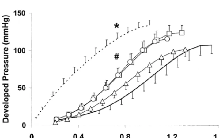
veloped pressures in both untreated and losartan treated hearts relative to control MI hearts (Fig. 2).
3.4. Morphometry and cardiac fibrosis
Infarct size was large in all infarcted animals, with no significant difference among groups. Mid-septal wall thick-ness, an assessment of compensatory hypertrophy, was also similar for all infarct groups. Furthermore, infarction caused an increase in cardiac fibrosis in the non-infarct and infarct zone compared to the sham group, though collagen content in the scar region was lower in the losartan-treated animals. Infarct thickness, however, was only reduced in exercise trained animals relative to control and exercise1 losartan treated animals (Table 2). Therefore post-MI exercise training induced further scar thinning.
Fig. 1. Diastolic pressure–volume relationship for Sham (- - -), MI (—), MI–Ex (&), MI–Los (^) and MI–Ex–Los (N) hearts. MI resulted in ventricular dilation in all animals, shifting all MI groups significantly
rightward of sham animals. Infarcted hearts receiving exercise, losartan, 4. Discussion or exercise plus losartan therapy had reduced cavity dilation and a
leftward shift in the P –V curve relative to infarcted control animals;
We tested in a rodent model of chronic infarction how
*5P ,0.05.
treadmill exercise training affects post-MI remodelling and
whether exercise in conjunction with AT receptor antago-1
nist losartan, attenuates deleterious remodelling. We found
that AT receptor blockade, similarly to ACE inhibition,1
decreased LV dilation post-infarction, and exercise training resulted in a comparable attenuation of ventricular dilation. Combination therapy of both exercise and AII receptor blockade, however, had no additive effect on ventricular dimensions. Furthermore, post-MI exercise increased LV developed pressure over a range of diastolic volumes, as well as resulted in additional scar thinning and pulmonary congestion. Concurrent losartan treatment, however, at-tenuated exercise-induced increases in scar thinning. 4.1. Exercise training
Fig. 2. Developed pressure–volume relationship for Sham (- - -), MI Previous studies of exercise in infarcted rats have (—), MI–Ex (&), MI–Los (^) and MI–Ex–Los (N) hearts. MI generally shown an increase in ventricular enlargement resulted in diminished contractile function in all MI hearts relative to
[8,9,11], with only one study describing a decrease in LV
sham animals (*P ,0.05). Exercise training augmented LV developed
dilation [12]. It was therefore unexpected that treadmill
pressures in untreated and losartan treated hearts relative to infarcted
Table 2
a
Morphometry and fibrosis
Group Infarct Septal Thinnest Fibrosis (%)
(%) thickness region of
(mm) infarct (mm) Septum Infarct zone
Sham N /A 1.6360.08 N /A 2.6760.14 2.760.6 MI 45.863.8 1.7160.15 0.9660.17 3.3960.22 76.062.2**†§ MI–Ex 44.161.6 1.8360.11 0.5960.04†§ 3.1460.35 76.262.6**†§ MI–LOS 50.762.5 1.5960.08 0.8560.08 3.3760.31 66.861.3** MI–Ex–LOS 46.263.0 1.7660.07 0.9660.08 4.1560.29** 61.762.8** a
N /A, not applicable; MI, post-MI controls; MI–Ex, post-MI rats with exercise; MI–LOS, post-MI rats treated with losartan; MI–Ex–LOS, post-MI rats treated with exercise and losartan.
**P ,0.01 vs. sham; †P ,0.05 vs. MI; ‡P ,0.05 vs. MI–Los; §P ,0.05 vs. MI–Ex–Los.
LV dilation. The deviation from previous exercise studies mean radius and wall thickness dimensions, and indeed
may be due to multiple factors, of which mode of exercise found exercise treated hearts to have elevated systolic wall
might be the most important. Previous studies have stresses [33]. The elevated systolic stress in the exercise
generally employed swimming as the exercise regimen. groups did not deleteriously affect ventricular dilation
Swimming is a very different, less quantifiable mode of during the 6-week observation period.
exercise stimulus. It often results in transient hypoxia and
hypothermia, as well as undo stress, all which could 4.2. Losartan
potentially influence experimental results [25]. We
attempt-ed to use an exercise protocol that best simulates current In this study, coronary ligation resulted in large
anterior-cardiovascular exercise rehabilitation for humans post-MI, lateral infarction of |45% of the LV and subsequent
namely, low intensity endurance exercise. Previous studies ventricular dilation. AT1 receptor blockade resulted in a
employing treadmill exercise training in rats post-MI have reduction of LV dilation. While other studies have reported
shown no detrimental effect on infarct size and aneurysm that AT antagonists decreased mortality and fibrosis, as1
expansion [26]. Also, exercise initiated early after infarc- well as increased capillary density in post-MI hearts to a
tion has been suggested to cause a more deleterious effect similar extent as ACE inhibition therapy, this study may be
[8], while onset of training after infarct healing has the first to report a similar attenuation of overall left
occurred, resulted in more favorable remodelling [12]. ventricular remodelling [13,20,21,34]. While these results
Furthermore, the moderate nature of our exercise regimen cannot definitively determine whether an ACE inhibitor or
might have contributed to its final beneficial effect. In- AII antagonist acts more favorable on LV remodelling,
creases in mid-septal fibrosis, wall thickness and heart they, along with others, do suggest that AII antagonists
weight associated with intense exercise, especially swim- may be of similar beneficial value as ACE inhibitors upon
ming exercise [27], were not seen in this study. Our study post-MI hemodynamics and remodelling.
therefore suggests that relative late onset of a moderately The mechanism by which AII-antagonists exert their
intense treadmill exercise regiment is beneficial for overall effects on post-MI remodelling is not well understood. The
post-MI dilation, although it may still cause scar thinning beneficial effects of ACE inhibition on left ventricular
[12]. We speculate that the effects of exercise on LV remodelling have been attributed to an attenuation of tissue
dilation may be influenced by beneficial adaptations of the and circulating levels of angiotensin II and / or inhibition of
peripheral and coronary vasculature, resulting in vasodilat- bradykinin breakdown [18,19,35–38]. Indeed, most of the
ion and a decrease in vascular resistance [28,29]. Further- beneficial effects of ACE inhibitors on post-MI
remodel-more, exercise has also been shown to attenuate catechol- ling can be blocked by inhibition of the kinin system
amine release and increases vagal tone [30–32]. [35–38]. If the kinin pathway contributes substantially to
In this study, exercise also improved contractile function the beneficial effects of ACE inhibition on post-MI
following MI, causing greater developed pressures over a remodelling, it was unclear what effect, if any, selective
range of diastolic volumes, relative to infarcted control AT blockade may have on post-MI remodelling.1
rats. Though elevated contractile function is believed to The possible mechanisms by which losartan influenced
generally be beneficial post-infarction, augmented de- LV remodelling post-MI include a reduction in afterload
veloped pressure, without additional compensatory hy- and preload, as well as, direct cardiac effects. Though we
pertrophy, would suggest that exercise treated hearts did not measure in vivo cardiac pressures in this
in-generated greater systolic wall stress. We previously esti- vestigation, preliminary studies in non-infarcted rats
mated mid-ventricular wall stress according to Laplace’s proved a therapy with losartan (at 10 mg / kg body weight /
Law (wall stress~pressure?chamber radius / wall thickness), day) was sufficient to cause a reduction in systolic
well as smaller doses, of losartan were found to decrease cavity dimensions. Furthermore, exercise training
aug-blood pressure in previous described post-MI rat studies mented contractile function post-MI in both untreated and
[18–20]. Therefore, it appears likely that losartan caused a losartan treated hearts. In addition, exercise therapy, in the
reduction in afterload and that this attenuation of afterload absence of losartan, resulted in the greatest amount of
might beneficially contribute to LV remodelling [18]. pulmonary congestion and scar thinning. AII receptor
Losartan, through induction of natiuresis and diuresis, has blockade therapy in conjunction with exercise therapy,
also been shown to reduce in vivo left ventricular diastolic however, attenuated the increase in pulmonary congestion
pressures, thereby reducing preload in post-MI hearts and and scar thinning. Therefore, the combination of exercise
possibly causing some of the beneficial effects of AII and losartan appears to provide the most beneficial therapy
antagonism [18,19,34]. on post-MI ventricular remodeling. Concurrent losartan
Furthermore, the beneficial effect of losartan might also treatment with exercise would still provide the
neuro-be in part due to direct cardiac effects [18]. AT receptor1 humoral [30–32] and vasculature benefits [28,29] of
antagonists enact their direct cardiac effects via a combina- exercise training, while reducing deleterious elevations in
tion of blockade of the AT1 receptor and an unhindered wall stress.
stimulation of the angiotensin type-2 (AT ) receptor.2 Though we attempted to mimic clinical episodes of
Blockade of the AT receptor results in increased plasma1 infarction and cardiac rehabilitation, including drug and
renin and circulating angiotensin II levels [39], and an moderate exercise therapy, the prognostic and therapeutic
increase in AII will activate AT2 receptors, which are implications of these results need to be examined with
already upregulated post-MI [40]. Liu et al., showed that caution. Nevertheless, our study suggests that even
follow-the processes mediated by AT receptors are important for2 ing a large anterior infarction, moderate endurance training
the beneficial effects of AT receptor blockade and sug-1 does not adversely affect overall remodelling. In addition,
gested that indirect AT -mediated activation of nitric2 blockade of the renin–angiotensin system, an
indispens-oxide, other autocoids, and the kinin system, might be able part of post-MI therapy, in conjunction with exercise
involved [18]. training might make the latter safer and more beneficial
We found a significant reduction in cardiac fibrosis in [7].
the infarcted portion of the LV with losartan treatment in both the exercise and non-exercise groups. As previously
seen with ACE inhibition [41], this effect did not cause Acknowledgements
additional scar thinning, and did not influence the overall
favorable effect on LV remodelling. Septal fibrosis did not This work was supported in part by NIH T32-HL07224
appear to be altered by losartan treatment, and was even (FRE), NIH R01-HL48715 (CSA), grant-in-aid from
slightly elevated in hearts treated with losartan and exer- Merck Pharmaceuticals (CSA), and AHA Medical Student
cise. The differential effect of losartan treatment on Fellowship (MJ).
infarcted and non-infarcted portion of the LV might be related to tissue specific alterations of the
renin–angioten-sin system post-MI. Tissue AII levels and AT -receptor1 References
mRNA expression were reported to be increased to a greater extent in the infarcted than in the non-infarcted
[1] Pfeffer MA, Braunwald E. Remodelling after myocardial infarction:
portion of the LV [40,42]. experimental observations and clinical implications. Circulation
Furthermore, in accordance with previous work [19,38] 1990;81:1161–1172.
but in contradiction to finding by Smits et al. [43] we did [2] Anversa P, Li P, Zhang X, Olivetti G, Capasso J. Ischaemic
myocardial injury and ventricular remodelling. Cardiovasc Res
not find a significant reduction in heart weight with
1993;27:145–157.
losartan treatment. This might be due to differences in drug
[3] Fletcher GF, Balady G, Blair SN et al. Statement of exercise:
dosage and method of application. Smits et al. infused 15 benefits and recommendations for physical activity programs for all
mg / kg / day of losartan subcutaneously, whereas, 10 mg / Americans. A statement for health professionals by the Committee
kg / day was delivered orally in our study. Whether the on exercise and cardiac rehabilitation of the council on clinical
cardiology. American Heart Association. Circulation 1996;94:857–
prevention of cardiac hypertrophy resulted from a more
862.
pronounced hemodynamic change or from a dose
depen-[4] Jugdutt BI, Michorowski BL, Kappagoda CT. Exercise training after
dent anti-trophic effect of the AII antagonist remains anterior Q wave myocardial infarction: importance of regional left
unclear. ventricular function and topography. J Am Coll Cardiol
1988;12:362–372.
[5] Giannuzzi P, Tavazzi L, Temporelli PL et al. EAMI Study Group.
4.3. Combination of exercise and losartan
Long terms physical training and left ventricular remodelling after anterior myocardial infarction: results of the exercise in anterior
In this study exercise and losartan independently at- myocardial infarction (EAMI) trial. J Am Coll Cardiol
tenuated LV dilation post-MI, though the combination of 1993;22:1821–1829.
myocardial remodelling in patients with reduced left ventricular [24] Whittaker P, Kloner RA, Boughner DR, Pickering JG. Quantitative function after myocardial infarction. Circulation 1997;95:2060– assessment of myocardial collagen with picrosirius red staining and
2067. circularly polarized light. Basic Res Cardiol 1994;89:397–410.
[7] Giannuzzi P, Temporelli PL, Corra U, Gattone M, Giordano A, [25] Flaim SF, Minteer WJ, Clark DP, Zelis R. Cardiovascular response Tavazzi L. for the ELVD Study Group. Attenuation of unfavorable to acute aquatic and treadmill exercise in the untrained rat. J Appl remodelling by exercise training in postinfarction patients with left Physiol 1979;46:302–308.
ventricular dysfunction. Circulation 1997;96:1790–1797. [26] Hochman JS, Healy B. Effect of exercise on acute myocardial [8] Gaudron P, Hu K, Schamberger R, Budin M, Walter B, Ertl G. infarction in rats. J Am Coll Cardiol 1986;7:126–132.
Effect of endurance training early or later after coronary artery [27] Musch TI, Moore RL, Leathers DJ, Bruno A, Zelis R. Endurance occlusion on left ventricular remodelling, hemodynamics and sur- training in rats with chronic heart failure induced by myocardial vival in rats with chronic transmural myocardial infarction. Circula- infarction. Circulation 1986;74:431–441.
tion 1994;89:402–412. [28] Niebauer J, Cooke JP. Cardiovascular effects of exercise: role of [9] Oh BH, Ono S, Rockman HA, Ross Jr. J. Myocardial hypertrophy in endothelial shear stress. J Am Coll Cardiol 1996;28:1652–1660.
the ischemic zone induced by exercise in rats after coronary [29] Froelicher VF, Jensen D, Genter F et al. A randomized trial of reperfusion. Circulation 1993;87:598–607. exercise training in patients with coronary artery disease. J Am Med [10] Kloner RA, Kloner JA. The effect of early exercise on myocardial Assoc 1984;252:1291–1297.
infarct scar formation. Am Heart J 1983;106:1009–1013. [30] Cooksey JD, Reilly P, Brown S, Bomze H, Cryer PL. Exercise [11] Hammerman H, Schoen FJ, Kloner RA. Short-term exercise has a training and plasma catecholamines in patients with ischemic heart
prolonged effect on scar formation after experimental acute myocar- disease. Am J Cardiol 1978;42:372–376.
dial infarction. J Am Coll Cardiol 1983;2:979–982. [31] Hull Jr. SS, Vanoli E, Adamson PB et al. Exercise training confers [12] Orenstein TL, Parker TG, Butany JW et al. Favorable left ventricular anticipatory protection from sudden death during acute myocardial
remodelling following large myocardial infarction by exercise ischemia. Circulation 1994;89:548–552.
training. J Clin Invest 1995;96:858–866. [32] Goldsmitt RL, Bigger Jr. JT, Steiman RC, Fleiss JL. Comparison of [13] Pfeffer JM, Pfeffer MA, Braunwald E. Influence of chronic captopril 24-h parasympathetic activity in endurance-trained and untrained
therapy on the infarcted left ventricle of the rat. Circ Res young men. J Am Coll Cardiol 1992;20:552–558.
1985;57:84–95. [33] Jain M, Apstein CA, Libonati JR, Ngoy S, Whittaker P, Eberli FR. [14] Litwin SE, Katz SE, Weinberg EO, Lorell BH, Aurigemma GP, Effects of Losartan and exercise on post-MI remodelling and
Douglas PS. Serial echocardiographic-doppler assessment of left contractility. Circulation 1997;96(Suppl):1924, Abstract.
ventricular geometry and function in rats with pressure-overload [34] Sladek T, Sladkova J, Kolar F. The effect of AT1 receptor hypertrophy. Circulation 1995;91:2642–2654. anatagonist on chronic cardiac response to coronary artery ligation [15] Pfeffer MA, Braunwald E, Moye LA et al. Effect of captopril on in rats. Cardiovasc Res 1996;31:568–576.
mortality and morbidity in patients with left ventricular dysfunction [35] Mortorana PA, Kettenback B, Brelpohl G, Linz W, Scholkens BA. after myocardial infarction. Results of the survival and ventricular Reduction of infarct size by local angiotension coverting enzyme enlargement trial. New Engl J Med 1992;327:669–677. inhibition is abolished by bradykinin antagonist. Eur J Pharmacol [16] Consensus Trial Study Group. Effects of enalapril on mortality in 1990;182:395–396.
severe congestive heart failure: results of the cooperative North [36] Linz W, Scholkens BA. Role of bradykinin in the cardiac effects of Scandinavian enalapril survival study (Consensus). New Engl J Med angiotensin converting enzyme inhibitors. J Cardiovasc Pharmacol
1987;316:1429–1435. 1992;20(Suppl 9):S83–S90.
[17] Acute Infarction Ramipril Efficacy (AIRE) study investigators. [37] Hartmann JC, Wall TM, Hullinger TG, Shebuski RJ. Reduction of Effect of ramipril on mortality and morbidity of survivors of acute myocardial infarct size in rabbits by ramiprilat: reversal by the myocardial infarction with clinical evidence of heart failure. Lancet bradykinin antagonist HOE 140. J Cardiovasc Pharmacol
1993;342:821–828. 1993;21:996–1003.
[18] Liu YE, Yang XP, Sharov VG et al. Effects of angiotensin-coverting [38] Stauss HM, Zhu YC, Redlich T et al. Angiotensin-converting enzyme inhibitors and angiotensin II type 1 receptor antagonists in enzyme inhibition in infarct-induced heart failure in rats: bradykinin rats with heart failure. J Clin Invest 1997;99:1926–1935. versus angiotensin II. J Cardiovasc Risk 1994;1:255–262. [19] Raya TE, Fonken SJ, Lee RW et al. Hemodynamic effects of direct [39] Campbell DJ, Kladis A, Valentijn AJ. Effects of losartan on
angiotensin II blockade compared to converting enzyme inhibition angiotensin and bradykinin peptides and angiotensin-converting in rat model of heart failure. Am J Hypertens 1991;4:334S–340S. enzyme. J Cardiovasc Pharmacol 1995;26:233–240.
[20] Schieffer B, Wirger A, Meybrunn M et al. Comparative effects of [40] Nio Y, Matsubara H, Murasawa S, Kanasaki M, Inada M. Regulation chronic angiotensin-converting enzyme inhibition and angiotensin II of gene transcription of angiotensin II receptor subtypes in myocar-type 1 receptor blockade on cardiac remodelling after myocardial dial infarction. J Clin Invest 1995;95:46–54.
infarction in the rat. Circulation 1994;89:2273–2282. [41] Jugdutt BI, Lucas A, Khan MI. Effect of angiotensin-converting [21] Pitt B, Segal R, Martinez FA et al. Randomised trial of losartan enzyme inhibition on infarct collagen deposition and remodelling versus captopril in patients over 65 with heart failure (Evaluation of during healing after transmural canine myocardial infarction. Can J Losartan in the Elderly Study (ELITE). Lancet 1997;349:747–752. Cardiol 1997;13:657–668.
[22] Eberli FR, Weinberg EO, Grice WM, Horowitz G, Apstein CS. [42] Yamagishi H, Kim S, Niskikimi T, Takeuchi K, Takeda T. Contribu-Protective effect of increased glycolytic substrate against systolic tion of cardiac renin–angiotensin system to ventricular remodelling and diastolic dysfunction and increased coronary resistance from in myocardial-infarcted rats. J Moll Cell Cardiol 1993;25:1369– prolonged global underperfusion and reperfusion in isolated rabbit 1380.
hearts perfused with erythrocyte suspensions. Circ Res [43] Smits JF, van Krimpen C, Schoemaker RG, Cleutjens JP, Daemen 1991;68:466–481. MJ. Angiotensin II receptor blockade after myocardial infarction in [23] Fletcher PJ, Pfeffer JM, Pfeffer MA, Braunwald E. Left ventricular rats: effects on hemodynamics, myocardial DNA synthesis, and diastolic pressure–volume relations in rats with healed myocardial interstitial collagen content. J Cardiovasc Pharmacol 1992;20:772– infarctions. Effects on systolic function. Circ Res 1981;49:618–626. 778.