ORIGINAL ARTICLE
Bacterial expression of mutant argininosuccinate lyase
reveals imperfect correlation of
in-vitro enzyme activity
with clinical phenotype in argininosuccinic aciduria
Katharina Engel&Jean-Marc Vuissoz&Sandra Eggimann&Murielle Groux&Christoph Berning&Liyan Hu&Vera Klaus&Dorothea Moeslinger&
Saadet Mercimek-Mahmutoglu&Sylvia Stöckler&Bendicht Wermuth&
Johannes Häberle&Jean-Marc Nuoffer
Received: 17 December 2010 / Revised: 20 May 2011 / Accepted: 25 May 2011 / Published online: 11 June 2011 # SSIEM and Springer 2011
Abstract
Background The urea cycle defect argininosuccinate lyase (ASL) deficiency has a large spectrum of presentations from highly severe to asymptomatic. Enzyme activity assays in red blood cells or fibroblasts, although diagnostic of the deficiency, fail to discrimi-nate between severe, mild or asymptomatic cases. Mutation/phenotype correlation studies are needed to characterize the effects of individual mutations on the activity of the enzyme.
Methods Bacterial in-vitro expression studies allowed the enzyme analysis of purified mutant ASL proteins p.I100T (c.299 T>C), p.V178M (c.532 G>A), p.E189G (c.566A>
G), p.Q286R (c.857A>G), p.K315E (c.943A>G), p.R379C (c.1135 C>T) and p.R385C (c.1153 C>T) in comparison to the wildtype protein.
Results In the bacterial in-vitro expression system, ASL wild-type protein was successfully expressed. The known classical p.Q286R, the novel classical p.K315E and the known mutations p.I100T, p.E189G and p.R385C, which all have been linked to a mild phenotype, showed no significant residual activity. There was some enzyme activity detected with the p.V178M (5 % of wild-type) and p.R379C (10 % of wild-type) mutations in which Km
values for argininosuccinic acid differed significantly from the wild-type ASL protein.
Communicated by: Matthias Baumgartner Competing interest: None declared.
K. Engel
:
C. Berning:
V. Klaus:
J. HäberleKlinik und Poliklinik für Kinder- und Jugendmedizin, Universitätsklinikum Münster,
Albert-Schweitzer-Str. 33, 48149 Muenster, Germany J.-M. Vuissoz
Clinique de Pédiatrie, Hôpital cantonal de Fribourg, 1700 Fribourg, Switzerland
S. Eggimann
:
M. Groux:
B. Wermuth:
J.-M. Nuoffer (*)Universitätsinstitut für Klinische Chemie Universität Bern, Freiburgerstr. 15,
3010 Bern, Switzerland
e-mail: [email protected] D. Moeslinger
Department of Pediatrics, Medical University of Vienna, Vienna, Austria
S. Mercimek-Mahmutoglu
:
S. StöcklerDepartment of Paediatrics, Division of Biochemical Diseases, University of British Columbia,
4480 Oak Street,
Vancouver, BC, Canada V6H 3V4
L. Hu
:
J. HäberleDivision of Metabolism, Kinderspital Zürich, Steinwiesstr. 75,
8032 Zurich, Switzerland
L. Hu
:
J. HäberleChildren’s Research Center, Steinwiesstr. 75,
8032 Zurich, Switzerland
J.-M. Vuissoz
:
J.-M. NuofferUniversitäts Kinderspital Bern, Freiburgerstr. 15,
Conclusion The bacterially expressed enzymes proved that the mutations found in patients and studied here indeed are detrimental. However, as in the case of red cell ASL activity assays, some mutations found in genetically homozygous patients with mild presentations resulted in virtual loss of enzyme activity in the bacterial system, suggesting a more protective environment for the mutant enzyme in the liver than in the heterologous expression system and/or in the highly dilute assays utilized here.
Introduction
Argininosuccinate lyase (ASL, EC 4.3.2.1, MIM *608310) catalyzes the cleavage of argininosuccinate to fumarate and arginine which is further metabolized to ornithine and urea in the urea cycle. Additionally, it is involved in the synthesis of arginine as substrate of the nitric oxide (NO) synthase to yield citrulline and NO. ASL was first purified and characterized from human liver (Palekar and Mantagos
1981), is expressed highly in liver and kidney and at much lower levels in many other tissues and cells including red blood cells and fibroblasts (O'Brien and Barr1981).
ASL deficiency, also named argininosuccinic aciduria (ASLD, MIM #207900), is inherited in an autosomal-recessive trait (Brusilow and Horwich 2001). This is the second most common urea cycle disorder with an incidence of approximately 1 in 70,000 live births (Levy et al.1984). The phenotype is highly variable ranging from acute neonatal-onset with severe hyperammonemic encephalopa-thy, often causing severe mental retardation in surviving patients to subacute and late-onset forms with less severe symptoms such as mild mental retardation and learning disabilities (Kleijer et al.2002). Progressive liver disease is one of the long-term complications which is poorly understood (Zimmermann et al.1986).
ASL deficiency is diagnosed by hyperammonemia and increased levels of argininosuccinic acid and its anhydrides in blood and urine. The diagnosis can be confirmed by genetic means both on the RNA and DNA level. So far more than 40 known disease-causing mutations have been reported (Al-Sayed et al. 2005; Barbosa et al. 1991a; Christodoulou et al.2006; Kleijer et al.2002; Linnebank et al. 2000, 2002; Mercimek-Mahmutoglu et al. 2010; Sampaleanu et al.2001; Tanaka et al. 2002; Trevisson et al.2007; Walker et al.1990).
As an alternative to mutation analysis or in cases with no detected mutations in the ASL gene, ASL activity can be determined in erythrocytes and skin fibroblasts by two types of assay. In the direct assay, the immediate reaction products arginine or fumarate, respectively, are determined in-vitro (Nuzum and Snodgrass1976). In the indirect assay, ASL activity is estimated from the amount of radioactive
citrulline incorporated into acid precipitable proteins in cultured fibroblasts (Kleijer et al.2002). In previous studies with patients presenting mild or asymptomatic ASLD, red cell and fibroblast activity assays tended to reveal higher activity decreases than citrulline incorporation assays in fibroblasts, which in some cases gave even normal results (Ficicioglu et al. 2009; Kleijer et al. 2002). This discrep-ancy may, at least in part, be due to an overestimation of ASL activity in the indirect assay (Kleijer et al.2002) and/ or to a limited accuracy of the direct enzyme assay due to the low ASL activity in fibroblasts.
Previous studies showed that recombinant ASL amena-ble to kinetic and structural analysis may be obtained in high yield in a bacterial expression system (Sampaleanu et al. 2001). In order to better understand the clinical heterogeneity, we expressed mutant proteins from patients with variable severity of ASLD in E. coli and determined the enzymatic activity of ASL in-vitro.
Material and methods Patients
Information was collected from 28 homozygous patients of Turkish, German, Italian, Austrian and Finish origin. This study focuses on the seven homozygous genotypes found in this cohort of which six were already reported (Barbosa et al.1991a; Linnebank et al.2002; Mercimek-Mahmutoglu et al.2010; Walker et al.1997) and one novel mutation. Two mutations were associated with neonatal-onset and five with late-onset or asymptomatic ASLD. Among the patients with neonatal-onset, two carried the known mutation c.857A>G (p.Q286R) (Walker et al. 1990). In another patient who was detected by newborn screening the novel change c.943A>G (p.K315E) was found in a homozygous state. This patient was consequently treated from the second week of life and never experienced a hyperammonemic crisis but still showed a poor neurological outcome with severe psychomotor retardation.
In the group of patients with late-onset ASLD, 12 patients carried the mutation c.1153 C > T (p.R385C) and had mild to moderate symptoms (Keskinen et al. 2008; Kleijer et al. 2002). Similarly, four patients with the c.299 T> C (p.I100T) mutation with late-onset ASLD had only a learning disability (Keskinen et al. 2008). In two more patients with late-onset ASLD, c.1135 C > T (p. R379C) and c.532 G > A (p.V178M) mutations were identified. In addition, the latter mutation and the known mutation c.566A > G (p.E189G) were found by newborn screening in three and two children, respectively, who have remained asymptomatic so far (Mercimek-Mahmutoglu et al.
The study was approved by the Ethics Committee of the University of Münster. All parents signed a written informed consent form prior to mutation analysis. All mutant alleles detected in patients from this study were identified in parents confirming their carrier status. Mutation analysis
Genomic DNA was purified from whole blood using the QIAamp DNA blood kit (Qiagen, Hilden, Germany). For mutation screening, exon-wise PCR amplification was carried out as described previously (Linnebank et al.2002). Construction of wildtype and mutant expression plasmids ASL wild-type cDNA was prepared from control cultured fibroblasts (RNeasy Mini Kit, Invitrogen, Groningen, The Netherlands). Amplification of the ASL full-length cDNA as initial template was performed using oligonucleotides 5 ′-ATGTGGATCCCCTCGGAGGTGAGTGGGACC (forward) and 5′- ACGTGGTACCCTAGGCCTGCTGTGCCTGCAG (reverse) which carried a BamHI and KpnI restriction site. After denaturation at 94 °C, amplification with PWO-polymerase was accomplished at an annealing temperature of 65 °C for 35 cycles. The amplified fragments were purified and subcloned into TA cloning vector pCR2.1 (Invitrogen). Plasmids were control-sequenced using the ABI Prism 3700 DNA sequencer (Applied Biosystems, Foster City, CA), digested by BamHI and KpnI and ligated into the expression vector pQE30 (Qiagen). This plasmid introduces the 6-His containing tag at the N-terminus of the polypeptide (sequence then reads Met-Arg-Gly-Ser-6xHis-ASL protein). The pQE30 plasmid containing wildtype Met-Arg-Gly-Ser-6xHis-ASL was used for in-vitro site-directed mutagenesis (GeneTailor Site-Directed Mutagenesis System, Invitrogen) following the manufacturer’s instructions. Oligonucleotide primers
carry-ing the desired mutations are indicated in Table1. Mutagen-esis was performed at an annealing temperature of 55 °C for 29 cycles using Platinum Taq High Fidelity DNA Polymer-ase (Invitrogen). Mutant plasmids grown in and isolated from DH5α™-T1® E. coli (Invitrogen) were control sequenced.
Expression of human wildtype and mutant ASL in E. coli Purified and sequence-controlled plasmids were trans-formed into E. coli host strain M15 [pREP4] (Qiagen). Positive clones of each mutant and wild-type ASL were grown in LB media containing 50 μg/ml ampicillin and 25 μg/ml kanamycin overnight at 37 °C. 5 ml of this overnight culture were inoculated in 100 ml fresh LB medium containing identical concentrations of antibiotics. Expression of recombinant protein was induced by adding isopropyl thiogalactoside (IPTG) to a final concentration of 1 mM when OD600 of the culture reached 0.5 to 0.8 and
temperature was lowered to 25 °C. On the following morning, cells were harvested by centrifugation and the pellets suspended in each 3 ml lysis buffer (300 mM NaCl, 200 mM NaH2PO4, 5 mM imidazol, pH 8) and lysed by 10
cycles sonification 10 sec (Kontes Micro-Ultrasonic Cell Disruptor, Kontes, Vineland, N.J., U.S.A.). Cell lysates were then centrifuged at 4 °C and 11.000 rpm for 30 min and the supernatant used for purification.
Purification of recombinant wild-type and mutant ASL proteins
Expressed wildtype and mutant ASL proteins were purified under native conditions using nickel agarose affinity columns according to the manufacturer’s instructions (The QIAexpressionist, Qiagen). In brief, polypropylene col-umns were prepared containing each 1.5 ml nickel agarose
Table 1 Details of the mutations investigated in this study, clinical course of disease and results of enzymatic analysis of mutated recombinant proteins
Nucleotide level WT c.299 T>C c.532 G>A c.566 A>G c.857 A>G c.943 A>G c.1135 C>T c.1153 C>T
Protein level p.I100T p.V178M p.E189G p.Q286R p.K315E p.R379C p.R385C
Exon 4 7 7 11 12 14 15
Clinical course mild mild mild severe severe mild mild
ASL activitya 4227 20 208 41 23 5 280 3
Vmaxb 12112 n.d. 254 n.d. n.d. n.d. 488 n.d.
Km(mM)b 2.6 n.d. 0.2 n.d. n.d. n.d. 0.23 n.d.
a
ASL activity (mU/mg protein) measured under standard conditions using 13.6 mM argininosuccinate: activities are mean values of four and two independent experiments for the WT and mutant proteins, respectively, each measured in triplicate.
b
Vmax(mU/mg protein) and Kmwas only measured in proteins with residual activities above 5%. Vmaxand Kmwere extrapolated from the linear
part of the double-reciprocal plots of two experiments. n.d.: not determined
and loaded with about 3 ml supernatant. Columns were then gently mixed on ice using a rotary shaker for 90 min. Then, each column was washed five times using each 4 ml washing buffer (300 mM NaCl, 200 mM NaH2PO4, 10 mM
imidazole, pH 8). Then, recombinant proteins were eluted in fractions using four times 1 ml elution buffer (50 mM NaPi, 300 mM NaCl, 250 mM imidazol, no protease inhibitors, pH7). Purified proteins were immediately frozen in 50% glycerol at−80 °C. Ten %-SDS-PAGE analysis was carried out using 15μl of eluate to estimate the purity of the proteins. In order to analyse the tetramer stability of the ASL proteins we performed an 11.5% native PAGE. For the silver staining 15 μl of recombinant proteins, for the Western blot 0.5 μl of recombinant proteins and 8 μg human liver-protein were loaded. As marker we used the NativeMark™ Unstained Protein Standard from Invitrogen. For immunoblotting, the membrane was blocked over night (4°C) with 5% non-fat dried milk in T-TBS (T-TBS: 20 mM TRIS, 137 mM NaCl, 0.1% Tween-20, pH 8.3) and then incubated for 2 h at room temperature with rabbit antiserum ASL, diluted 1:1000 in 1% non fat dried milk in T-TBS. The membrane was then washed thrice in T-TBS and incubated in a 1:5000 dilution of peroxidase conjugate anti-rabbit IgG (Amersham) in T-TBS (1 % non fat dried milk) for 1 h at room temperature. Blots were washed finally in T-TBS and bound ASL antibodies visualized onto Amersham Hyperfilm ECL by chemiluminescence (ECL kit, Amersham) with different exposure times. The film was scanned on a conventional scanner.
The concentration of purified proteins was determined with the Bio-Rad DC Protein Assay (Bio-Rad Nr 500– 0116) using BSA (Sigma-Aldrich, Buchs, Switzerland) as standard. Protein concentrations were corrected with the blank (protein elution puffer and 50 % glycerol).
Determination of enzyme activity
ASL activity of recombinant proteins was determined spectrophotometrically using a coupled assay with arginase and measuring urea production as described (Ceriotti and Spandrio1963). The reaction was initiated by adding 50μl ASL protein solution (8–16 μg enzyme) to 100 μl argininosuccinate (13.6 mM final concentration) in water and 100 μl arginase (50 units) (both Sigma-Aldrich) in 66.7 mM Na-phosphate (at pH 7.5) and stopped after 30 min at 37 °C by the addition of perchloric acid 2% final concentration. Enzyme activities of wild-type and mutant proteins are expressed as mU/mg protein. Vmax and Km,
were determined by extrapolation from double-reciprocal plots of 1/v versus 1/[argininosuccinate] at argininosucci-nate concentrations of 0.544 mM, 0.272 mM, 0.181 mM, 0.136 mM and 0.068 mM, respectively. All assays were carried out in triplicate.
Structural analysis of the ASL protein
Structural analysis was performed on the basis of the crystal structure of the ASL mutant Q286R obtained from the Protein Data Bank PDB Entry 1 K62 (http:// www.rcsb.org/pdb/). The reconstitution of the ASL tet-ramers was carried out in CHIMERA (Pettersen et al.
2004) using the data of the PDB file. The location of one argininosuccinate substrate molecule was superimposed to the model based on its known location in the highly homologous duck delta-2-crystallin (PDB Entry 1DCN). The Pymol software with the hollow utility was used to produce the pictures (The PyMOL Molecular Graphics System Version 1.3, Schrödinger, LLC).
Results
Expression and enzymatic characterization of recombinant ASL proteins
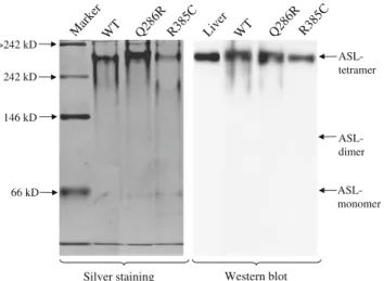
Recombinant wild-type and all seven different mutant ASL proteins were obtained with a purity of >90% as judged by SDS-PAGE (Fig. 1). Silver staining and western blot of native polyacrylamide gel of the wild-type and mutant ASL proteins Q286R and R385C illustrates the purity of the investigated proteins and shows that the vast majority of investigated ASL proteins are present in the tetrameric form (Fig. 2).
Activity of recombinant wildtype ASL protein from four independent experiments was 4227±321 mU/ mg protein (mean±SD; triplicate measurements) under standard assay conditions (13.6 mM argininosuccinate). At tenfold higher substrate concentration, activity was decreased by 18% indicating substrate inhibition. Further measurements at varying substrate concentrations (range 0.068–13.6 mM) yielded nonlinear double-reciprocal plots of 1/v versus 1/ [argininosuccinate] with marked deviation from linearity already at standard assay conditions. Vmax and Km,
extrapolated from the linear part of the double-reciprocal plots of two experiments were 12112 mU / mg protein and 2.6 mM, respectively (Table1).
Recombinant proteins carrying the mutations p.Q286C and p.K315E which were associated with severe ASL deficiency and the mild mutations p.I100T, p.E189G and p. R385C exhibited less than 1% of the wildtype activity under standard assay conditions, respectively, and were not subjected to further kinetic analysis. There was also no activity detectable with reducing the argininosuccinate concentration to 7.8 mM.
In contrast, recombinant proteins carrying the mild mutations p.V178M and p.R379C yielded residual activi-ties of 4.9% and 6.6% under standard assay conditions,
respectively. Both wildtype and mutant proteins p.V178M and p.R379C were inhibited by higher substrate concen-trations. Vmaxand Kmvalues of the mutant enzymes were
between one and two orders of magnitude lower than the corresponding values of the wildtype enzyme (Table1). Localization of mutations in the homotetrameric protein Localization of the mutated amino acids in the quaternary structure of the enzyme revealed two distinct distribution patterns. Amino acids I100, Q286, R379 and R385 are in close vicinity to the active site and/or the argininosuccinate binding pocket, whereas amino acids V178, E189 and K315 are all at the contact surface between different monomers of the tetramer (Fig.3).
Discussion
ASLD exhibits a highly variable phenotypic pattern ranging from life-threatening neonatal onset to mild developmental delay and learning disabilities to asymptomatic phenotype. Unfortunately, with the exception of the severe neonatal-onset cases which generally have a poor prognosis, long-term outcome is difficult to predict. Widhalm et al. followed 12 patients with biochemically confirmed ASLD identified by newborn screening. All patients were treated with arginine supplementation and, some with protein restricted diet (Widhalm et al. 1992). All patients showed normal intellectual and psychomotor development, however the degree of residual ASL activity was not reported. More recently, Shih and coworkers followed another 13 patients with ASLD identified by newborn screening and confirmed by enzymatic analysis in red blood cell hemolysates and/or fibroblast extracts (Ficicioglu et al. 2009). In their long-term follow-up study, the outcome of patients with neonatal-onset and late-onset disease identified by selective screening was compared: patients identified by newborn screening had higher enzyme activities as measured by the
14
C-citrulline incorporation assay and had a better long-term outcome than patients identified by selective
screen-ing. In fact, with one exception14C-citrulline incorporation in the newborn screening group was not different from a control group. In contrast, red blood cells and fibroblasts of patients from both the newborn screening group and the late-onset group exhibited markedly decreased enzyme activities ranging from not detectable to 10–20% of controls by the direct enzyme assay method. Similar discrepancies between the two assays had previously been observed by Kleijer et al. in five patients with late-onset disease (Kleijer et al.2002). These authors also showed that the relative residual activity in the citrulline incorporation assay decreased with increasing citrulline concentration, probably because ASL activity is not the rate-limiting step in the citrulline incorporation assay. Nevertheless, the discrepancy between the two assays remained even at near saturating citrulline concentrations.
There is a known broad genetic heterogeneity at the ASL locus, which might determine diversity in residual enzyme function. The activity of mutant ASL proteins has been assessed in human fibroblasts (Kleijer et al. 2002), eukaryotic COS-1 cells (Howell et al. 1998; Walker et al.
1997), erythrocytes (Bastone et al. 1990; Trevisson et al.
2007) and rat hepatocytes (Bastone et al. 1990) but bacterial in-vitro expression has only been performed for a few mutations (Sampaleanu et al.2001; Yu et al.2001).
The aim of our study was to test whether varying levels of residual enzyme activity contribute to the severity of the clinical course of ASLD. It should be noted that we investigated recombinant proteins that comprised, in addi-tion to the wildtype or mutant ASL sequence, another three amino acids (Arg-Gly-Ser) as well as the 6xHis-tag. However, the relatively high activity in-vitro, albeit some-what lower than previously reported in human liver
Fig. 1 SDS-PAGE analysis of 15μl of in E. coli expressed wild-type
and mutant ASL proteins investigated in the present study illustrating a level of purity of>90%. kDa: Kilo-Daltons; WT: wild-type
WT Mar ker Q28 6R R385 C 66 kD 146 kD >242 kD 242 kD ASL- tetramer ASL- monomer ASL- dimer Silver staining Live r WT Q28 6R R38 5C Western blot
Fig. 2 Native polyacrylamide gel electrophoresis of the wild-type and mutant ASL proteins Q286R and R385C. The silver staining illustrates the purity of the investigated proteins and the western blot confirm that the vast majority of recombinant proteins are present in the tetrameric form
(Palekar and Mantagos 1981) or recombinantly expressed in bacteria (Yu et al.2001), suggests that the added amino acid sequence to N-terminus does not interfere with the protein folding and formation of the intact ASL homote-tramer. In contrast to previous literature (Palekar and Mantagos 1981; Yu et al. 2001) we found a non-hyperbolic dependency of the ASL activity on the [argini-nosuccinate] and 20-fold higher Km of the wild-type
protein. Native PAGE of the wild-type and mutant ASL proteins Q286R and R385C shows that the vast majority of these recombinant proteins are present in the tetrameric form and excludes cold-dissociation as explanation. The discrepancies of absolute activity levels in our study and those reported in the literature may be due to different
activity assay used; e.g., we did not continuously monitor fumarate in our assay.
The mutant p.Q286R was used as negative control because it has been identified before in neonatal-onset patients (Barbosa et al. 1991b; Trevisson et al. 2007) and demonstrated only minimal residual ASL activity in trans-fected COS-1 cells (Walker et al. 1997) and only 3.0% residual activity in E. coli (Yu et al.2001). In our assay, this mutation did not exhibit any residual activity. Likewise, the novel mutation c.943A > G (p.K315E), identified in a Turkish patient detected by newborn screening who showed severe retardation despite absence of hyperammonemic episodes, yielded no relevant residual ASL activity.
In a pair of siblings, who were identified by newborn screening and remained asymptomatic after initiation of treatment prior to onset of symptoms, the mutation c.566A> G (p.E189G) was found in a homozygous state (Mercimek-Mahmutoglu et al. 2010). Since no other cases with this mutation are known to date, the natural course of this specific mutation remains unknown. As in p.K315E muta-tion, there was only irrelevant low residual activity in the bacterial in-vitro expression. Thus, the cause of the signif-icantly better outcome in comparison to the one patient affected by mutant p.K315E remains obscure.
Another four mutations investigated in this study (p. I100T, p.V178M, p.R379C, p.R385C) have been described in relation to mild phenotype of ASLD (Linnebank et al.
2002). Other than in the direct assay (0%), enzymatic activities of 7-20% were identified by the indirect assay in fibroblasts from patients who were homozygous for p. R385C mutation (Kleijer et al. 2002). However, the bacterial in-vitro expression of p.R385C mutation yielded no relevant residual activity. Similarly, p.I100T mutation did not result in relevant residual activity but there are no data from enzyme measurements in other cells for comparison.
A striking difference was found for protein variants p. V178M and p.R379C which were both identified in patients with a mild phenotype (Kleijer et al. 2002). For mutant p.V178M, residual ASL activity in fibroblasts in a direct assay and in an indirect assay yielded relevant residual activity (<10% and 8%, respectively) (Kleijer et al. 2002). In accordance with these findings, mutant p. V178M showed a similar level of residual activity (5% of wildtype) in the bacterial in-vitro expression. Similarly, the residual enzyme activity of mutant p.R379C measured in the bacterial in-vitro expression system was 10% of wild-type activity which is only slightly lower than the ASL activity in fibroblasts as measured by the indirect assay (15-28%; (Kleijer et al.2002)).
Measurement of ASL activity in cultured skin fibroblasts has served as diagnostic means in patients with ASLD. The indirect enzyme assay has been proven to be superior to the
Fig. 3 View on the ASL-homotetramer with one argininosuccinate depicted in its binding-cavity. Two of the oligomer are drawn in ribbon view (orange and mauve) one in surface (green) and one in ribbon and transparent surface (blue). Amino acids of interest in this study are shown in red. The center of the figure show an overview of the homotetramer, on two different point of view ( rotation of about 90 degree on an vertical axis). (AS=argininosuccinate)
direct assay (Ficicioglu et al. 2009; Kleijer et al. 2002). However, ASL activity in late-onset patients has been shown to be as low as in neonatal-onset patients indicating that the level of residual activity in fibroblasts has only limited prognostic value (McInnes et al.1984). In contrast, increased levels of residual activity were only found in patients with late-onset ASLD. In line with this, residual activity levels of the mutant proteins p.V178M and p. R379C in the bacterial in-vitro expression system are consistent with the data obtained from cultured fibroblasts. However, other mutant proteins associated with a mild phenotype were not distinguishable from severely defective proteins. Thus, the expression system presented here does not reliably predict a mild phenotype of ASLD.
Part of the variability of the clinical courses in ASLD might be explained by tissue specific expression of the ASL protein as suggested by the finding of substantial ASL activity in brain tissues of neonatal-onset patients with loss of activity in liver (Glick et al.1976; Perry et al.1980).
Besides tissue specific expression, DNA methylation has been shown to influence the level of ASL gene expression. This mechanism seems to be of particular importance for the different levels of ASL protein activity and ASL mRNA synthesis in adult and fetal rat liver (Renouf et al.1998). However, the role of this developmental regulation mech-anism in tissue specific expression in the mature human organism is as yet unclear.
The localization of the mutated amino acids within the ASL homotetramer allows us to speculate on their functional consequences. Mutants located near the argini-nosuccinate binding pocket are likely to affect ASL enzyme activity to varying degrees. Likewise, mutants that affect the formation of the monomer surface may lead to heterodimer instability ultimately affecting homotetramer stability. Thus, the mutant proteins investigated in this study may lead to an impairment of ASL function by different mechanisms.
It is essential to make a note of the fact that our experiments only reflect patients who are homozygous for a certain mutation. In cases of compound heterozygosity extensive intragenic complementation is known to contrib-ute to different degrees of ASLD severity (Howell et al.
1998; McInnes et al. 1984). The effect of this mechanism varies with each set of mutations.
In summary, the bacterial in-vitro expression of ASL mutants is feasible and might detect variants with only partial impairment of ASL activity. However, the sensitivity of the enzyme analysis of mutant proteins derived from this artificial system does not allow for a reliable prediction of all ASL mutants associated with mild ASLD. Insofar, there is still a lack of an unfailing prognostic factor for patients with ASLD and the basis of their clinical heterogeneity remains to be fully elucidated.
Acknowledgments The technical assistance of Ilka Neumann was a
great help for cloning and expression experiments. The authors also thank the referring physicians from many institutions for sending
patient’s samples, in particular Dr. F. Trefz from Reutlingen, Germany.
This study was in part supported by the Swiss National Science Foundation (Grant 310030_127184/1 to J.H.).
References
Al-Sayed M, Alahmed S, Alsmadi O, Khalil H, Rashed MS, Imtiaz F, Meyer BF (2005) Identification of a common novel mutation in Saudi patients with argininosuccinic aciduria. J Inherit Metab Dis
28:877–883
Barbosa P, Cialkowski M, O'Brien WE (1991a) Analysis of naturally occurring and site-directed mutations in the argininosuccinate
lyase gene. J Biol Chem 266:5286–5290
Barbosa P, Wistow GJ, Cialkowski M, Piatigorsky J, O'Brien WE (1991b) Expression of duck lens delta-crystallin cDNAs in yeast and bacterial hosts. Delta 2-crystallin is an active argininosucci-nate lyase. J Biol Chem 266:22319–22322
Bastone A, Diomede L, Parini R, Carnevale F, Salmona M (1990) Determination of argininosuccinate lyase and arginase activ-ities with an amino acid analyzer. Anal Biochem 191:384– 389
Brusilow S, Horwich A (2001) Urea cycle enzymes. In: Scriver C, Beaudet A, Sly W, Valle D (eds) The metabolic & molecular bases of inherited disease, 8th edn. McGraw-Hill, New York, pp
1909–1963
Ceriotti G, Spandrio L (1963) A spectrophotometric method for
determination of urea. Clin Chim Acta 8:295–299
Christodoulou J, Craig HJ, Walker DC, Weaving LS, Pearson CE, McInnes RR (2006) Deletion hotspot in the argininosuccinate lyase gene: association with topoisomerase II and DNA
poly-merase alpha sites. Hum Mutat 27:1065–1071
Ficicioglu C, Mandell R, Shih VE (2009) Argininosuccinate lyase deficiency: longterm outcome of 13 patients detected by newborn
screening. Mol Genet Metab 98:273–277
Glick NR, Snodgrass PJ, Schafer IA (1976) Neonatal argininosuccinic aciduria with normal brain and kidney but absent liver arginino-succinate lyase activity. Am J Hum Genet 28:22–30
Howell PL, Turner MA, Christodoulou J, Walker DC, Craig HJ, Simard LR, Ploder L, McInnes RR (1998) Intragenic comple-mentation at the argininosuccinate lyase locus: reconstruction of
the active site. J Inherit Metab Dis 21(Suppl 1):72–85
Keskinen P, Siitonen A, Salo M (2008) Hereditary urea cycle diseases
in Finland. Acta Paediatr 97:1412–1419
Kleijer WJ, Garritsen VH, Linnebank M, Mooyer P, Huijmans JG, Mustonen A, Simola KO, Arslan-Kirchner M, Battini R, Briones P, Cardo E, Mandel H, Tschiedel E, Wanders RJ, Koch HG (2002) Clinical, enzymatic, and molecular genetic characterization of a biochemical variant type of argininosuccinic aciduria: prenatal and postnatal diagnosis in five unrelated families. J Inherit Metab
Dis 25:399–410
Levy HL, Mitchell ML, Ridley SE (1984) Newborn screening. Pediatrics 73:417
Linnebank M, Homberger A, Rapp B, Winter C, Marquardt T, Harms E, Koch HG (2000) Two novel mutations (E86A, R113W) in argininosuccinate lyase deficiency and evidence for highly variable splicing of the human argininosuccinate lyase gene. J
Inherit Metab Dis 23:308–312
Linnebank M, Tschiedel E, Häberle J, Linnebank A, Willenbring H, Kleijer WJ, Koch HG (2002) Argininosuccinate lyase (ASL) deficiency: mutation analysis in 27 patients and a completed
McInnes RR, Shih V, Chilton S (1984) Interallelic complementation in an inborn error of metabolism: genetic heterogeneity in
argini-nosuccinate lyase deficiency. Proc Natl Acad Sci U S A 81:4480–
4484
Mercimek-Mahmutoglu S, Moeslinger D, Häberle J, Engel K, Herle M, Strobl MW, Scheibenreiter S, Muehl A, Stockler-Ipsiroglu S (2010) Long-term outcome of patients with argininosuccinate lyase deficiency diagnosed by newborn screening in Austria. Mol Genet Metab 100:24–28
Nuzum CT, Snodgrass PJ (1976) Multiple assays of the 5 urea cycle enzymes in human liver homogenates. In: Grisolia S, Mayor F,
Baguena RB (eds) The urea cycle. Wiley, New York, pp 325–349
O'Brien WE, Barr RH (1981) Argininosuccinate lyase: purification
and characterization from human liver. Biochemistry 20:2056–
2060
Palekar AG, Mantagos S (1981) Human liver argininosuccinase purification and partial characterization. J Biol Chem
256:9192–9194
Perry TL, Wirtz ML, Kennaway NG, Hsia YE, Atienza FC, Uemura HS (1980) Amino acid and enzyme studies of brain and other tissues in an infant with argininosuccinic aciduria. Clin Chim Acta 105:257–267
Pettersen EF, Goddard TD, Huang CC, Couch GS, Greenblatt DM, Meng EC, Ferrin TE (2004) UCSF Chimera–a visualization system for exploratory research and analysis. J Comput Chem 25:1605–1612
Renouf S, Fairand A, Husson A (1998) Developmental control of argininosuccinate lyase gene by methylation. Biol Neonate
73:190–197
Sampaleanu LM, Vallee F, Thompson GD, Howell PL (2001) Three-dimensional structure of the argininosuccinate lyase frequently
complementing allele Q286R. Biochemistry 40:15570–15580
Tanaka T, Nagao M, Mori T, Tsutsumi H (2002) A novel stop codon mutation (X465Y) in the argininosuccinate lyase gene in a patient with argininosuccinic aciduria. Tohoku J Exp Med 198:119–124 Trevisson E, Salviati L, Baldoin MC, Toldo I, Casarin A, Sacconi S, Cesaro L, Basso G, Burlina AB (2007) Argininosuccinate lyase deficiency: mutational spectrum in Italian patients and
identifi-cation of a novel ASL pseudogene. Hum Mutat 28:694–702
Walker DC, McCloskey DA, Simard LR, McInnes RR (1990) Molecular analysis of human argininosuccinate lyase: mutant characterization and alternative splicing of the coding region.
Proc Natl Acad Sci U S A 87:9625–9629
Walker DC, Christodoulou J, Craig HJ, Simard LR, Ploder L, Howell PL, McInnes RR (1997) Intragenic complementation at the human argininosuccinate lyase locus. Identification of the major
complementing alleles. J Biol Chem 272:6777–6783
Widhalm K, Koch S, Scheibenreiter S, Knoll E, Colombo JP, Bachmann C, Thalhammer O (1992) Long-term follow-up of 12 patients with the late-onset variant of argininosuccinic acid lyase deficiency: no impairment of intellectual and psychomotor development during therapy. Pediatrics 89:1182–1184
Yu B, Thompson GD, Yip P, Howell PL, Davidson AR (2001) Mechanisms for intragenic complementation at the human argininosuccinate lyase locus. Biochemistry 40:15581–15590 Zimmermann A, Bachmann C, Baumgartner R (1986) Severe liver
fibrosis in argininosuccinic aciduria. Arch Pathol Lab Med