HAL Id: hal-01414544
https://hal.sorbonne-universite.fr/hal-01414544
Submitted on 12 Dec 2016HAL is a multi-disciplinary open access archive for the deposit and dissemination of sci-entific research documents, whether they are pub-lished or not. The documents may come from teaching and research institutions in France or abroad, or from public or private research centers.
L’archive ouverte pluridisciplinaire HAL, est destinée au dépôt et à la diffusion de documents scientifiques de niveau recherche, publiés ou non, émanant des établissements d’enseignement et de recherche français ou étrangers, des laboratoires publics ou privés.
Blood-brain barrier, cytotoxic chemotherapies and
glioblastoma
Antonin Dréan, Lauriane Goldwirt, Maïté Verreault, Michael Canney,
Charlotte Schmitt, Jeremy Guehennec, Jean-Yves Delattre, Alexandre
Carpentier, Ahmed Idbaih
To cite this version:
Antonin Dréan, Lauriane Goldwirt, Maïté Verreault, Michael Canney, Charlotte Schmitt, et al.. Blood-brain barrier, cytotoxic chemotherapies and glioblastoma. Expert Review of Neurotherapeutics, Expert Reviews (formerly Future Drugs), 2016, 16 (11), pp.1285 - 1300. �10.1080/14737175.2016.1202761�. �hal-01414544�
Blood-brain barrier, cytotoxic chemotherapies and glioblastoma
Dréan Antonin1,2, Goldwirt Lauriane3, Verreault Maïté1, Canney Michael2, Schmitt Charlotte1,
Guehennec Jeremy1, Delattre Jean-Yves1,4, Carpentier Alexandre2,5 and Idbaih Ahmed1,4.
1Inserm U 1127, CNRS UMR 7225, Sorbonne Universités, UPMC Univ Paris 06 UMR S 1127,
Institut du Cerveau et de la Moelle épinière, ICM, F-75013, Paris, France.
2Carthera SAS, Institut du Cerveau et de la Moelle épinière, ICM, F-75013, Paris, France.
3AP-HP, Hôpital Universitaire Saint Louis, Service de Pharmacologie, F-75013, Paris, France.
4AP-HP, Hôpital Universitaire La Pitié Salpêtrière, Service de Neurologie 2-Mazarin, F-75013,
Paris, France.
5AP-HP, Hôpital Universitaire La Pitié Salpêtrière, Service de Neurochirurgie, F-75013, Paris,
France.
Corresponding author
Ahmed Idbaih. Service de Neurologie 2-Mazarin, Groupe Hospitalier Pitié-Salpêtrière. 47-83,
Boulevard de l'Hôpital, 75013 Paris, France. Tel: 01-42-16-03-85. Fax: 01-42-16-04-18. Email:
ahmed.idbaih@gmail.com or ahmed.idbaih@aphp.fr
Acknowledgments
The research leading to these results has received funding from the program "Investissements d’avenir” ANR-10-IAIHU-06. Institut Universitaire de Cancérologie (IUC). Antonin Dréan is funded by Carthera SAS. This work is part of GlioTex (i.e. Glioblastoma and Experimental
Abstract
Introduction.
Glioblastoma (GBM) is the most common and aggressive primary malignant brain tumors in
adults. The blood brain barrier (BBB) is a major limitation reducing efficacy of anti-cancer
drugs in the treatment of GBM patients.
Areas covered.
Virtually all GBM recur after the first-line treatment, at least partly, due to invasive tumor cells
protected, from chemotherapeutic agents, by the intact BBB in the brain adjacent to tumor. The
passage, through the BBB, by antitumor drugs is poorly and heterogeneously documented in
the literature. In this review, we have focused our attention on: (i) the BBB, (ii) the passage of
chemotherapeutic agents across the BBB and (iii) the strategies investigated to overcome this
barrier.
Expert commentary.
A better preclinical knowledge of the crossing of the BBB by antitumor drugs will allow
optimizing their clinical development, alone or combined with BBB bypassing strategies,
towards an increased success rate of clinical trials.
Keywords: glioblastoma, blood-brain barrier, cytotoxic chemotherapy, pharmacokinetics,
1. Introduction
Glioblastoma (GBM) is the most frequent primary brain cancer in adults. Indeed, GBM has an
annual incidence from 0.6 to 3.7/100,000 individuals, with the highest incidences in European
countries, United States, and Australia [1]. The median overall survival of newly diagnosed
GBM patients is 12 to 18 months despite very intensive therapeutic regimens. The standard of
care in newly diagnosed GBM patients, under 70 years old and in good clinical conditions, is
maximal safe resection surgery followed by concurrent radiochemotherapy and adjuvant
treatment with temozolomide (TMZ), an alkylating agent [2].
Virtually all GBM patients experience tumor recurrence. Several issues are known to limit the
immediate and longterm efficacies of anticancer drugs in GBM: (i) the bloodbrain barrier
-BBB- limiting penetration of drugs within the tumor and the brain adjacent to tumor -BAT-,
(ii) primary or intrinsic molecular resistance, and (iii) secondary or acquired resistance after
drug exposure.
In this review, we will focus on the BBB in the setting of primary brain cancers. Indeed, the
BBB is a physical and biological barrier limiting drug penetration within the brain, and
therefore within GBM cells. Although the BBB is disrupted in the tumor core, allowing a partial
penetration of anti-tumor drugs, the BBB is widely intact around the BAT where
invasive/escaping GBM cells can be found [3]. Reaching efficiently and safely these
invasive/escaping GBM cells is one of the main challenges in GBM treatment, and developing
strategies to overcome this limit will undoubtedly open new therapeutic perspectives using
well-known cytotoxic drugs or innovative drugs.
In this review, we have focused our attention on: (i) the BBB, (ii) our knowledge of the passage
of chemotherapeutic agents across the BBB and (iii) the strategies investigated to overcome
2. Methods
Our review of public data was performed using: (i) Pubmed
(http://www.ncbi.nlm.nih.gov/pubmed), (ii) Google (https://www.google.fr/), (iii) Google
Scholar (https://scholar.google.fr/) and, (iv) University library.
Data related to the ability of drugs to cross the BBB were searched using the following formula
(e.g. for CCNU): (CCNU OR belustine OR lomustine) AND ("brain/blood" OR "brain/plasma"
OR "CSF/blood" OR "CSF/plasma" OR "brain:blood" OR "brain:plasma" OR "CSF:blood" OR
"CSF:plasma") ratio.
Data related to the physicochemical characteristics of drugs were collected using public
databases chEMBL (https://www.ebi.ac.uk/chembl/) and drugbank (http://www.drugbank.ca/)
In silico data prediction was performed using http://www.cbligand.org/BBB/index.php.
Data related to cytotoxicity of anti-cancer drugs were collected from chEMBL database
(https://www.ebi.ac.uk/chembl/).
The figures were made using the Servier Medical Art
(http://www.servier.fr/smart/banque-dimages-powerpoint.
3. Brain barriers
3.1. The normal BBB
The BBB is a physical and biological barrier: (i) protecting the brain from pathogens and toxic
molecules circulating in the blood flow and, (ii) regulating hydrometabolic exchanges between
the brain and blood to maintain brain homeostasis.
The BBB includes several cellular and molecular actors: (i) endothelial cells, (ii) pericytes, (iii)
astrocytes, and (iv) extracellular matrix (Figure 1B). The barrier function of the BBB is mainly
endorsed by the endothelial cells of blood vessels. The BBB functioning is also influenced by
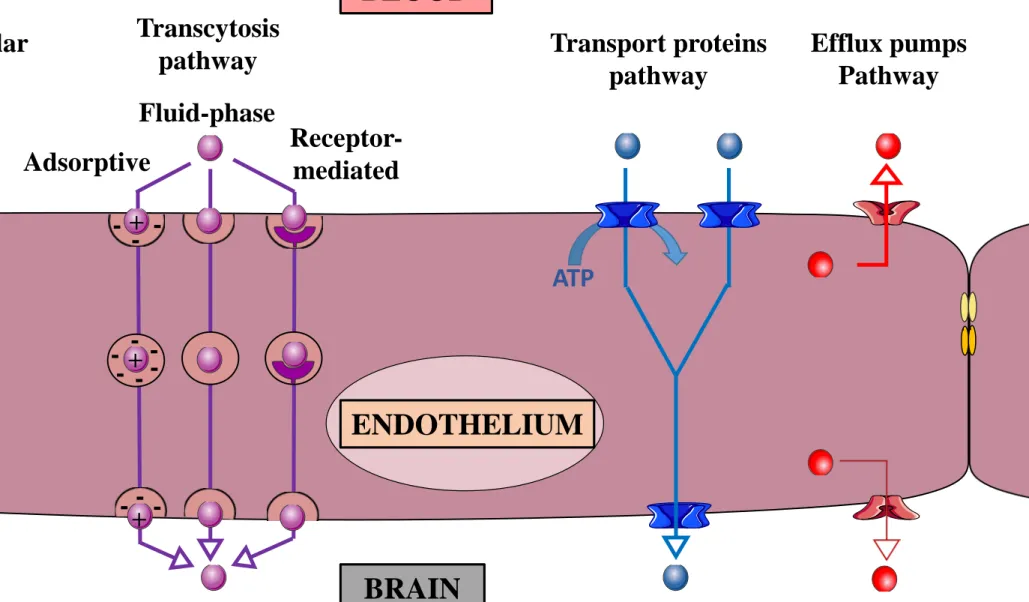
There are five main mechanisms or pathways driving molecular penetration through the BBB:
(i) passive paracellular pathway, (ii) transcellular lipophilic pathway, (iii) transcytosis pathway,
(iv) transport protein pathway, and (v) efflux pumps pathway (Figure 2).
Passive paracellular diffusion of molecules between endothelial cells is hampered by the tight
junctions (TJ) and adherens junctions (AJ). Only few small highly liposoluble molecules can
cross the BBB by passive paracellular diffusion [4].
Some small gaseous or lipophilic molecules are also able to cross the BBB by passive
transcellular diffusion across endothelial cells themselves [4].
The transcytosis pathway refers to successive endocytosis from one side and exocytosis from
the other side of endothelial cells. The three main transcytosis types are: (i) constitutive and
non-specific -i.e. fluid-phase endocytosis: micropinocytosis, macropinocytosis-, (ii) ligand's
charges mediated and non-specific -i.e. adsorptive endocytosis- and (iii) specific
receptor-mediated. The non-specific transcytosis mechanisms are less represented in the BBB than in
peripheral blood vessels [4,5].
The transport protein pathway is an active and specific transport mechanism of molecules
across the BBB. This transport pathway is predominant in the BBB. A large variety of
transporters are expressed by endothelial cells including transporters from the solute carrier
family (SLC). SLC2A1 (GLUT-1), involved in the crossing of glucose, is one of the most
abundant transport of the SLC family [4,6,7].
The last mechanism is the efflux pumps pathway, a crucial mechanism for detoxification.
Mainly ABCB1 (P-gp), ABCG2 (BCRP) and MRP 1 to 5 reject potential harmful xenobiotics
from the endothelial cells to the blood (Figure 2) [4,8].
The BBB is disrupted in restricted zones of the brain close to the 3rd and the 4th ventricles: the
cells (tanycytes) and from the brain by a dense layer of astrocytes, tanycytes and extracellular
matrix [9–11] (figure 1E).
3.2. The blood-tumor barrier
In GBM, the tumor bulk is schematically organized in three major parts: (i) the necrotic central
area, (ii) the proliferative/angiogenic forehead, and (iii) the BAT including invasive/escaping
tumor cells (Figure 3).
The blood tumor barrier (BTB) refers to a histologically and/or biologically altered BBB with
increased permeability. In the BTB, the blood vessels are anarchic, disorganized, sinuous,
irregularly shaped, large and leaky, mainly due to an imperfect angiogenesis and inflammation
[12,13].
These modifications are due to both: (i) pro-angiogenic and immune-modulating factors
secreted by GBM cells, and (ii) tumor-induced micro-environment changes [14–16].
3.3. CSF-related barriers
As discussed by Saunders et al., CSF is both isolated from the blood and the brain. In the
ventricular system, CSF is isolated from the blood in the choroid plexus by epithelial cells that
play a barrier role similar to the endothelial cells in the BBB (figure 1C). Even if the
mechanisms are similar, specific transporters and efflux pumps are different from the ones
expressed in the BBB [17,18]. Ependymal cells lining the ventricle are not tightly attached in
adults. The ependyma is therefore not thought to hamper the diffusion from the CSF to the brain
(figure 1C). However, transport systems and CSF flow limit diffusion to 1-2 mm [17,19]. CSF
is also isolated from brain and blood by the arachnoid and pia matters that both present tightly
packet cell layers that prevent diffusion from the blood to CSF and from CSF to the brain [20]
4. The BBB limits drug penetration to both normal brain and tumors
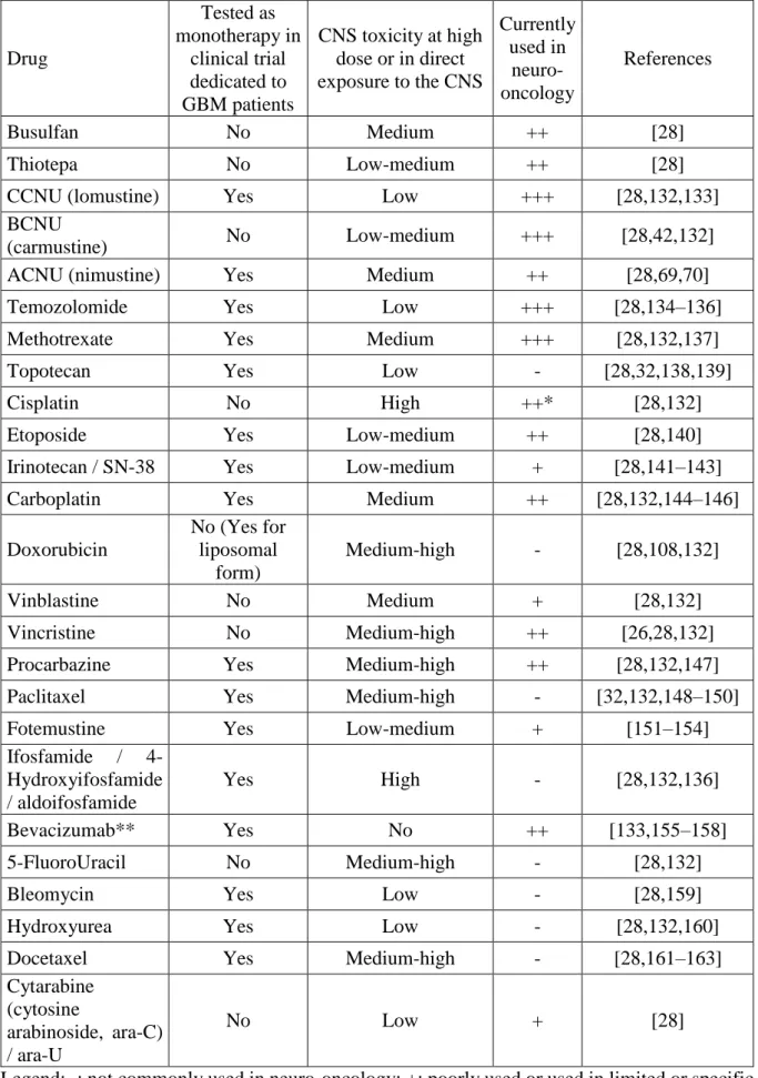
Currently, the most frequently used chemotherapy agent in GBM is temozolomide (TMZ), a
drug that is able to cross the BBB [2]. Table 1 indicates several drugs according to their clinical
use and their relevance in treatment of central nervous system (CNS) tumors.
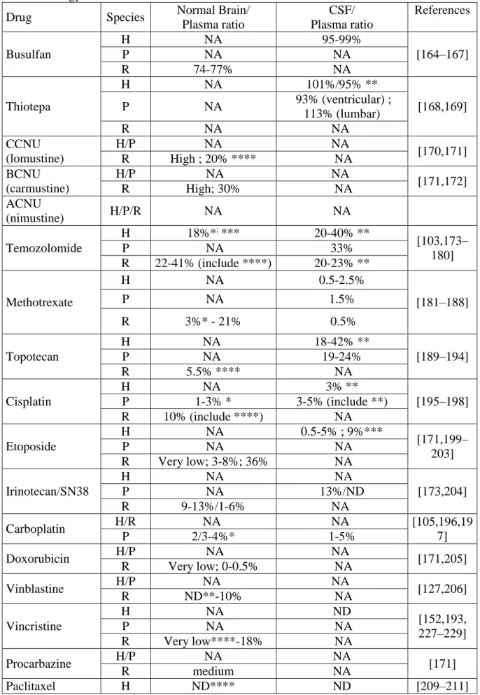
Table 2 shows experimental brain and CSF penetration data for several drugs. Recently, Jacus
et al. reviewed the pharmacokinetic properties of several anticancer agents, and assessed their
penetration in CSF and/or in brain tissue of patients with CNS tumors [21].
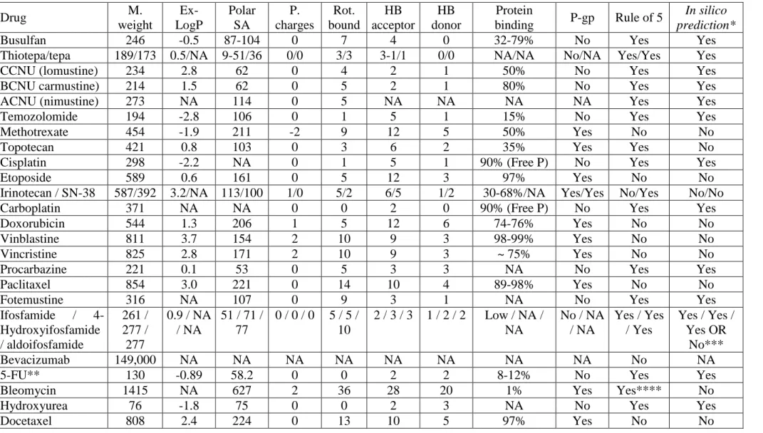
Several physicochemical parameters are involved in the ability of drugs to cross the normal
BBB: (i) size, (ii) liposolubility, (iii) charge, (iv) interactions with plasma proteins, and (v)
interactions with efflux pumps and transporters. According to these parameters, several groups
have suggested a way to predict in silico their ability to cross the BBB. The rule of 5 developed
by Lipinski is the theoretical basis of these predictions [22]. According to this rule, “poor
absorption or permeation is more likely when: (i) > 5 hydrogen bond donors, (ii) MWt > 500,
(iii) logP > 5, (iv) > 10 hydrogen bond acceptors, and (v) substrates for biological transporters
are exceptions to this rule”. Although this modeling has been significantly improved over time,
predictions are not always consistent with the experimental data [23]. Table 3 shows several
parameters used for prediction of BBB crossing by drugs. As an example of the limits of
predictive models, irinotecan is predicted not to cross the BBB and cisplatin is predicted to
cross the BBB (Table 3), while the in vivo data reported in Table 2 shows that irinotecan is
more likely to cross the BBB than cisplatin.
Predicting accurately the ability of anti-cancer drugs to cross the BBB and to penetrate in brain
patients, based on our currents preclinical models, remains challenging. Combining in silico, in
5. Overcoming the BBB for better drug delivery within the tumor core and the BAT
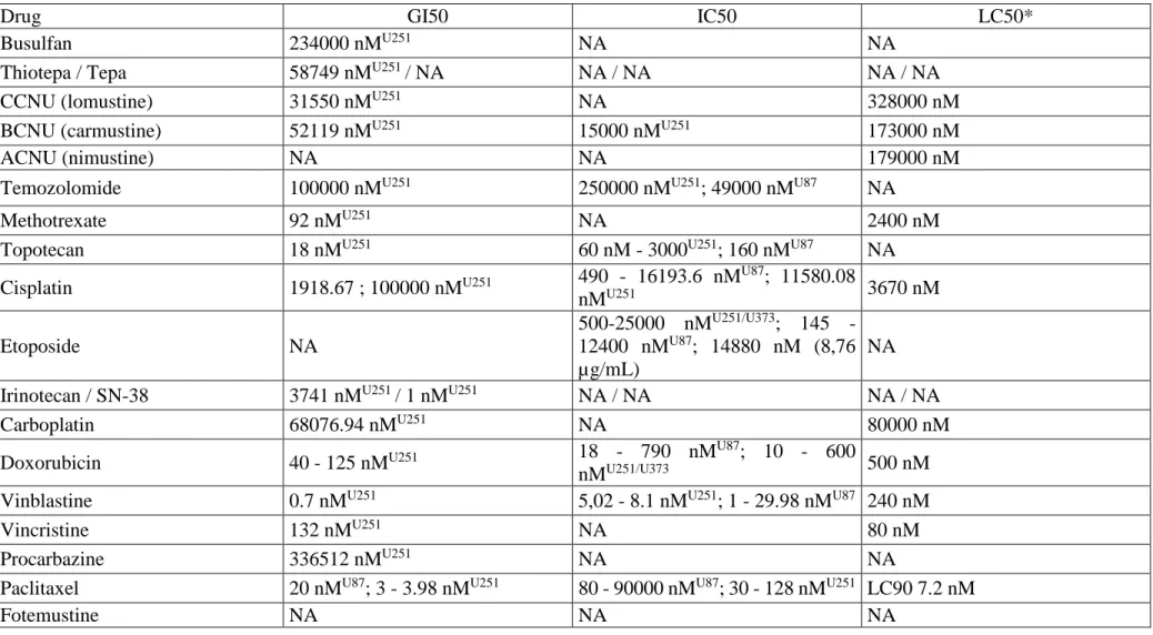
Table 4 indicates in vitro efficacy of chemotherapeutic cytotoxic agents investigated against
GBM cells. Significant inter-laboratories variability is observed (e.g. the IC50 for paclitaxel on
U87 cells ranges from 80 to 90000 nM). TMZ, the most commonly used chemotherapy agent
in GBM, is inconsistently cytotoxic on GBM cell lines, while vincristine, vinblastine, paclitaxel
and doxorubicin are up to 10,000 – 100,000 times more cytotoxic than TMZ. Integrated
therapeutic strategies including improved brain delivery of the most efficient drugs and
molecular biomarkers of response to these drugs (e.g. MGMT for TMZ) will significantly
improve the outcome for GBM patients [24]. Several approaches have been developed or are
still under development.
5.1. Intra-tumor injection
Direct delivery of chemotherapy within the tumor and the BAT require insertion of a catheter
within the tumor site. Imaging prior to drug administration is thus necessary to locate
specifically the target site. Any molecule, regardless of its physicochemical characteristics, is
deliverable using this method. The main limitations are local injuries: (i) infection, (ii)
inflammatory reaction, and (iii) direct neurotoxicity (Table 1) [25,26]. Indeed, neurotoxicity of
chemotherapeutic agents is a major issue when increasing local delivery (e.g. vincaalcaloids
may induce seizure, encephalopathy, ataxia, and/or movement disorders; taxanes may induce
seizure; and platinum derivatives may induce seizure, encephalopathy, stroke, ataxia and/or
myelopathy) [27,28]. The main advantage of catheter-based drug delivery is an increase of local
drug concentration without increasing systemic concentration and drug toxicity. The use of this
method for nitrosoureas (i.e. BCNU and CCNU) has shown efficacy and few side effects in
by DTI-015 (BCNU in 100% ethanol) (NCT00038441) [29]. However, the invasiveness and
the direct exposure of the brain to the drug toxicity limit the use of this method.
5.2. Convection enhanced delivery (CED)
Convection Enhanced Delivery (CED) is based on a catheter inserted, during a neurosurgical
procedure, within the tumor or the BAT. The catheter is linked to an Ommaya/Rickham
reservoir or to an external pump maintaining a positive pressure and flow. It allows a slow drug
delivery over a longer period of time to reduce the potential acute neurotoxicity of
chemotherapy [31]. CED has been tested for cisplatin, methotrexate, paclitaxel, nimustine,
topotecan and carboplatin [32]. The capacity of a drug to diffuse within the brain parenchyma
is heterogeneous and depends on : (i) the drug, (ii) the tumor site, and (iii) the administration
parameters [32–35]. Cisplatin was reported to diffuse 1 cm around the needle tip in 1982 [36].
A more recent study indicated the mean volume of distribution is between 12.8 to 22.9 cm3 for
paclitaxel [35]. Neurotoxicity was reported in some trials (e.g. paclitaxel). Despite the
limitations of this invasive procedure, clinical benefits were observed brain tumor patients in
some trials (e.g. nimustine, topotecan, carboplatin) [32]. Recently, an implantable catheter
system was recently developed and tested with carboplatin in a recurrent GBM patient, and induced a 58% tumor shrinkage and a stabilization of the patient’s clinical condition (NCT01317212) [37].
5.3. CSF delivery
Drugs can be directly injected within the CSF. This method is mainly used to treat spinal cord
tumors, leptomeningeal tumors, and tumor meningitis. As the CSF volume is lower than the
blood volume, the intrathecal injection of chemotherapy leads to a higher concentration of
from CSF to the CNS parenchyma reduces the impact of this strategy in the treatment of
intraparenchymal GBM patients [38]. Although some drugs are commonly used through the
CSF route (e.g. methotrexate, cytarabine) with acceptable side effects, other drugs (e.g.
vincristine) are contra-indicated for direct CSF delivery due to the high risk a severe
neurotoxicity or death. [39,40].
5.4. In situ biodegradable polymer, gels, microships or microcarrier
After surgical resection of the brain tumor, a cytotoxic agent-impregnated biodegradable
polymer can be deposited in the tumor resection cavity. Carmustine impregnated wafers
(Gliadel®) continuously deliver the drug directly in the brain parenchyma over 3 weeks [41].
Although initial results were promising, more recent data suggest a limited survival benefit in
GBM patients (NCT00003876) [42–44]. Increasing drug concentration within the wafers might
increase efficacy with acceptable toxicity as shown in a phase I clinical trial [45]. Adverse
effects such as seizures, convulsions, confusion, brain edema, infection, hemiparesis, aphasia,
and visual field defects were reported with this treatment [41–44,46,47]. High dose BCNU was
detected 5 to 6.1 mm around the wafer on day 1, and between 1.1 to 3.6 mm from days 3 to 30.
Several drugs were used within this delivery system. They were detected at low concentrations
up to 5 cm around the wafer, but their concentration dropped below the LC90 (lethal
concentration) within 1 cm around the wafer [46,48,49]. However, tumor recurrence was
reported to occur mainly in the 2 cm around the BAT. The use of gels to fill the postsurgical
cavity, micro-chips and micro-carriers instead of wafers has also been evaluated with quite
similar efficacy and limitations mainly in murine models [50–53]. Gels have been investigated
5.5. Transnasal epithelium drug delivery
A drug can also cross the nasal epithelium at least in some regions of the brain [54], and reach
CSF and brain. Transnasal drug delivery has been tested in animals for various treatments
including methotrexate or 5-FU [54–56]. However, it has not been used in human to treat brain
tumors so far.
5.6. High-dose and dose-dense chemotherapy delivered using intravenous (i.v.) peripheral
route
As mentioned above, some drugs are very efficient against GBM cells in vitro, but exhibit
limited effects in vivo due to their low ability to cross the BBB (e.g. doxorubicin, vincristine,
vinblastine, paclitaxel). High-dose and dose-dense chemotherapy regimens aim, with or without
bone marrow transplant, increasing drug concentrations within the brain tumor using higher
dose of chemotherapy delivered in a peripheral vein. The benefits of these procedures,
associated with significant toxicity, is debated and heterogeneous across patients (e.g.
NCT00304031 and NCT01364064) [57–62].
5.7. Intra-arterial (i.a.) drug delivery
The i.a. delivery of drugs via the carotid artery has been shown to improve drug delivery within
CNS. I.a. injection of cisplatin and etoposide led to a 2- and 4- fold increase of drug delivery
to the brain compared to i.v. injection, respectively [63,64]. Penetration of methotrexate,
aminoisobutyric acid and dextran 70 within the brain was 2 to 2.5 higher with i.a. delivery
compared to i.v. delivery to tumor-bearing rats [65]. BCNU i.a. delivery achieved an 50-fold
improvement of delivery in glioma patients [66]. ACNU i.a. delivery was not associated with
an improvement of survival in glioma patients but showed a lower chemotherapy-related
associated with a high risk of neurological, ophtalmological, and vascular toxicities limiting its
use [67–73].
5.8. Efflux pumps inhibition
P-gp and BCRP can be inhibited by various drugs (e.g. cyclosporin A, elacridar, valspodar,
tariquidar, or zosuquidar trihydrochloride) [74–76]. The association of paclitaxel and valspodar
reduced the tumor volume up to 90%, while paclitaxel alone had no effect. A prolonged 1.7
fold increase of brain concentration of paclitaxel was observed when combined with several of
these inhibitors [77–79]. Colchicine and vinblastine uptake was enhanced 8.42- and 9.08-fold,
respectively, when they were co-injected with valspodar in rats [80]. Cyclosporin A treatment
also increases brain delivery of doxorubicin in rats [81]. However, cyclosporin A injection in
non-human primates did not appear to improve the CSF delivery of doxorubicin [82]. Docetaxel
brain concentration was also increased by elacridar, valspodar and cyclosporine A in mice [83].
Such inhibitors showed no or poor effect on different non-CNS tumors expressing P-gp in
clinical trials enrolling patients (NCT00069160) [75]. However, due to the BBB and to high
expression of efflux pumps in brain normal cells, this approach might be interesting. For
example, combination of verapamil to an antiepileptic treatment in a patients with
pharmacoresistant seizures doubled the time interval between hospitalizations, improved the
overall control of seizures and the quality of life of patients [84]. An improved brain/plasma
ratio was also obtained for loperamide when associated with tariquidar and elacridar [85].
Moreover, even if a method allows a molecule to cross the BBB, the therapeutic impact would
be decreased by the efflux of the drug to the blood if it is substrate of efflux pumps [86]. Any
method developed to delivery drugs to the brain could benefit the addition of an adjuvant efflux
5.9. BBB opening
Interestingly, beside their direct antitumor effect, some anti-tumor therapeutic strategies already
used in clinics are able to open the BBB (e.g. etoposide, morphine and radiotherapy) [2,87–89].
The opening of the BBB can also be obtained by i.a. injection of hypotonic solutions or
hyperosmotic solutions (i.e. mannitol). These two methods induce a water flow from the
endothelial cells to the blood, leading to shrinkage and subsequent opening of TJs [90–92].
Interestingly, complete tumor response was reported for patients receiving carboplatin and
etoposide after i.a. administration of mannitol [93]. Methotrexate, aminoisobutyric acid and
dextran 70 delivery to the brain was improved by 2.5 to 7.6 fold by mannitol-induced BBB
disruption [65]. In the same line, intra-carotid hyperosmolar perfusion in rats allowed
240-500% increase for antibodies [94]. Bradykinin or its agonist (i.e. RMP-7) or histamine also
opens the BBB [95–97]. Intra-carotid infusion of RMP-7 improved the delivery of carboplatin
by 2.7 fold in rats [98]. For methotrexate, aminoisobutyric acid and dextran 70, Neuwelt et al.
reported an increase of drug delivery to the tumor and the BAT in rats by: (i) 2.2 to 2.5-fold
after i.a. injection compared to i.v. injection, (ii) 2.5 to 7.6-fold after mannitol-induced BBB
disruption compared to saline injection and, (iii) 6.3 to 16.7-fold combining both methods (i.a.
+ mannitol vs i.v. + saline) [65].
Ultrasounds can also be used to open the BBB [92,99]. Indeed, association of low frequency
ultrasounds with microbubble contrast agents was shown to open the BBB, a technics that was
described to be minimally/non-invasive and safe [100–102]. The ultrasound-induced opening
of the BBB was reported to improve the TMZ CSF/plasma ratio from 22.7% to 38.6% in tumor
bearing rats [103]. Irinotecan delivery was increased by 206% to 331% in healthy rabbits [104].
In a primate model, the mean platinum brain distribution was 5.2-fold higher in the US field
(0-5mm section) than in the contralateral hemisphere [105]. A phase I clinical trial
recurrent GBM patients. Thermal ablation of GBM on patients has also been performed with
transcranial high-frequency focused ultrasound [106]. The use of the same device with different
ultrasound parameters could therefore lead to a BBB opening [107].
5.10. Drug design, modification and encapsulation
Biochemical modifications (e.g. addition of ligand to receptor mediated transcytosis, lipophilic
molecules, nanovectors and/or positively charged molecules) of existing drugs are explored to
improve their capacity to cross the BBB and their anti-tumor efficacy.
Doxil®/Caelix® consists of doxorubicin encapsulated in a PEGylated liposome. A stabilization
in malignant gliomas patients was obtained with Doxil® [108]. The modification of Doxil® with
glutathione groups led to a 4.8-fold increase of the brain-to-blood ratio compared to
Doxil®/Caelix® (~0.08% vs. ~0.02% respectively) in preclinical models [109,110]. Various
other methods have been tested, such as the modification of drugs with fatty acids to increase
their liposolubility and to improve their diffusion through the BBB [111]. The modification of
a drug with a molecule that is recognized by specific receptors/proteins on endothelial cells can
also promote its passage through receptor mediated transcytosis (e.g. complexation of drugs
with transferrin) [5,112,113].
Interestingly, drug modification can be combined with other strategies to increase drug delivery
within the brain, improve its stability, or reduce its elimination [114].
5.11. Magnetic delivery
Magnetic nanoparticles can be included in liposomes, forming thus magnetoliposomes. These
magnetoliposomes can be modified with molecules such as transferrin to promote their
interaction with the brain endothelium. Drugs can be loaded in these magnetoliposomes.
the blood vessels, through the BBB, and deliver the drug in the brain parenchyma [115]. The
use of paclitaxel-loaded anti-GPNMB antibodies-decorated magnetoliposomes improved the
brain delivery of paclitaxel by 4 fold in rats. Paclitaxel concentration was still high 48h after
treatment for the liposomal form, while it was not detected after 6h in animals treated with
unmodified paclitaxel [116].
5.12. Electric fields and Electromagnetic fields
In 1977, application of low intensity electric fields to the brain was shown to induce BBB
opening and to improve passage of dyes and drugs from the blood to the brain. This passage of
drugs can also implicate multiple putative mechanisms : (i) iontophoresis (charge-mediated
displacement of charged molecules), (ii) electro-osmotic, (iii) convection flows, and/or (iv)
electroporation [117]. Recently, the use of intracranial irreversible electroporation in rats’ brain
was shown to induce a BBB opening [118].
The Novocure device delivers “tumor-treating fields” (TTF) to the brain, and was shown to
induce tumor cells death in preclinics and to increase survival of newly diagnosed GBM patients
(NCT00916409) [120]. The mechanism of tumor cells death remains unclear, but it is at least
partly due to interaction of TTF with the cytoskeleton. Indeed, TTF impede polymerization and
functions of tubulin resulting in abnormal mitoses and cytokinesis. Finally, the electroporation
of tumor cells was also observed. Electroporation was proposed to explain the synergic effect
of TTF and chemotherapy observed in some patients [120]. Moreover, a BBB opening was
observed after non-thermal irreversible electroporation as a tumor ablation method [92].
Interactions between electromagnetic fields and BBB are still unclear and under investigations.
[121].
GBM is a lethal disease and more efficient therapeutic strategies are urgently needed. Despite
the fact that multiple efficient cytotoxic chemotherapeutic agents are available, as demonstrated
in in vitro preclinical models (i.e. GBM cell lines) and in vivo preclinical models without BBB
(i.e. GBM subcutaneous xenografts), their efficacy is dramatically reduced in GBM orthotopic
xenografts and in GBM patients [122]. Several reasons might explain this reduced efficacy in
patients, one of them being the limited penetration of drugs within the tumor and the BAT.
The passage of cytotoxic drugs across the BBB is commonly admitted to be limited in patients
and to be well-documented in the literature. However, data are scarce and heterogeneous in the
literature, limiting comparisons across studies. Our review of the literature reports, in Table 2,
the ability of anti-tumor drugs to cross the brain barriers (i.e. brain/plasma and CSF/plasma
ratio). However, preclinical and clinical studies are heterogeneous in terms of material and
methods: (i) heterogeneity of models and patients -tumor or not, tumor type, CNS involvement
or not- and, (ii) heterogeneity of methods - i.e. route of administration of the drug, total dose,
time between treatments, biological samples management, assays, cell lines used-. These
heterogeneities, also raised by Jacus et al., highlight the difficulties to compare and to interpret
studies in robust manner [21]. Therefore, major efforts need to be conducted by the community
to standardize preclinical evaluation of drug efficacy and preclinical evaluation of drug
penetration within the tumor, the BAT and the CSF.
Indeed, these data are critical to optimize drug delivery of current cytotoxic agents and to take
advantage of efficient drugs that would be otherwise disregarded due to their incapacity to reach
GBM cells. Indeed, one of the major therapeutic advances that have been accomplished over
the last years in neuro-oncology came from very old cytotoxic drugs [123,124]. Therefore, “old
drugs” might have unexpected efficacy if used in the right indication, in the right therapeutic
Assessment of brain distribution is challenging in practice in preclinical and clinical settings.
In silico prediction is the most efficient approach to perform a high throughput analysis.
However, the limited accuracy of this method hampers the benefits of such evaluations. On a
smaller scale, a limited number of molecules can also be analyzed using in vitro models of
BBB. Various models exist, and they all rely on the transwell system. Endothelial cells are
cultured on a porous membrane delimiting two compartments, representing the blood and the
brain [4,125]. The drug can then be deposited in one compartment, and measured in the two
compartments to quantify the passage of the drug. The most commonly used human endothelial
cells are the immortalized human brain endothelial cells hCMEC/D3 cells [126]. These cells
were shown to retain the normal gene expression pattern of endothelial cells of the BBB,
making them one of the easiest to use, the most reproducible and the most reliable models.
More accurate models are available but cannot be used in medium to large-scale studies [127].
The optimal animal model remains the non-human primate (e.g. Rhesus monkey). The BBB of
rodents (mice, rats) is different from the human BBB. More specifically, the expression of
efflux pumps such as ABCB1 (P-gp) and ABCG2 (BCRP) is qualitatively similar, but
quantitatively different [128]. The pathway of in silico, preclinical in vitro, preclinical in vivo
analysis allows preselection of the best candidate drugs at each step, and reduces the cost of
drugs screening [129].
In clinical trials, most often, the passage of drugs is measured in the CSF. However, the
drug concentration is not always well-correlated between the CSF and the brain. The most
robust data are obtained with biopsies of the brain after chemotherapy administration. However,
biopsies are mainly used for diagnosis rather than for drug dosage, and are thus performed prior
to initiation of treatment. Phase 0 clinical trials may help to better understand CNS
7. Expert commentary
Treating brain diseases including glioblastoma is challenging due, at least partly, to the BBB.
It will be virtually impossible to test, in human patients, for each anti-GBM candidate drug and
for each drugs combination, their ability to cross the BBB, their therapeutic efficacy and their
toxicity. Therefore, robust, reproducible and consensual models to assess the ability of
anti-GBM candidate drugs to cross the BBB in preclinical settings (i.e. in vitro and in vivo) needs
to be optimized and validated-admitted across the research teams involved in the field. These
models, including the complexity of the human BBB, will undoubtedly better rationalize our
selection of anti-GBM candidate drugs to be tested, alone or combined with a BBB-opening
procedure, in clinical trials enrolling GBM patients. This strategy will improve our success rate
in clinical trials dedicated to GBM patients and will benefit to GBM patients.
8. Five-years view
Currently, two major axis of therapeutic research are converging to increase efficacy of
cancer treatments in the field of primary malignant brain tumors. Innovative smart
anti-tumor drugs are developed and some of them have demonstrated dramatic efficacy in systemic
cancers raising hope in the treatment of GBM patients. Some of these promising drugs are large
molecules (e.g. monoclonal antibodies) or highly hydrophilic. Obviously, although these
anti-cancer drugs are efficient, they will not be able to reach GBM cells located in the BAT.
Significant efforts are ongoing to increase, using chemical or physical approaches,
bioavailability of these drugs with the GBM bulk and the BAT. Merging these two axis of
therapeutic research will undoubtedly improve treatments of GBM patients.
The blood brain/tumor barrier is a major obstacle limiting efficacy of anti-cancer agents
in GBM. Multiple classic cytotoxic agents and innovative drugs showed promising therapeutic
activity in GBM cells in the absence of the BBB or BTB (i.e. in vitro experiments or
subcutaneous xenografts). Their ability to cross the BBB and the BTB has been poorly or
heterogeneously documented in the literature. A better comprehensive and standardize
evaluation of the ability of drugs to cross the BBB and their anti-tumor efficacy is needed. In
parallel, multiples innovative physical and chemical strategies are under development to bypass
the BBB and the BTB particularly in the BAT. A better knowledge of the ability of drugs to
cross the BBB and a better ability to open the BBB will undoubtedly improve treatments of
GBM patients.
10. Key issues
Escaping/invasive GBM cells, located in the BAT which is protected by intact BBB, are often the source of GBM recurrence
The BBB remains a major obstacle to obtain therapeutic drug bio-availability within the GBM bulk and the BAT
Systematic, comparable and comprehensive pharmacokinetics and pharmacodynamics data for anti-cancer drugs are lacking.
Physical strategies to open the BBB in a reproducible, large, transient and safe are under investigations.
Chemical strategies to increase drug penetration through the BBB are under investigations
11. References
Papers of special note have been highlighted as:
* of interest
** of considerable interest
1. Ostrom QT, Bauchet L, Davis FG, et al. The epidemiology of glioma in adults: a “state of the science” review. Neuro-Oncol. 16(7), 896–913 (2014).
2. Stupp R, Mason WP, van den Bent MJ, et al. Radiotherapy plus concomitant and adjuvant temozolomide for glioblastoma. N. Engl. J. Med. 352(10), 987–996 (2005).
3. Scherer H. Structural development in gliomas. Am J Cancer. , 333–351 (1938).
4. Wong AD, Ye M, Levy AF, Rothstein JD, Bergles DE, Searson PC. The blood-brain barrier: an engineering perspective. Front. Neuroengineering. 6, 7 (2013).
5. Preston JE, Joan Abbott N, Begley DJ. Transcytosis of macromolecules at the blood-brain barrier. Adv. Pharmacol. San Diego Calif. 71, 147–163 (2014).
6. Shawahna R, Uchida Y, Declèves X, et al. Transcriptomic and quantitative proteomic analysis of transporters and drug metabolizing enzymes in freshly isolated human brain microvessels. Mol. Pharm. 8(4), 1332–1341 (2011).
7. Uchida Y, Ohtsuki S, Katsukura Y, et al. Quantitative targeted absolute proteomics of human blood-brain barrier transporters and receptors. J. Neurochem. 117(2), 333–345 (2011).
8. Hartz AMS, Bauer B. ABC transporters in the CNS - an inventory. Curr. Pharm. Biotechnol. 12(4), 656–673 (2011).
9. Horsburgh A, Massoud TF. The circumventricular organs of the brain: conspicuity on clinical 3T MRI and a review of functional anatomy. Surg. Radiol. Anat. SRA. 35(4), 343–349 (2013).
10. Langlet F, Mullier A, Bouret SG, Prevot V, Dehouck B. Tanycyte-like cells form a blood-cerebrospinal fluid barrier in the circumventricular organs of the mouse brain. J. Comp. Neurol. 521(15), 3389–3405 (2013).
11. Morita S, Furube E, Mannari T, et al. Heterogeneous vascular permeability and alternative diffusion barrier in sensory circumventricular organs of adult mouse brain. Cell Tissue Res. (2015).
12. Hillen F, Griffioen AW. Tumour vascularization: sprouting angiogenesis and beyond. Cancer Metastasis Rev. 26(3–4), 489–502 (2007).
13. Dubois LG, Campanati L, Righy C, et al. Gliomas and the vascular fragility of the blood brain barrier. Front. Cell. Neurosci. 8, 418 (2014).
14. Lee J, Lund-Smith C, Borboa A, Gonzalez AM, Baird A, Eliceiri BP. Glioma-induced remodeling of the neurovascular unit. Brain Res. 1288, 125–134 (2009).
15. Schneider SW, Ludwig T, Tatenhorst L, et al. Glioblastoma cells release factors that disrupt blood-brain barrier features. Acta Neuropathol. (Berl.). 107(3), 272–276 (2004).
16. Wolburg H, Noell S, Fallier-Becker P, Mack AF, Wolburg-Buchholz K. The disturbed blood-brain barrier in human glioblastoma. Mol. Aspects Med. 33(5–6), 579–589 (2012). 17. de Lange ECM. Potential role of ABC transporters as a detoxification system at the
blood-CSF barrier. Adv. Drug Deliv. Rev. 56(12), 1793–1809 (2004).
18. Saunders NR, Daneman R, Dziegielewska KM, Liddelow SA. Transporters of the blood-brain and blood-CSF interfaces in development and in the adult. Mol. Aspects Med. 34(2– 3), 742–752 (2013).
19. Pardridge WM. Drug transport in brain via the cerebrospinal fluid. Fluids Barriers CNS. 8(1), 7 (2011).
20. Patel N, Kirmi O. Anatomy and imaging of the normal meninges. Semin. Ultrasound. CT MR. 30(6), 559–564 (2009).
21. Jacus MO, Daryani VM, Harstead KE, Patel YT, Throm SL, Stewart CF. Pharmacokinetic Properties of Anticancer Agents for the Treatment of Central Nervous System Tumors: Update of the Literature. Clin. Pharmacokinet. (2015).
22. Lipinski CA, Lombardo F, Dominy BW, Feeney PJ. Experimental and computational approaches to estimate solubility and permeability in drug discovery and development settings. Adv. Drug Deliv. Rev. 46(1–3), 3–26 (2001).
23. Lanevskij K, Japertas P, Didziapetris R. Improving the prediction of drug disposition in the brain. Expert Opin. Drug Metab. Toxicol. 9(4), 473–486 (2013).
24. van Nifterik KA, van den Berg J, van der Meide WF, et al. Absence of the MGMT protein as well as methylation of the MGMT promoter predict the sensitivity for temozolomide. Br. J. Cancer. 103(1), 29–35 (2010).
25. Tomita T. Interstitial chemotherapy for brain tumors: review. J. Neurooncol. 10(1), 57– 74 (1991).
26. Oliver AS, Firth G, McKeran RO. Studies on the intracerebral injection of vincristine free and entrapped within liposomes in the rat. J. Neurol. Sci. 68(1), 25–30 (1985). 27. Stone JB, DeAngelis LM. Cancer-treatment-induced neurotoxicity--focus on newer
treatments. Nat. Rev. Clin. Oncol. 13(2), 92–105 (2016).
28. Newton HB. Neurological complications of chemotherapy to the central nervous system. Handb. Clin. Neurol. 105, 903–916 (2012).
29. Hassenbusch SJ, Nardone EM, Levin VA, Leeds N, Pietronigro D. Stereotactic injection of DTI-015 into recurrent malignant gliomas: phase I/II trial. Neoplasia N. Y. N. 5(1), 9– 16 (2003).
30. Tator CH, Day A, Ng R, Liberman L. Chemotherapy of an experimental glioma with nitrosoureas. Cancer Res. 37(2), 476–481 (1977).
31. Ommaya AK. Subcutaneous reservoir and pump for sterile access to ventricular cerebrospinal fluid. Lancet. 2(7315), 983–984 (1963).
32. Healy AT, Vogelbaum MA. Convection-enhanced drug delivery for gliomas. Surg. Neurol. Int. 6(Suppl 1), S59-67 (2015).
33. Kroll RA, Pagel MA, Muldoon LL, Roman-Goldstein S, Neuwelt EA. Increasing volume of distribution to the brain with interstitial infusion: dose, rather than convection, might be the most important factor. Neurosurgery. 38(4), 746-752-754 (1996).
34. Sampson JH, Brady ML, Petry NA, et al. Intracerebral infusate distribution by convection-enhanced delivery in humans with malignant gliomas: descriptive effects of target anatomy and catheter positioning. Neurosurgery. 60(2 Suppl 1), ONS89-98-99 (2007).
35. Tanner PG, Holtmannspötter M, Tonn J-C, Goldbrunner R. Effects of drug efflux on convection-enhanced paclitaxel delivery to malignant gliomas: technical note. Neurosurgery. 61(4), E880–882; discussion E882 (2007).
36. Kroin JS, Penn RD. Intracerebral chemotherapy: chronic microinfusion of cisplatin. Neurosurgery. 10(3), 349–354 (1982).
37. Barua NU, Hopkins K, Woolley M, et al. A novel implantable catheter system with transcutaneous port for intermittent convection-enhanced delivery of carboplatin for recurrent glioblastoma. Drug Deliv. , 1–7 (2015).
38. Kerr JZ, Berg S, Blaney SM. Intrathecal chemotherapy. Crit. Rev. Oncol. Hematol. 37(3), 227–236 (2001).
39. Qweider M, Gilsbach JM, Rohde V. Inadvertent intrathecal vincristine administration: a neurosurgical emergency. Case report. J. Neurosurg. Spine. 6(3), 280–283 (2007).
40. Saiki JH, Thompson S, Smith F, Atkinson R. Paraplegia following intrathecal chemotherapy. Cancer. 29(2), 370–374 (1972).
41. Brem H, Mahaley MS, Vick NA, et al. Interstitial chemotherapy with drug polymer implants for the treatment of recurrent gliomas. J. Neurosurg. 74(3), 441–446 (1991).
42. Brem H, Piantadosi S, Burger PC, et al. Placebo-controlled trial of safety and efficacy of intraoperative controlled delivery by biodegradable polymers of chemotherapy for recurrent gliomas. The Polymer-brain Tumor Treatment Group. Lancet. 345(8956), 1008–1012 (1995).
43. Perry J, Chambers A, Spithoff K, Laperriere N. Gliadel wafers in the treatment of malignant glioma: a systematic review. Curr. Oncol. Tor. Ont. 14(5), 189–194 (2007).
44. Subach BR, Witham TF, Kondziolka D, Lunsford LD, Bozik M, Schiff D. Morbidity and survival after 1,3-bis(2-chloroethyl)-1-nitrosourea wafer implantation for recurrent
glioblastoma: a retrospective case-matched cohort series. Neurosurgery. 45(1), 17-22-23 (1999).
45. Olivi A, Grossman SA, Tatter S, et al. Dose escalation of carmustine in surgically implanted polymers in patients with recurrent malignant glioma: a New Approaches to Brain Tumor Therapy CNS Consortium trial. J. Clin. Oncol. Off. J. Am. Soc. Clin. Oncol. 21(9), 1845–1849 (2003).
46. Engelhard HH. Tumor bed cyst formation after BCNU wafer implantation: report of two cases. Surg. Neurol. 53(3), 220–224 (2000).
47. Weber EL, Goebel EA. Cerebral edema associated with Gliadel wafers: Two case studies. Neuro-Oncol. 7(1), 84–89 (2005).
48. Fung LK, Shin M, Tyler B, Brem H, Saltzman WM. Chemotherapeutic drugs released from polymers: distribution of 1,3-bis(2-chloroethyl)-1-nitrosourea in the rat brain. Pharm. Res. 13(5), 671–682 (1996).
49. Fung LK, Ewend MG, Sills A, et al. Pharmacokinetics of interstitial delivery of carmustine, 4-hydroperoxycyclophosphamide, and paclitaxel from a biodegradable polymer implant in the monkey brain. Cancer Res. 58(4), 672–684 (1998).
50. Akbar U, Jones T, Winestone J, et al. Delivery of temozolomide to the tumor bed via biodegradable gel matrices in a novel model of intracranial glioma with resection. J. Neurooncol. 94(2), 203–212 (2009).
51. Elstad NL, Fowers KD. OncoGel (ReGel/paclitaxel)--clinical applications for a novel paclitaxel delivery system. Adv. Drug Deliv. Rev. 61(10), 785–794 (2009).
52. Kim GY, Tyler BM, Tupper MM, et al. Resorbable polymer microchips releasing BCNU inhibit tumor growth in the rat 9L flank model. J. Control. Release Off. J. Control. Release Soc. 123(2), 172–178 (2007).
53. Li KW, Dang W, Tyler BM, et al. Polilactofate microspheres for Paclitaxel delivery to central nervous system malignancies. Clin. Cancer Res. Off. J. Am. Assoc. Cancer Res. 9(9), 3441–3447 (2003).
54. Illum L. Transport of drugs from the nasal cavity to the central nervous system. Eur. J. Pharm. Sci. Off. J. Eur. Fed. Pharm. Sci. 11(1), 1–18 (2000).
55. Sakane T, Yamashita S, Yata N, Sezaki H. Transnasal Delivery of 5-Fluorouracil to the Brain in the Rat. J. Drug Target. 7(3), 233–240 (1999).
56. Shingaki T, Inoue D, Furubayashi T, et al. Transnasal delivery of methotrexate to brain tumors in rats: a new strategy for brain tumor chemotherapy. Mol. Pharm. 7(5), 1561– 1568 (2010).
57. Bay J-O, Jacques-Olivier B, Linassier C, et al. Does high-dose carmustine increase overall survival in supratentorial high-grade malignant glioma? An EBMT retrospective study. Int. J. Cancer J. Int. Cancer. 120(8), 1782–1786 (2007).
58. Grill J, Kalifa C, Doz F, et al. A high-dose busulfan-thiotepa combination followed by autologous bone marrow transplantation in childhood recurrent ependymoma. A phase-II study. Pediatr. Neurosurg. 25(1), 7–12 (1996).
59. Valteau-Couanet D, Fillipini B, Benhamou E, et al. High-dose busulfan and thiotepa followed by autologous stem cell transplantation (ASCT) in previously irradiated medulloblastoma patients: high toxicity and lack of efficacy. Bone Marrow Transplant. 36(11), 939–945 (2005).
60. Armstrong TS, Wefel JS, Wang M, et al. Net clinical benefit analysis of radiation therapy oncology group 0525: a phase III trial comparing conventional adjuvant temozolomide with dose-intensive temozolomide in patients with newly diagnosed glioblastoma. J. Clin. Oncol. Off. J. Am. Soc. Clin. Oncol. 31(32), 4076–4084 (2013).
61. Gilbert MR, Wang M, Aldape KD, et al. Dose-dense temozolomide for newly diagnosed glioblastoma: a randomized phase III clinical trial. J. Clin. Oncol. Off. J. Am. Soc. Clin. Oncol. 31(32), 4085–4091 (2013).
62. Massimino M, Gandola L, Luksch R, et al. Sequential chemotherapy, high-dose thiotepa, circulating progenitor cell rescue, and radiotherapy for childhood high-grade glioma. Neuro-Oncol. 7(1), 41–48 (2005).
63. Nakagawa H, Fujita T, Izumoto S, et al. cis-diamminedichloroplatinum (CDDP) therapy for brain metastasis of lung cancer. I. Distribution within the central nervous system after intravenous and intracarotid infusion. J. Neurooncol. 16(1), 61–67 (1993).
64. Savaraj N, Lu K, Feun LG, Burgess MA, Loo TL. Comparison of CNS penetration, tissue distribution, and pharmacology of VP 16-213 by intracarotid and intravenous administration in dogs. Cancer Invest. 5(1), 11–16 (1987).
65. Neuwelt EA, Barnett PA, McCormick CI, Remsen LG, Kroll RA, Sexton G. Differential permeability of a human brain tumor xenograft in the nude rat: impact of tumor size and method of administration on optimizing delivery of biologically diverse agents. Clin. Cancer Res. Off. J. Am. Assoc. Cancer Res. 4(6), 1549–1555 (1998).
66. Tyler JL, Yamamoto YL, Diksic M, et al. Pharmacokinetics of superselective intra-arterial and intravenous [11C]BCNU evaluated by PET. J. Nucl. Med. Off. Publ. Soc. Nucl. Med. 27(6), 775–780 (1986).
67. Chauveinc L, Sola-Martinez MT, Martin-Duverneuil M, et al. Intra arterial chemotherapy with ACNU and radiotherapy in inoperable malignant gliomas. J. Neurooncol. 27(2), 141–147 (1996).
68. Hirano Y, Mineura K, Mizoi K, Tomura N. Therapeutic results of intra-arterial chemotherapy in patients with malignant glioma. Int. J. Oncol. 13(3), 537–542 (1998).
69. Imbesi F, Marchioni E, Benericetti E, et al. A randomized phase III study: comparison between intravenous and intraarterial ACNU administration in newly diagnosed primary glioblastomas. Anticancer Res. 26(1B), 553–558 (2006).
70. Kochii M, Kitamura I, Goto T, et al. Randomized comparison of intra-arterial versus intravenous infusion of ACNU for newly diagnosed patients with glioblastoma. J. Neurooncol. 49(1), 63–70 (2000).
71. Poisson M, Chiras J, Fauchon F, Delattre JY. [Treatment of supratentorial glioma in adults by intra-arterial HECNU. Experience of the Pitié-Salpétrière group]. Rev. Neurol. (Paris). 148(6–7), 441–447 (1992).
72. Roosen N, Lins E, Kiwit JC, Schirmer M, Bock WJ. Intra-arterial chemotherapy with ACNU for the treatment of glioblastoma. Preliminary experience. Acta Radiol. Suppl. 369, 220–222 (1986).
73. Vega F, Davila L, Chatellier G, et al. Treatment of malignant gliomas with surgery, intraarterial chemotherapy with ACNU and radiation therapy. J. Neurooncol. 13(2), 131– 135 (1992).
74. Bankstahl JP, Bankstahl M, Römermann K, et al. Tariquidar and elacridar are dose-dependently transported by P-glycoprotein and Bcrp at the blood-brain barrier: a small-animal positron emission tomography and in vitro study. Drug Metab. Dispos. Biol. Fate Chem. 41(4), 754–762 (2013).
75. Fox E, Bates SE. Tariquidar (XR9576): a P-glycoprotein drug efflux pump inhibitor. Expert Rev. Anticancer Ther. 7(4), 447–459 (2007).
76. International Transporter Consortium, Giacomini KM, Huang S-M, et al. Membrane transporters in drug development. Nat. Rev. Drug Discov. 9(3), 215–236 (2010).
77. Kemper EM, Cleypool C, Boogerd W, Beijnen JH, van Tellingen O. The influence of the P-glycoprotein inhibitor zosuquidar trihydrochloride (LY335979) on the brain penetration of paclitaxel in mice. Cancer Chemother. Pharmacol. 53(2), 173–178 (2004).
78. Kemper EM, van Zandbergen AE, Cleypool C, et al. Increased penetration of paclitaxel into the brain by inhibition of P-Glycoprotein. Clin. Cancer Res. Off. J. Am. Assoc. Cancer Res. 9(7), 2849–2855 (2003).
79. Fellner S, Bauer B, Miller DS, et al. Transport of paclitaxel (Taxol) across the blood-brain barrier in vitro and in vivo. J. Clin. Invest. 110(9), 1309–1318 (2002).
80. Drion N, Lemaire M, Lefauconnier JM, Scherrmann JM. Role of P-glycoprotein in the blood-brain transport of colchicine and vinblastine. J. Neurochem. 67(4), 1688–1693 (1996).
81. Hughes CS, Vaden SL, Manaugh CA, Price GS, Hudson LC. Modulation of doxorubicin concentration by cyclosporin A in brain and testicular barrier tissues expressing P-glycoprotein in rats. J. Neurooncol. 37(1), 45–54 (1998).
82. Warren KE, Patel MC, McCully CM, Montuenga LM, Balis FM. Effect of P-glycoprotein modulation with cyclosporin A on cerebrospinal fluid penetration of doxorubicin in non-human primates. Cancer Chemother. Pharmacol. 45(3), 207–212 (2000).
83. Kemper EM, Verheij M, Boogerd W, Beijnen JH, van Tellingen O. Improved penetration of docetaxel into the brain by co-administration of inhibitors of P-glycoprotein. Eur. J. Cancer Oxf. Engl. 1990. 40(8), 1269–1274 (2004).
84. Asadi-Pooya AA, Razavizadegan SMA, Abdi-Ardekani A, Sperling MR. Adjunctive use of verapamil in patients with refractory temporal lobe epilepsy: a pilot study. Epilepsy Behav. EB. 29(1), 150–154 (2013).
85. Choo EF, Kurnik D, Muszkat M, et al. Differential in vivo sensitivity to inhibition of P-glycoprotein located in lymphocytes, testes, and the blood-brain barrier. J. Pharmacol. Exp. Ther. 317(3), 1012–1018 (2006).
86. Mason WP. Blood-brain barrier-associated efflux transporters: a significant but underappreciated obstacle to drug development in glioblastoma. Neuro-Oncol. 17(9), 1181–1182 (2015).
87. Hollis PH, Zappulla RA, Spigelman MK, et al. Physiological and electrophysiological consequences of etoposide-induced blood-brain barrier disruption. Neurosurgery. 18(5), 581–586 (1986).
88. Sardi I. Morphine facilitates doxorubicin penetration in the central nervous system: a new prospect for therapy of brain tumors. J. Neurooncol. 104(2), 619–620 (2011).
89. Cao Y, Tsien CI, Shen Z, et al. Use of magnetic resonance imaging to assess blood-brain/blood-glioma barrier opening during conformal radiotherapy. J. Clin. Oncol. Off. J. Am. Soc. Clin. Oncol. 23(18), 4127–4136 (2005).
90. Rapoport SI, Robinson PJ. Tight-junctional modification as the basis of osmotic opening of the blood-brain barrier. Ann. N. Y. Acad. Sci. 481, 250–267 (1986).
91. Joshi S, Ergin A, Wang M, et al. Inconsistent blood brain barrier disruption by intraarterial mannitol in rabbits: implications for chemotherapy. J. Neurooncol. 104(1), 11–19 (2011).
92. Rodriguez A, Tatter SB, Debinski W. Neurosurgical Techniques for Disruption of the Blood-Brain Barrier for Glioblastoma Treatment. Pharmaceutics. 7(3), 175–187 (2015).
93. Neuwelt EA, Wiliams PC, Mickey BE, Frenkel EP, Henner WD. Therapeutic dilemma of disseminated CNS germinoma and the potential of increased platinum-based chemotherapy delivery with osmotic blood-brain barrier disruption. Pediatr. Neurosurg. 21(1), 16–22 (1994).
94. Bullard DE, Bourdon M, Bigner DD. Comparison of various methods for delivering radiolabeled monoclonal antibody to normal rat brain. J. Neurosurg. 61(5), 901–911 (1984).
95. Gregor A, Lind M, Newman H, et al. Phase II studies of RMP-7 and carboplatin in the treatment of recurrent high grade glioma. RMP-7 European Study Group. J. Neurooncol. 44(2), 137–145 (1999).
96. Prados MD, Schold SC JR SC, Fine HA, et al. A randomized, double-blind, placebo-controlled, phase 2 study of RMP-7 in combination with carboplatin administered
intravenously for the treatment of recurrent malignant glioma. Neuro-Oncol. 5(2), 96– 103 (2003).
97. Qureshi AI, Suri MF, Khan J, et al. Superselective intra-arterial carboplatin for treatment of intracranial neoplasms: experience in 100 procedures. J. Neurooncol. 51(2), 151–158 (2001).
98. Matsukado K, Inamura T, Nakano S, Fukui M, Bartus RT, Black KL. Enhanced tumor uptake of carboplatin and survival in glioma-bearing rats by intracarotid infusion of bradykinin analog, RMP-7. Neurosurgery. 39(1), 125-133-134 (1996).
99. Vykhodtseva NI, Hynynen K, Damianou C. Histologic effects of high intensity pulsed ultrasound exposure with subharmonic emission in rabbit brain in vivo. Ultrasound Med. Biol. 21(7), 969–979 (1995).
100. Horodyckid C, Canney M, Vignot A, et al. Safe long-term repeated disruption of the blood-brain barrier using an implantable ultrasound device : a multiparametric study in primates. J Neurosurg. (2015).
101. McDannold N, Vykhodtseva N, Raymond S, Jolesz FA, Hynynen K. MRI-guided targeted blood-brain barrier disruption with focused ultrasound: histological findings in rabbits. Ultrasound Med. Biol. 31(11), 1527–1537 (2005).
102. Hynynen K, McDannold N, Sheikov NA, Jolesz FA, Vykhodtseva N. Local and reversible blood-brain barrier disruption by noninvasive focused ultrasound at frequencies suitable for trans-skull sonications. NeuroImage. 24(1), 12–20 (2005). 103. Wei K-C, Chu P-C, Wang H-YJ, et al. Focused ultrasound-induced blood-brain barrier
opening to enhance temozolomide delivery for glioblastoma treatment: a preclinical study. PloS One. 8(3), e58995 (2013).
104. Beccaria K, Canney M, Goldwirt L, et al. Ultrasound-induced opening of the blood-brain barrier to enhance temozolomide and irinotecan delivery: an experimental study in rabbits. J. Neurosurg. , 1–9 (2015).
105. Goldwirt L, Canney M, Horodyckid C, et al. Enhanced brain distribution of carboplatin in a primate model after blood-brain barrier disruption using an implantable ultrasound device. Cancer Chemother. Pharmacol. (2015).
106. McDannold N, Clement GT, Black P, Jolesz F, Hynynen K. Transcranial magnetic resonance imaging- guided focused ultrasound surgery of brain tumors: initial findings in 3 patients. Neurosurgery. 66(2), 323–332; discussion 332 (2010).
107. McDannold N, Arvanitis CD, Vykhodtseva N, Livingstone MS. Temporary disruption of the blood-brain barrier by use of ultrasound and microbubbles: safety and efficacy evaluation in rhesus macaques. Cancer Res. 72(14), 3652–3663 (2012).
108. Fabel K, Dietrich J, Hau P, et al. Long-term stabilization in patients with malignant glioma after treatment with liposomal doxorubicin. Cancer. 92(7), 1936–1942 (2001).
109. Birngruber T, Raml R, Gladdines W, et al. Enhanced doxorubicin delivery to the brain administered through glutathione PEGylated liposomal doxorubicin (2B3-101) as
compared with generic Caelyx,(®)/Doxil(®)--a cerebral open flow microperfusion pilot study. J. Pharm. Sci. 103(7), 1945–1948 (2014).
110. Gaillard PJ, Appeldoorn CCM, Dorland R, et al. Pharmacokinetics, brain delivery, and efficacy in brain tumor-bearing mice of glutathione pegylated liposomal doxorubicin (2B3-101). PloS One. 9(1), e82331 (2014).
111. Bradley MO, Webb NL, Anthony FH, et al. Tumor targeting by covalent conjugation of a natural fatty acid to paclitaxel. Clin. Cancer Res. Off. J. Am. Assoc. Cancer Res. 7(10), 3229–3238 (2001).
112. Lajoie JM, Shusta EV. Targeting receptor-mediated transport for delivery of biologics across the blood-brain barrier. Annu. Rev. Pharmacol. Toxicol. 55, 613–631 (2015).
113. Zhang F, Xu C-L, Liu C-M. Drug delivery strategies to enhance the permeability of the blood-brain barrier for treatment of glioma. Drug Des. Devel. Ther. 9, 2089–2100 (2015).
114. Fan C-H, Ting C-Y, Lin H-J, et al. SPIO-conjugated, doxorubicin-loaded microbubbles for concurrent MRI and focused-ultrasound enhanced brain-tumor drug delivery. Biomaterials. 34(14), 3706–3715 (2013).
115. Thomsen LB, Thomsen MS, Moos T. Targeted drug delivery to the brain using magnetic nanoparticles. Ther. Deliv. (2015).
116. Dilnawaz F, Singh A, Mewar S, Sharma U, Jagannathan NR, Sahoo SK. The transport of non-surfactant based paclitaxel loaded magnetic nanoparticles across the blood brain barrier in a rat model. Biomaterials. 33(10), 2936–2951 (2012).
117. Mitragotri S. Devices for overcoming biological barriers: the use of physical forces to disrupt the barriers. Adv. Drug Deliv. Rev. 65(1), 100–103 (2013).
118. Garcia PA, Rossmeisl JH, Robertson JL, et al. 7.0-T magnetic resonance imaging characterization of acute blood-brain-barrier disruption achieved with intracranial irreversible electroporation. PloS One. 7(11), e50482 (2012).
119. Stupp R, Taillibert S, Kanner AA, et al. Maintenance therapy with tumor-treating fields plus temozolomide vs temozolomide alone for glioblastoma: A randomized clinical trial. JAMA. 314(23), 2535–2543 (2015).
120. Rehman AA, Elmore KB, Mattei TA. The effects of alternating electric fields in glioblastoma: current evidence on therapeutic mechanisms and clinical outcomes. Neurosurg. Focus. 38(3), E14 (2015).
121. Stam R. Electromagnetic fields and the blood-brain barrier. Brain Res. Rev. 65(1), 80– 97 (2010).
122. Pokorny JL, Calligaris D, Gupta SK, et al. The Efficacy of the Wee1 Inhibitor MK-1775 Combined with Temozolomide Is Limited by Heterogeneous Distribution across the Blood–Brain Barrier in Glioblastoma. Clin. Cancer Res. 21(8), 1916–1924 (2015). 123. van den Bent MJ, Brandes AA, Taphoorn MJB, et al. Adjuvant procarbazine, lomustine,
long-term follow-up of EORTC brain tumor group study 26951. J. Clin. Oncol. Off. J. Am. Soc. Clin. Oncol. 31(3), 344–350 (2013).
124. Cairncross G, Wang M, Shaw E, et al. Phase III trial of chemoradiotherapy for anaplastic oligodendroglioma: long-term results of RTOG 9402. J. Clin. Oncol. Off. J. Am. Soc. Clin. Oncol. 31(3), 337–343 (2013).
125. Naik P, Cucullo L. In vitro blood-brain barrier models: current and perspective technologies. J. Pharm. Sci. 101(4), 1337–1354 (2012).
126. Weksler BB, Subileau EA, Perrière N, et al. Blood-brain barrier-specific properties of a human adult brain endothelial cell line. FASEB J. Off. Publ. Fed. Am. Soc. Exp. Biol. 19(13), 1872–1874 (2005).
127. Garberg P, Ball M, Borg N, et al. In vitro models for the blood-brain barrier. Toxicol. Vitro Int. J. Publ. Assoc. BIBRA. 19(3), 299–334 (2005).
128. Warren MS, Zerangue N, Woodford K, et al. Comparative gene expression profiles of ABC transporters in brain microvessel endothelial cells and brain in five species including human. Pharmacol. Res. Off. J. Ital. Pharmacol. Soc. 59(6), 404–413 (2009).
129. Abbott NJ. Prediction of blood-brain barrier permeation in drug discovery from in vivo, in vitro and in silico models. Drug Discov. Today Technol. 1(4), 407–416 (2004).
130. Sigmond J, Honeywell RJ, Postma TJ, et al. Gemcitabine uptake in glioblastoma multiforme: potential as a radiosensitizer. Ann. Oncol. 20(1), 182–187 (2009).
131. Langlet F. [Role of tanycytes within the blood-hypothalamus interface]. Biol. Aujourdhui. 208(3), 225–235 (2014).
132. Cherubini GB, Platt S. Neurological complications of chemotherapy. Companion Anim. 13(5), 76–82 (2008).
133. Taal W, Oosterkamp HM, Walenkamp AME, et al. Single-agent bevacizumab or lomustine versus a combination of bevacizumab plus lomustine in patients with recurrent glioblastoma (BELOB trial): a randomised controlled phase 2 trial. Lancet Oncol. 15(9), 943–953 (2014).
134. Brada M, Hoang-Xuan K, Rampling R, et al. Multicenter phase II trial of temozolomide in patients with glioblastoma multiforme at first relapse. Ann. Oncol. Off. J. Eur. Soc. Med. Oncol. ESMO. 12(2), 259–266 (2001).
135. Malmström A, Grønberg BH, Marosi C, et al. Temozolomide versus standard 6-week radiotherapy versus hypofractionated radiotherapy in patients older than 60 years with glioblastoma: the Nordic randomised, phase 3 trial. Lancet Oncol. 13(9), 916–926 (2012).
136. Paulsen F, Hoffmann W, Becker G, et al. Chemotherapy in the treatment of recurrent glioblastoma multiforme: ifosfamide versus temozolomide. J. Cancer Res. Clin. Oncol. 125(7), 411–418 (1999).
137. Luyendijk W, van Beusekom GT. Chemotherapy of cerebral gliomas with intra-carotid methotrexate-infusion. Acta Neurochir. (Wien). 15(3), 234–248 (1966).
138. Bruce JN, Fine RL, Canoll P, et al. Regression of recurrent malignant gliomas with convection-enhanced delivery of topotecan. Neurosurgery. 69(6), 1272-1279-1280 (2011).
139. Macdonald D, Cairncross G, Stewart D, et al. Phase II study of topotecan in patients with recurrent malignant glioma. National Clinical Institute of Canada Clinical Trials Group. Ann. Oncol. Off. J. Eur. Soc. Med. Oncol. ESMO. 7(2), 205–207 (1996).
140. Fulton D, Urtasun R, Forsyth P. Phase II study of prolonged oral therapy with etoposide (VP16) for patients with recurrent malignant glioma. J. Neurooncol. 27(2), 149–155 (1996).
141. Friedman HS, Petros WP, Friedman AH, et al. Irinotecan therapy in adults with recurrent or progressive malignant glioma. J. Clin. Oncol. Off. J. Am. Soc. Clin. Oncol. 17(5), 1516–1525 (1999).
142. Prados MD, Lamborn K, Yung WKA, et al. A phase 2 trial of irinotecan (CPT-11) in patients with recurrent malignant glioma: a North American Brain Tumor Consortium study. Neuro-Oncol. 8(2), 189–193 (2006).
143. Turner CD, Gururangan S, Eastwood J, et al. Phase II study of irinotecan (CPT-11) in children with high-risk malignant brain tumors: the Duke experience. Neuro-Oncol. 4(2), 102–108 (2002).
144. Cloughesy TF, Gobin YP, Black KL, et al. Intra-arterial carboplatin chemotherapy for brain tumors: a dose escalation study based on cerebral blood flow. J. Neurooncol. 35(2), 121–131 (1997).
145. Prados MD, Warnick RE, Mack EE, et al. Intravenous carboplatin for recurrent gliomas. A dose-escalating phase II trial. Am. J. Clin. Oncol. 19(6), 609–612 (1996).
146. Stewart DJ, Belanger JM, Grahovac Z, et al. Phase I study of intracarotid administration of carboplatin. Neurosurgery. 30(4), 512-516-517 (1992).
147. Yung WK, Albright RE, Olson J, et al. A phase II study of temozolomide vs. procarbazine in patients with glioblastoma multiforme at first relapse. Br. J. Cancer. 83(5), 588–593 (2000).
148. Chamberlain MC, Kormanik P. Salvage chemotherapy with paclitaxel for recurrent primary brain tumors. J. Clin. Oncol. Off. J. Am. Soc. Clin. Oncol. 13(8), 2066–2071 (1995).
149. Lidar Z, Mardor Y, Jonas T, et al. Convection-enhanced delivery of paclitaxel for the treatment of recurrent malignant glioma: a phase I/II clinical study. J. Neurosurg. 100(3), 472–479 (2004).
150. Postma TJ, Heimans JJ, Luykx SA, et al. A phase II study of paclitaxel in chemonaïve patients with recurrent high-grade glioma. Ann. Oncol. Off. J. Eur. Soc. Med. Oncol. ESMO. 11(4), 409–413 (2000).