HAL Id: hal-01690373
https://hal.univ-reunion.fr/hal-01690373
Submitted on 23 Jan 2018
HAL is a multi-disciplinary open access
archive for the deposit and dissemination of
sci-entific research documents, whether they are
pub-lished or not. The documents may come from
teaching and research institutions in France or
abroad, or from public or private research centers.
L’archive ouverte pluridisciplinaire HAL, est
destinée au dépôt et à la diffusion de documents
scientifiques de niveau recherche, publiés ou non,
émanant des établissements d’enseignement et de
recherche français ou étrangers, des laboratoires
publics ou privés.
Distributed under a Creative Commons Attribution - NonCommercial - NoDerivatives| 4.0
International License
Stem cells and the circadian clock
Meltem Weger, Nicolas Diotel, Anne-Claire Dorsemans, Thomas Dickmeis,
Benjamin Weger
To cite this version:
Meltem Weger, Nicolas Diotel, Anne-Claire Dorsemans, Thomas Dickmeis, Benjamin Weger.
Stem cells and the circadian clock. Developmental Biology, Elsevier, 2017, 431 (2), pp.111-123.
�10.1016/j.ydbio.2017.09.012�. �hal-01690373�
Contents lists available atScienceDirect
Developmental Biology
journal homepage:www.elsevier.com/locate/developmentalbiology
Stem cells and the circadian clock
Meltem Weger
a,1, Nicolas Diotel
b, Anne-Claire Dorsemans
b, Thomas Dickmeis
c,
Benjamin D. Weger
d,⁎aCentre for Endocrinology, Diabetes and Metabolism, University of Birmingham, Birmingham, United Kingdom
bUniversité de La Réunion, INSERM, UMR 1188, Diabète athérothrombose Thérapies Réunion Océan Indien (DéTROI), Saint-Denis de La Réunion, France cInstitute of Toxicology and Genetics, Karlsruhe Institute of Technology, Hermann-von-Helmholtz-Platz 1, 76344 ,Eggenstein-Leopoldshafen, Germany dNestlé Institute of Health Sciences SA, EPFL Innovation Park, Bâtiment H, 1015 Lausanne, Switzerland
A R T I C L E I N F O
Keywords: Circadian clock Stem cell Development Vertebrate Adult neurogenesisA B S T R A C T
The circadian timing system is a complex biological network of interacting circadian clocks that regulates 24 h rhythms of behavioral and physiological processes. One intriguing observation is that stem cell homeostasis is subject to circadian clock regulation. Rhythmic oscillations have been observed in a variety of embryonic and adult stem cell dependent processes, such as hematopoietic progenitor cell migration, the hair follicle cycle, bone remodeling, regenerative myogenesis and neurogenesis. This review aims to discuss the nature of the circadian clock in embryonic stem cells and how it changes during differentiation. Furthermore, it will examine how the circadian clock contributes to adult stem cell function in different tissues of the body with an emphasis on the brain and adult neurogenesis.
1. The circadian clock
1.1. The hallmarks of the circadian timing system
Organisms face regular changes in their environment linked to day and night cycles, including, for example, variations in the availability of food or the activity of predators. In order to adapt to these cyclical daily changes, organisms possess an internal timing system, proactively orchestrating their behavior and physiology. In modern societies, humans are no longer subjected to variations in prey and predator presence, but many aspects of human behavior (e.g., sleep/wake cycle) and physiology (e.g., hormone secretion, body temperature, metabo-lism) are still regulated by the same timing system. This system consists of biological clocks that can be found in almost every cell of the body. Via regulatory mechanisms including rhythmic transcrip-tional, post-transcriptional and post-translational modulation of gene expression and function such clocks produce rhythmic changes in behavior and physiology (Atger et al., 2017; Lim and Allada, 2013; Reddy et al., 2006a, 2006b). These clocks have particular hallmarks: they conduct rhythms with a periodicity of approximately 24 h and, thus, are called “circadian” clocks (coined from Latin: circa-diem = around a day). Circadian clocks are endogenous and self-sustained, leading to rhythms that persist in constant conditions such as sustained darkness. However, to remain synchronized with their environment,
they are“entrainable” or “resettable” by external time cues, the most prevailing one being light (Roenneberg et al., 2013). In chronobiology, these cues are called Zeitgeber (German, literally:“time giver”). This property of the clock becomes obvious during "jet-lag", which causes a temporary disruption of the sleep/wake cycle that soon adapts to the new environmental light conditions.
1.2. The organization of circadian clocks in vertebrates
The first experiments aiming to locate the clock that drives circadian rhythms in mammals pointed to the suprachiasmatic nucleus (SCN). This small region of the brain is a paired neuronal structure located in the anteroventral hypothalamus above the optic chiasm (Brancaccio et al., 2014). Ablating the SCN in rodents resulted in abolished circadian locomotor and endocrine rhythms (Moore and Eichler, 1972), as well as in circadian feeding (Nagai et al., 1978) and drinking behavior (Stephan and Zucker, 1972). A transplantation of SCN tissue can restore these rhythms (Lehman et al., 1987). Moreover, the donor tissue dictates its period length to the restored rhythms of the recipient (Ralph et al., 1990). The discovery of thefirst circadian clock genes led to the observation that their self-sustained oscillatory expression is not restricted to neural structures such as the SCN, but can also be found in virtually all cells of the body (Dibner et al., 2010). In this network of oscillating cells, the mammalian SCN fulfils the role
http://dx.doi.org/10.1016/j.ydbio.2017.09.012
Received 1 June 2017; Received in revised form 11 August 2017; Accepted 8 September 2017
⁎Corresponding author.
1Current address: Brain Mind Institute, École polytechnique fédérale de Lausanne, 1015 Lausanne, Switzerland.
E-mail address:[email protected](B.D. Weger).
Available online 09 September 2017
0012-1606/ © 2017 The Authors. Published by Elsevier Inc. This is an open access article under the CC BY-NC-ND license (http://creativecommons.org/licenses/BY-NC-ND/4.0/).
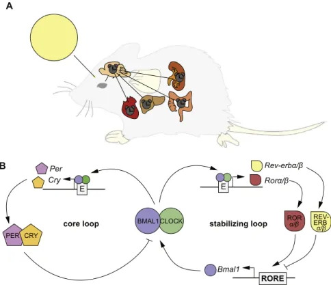
of a“central” or “master” pacemaker orchestrating the tissue clocks in peripheral organs (Fig. 1A). Environmental light sensed by the retina leads to entrainment of the central pacemaker clocks in the SCN. This timing information is then forwarded via neuronal and humoral signals, to other areas of the brain, such as the pineal gland responsible for melatonin release, and to the peripheral organ clocks (Dibner et al., 2010; Hastings et al., 2007). Interestingly, under certain conditions, conflicting systemic signals can lead to a decoupling of peripheral clocks from synchronization with the central pacemaker. For example, when mice are fed only during daytime (their rest phase) their liver clocks show a phase shift of up to 12 h compared to the SCN, which remains locked to the light phase (Damiola et al., 2000). This indicates that the peripheral clocks are able to integrate various physiological signals in order to mount appropriate rhythms in their tissues.
It has been proposed that mammals possess a more centralized organization of their circadian clocks than non-mammalian vertebrates (Cahill, 2002; Falcon et al., 2010; Menaker et al., 1997). In fish, amphibians, reptiles and birds, the retina and the pineal gland serve as central pacemaker structures, acting together with or even dominating the SCN or other brain clocks. In zebrafish, peripheral tissue clocks are directly light sensitive, reflecting expression of photoreceptors such as opsins in a wide variety of tissues (Cavallari et al., 2011; Whitmore et al., 2000). In contrast to the SCN-centered mammalian brain (Wilsbacher et al., 2002), the anatomically defined SCN equivalent in zebrafish is only one of many brain nuclei showing a high clock gene expression and activity (Moore and Whitmore, 2014; Weger et al., 2013). However, under some conditions, the SCN seem to be dis-pensable for systemic rhythm generation also in mammals, and other ill-defined oscillators take over. For example, a food-entrainable oscillator drives food anticipatory activity rhythms (Guilding and
Piggins, 2007; Mistlberger, 2011; Patton and Mistlberger, 2013). The precise relationship of this oscillator mechanism to the SCN still needs to be defined. It has recently been suggested that a larger neural network, that comprises the SCN, generates food anticipatory activity (Acosta-Galvan et al., 2011). In this view, it is tempting to speculate that both mammalian and non-mammalian circadian systems possess decentralized oscillator networks. Several modes of centralization may have evolved in the different vertebrate lineages starting from a highly decentralized system infish to a centralized system with dominance of the SCN pacemaker being a unique innovation of mammals.
1.3. The molecular clockwork
The molecular mechanism underlying circadian clock rhythms consists of a transcriptional-translational feedback loop that takes approximately 24 h to complete. The circadian clock genes themselves are not conserved between the different groups of organisms, but a common principle in all organisms is the generation of circadian rhythms by such a transcriptional-translational feedback loop ( Bell-Pedersen et al., 2005; Mohawk et al., 2012). In vertebrates, the molecular“clockwork” can be subdivided into the so-called core loop and the stabilizing loop (Fig. 1B). In the core loop, a heterodimer of the “positive” factors of the circadian clock, CLOCK (Circadian Locomotor Output Cycles Kaput) and BMAL1 (Brain and Muscle Arnt-Like protein), binds to E-box enhancer elements to activate transcription of their target genes. Among these target genes are the “negative” factors Cryptochrome (Cry) and Period (Per), acting as inhibitors of their own expression. After the translation and dimerization of the PER and CRY proteins, the PER/CRY complex translocates into the nucleus, where it inhibits the transcriptional activity of the CLOCK/BMAL1
RORE Bmal1 p o o l g n i z il i b a t s p o o l e r o c Rev-erbα/β Rorα/β ROR α/β REV-ERB α/β CRY PER BMAL1 CLOCK Per Cry
A
B
Fig. 1. The circadian timing system in mammals. (A) Schematic overview of the circadian clock system in mammals. The suprachiasmatic nucleus (SCN), a small region in the brain, is hierarchically at the top of all body clocks. After receiving light (yellow circle representing the sun) entrainment information from the eyes, the SCN acts as a central pacemaker to synchronize the circadian clocks outside the SCN, including the circadian clock of, for example, the liver and the heart via systemic cues. (B) Schematic of the molecular mechanism of the circadian clock oscillator. On a molecular level, circadian clocks consist of a core loop and accessory loops, such as the stabilizing loop. In the core loop, a heterodimer of bHLH/PAS transcription factors, namely CLOCK (green) and BMAL1 (blue), bind to E-box enhancer elements (E; gray) of the Per (purple) and Cry (orange) genes in order to initiate transcription. The PER and CRY proteins are translated and accumulate in the cytosol. Here, they heterodimerize and are translocated into the nucleus to repress CLOCK/BMAL1 activity, causing the repression of their own transcription. This mechanism is regulated by several posttranslational modifications that cause delays in the process such that a cycle takes about 24 h to complete. In the stabilizing loop, CLOCK/BMAL1 activity leads to the expression of REV-ERBα/β (yellow) and RORα/β (red), which regulate the rhythmic expression of Bmal1 by binding to the RORE (gray).
complex. As a consequence, Per and Cry transcripts decrease and subsequently less PER and CRY proteins are synthesized. This together with their degradation by the 26S proteasomal pathway releases the inhibition and starts a new cycle of transcription (Takahashi, 2015). The so-called stabilizing loop is an integral part of the circadian clock (Fig. 1B), as its impairment can lead to circadian clock arrhythmicity (Bugge et al., 2012; Cho et al., 2012). In this loop, Bmal1 expression is regulated by two types of nuclear orphan receptors, namely the REV-ERB [NR1D1 (REV-REV-ERBα), NR1D2 (REV-ERBβ)], for nuclear receptor subfamily 1, group d, member 1/2) and ROR isoforms (RORα, RORβ for RAR-related orphan receptorα/β). REV-ERBs can inhibit Bmal1 expression, whereas RORs compete with REV-ERBs for shared DNA binding sites (ROREs) and promote Bmal1 expression. Closing the stabilizing loop, expression of both factors is regulated by the core loop (Bell-Pedersen et al., 2005).
2. Circadian regulation of stem cells
The circadian clock controls a huge variety of physiological processes including the sleep/wake cycle, metabolism, and cell pro-liferation. One intriguing role of the circadian clock is its involvement in stem cell homeostasis, which is important throughout an organism's life. Even though it is well known that stem cell homeostasis and function are subject to circadian clock regulation, many aspects of how precisely this is managed are only beginning to be explored. Herein, we will first give an overview about stem cell properties and then will continue to discuss the current literature about how embryonic and adult stem cells are subject to circadian clock regulation.
2.1. Stem cells
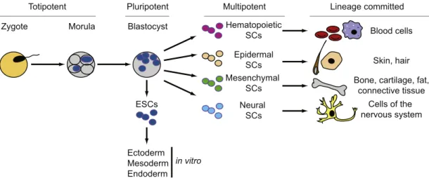
Stem cells are essential for the development of tissues during embryogenesis, and they allow in postnatal stages tissue homeostasis and regeneration due to general turnover or tissue injury. Stem cells are defined as primal (for “first” or “original”) cells that are basically undifferentiated (or unspecialized), and give rise to different cell lineages. A crucial property of stem cells is their capacity to self-renew, with cell divisions producing new stem cells and cells embarking on differentiation. During embryonic development, the fusion of an egg with a sperm initiates cell division that results in the formation of cells at the morula stage that are totipotent and able to differentiate into embryonic and extra-embryonic cells (Fig. 2). These totipotent stem cells produce pluripotent stem cells that are able to generate cells of the
three germ layers (ectoderm, mesoderm, and endoderm) that will then differentiate into all embryonic tissues. Embryonic stem cells (ESCs) derived from the inner cell mass of a blastocyst also exhibit these pluripotent properties upon culture in vitro, as do induced pluripotent stem (iPS) cells generated in vitro from differentiated cells. Finally, multipotent stem cells only produce cells of a restricted type, such as, for example, hematopoietic or neural stem cells that give rise solely to red and white blood cells and to neurons, respectively (Wobus and Boheler, 2005).
2.2. Circadian clock regulation of embryonic stem cells
One of the most intriguing questions is whether a functional circadian clock exists in pluripotent stem cells such as ESCs and if so, what role might the circadian clock play in these cells during development. Several studies in bothfish and mammals showed that clock gene products are deposited in eggs by the mother and, therefore, are already present before activation of the zygotic genome (Dekens and Whitmore, 2008; Delaunay et al., 2003; Hamatani et al., 2004; Ko et al., 2000). However, these gene products may not yet form a functional circadian feedback loop during early stages of development (Amano et al., 2009; Dekens and Whitmore, 2008; Johnson et al., 2002; Ko et al., 2000; Weger et al., 2013).
Along the same lines, experiments byYagita et al. (2010) revealed that undifferentiated mouse ESCs expressing a luciferase reporter gene driven by a clock gene promoter do not show any circadian biolumi-nescence oscillations. Differentiation of these cells towards a neural fate induced by all-trans retinoic acid initiated circadian reporter oscillations. In contrast, the induced dedifferentiation of neural stem cells using Oct3/4, Sox2, Klf4 and c-Myc factors led to the loss of circadian oscillation (Yagita et al., 2010). More recent work has implicated high expression levels of the Kpna2/Importin-α2 gene, which correlates with cytoplasmic accumulation of PER proteins, as one molecular mechanism underlying the absence of clock oscillations in ESCs (Umemura et al., 2014). These observations strongly suggest that the differentiation process of ESCs is an important feature for the development of a functional circadian clock and that a functional self-sustaining oscillator is generated gradually during development, as also seen in other studies (Dierickx et al., 2017; Kowalska et al., 2010; Umemura et al., 2013).
Strikingly, another recent publication demonstrated that even though ESCs lack a functional circadian transcriptional-translational feedback loop, they show circadian rhythms of glucose utilization and
Zygote Totipotent Morula Blastocyst Pluripotent ESCs Ectoderm Mesoderm Endoderm in vitro Multipotent Neural SCs Cells of the nervous system Hematopoietic SCs Blood cells Mesenchymal
SCs Bone, cartilage, fat,connective tissue
Lineage committed
Epidermal
SCs Skin, hair
Fig. 2. The stem cell hierarchy. The fertilization of an egg by a sperm causes the formation of the zygote. In mammalian development, the zygote stage leads after some division rounds to a solid ball of cells called the morula stage. The undifferentiated cells between zygote and morula are defined as totipotent stages, as they will give rise to a complete organism. The morula stage will lead to the blastocyst stage, in which only the cells of the inner cell mass have the capacity to give rise to the three germ layers, ectoderm, mesoderm, and endoderm. Embryonic stem cells (ESCs), derived from the inner cell mass, have the developmental capacity to differentiate in vitro into cells of all somatic cell lineages. In adult tissues, multipotent stem cells in different tissues and organs allow for the replacement of lost or injured cells of their corresponding tissues or organs. For example, hematopoietic stem cells are lineage committed to blood cells, whereas neural stem cells are lineage committed to cells of the nervous system.
of transcription of glucose transporter mRNA (Paulose et al., 2012). Thus, it is unclear whether rhythmic transcription of clock genes is indeed required for rhythmic physiological outputs such as glucose uptake and utilization. Importantly, there is some evidence that oscillations in clock protein abundance are not always required for the generation of circadian rhythms (Hastings et al., 2008; Lakin-Thomas, 2006; Putker and O'Neill, 2016). Thesefindings, together with the recent discovery of transcription-independent circadian redox cycles [as revealed, for example, by circadian changes of peroxiredoxin hyperoxidation in cultured human erythrocytes (O'Neill and Reddy, 2011;Putker and O'Neill, 2016;Reddy and Rey, 2014)], indicate non-canonical clock mechanisms that may underlie circadian rhythms in ESCs.
Even though clock gene expression oscillations are absent from ESCs and apparently not required for circadian regulation of glucose metabolism, clock genes may have functions also in these cells. A recent study reported that ESCs in which the Clock gene had been deleted exhibit decreased proliferation and increased apoptosis (Lu et al., 2016). This may indicate some non-clock function for this gene, as we will also discuss in other contexts further below.
2.3. Circadian clock regulation of adult stem cells 2.3.1. Hematopoietic stem cells
Hematopoietic stem cells (HSC) and hematopoietic stem/progeni-tor cells (HSPCs) are important for the regular formation and renewal of blood (erythrocytes, platelets) and immune cells (granulocytes, macrophages, dendritic, T-, B- and NK cells) throughout life [for a detailed overview about the hematopoietic hierarchy, see (Bryder et al., 2006)]. Their niches are quite dispersed in the fetus, with cells found in the yolk sac, placenta, the bone marrow and the spleen and liver. In the adult, the bone marrow forms the main site for HSPC niches. HSCs and HSPCs are mobile. They regularly exit the bone marrow and enter the circulation (egression), and then re-enter the hematopoietic tissues (homing) (Morrison and Scadden, 2014; Shiozawa and Taichman, 2012). The physiological role of this behavior remains elusive, but was suggested to be important for normal immunosurveillance and for maintaining homeostasis (Massberg et al., 2007; McKelvie, 1994). The mobilization of HSCs into and out of the circulation is regulated by Chemokine (C-X-C Motif) Ligand 12 (CXCL12) via its receptor Chemokine (C-X-C Motif) Receptor 4 (CXCR4). CXCL12 expressed in the stromal cells of the bone marrow niche forms a retention signal for HSCs to remain in the bone marrow and an attractant to re-enter it from circulation (Hoggatt et al., 2014; Link, 2010; Shiozawa and Taichman, 2012).
The circulation of HSCs and HSPCs under steady-state conditions is subject to circadian clock regulation. Diurnal variations in the presence of HSCs in the circulation were observed in human and mouse blood samples in early studies (Haus et al., 1983; Ross et al., 1980; Verma et al., 1980), and later work verified that the mobilization of HSCs and HSPCs into the bloodstream is under circadian clock regulation (Haus and Smolensky, 1999; Lucas et al., 2008; Mendez-Ferrer et al., 2008; Scheiermann et al., 2012). In both mouse and human, the highest blood HSC levels are found while they are resting (Lucas et al., 2008). Cxcl12 expression in the bone marrow shows a circadian pattern inversely correlated with the circadian circulation pattern of HSCs, suggesting that changes in the availability of this retention and homing cue underlie the HSC mobilization rhythms. In line with this observa-tion, circadian expression of Cxcl12 is lost in the arrhythmic Bmal1 knockout (KO) mice, as are the HSC release rhythms (Mendez-Ferrer et al., 2010, 2008). This circadian regulation seems to be systemic, because the circadian expression of Cxcl12 requires β-adrenergic receptor activation by local noradrenalin release from sympathetic nervous system neurons in the bone marrow (Mendez-Ferrer et al., 2010, 2008). Activation ofβ-adrenergic signaling leads to the degrada-tion of the transcripdegrada-tion factor Sp1 in stromal cells, which seems to be
more important for the circadian transcription of Cxcl12 than the non-canonical E-boxes also present in the Cxcl12 promoter. Thus, sympa-thetic nervous system activity dependent circadian changes in tran-scription of the retention and homing cue Cxcl12 in the niche would generate circadian patterns of HSPC mobilization (Mendez-Ferrer et al., 2009, 2008). Expression of the cognate receptor of Cxcl12, Cxcr4, was also reported to be regulated by the circadian clock in a pattern synchronized with its ligand (Lucas et al., 2008).
HSPC egression also appears to be linked with neutrophil turn-over (Casanova-Acebes et al., 2013). Under steady state conditions, young neutrophils are released into the blood mainly during the second half of the murine active phase, while aged neutrophils are taken up again into the bone marrow and thereby eliminated from the circulation during the second half of the murine rest phase. The uptake is dependent on the CXCR4 receptor of the neutrophils themselves, again implicating the CXCL12 chemokine system in this process (Casanova-Acebes et al., 2013). After their uptake, the authors suggest that aged neutrophils are phagocytosed by macrophages. This in turn seems to cause modula-tions of the hematopoietic niche, which eventually result in reduction of CXCL12 protein levels produced by the niche and an increase in HSC egression. This process involves signaling by the cholesterol-sensing LXR nuclear receptors, and indeed, LXR deficient mice do not show increased HSC egression upon stimulation with aged neutrophils. Importantly, depletion of either circulating neutrophils or of macro-phages abolishes diurnal HSC changes in the bloodstream as well as diurnal expression of an LXR target gene. Thus, the diurnal clearance of aged neutrophils by macrophages seems to be a crucial mechanism regulating HSC egression.
A number of further signaling mechanisms that show connections to circadian clock regulation and function have been implicated in cell autonomous and systemic regulation of HSC/HSPC numbers in the blood. These include glycogen synthase kinase 3β (GSK3β), a negative regulator of the Wnt/β-Catenin pathway that also contributes to the regulation of clock proteins (Reischl and Kramer, 2011). GSK3β seems to modulate egression behavior in a cell autonomous fashion, acting in parallel to the CXCL12/CXCR4 system (Lapid et al., 2013; Voermans et al., 2001). Furthermore, diurnal corticosterone oscillations were linked to the diurnal regulation of HSC/HSPC proliferation through modulation of Cxcl12 expression (Kollet et al., 2013). Low levels of corticosterone promote HSPC proliferation without influencing their differentiation, while high levels lead to a reduction of HSPCs. This regulation involves signaling by NOTCH1 in the HSPCs and modula-tion of the stem cell niche of the bone marrow, where corticosterone affects proliferation of mesenchymal and stromal progenitors and thus Cxcl12 expression (Kollet et al., 2013).
In summary, circadian changes in HSPC levels in the blood are driven by both changes in egression and by changes in proliferation. A key player in the regulation of egression is the peripheral nervous system, which acts on expression of the homing chemokine CXCL12 via both direct and indirect mechanisms, also involving the clearing of aged neutrophils. How precisely cell autonomous, local (niche) and systemic cues interplay in this process is not entirely understood. It may be important that two systems participating in the stress response, the noradrenergic peripheral nervous system and the glucocorticoid-producing adrenal gland, are involved in the circadian regulation of HSPC behavior. This raises a number of questions, for example: To what extent does the circadian system affect stress responses in the immune system via daily changes in stem cell behavior and how are these rhythms affected by stress? Are the circadian changes in HSPC behavior adaptive in their own right, or are they merely a side-effect of other adaptive functions for circadian dynamics in the stress systems? Clearly, studying the interface of stress, immune system function, the circadian clock and hematopoietic stem cells promises many fascinat-ing biomedical insights.
2.3.2. Epidermal stem cells
Functional circadian clocks were also described in the skin (Bjarnason et al., 2001; Brown et al., 2005; Kawara et al., 2002; Shiriaev et al., 1990; Tanioka et al., 2009; Zanello et al., 2000). The adult skin fulfils various important functions: it acts as a protective permeability barrier and is important for thermoregulation. It is a complex organ consisting of several layers. The outermost layer, the epidermis, is a squamous epithelium composed of the interfollicular epidermis containing mainly keratinocytes and the so-called pilosebac-eous unit with its hair follicles and sebacpilosebac-eous glands for producing hair and sebum, respectively. Skin and hair renew throughout adult life, maintaining normal homeostasis and repair after injury. Therefore, the skin includes stem cells located in several niches within the inter-follicular epidermis and in the pilosebaceous unit (Blanpain and Fuchs, 2009; Forni et al., 2012; Plikus et al., 2015; Sotiropoulou and Blanpain, 2012).
2.3.2.1. Skin. Highly proliferative stem or progenitor cells of the interfollicular epidermis are located in the basal layer of the skin. The progeny of these basal stem cells exit the cell cycle and differentiate into keratinocytes as they move up towards the skin surface to form a protective barrier (Plikus et al., 2015). Importantly, the circadian clock regulates cell proliferation of epidermal basal stem cells [(Bjarnason and Jordan, 2002; Gaddameedhi et al., 2011; Geyfman et al., 2012; Scheving, 1959)], reviewed inKumar et al. (2013), Plikus et al. (2015). Deeper insight into this regulation was obtained using an in vitro model of normal human epidermal keratinocytes, which when cultivated under low Ca2+ concentrations resemble basal layer
progenitors. The transcriptome of these cells is organized into five circadian waves of expression in phase with mRNA expression of different clock genes (Janich et al., 2013). Genes linked to keratinocyte differentiation are expressed from the late night to the early morning, whereas pathways linked to cell proliferation as well as DNA replication and repair peak in the afternoon and evening. Likewise, sensitivity to different signaling pathways is apparently gated to certain phases. Thus, induction of differentiation markers by TGFβ and Ca2+signaling
is higher in late night and early morning. The clock seems to play an instructive role in the differentiation process, as manipulation of clock gene expression led to premature differentiation of the cells in culture and defective transplantation behavior in vivo. Furthermore, the expression of metabolic genes was separated into distinct temporal domains from that of DNA replication and repair genes. This lends support to the idea that clock control may help to prevent reactive oxygen species (ROS) producing metabolic processes from damaging DNA, especially during replication. In line with this, ROS levels have been reported to change across the day in mouse skin, with an antiphasic peak of S-phase in the basal epidermis (Geyfman et al., 2012).
An elegant in vivo imaging analysis in mouse adult skin recently provided more direct evidence for this idea. This study correlated the circadian cell proliferative state of basal cells with their energy status by determining relative concentrations of free and bound NADH (Stringari et al., 2015). Free NADH was assumed to be indicative of glycolysis, whereas bound NADH indicated oxidative phosphorylation. Free NADH levels of the epidermal basal cells show a diurnal oscillation, with the highest levels at the end of the night, the murine activity phase, and significantly lower levels during the late day and early night. This oscillation correlates with the circadian clock phase on a single cell level and is lost in Bmal1 KO mice (Stringari et al., 2015). Importantly, these dailyfluctuations of free NADH peak in phase with the highest percentage of proliferating cells in S-phase. Thus, S-phase proliferation occurs at the times of higher glycolytic and lower oxidative phosphorylation activity, thereby avoiding the higher ROS levels accompanying higher oxidative phosphorylation activity. Such an
antiphase cycling of ROS producing metabolism and S-phase had previously been observed in yeast, although oscillations in this system occur with a shorter period (Causton et al., 2015; Chen et al., 2007; Klevecz et al., 2004). Temporal separation of opposing processes appears to be a powerful mechanistic principle that increases the performance and fitness of cells and is therefore phylogenetically conserved across distant lineages.
2.3.2.2. Hair. Hair production is cyclical, with hair follicles of the pilosebaceous unit following a cycle of degeneration (catagen phase of the hair cycle), rest (telogen) and growth (anagen) (Alonso and Fuchs, 2006). The epidermal stem cells located within the bulge of the hair follicle undergo periods of activation and dormancy (Plikus et al., 2015). These bulge stem cells produce the cell types of the lower portion of the follicle, including highly proliferative hair matrix cells. Another population of stem cells is located in the upper part of the follicle, the infundibulum and isthmus. These cells can contribute cells to the neighboring epidermis and show a proliferation behavior similar to that of the interfollicular epidermis stem cells.
Interestingly, the timing of the hair cycle is modulated by the circadian clock, even though the hair cycle itself takes much longer to complete than a 24 h period (Lin et al., 2009). Hair follicles of Bmal1 KO mice reveal a clear delay in anagen progression compared to their Bmal1 KO littermates while the overall duration of the entire hair cycle is not altered. The delay in anagen progression was initially suggested to be caused by a block of the G1 phase of the cell cycle (Lin et al., 2009). However, specific KO of Bmal1 in the keratinocytes of both interfollicular epidermis and hair follicles did not cause such a delay in anagen progression (Geyfman et al., 2012). This observation indicates that changes in systemic factors or in neighboring cell types contribute to the impairment of hair follicle cycling when Bmal1 is absent. Similar to global Bmal1 KO conditions, the tissue specific KO disrupted circadian rhythms in S-phase in both epidermis and upper hair follicle, with slightly increased numbers of S-phase cells in both tissues (Geyfman et al., 2012). Furthermore, the gating of mitosis in the hair matrix cells of the lower hair follicle to a later night time point was abolished in tissue-specific KOs (Plikus et al., 2013). The overtly normal hair follicle cycle despite these changes in rhythmicity of cell proliferation indicates that compensatory mechanisms can buffer these disturbances and ensure normal hair formation. In line with the existence of such mechanisms also in the interfollicular epidermis, Bmal1 KO mice do not exhibit any obvious changes in epidermal thickness regardless of the changed proliferation rhythms (Geyfman et al., 2012).
Another study suggested that clock function in the bulge stem cell compartment may play a role distinct from the regulation of coordi-nated circadian rhythms of, for example, proliferation or differentiation in the organ (Janich et al., 2011). Studies using Per1-Luciferase reporter mice revealed that simultaneously within the same bulge at telogen, one half of the stem cells show high levels of reporter activity and the other half low levels. Indeed, transcriptome analysis of these coexisting “clockhigh
” and “clocklow
” populations showed differential expression of circadian core clock genes. Interestingly, several key genes involved in signaling pathways linked with bulge stem cell homeostasis, including WNT and TGF-β signaling, were also differen-tially expressed between these two stem cell populations. The activity of bulge stem cells is regulated during the hair cycle by signals stimulating (such as WNT) and inhibiting (such as TGFβ) proliferation and differentiation (Janich et al., 2011). Strikingly, only a subset of the bulge stem cells responds to the activating stimuli during a hair cycle, whereas others remain dormant (Blanpain and Fuchs, 2009; Lin et al., 2009). This heterogeneity of bulge stem cells in their ability to respond to activating signals correlates with the heterogeneity in circadian clock state and the corresponding differential expression of signaling
path-way components (Janich et al., 2011). Thus,“clockhigh” cells expressed high levels of WNT pathway components and of TGFβ antagonizing genes and were more prone to get activated and to proliferate than the “clocklow” state. Indeed, CLOCK/BMAL1 complexes bind to E-boxes in
the promoters of several of these genes. Furthermore, a conditional Bmal1 KO in the keratinocytes of epidermis and hair follicle causes circadian arrhythmia of epidermal cells and keeps the cells in the “clocklow” condition, with pathway component expression indicative of
the dormant state. These observations strongly suggest that the coexistence of different clock phases dividing the epidermal stem cells into“clockhigh” or “clocklow” states allow certain stem cells to be ready in case of stimulation, whereas it prevents others from being activated at the same time (Janich et al., 2011). In this way, heterogeneity is adaptive in that a pool of dormant stem cells is retained by preventing their exhaustion through activating stimuli.
In summary, the circadian clock patterns the way stem cells of the hair bulge are activated and gates the subsequent proliferative behavior at certain times of day. The latter behavior is similar to the gating of S-phase in the interfollicular epidermis and upper follicle stem cells (Geyfman et al., 2012). However, the gating in the hair matrix cells seems to be centered on the timing of mitosis (Plikus et al., 2013). These differences may result from the different proliferative rates of the two compartments, with proliferation rates in the hair matrix cells higher than in interfollicular epidermis and upper follicle stem cells (Plikus et al., 2015). However, none of the mouse models of circadian disruption show a very strong hair or skin phenotype, suggesting that despite the strong clock effects on stem cell recruitment and prolifera-tion, non-clock dependent compensatory mechanisms can restore homeostasis. The beneficial effects of clock regulation in epidermal stem cells may become apparent only when the organism is challenged. In line with this assumption, Bmal1 KO mice show increased in-cidences of skin cancer and ageing related skin and hair phenotypes (Kondratov et al., 2006). Clearly, mapping the precise processes under clock control in the different stem cell types under a variety of different conditions will help to better understand the role of the clock in this process.
2.3.3. Mesenchymal stem cells and their descendants
Mesenchymal stem cells (MSCs) are characterized by their ability to differentiate in vitro into adipocytes, bone-forming osteoblasts, and cartilage-forming chondroblasts [reviewed in Murray et al. (2014)]. They have been isolated from a large variety of tissues, such as fat (Xu et al., 2005; Zuk et al., 2002), dental pulp (Shi and Gronthos, 2003), muscle (Asakura et al., 2001), placenta (Igura et al., 2004), and umbilical cord (Rogers and Casper, 2004). However, compared with a large body of in vitro studies, their precise origin and properties in vivo are much less well established (Ullah et al., 2015). Regardless of their precise developmental origin, circadian clock gene function has been implicated in the behavior of a variety of stem cells involved in homeostasis and repair of adipose tissue and bone. In the following sections, we will take a closer look at these aspects, which frequently reveal clock gene specific functions. Given the potentially heteroge-neous nature (and nomenclature) of the stem cells studied, however, one should bear in mind that many results may not reflect general MSC properties, but rather could be specific to the subset of cells under study.
2.3.3.1. Bone. Bone homeostasis is assured by the activities of the MSC derived forming osteoblasts and monocyte-derived bone-resorbing osteoclasts. Osteoblasts also give rise to osteocytes, the mature bone cells, which equally have important regulatory functions in bone remodeling.
Samsa et al. (2016) recently reported that bone derived mesench-ymal stem cells of Bmal1 KO mice showed an impairment of
differentiation under osteogenic conditions. This finding is in line with the observation that Bmal1 KO mice possess a reduced number of both active osteoblasts and of osteocytes in vivo. The authors link this observation to the low bone mass phenotype observed in Bmal1 KO mice. In contrast, an earlier study had described an increased amount of osteoblast activity in Bmal1 KO mice (Fu et al., 2005). The difference between the two studies may stem from the difference of age of the mice. Increased osteoblast proliferation was also observed in mice deficient in other clock genes, namely Per1/Per2 and Cry1/Cry2 double KOs (Fu et al., 2005). Based on their detailed analysis of the Per1/Per2 double KOs, the authors proposed a model in which the osteoblast overproliferation phenotype is due to a relief of an inhibitory action of clock genes on C-myc expression. The clock genes as well as C-myc itself are under regulation byβ2-adrenergic signaling ultimately activated by the peptide hormone leptin, which is itself under circadian regulation. Thus, various levels of systemic and cell autonomous control mechanisms appear to regulate the proliferative activity of osteoblasts.
Clock gene specific effects also seem to play a role, as suggested by the analysis of single and combined Per2 and Cry2 KO (Maronde et al., 2010). Both single KOs exhibit increased bone volume at 12 weeks of age, and this was linked by the authors to increased osteoblast activity in Per2 KOs, as indicated by increased bone formation rate, and to decreased osteoclast activity in the Cry2 KOs. However, bone volume in the double KOs is indistinguishable from wild-type. The deficient osteoclast activity of Cry2 single KOs persists, and bone formation rate is reduced as in the Per2 KOs accompanied by a reduced osteoblast number. The mechanistic basis of these differential clock gene activities is still unclear and may involve both cell autonomous and systemic mechanisms. The reduced activity and number of osteoblasts in the double KOs may result from hampered proliferation or differentiation functions of the bone mesenchymal stem cells, but it has not yet been directly examined if the double mutation affects stem cell behavior.
Genes of the accessory loop have equally been suggested to play a role in MSC behavior. Both RORs (Meyer et al., 2000) and REV-ERBs (Wu et al., 2008) are expressed in bone MSCs. RORα levels increase during osteogenic differentiation, while REV-ERBα levels decrease (He et al., 2015; Meyer et al., 2000). Mice with a deletion in the Rorα gene, show reduced bone mineral content and density, consistent with a function in bone formation for this gene also in vivo (Meyer et al., 2000). However, it is unclear how precisely RORα functions in this context. As in the clock loop, REV-ERBα function may have opposing roles, since overexpression in bone MSCs negatively affected their proliferation and (late) osteogenic potential (He et al., 2015). Loss of function and especially in vivo studies are needed to provide more support to this idea.
Overall, a complication for the interpretation of many of the studies is the variation in ages and sexes of the examined animals, which may introduce confounding factors affecting bone metabolism such as age dependent changes in sex steroids. In addition, a precise dissection of systemic vs. cell-autonomous effects will require targeted genetic manipulation of specific cell types in vivo. Finally, it remains unclear whether the function of the genes in bone formation is related to their role in the clock or independent of it. This question is particularly difficult to tackle, as it will require a detailed understanding of interaction among multiple genes in different contexts, as well as precise manipulation of (rhythmic) expression levels.
2.3.3.2. Adipose tissue. Adipose tissue is a loose connective tissue composed of fat cells (called adipocytes) and pre-adipocytes. The latter act like stem cells by generating new adipocytes upon a wide variety of stimulations (e.g., hormonal signaling, energy balance). Two types of adipose tissues have been characterized in mammals: white adipose tissue, playing a role in energy storage, and brown adipose tissue, playing a role in thermogenesis (Haas et al., 2012).
More than 20% of the genes expressed in adipose tissue exhibit circadian oscillations in mice (Ptitsyn et al., 2006; Zvonic et al., 2006). An involvement of the circadian clock in the regulation of adipose tissue metabolism and differentiation (Grimaldi et al., 2010; Shimba et al., 2005) is not surprising, given that the circadian clock is a well-known regulator of metabolism and that its dysregulation can be linked with an increased risk to develop obesity and metabolic syndrome (Marcheva et al., 2013). However, only few studies exist that investi-gated into the role of the circadian clock in the regulation of adipose stem cell function or in adipogenesis. In cultured murine pre-adipo-cytes, robust rhythms have been observed only for some clock genes (i.e., Per2, Rev-erba and Dbp), but not for other genes such as Per1, Cry1, or Bmal1. The temporal expression profiles of clock genes are unaltered in mature adipocytes in comparison to pre-adipocytes with reduced amplitude for Per2 and Dbp (Otway et al., 2009). In undifferentiated human adipose stem cells, treatment with the gluco-corticoid dexamethasone, as well as serum shocks, generate robust and synchronized oscillations of clock gene expression (Wu et al., 2007). In addition, treatment with lithium chloride, which is known for its period lengthening effects on the circadian clock in vertebrates (Iwahana et al., 2004; LeSauter and Silver, 1993; Li et al., 2012; Weger et al., 2013; Welsh and Moore-Ede, 1990), also lengthens the period of Per3 and Rev-erbα mRNA expression in human adipose stem cells (Wu et al., 2007). Such data are of interest given that lithium chloride has been shown to inhibit adipose stem cell adipogenesis in vitro and to disrupt the cell proliferation occurring before adipocyte differentiation (Aratani et al., 1987). Clocks of adipocytes drive rhythmic expression and secretion of various cytokines secreted by adipose tissue, the so-called adipokines (van der Spek et al., 2012). These adipokines have important functions in general physiology, but also pathology including obesity, cardiovascular diseases, and pathophysiology of the central nervous system (Leal Vde and Mafra, 2013; Ouchi et al., 2011). Some of the adipokines are known to modulate adipogenesis (Korbonits, 2008; Roh et al., 2007; Shimba et al., 2005) and might constitute an indirect way how the circadian clock impact on the regulation of adipose stem cell activity.
Other studies suggest that the circadian clock components can more directly participate in the regulation of adipose stem cell activity. PER3 has been reported to have an inhibitory role in adipogenesis. Thus, Per3 KO mice display increased adipose and decreased muscle tissue (Costa et al., 2011). In vitro differentiation experiments with Per3 KO mesenchymal stem cells confirmed the inhibitory action of Per3 on adipocyte differentiation and linked this process to direct PER3-mediated inhibition of PPARγ. As rhythmic Cry1 and Rev-erba expression was not changed in mesenchymal stem cell cultures of Per3 KO mice, the authors suggested a non-clock function for Per3 (Costa et al., 2011). This is in line with the minor circadian phenotype observed in Per3 KO mice (Liu et al., 2007; Shearman et al., 2000) and would be compatible with PER3 functioning rather as a circadian clock output gene than as a component of the clock itself.
The clock gene BMAL1 seems to play an even more important role in adipogenesis. BMAL1 has been shown in vitro to regulate adipose differentiation and lipogenesis in adipocytes (Shimba et al., 2005). In line with this observation, Bmal1 KO mice display a 30% increase in the amount of brown fat (Nam et al., 2015). Similarly, a tissue-specific KO of Bmal1 leads to a similar phenotype indicating a cell-autonomous role of Bmal1 in brown adipogenesis (Nam et al., 2015).Nam et al. (2015) have furthermore shown that BMAL1 inhibits lineage commit-ment and the terminal differentiation of mesenchymal precursors and committed progenitors, which give rise to brown adipocytes. Bmal1 gene disruption favors mesenchymal precursor differentiation to brown adipocytes, which increases the expression of the brown-adipocytes differentiation marker Ucp-1, early brown progenitor genes (i.e. Myf5 and Prdm16) and adipogenic genes. Conversely, Bmal1 overexpression
in brown pre-adipocytes inhibits their terminal differentiation whereas they exhibit a higher differentiation rate when Bmal1 deficient. This BMAL1-regulated brown adipogenesis is mediated by the direct transcriptional regulation of components of the WNT, TGF-β and BMP pathways by BMAL1 (Guo et al., 2012; Nam et al., 2015).
Finally, the clock gene Rev-erbα has been shown to promote adipocyte differentiation (Fontaine et al., 2003). Rev-erba is a direct BMAL1 target gene and is decreased in Bmal1 KO mice which show, as mentioned above, increased adipogenesis (Nam et al., 2015). The opposing effects of BMAL1 and REV-ERBα on adipogenesis suggest a rather complex mechanism by which these clock components influ-ences adipogenesis. Indeed, the regulation of adipogenesis by Rev-erbα seems to depend on well-orchestrated dynamical changes of REV-ERBα levels during adipogenesis that involves posttranslational de-gradation (Wang and Lazar, 2008).
2.3.4. Satellite cells of the skeletal muscle
Skeletal muscle consists of multinucleated cells, called myofibers, and is one of the few organs that retain a high regenerative capacity throughout most of life, with adult myogenesis replacing fibers damaged upon hard exercise or injury, for example. Satellite cells are the stem cells in skeletal muscle and convey this high regeneration capacity. Satellite cells are located between the muscle sarcolemma and the basal lamina of individual myofibers (Mauro, 1961). The interplay between the satellite cells and this environment (their stem cell niche) plays an important role in the regeneration process.
The clock gene BMAL1 has been implicated in myogenesis, as ex vivo differentiation of primary myoblasts isolated from Bmal1 KO mice is impaired in (Chatterjee et al., 2013). Moreover, Bmal1 overexpres-sion in C2C12 myoblasts promotes their differentiation. This was shown to be at least partially dependent on BMAL1's ability to control expression of WNT signaling components, a signaling pathway that plays an important role in muscle growth (Chatterjee et al., 2013). Also, expression of the differentiation promoting factor MYOD is under BMAL1 regulation (Andrews et al., 2010), and overexpression of MyoD in Bmal1 KO cells, alone or in concert with WNT activation, enhances expression of the muscle differentiation marker myosin heavy chain (Chatterjee et al., 2013). However, fusion of the cells into myotubes was not efficiently promoted. Thus, full restoration of the WNT signaling pathway components deregulated in the absence of BMAL1 or of other BMAL1 dependent pathways is likely to be required for full-blown differentiation. In line with these observations, a recent study indicated that BMAL1 is also required for proper muscle regeneration after injury by contributing to the expansion of satellite cells (Chatterjee et al., 2015). It is currently not clear whether the effect of BMAL1 on myogenesis is a non-clock-related function of this gene. Answering this question will require further detailed studies of myoblast differentiation in other clock gene KOs with distinct defects in rhythmicity.
2.3.5. Neural stem cells
Neurogenesis is a process that generates neurons from neural precursors and is not restricted to embryogenesis and perinatal stages in mammals but also takes place in the adult brain (Ming and Song, 2005). Adult neurogenesis plays a key role in physiological brain function and is important for certain forms of memory and learning. Decline or failure of adult neurogenesis is associated with cognitive decline and severe brain diseases such as depression or Alzheimer's disease (Braun and Jessberger, 2014a, 2014b).
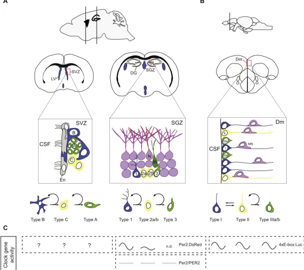
Two main neurogenic regions are found in adult mammalian brains: the subventricular zone (SVZ) of the lateral ventricle and the subgranular zone (SGZ) of the dentate gyrus (DG) in the hippocampus (Grandel and Brand, 2013; Lindsey and Tropepe, 2006). In these two neurogenic niches, different types of neural stem/progenitor cells (NSPCs) have been identified. In striking contrast to mammals and birds, adult teleost fish such as the zebrafish display widespread
neurogenesis from the most anterior to the most caudal regions in the brain (Adolf et al., 2006; Chapouton et al., 2007; Diotel et al., 2010; Grandel and Brand, 2013; Kah et al., 2009; Marz et al., 2010; Pellegrini et al., 2007; Zupanc, 2008). This widespread neurogenic activity is due to the persistence of numerous and distinct neural progenitors throughout the whole brain during adulthood (Lindsey et al., 2012; Marz et al., 2010). The best characterized brain region in the fish known for its high proliferative capacity is the telencephalon. For an overview about the different stem cells of the neurogenic regions in mammals and zebrafish, please seeFig. 3.
The circadian clock seems to influence adult neurogenesis. Rhythmic alteration of adult neurogenesis was first detected in an arthropod (Homarus americanus) (Goergen et al., 2002) in the early 2000's, and has been later described also in the mammalian
hippo-campus (Kochman et al., 2006; Tamai et al., 2008). Furthermore, studies in rodents have shown that jet-lag, a misalignment of the circadian clock with its environment, can inhibit adult neurogenesis and lead to cognitive deficits (Gibson et al., 2010; Kott et al., 2012). These effects may involve clock function in the neurogenic regions themselves. Indeed, clock gene expression in mammals was reported in the neurogenic regions of SVZ and the hippocampus (SGZ of the DG) (Borgs et al., 2009; Bouchard-Cannon et al., 2013; Kochman et al., 2006; Masubuchi et al., 2000; Wilsbacher et al., 2002; Yan et al., 2000), and also shows prominent expression in the neurogenic regions of the adult zebrafish brain (Moore and Whitmore, 2014; Weger et al., 2013). In vitro studies in isolated SVZ and DG spheres have reported that circadian rhythms emerge during neurosphere differentiation and that circadian clock genes may play a role in neurogenesis by affecting
A B
Type 1 Type 2a/b Type 3 Type I Type II Type IIIa/b Type B Type C Type A
B SVZ CSF C En A SVZ N 1 2b 3 SGZ 2a IN SGZ DG Dm CSF I IIIa II MN IIIb Dm ? ? ? n.d.
Clock gene activity
4xE-box:Luc Per2:DsRed
Per2/PER2 LV
C
Fig. 3. Neural stem/progenitor cells in rodents and zebrafish. (A) Sagittal and coronal mouse brain sections illustrating the main neurogenic regions: the subventricular zone (SVZ) of the lateral ventricle (LV) and the subgranular region (SGZ) of the dentate gyrus (DG) of the hippocampus. The SVZ neurogenic niche is composed of astrocytes, B-cells, localized just below the ependymocyte (En) layer that act as neural stem cells. B-cells express the intermediatefilament Nestin and the glial fibrillary acidic protein (GFAP) and only divide rarely to self-renew and give rise to transiently amplifying C-cells. C-cells divide actively and generate A-cells (neuroblasts expressing polysialylated neural cell adhesion molecule (PSA-NCAM)) that migrate along the rostral migratory stream to the olfactory bulbs where their terminally differentiate. The SGZ of the DG is the main other neurogenic niche. It is composed of quiescent type 1 radial-glial like cells that are thought to be the neural stem cells in the DG. Type 1 cells express GFAP, Nestin and SOX2. They can generate proliferating intermediate progenitor cells with transient amplifying characteristics, the type 2 cells. A subset of these cells, type 2a, still expresses radial glial markers (Nestin, SOX2), but lack the characteristic morphology of radial glial cells. Another subset of type 2 cells, the type 2b, express Nestin, but also reveal markers indicating neuronal lineage (doublecortin (DCX), PSA-NCAM, NeuroD). Type 2a/b progenitor cells divide actively and give rise to neuroblasts (type 3) that express neuronal markers. These neuroblasts will then give rise to immature neurons (IN) and eventually to new granule neurons (N). (B) Sagittal and coronal zebrafish brain section illustrating the dorsomedial (Dm) region of the telencephalon, a brain region best characterized for its neurogenic capacity infish. Type I cells correspond to quiescent radial glial cells and are thought to be equivalent to the subependymal astrocytes in mammals. They express radial glial cell markers such as Gfap, and progenitor markers like Nestin and Sox2. Type I cells can give rise to type II cells that are dividing radial glial cells (PCNA-positive). Type II cells then can give rise to type III progenitors (PSA-NCAM) characterized as neuroblasts that can produce neurons. Mn, migrating neuron; CSF, cerebrospinalfluid. (C) In vivo studies of rhythmic clock gene activity in the neurogenic regions in mammals and zebrafish. No in vivo studies on clock rhythmicity were reported for neural stem cells of the SVZ (“?”). Rhythmic Per2:Luc reporter gene expression in the SGZ was detected for type 1 (high amplitude) and type 2a (low amplitude) cells (Bouchard-Cannon et al., 2013). Per2/PER2 levels were reported to be stable in type 1, 2 and 3 cells (Borgs et al., 2009). In zebrafish rhythmic reporter expression (4xE-Box:Luc) was described in all 3 types of neural stem cells of the adult telencephalon (Weger et al., 2013).
neurosphere growth, proliferation and fate commitment (Malik et al., 2015a, 2015b). However, only a few studies have examined functional links between specific clock genes and neurogenesis in vivo. The circadian clock seems to be critically involved in the proper control of neurogenesis in the SGZ by restricting the expansion of both rapidly dividing committed and uncommitted neural precursors and by regulating the entry of quiescent neural stem cells into the cell cycle (Bouchard-Cannon et al., 2013). Specifically, Bouchard-Cannon and colleagues (Bouchard-Cannon et al., 2013) reported an increase of actively proliferating cells in the type 1/2a pools in both Per2 and Bmal1 KO mice. Moreover, Bmal1 and Per2 KO mice lack the circadian gating of cell cycle entry that is observed in wild-type animals, which leads to abolishment of circadian cell proliferation in the SGZ of the KOs (Bouchard-Cannon et al., 2013). In contrast to Per2 KO mice, in Bmal1 KO mice also cell cycle exit frequency is affected. This likely explains why only Bmal1 but not Per2 KO mice exhibited an increased number of proliferating type 2b cells, post-mitotic type 3 cells, and newborn neurons, and indicates that Bmal1 function buffers against overproduction of newborn granule neurons. In line with these observations done in Bmal1 KO, Rev-erbα KO mice have more DCX + (doublecortin expressing) immature neurons, exhibit a higher proliferation rate in the SGZ, and lack the diurnal rhythmicity of neurogenesis in the SGZ (Schnell et al., 2014). As Bmal1 mRNA expression is upregulated in Rev-erbα KOs (Preitner et al., 2002), this could imply that either up- or downregulation of Bmal1 has a similar effect on neurogenesis. However, a detailed mapping of the cell types and cell cycle processes affected in this model is still lacking.
The precise role of Per2 in adult neurogenesis remains controver-sial, as another study reported that the number of both dividing neural progenitors and newborn neurons in the DG are increased in Per2 KO animals (Borgs et al., 2009). Moreover, the role of PER2 in neurogen-esis might be independent of its function in the circadian clock loop. While Bouchard-Cannon and colleagues (Bouchard-Cannon et al., 2013) report rhythmic expression of a Per2 reporter gene in type 1 and weakly in type 2a cells, Borgs et al. (2009) observed constant expression of Per2 transcript and PER2 protein in all proliferating precursors and the subsequently formed immature and mature neu-rons in the DG. Constant expression had already been observed previously for the mRNA as well (Sun et al., 1997; Zheng et al., 1999). Protein expression rhythms in other parts of the hippocampus were more pronounced, and peaking at ZT22-4, roughly in antiphase to the SCN [peaking at ZT 12] (Borgs et al., 2009; Wang et al., 2009). The reasons for these discrepancies are elusive. However, as both endo-genous mRNA and protein levels of Per2 show a lack of, or at least a very low amplitude oscillation in the hippocampus, a non-oscillatory expression of Per2 in these regions is supported by two independent lines of observations. This raises the possibility that the function of Per2 in neurogenesis may be separable from its function in the clock and that the neurogenesis-related functions do not require changing expression levels.
The described neurogenesis studies in Bmal1 and Per2 KOs were carried out in relatively young mice (5 weeks after birth) in order to avoid indirect effects that might be caused by the accelerated aging observed in Bmal1 KO mice (Kondratov et al., 2006). One reason to suspect that age is an important variable is that another study examining neurogenesis in Bmal1 KO mice at 8 weeks of age did not observe significant differences in precursor proliferation compared to wild-type mice (Rakai et al., 2014). A study with even older Bmal1 KO mice (10–15 weeks of age), but still before the onset of age-related pathologies (at 16–18 weeks), showed a significantly reduced pool proliferating neural progenitor cells in the DG, both globally and among the DCX-positive fraction of cells (type 2b and 3 precursors). Given this information, the overproliferation in young Bmal1 KO mice appears to be transient and causes a “premature ‘division-coupled depletion’ of NSPC” in adult mice. This effect might lead to reduced proliferation in older Bmal1 KO mice. Accelerated aging of the
precursor cell populations, potentially involving ROS homeostasis deficiencies, may further aggravate this phenotype.
In addition to the reports in mice, recent studies have investigated the distribution of circadian clock gene expression and activity in the adult zebrafish brain. These studies showed that clock genes are widely expressed in a circadian manner throughout the zebrafish brain and can be detected in the neurogenic regions localized along the ventricle (Moore and Whitmore, 2014; Weger et al., 2013). Further insights were obtained by studies using a transgenic zebrafish line allowing the monitoring of core clock feedback loop activity in vivo (Weger et al., 2013). Mapping reporter expression showed that all types of neural progenitor cells of the adult zebrafish telencephalon possess circadian clock activity. It would be interesting to apply a similar core loop reporter construct in mammals, to examine if and how the differential patterns of clock gene expression described above affect core loop activity in the different cell types. This may give further hints as to whether some clock genes have non-clock-mechanism related functions during mammalian neurogenesis. It might also indicate whether the role of the clock in the more spatially restricted mammalian adult neurogenesis may be different from that in the widespread neurogen-esis in adultfish.
Another major gap of our understanding concerns the respective contribution of stem cell-autonomous clock functions and of systemic processes under clock control to neurogenesis. One such systemic cue might be the stress hormone cortisol that is released in an ultradian and circadian fashion [reviewed in Dickmeis et al. (2013)]. Glucocorticoids appear to regulate hippocampal neurogenesis invol-ving a balance of mineralocorticoid receptor and glucocorticoid recep-tor activation that affects both cell proliferation and apoptosis [re-viewed inDickmeis and Foulkes (2011)]. However, since the experi-ments leading to these conclusions involved adrenalectomy and pharmacological treatment with glucocorticoids or mineralocorticoids, it is unclear if and how the natural ultradian and circadian patterns of glucocorticoid availability are involved in these processes under normal conditions. It is also unknown to what extent glucocorticoid-clock interactions directly affect neurogenic processes and if their role includes the temporal gating of the function of other signals such as neurotransmitters involved in neurogenesis. Neurotransmitters are part of the neurogenic niche environment that can promote or inhibit neurogenesis in different ways [reviewed inBerg et al. (2013), Pardal and Lopez Barneo (2016)]. Circadian changes have been described for neurotransmitter systems including dopamine, serotonin, GABA or glutamate (Cardinali and Golombek, 1998; Castaneda et al., 2004; Kalsbeek et al., 2006; Parekh et al., 2015; Versteeg et al., 2015). One study has provided thefirst hints that interactions of serotonin with the circadian glucocorticoid rhythm participate in the regulation of neuro-genesis by showing that the neuroneuro-genesis-stimulating effect of fluox-etine, a selective serotonin reuptake inhibitor, is dependent on circadian glucocorticoid changes (Huang and Herbert, 2006). Future studies will elucidate whether and how rhythmic signals from systemic cues interact with NSPC clock on controlling cell proliferation in neurogenic niches.
3. Conclusion
It is intriguing that ESCs already show an oscillatory behavior and express components of the circadian clock, even though they appar-ently do not possess a proper functional circadian clock feedback loop. This observation may seem atfirst surprising, given for example that rapid cell divisions and differentiation on shorter time scales are a characteristic feature of embryonic development. One would not necessarily expect a selective advantage for synchronizing these early processes with the day-night cycle. However, it is tempting to speculate that the circadian clock may need to start early during development in order to allow time for its maturation over several day-night cycles, so that it is robustly functional when needed at later stages of
develop-ment. This view would be consistent with the gradual emergence of the transcriptional-translational feedback loop seen upon differentiation of ESCs (Kowalska et al., 2010; Yagita et al., 2010) and during embryonic development (Dekens and Whitmore, 2008; Martin-Robles et al., 2012; Vallone et al., 2007; Weger et al., 2013).
Another idea is that a functional cellular clock may allow for temporal compartmentalization of cellular processes, which otherwise would interfere with each other (Chen and McKnight, 2007; Johnson, 2010; Khapre et al., 2010; Kowalska et al., 2010). For example, oxidant-producing metabolism and the S-phase in mouse skin are precisely antiphasic to each other across the day-night cycle (Geyfman et al., 2012; Stringari et al., 2015). A related possible function for clocks in stem cells could be the avoidance of UV light, which equally generates ROS in cells (Ndiaye et al., 2014) and can damage DNA directly, potentially leading to mutations. Recent work in mice showed that at least one type of DNA repair, nuclear excision repair, is more effective during day time, when the fewest cells are in S-phase (Gaddameedhi et al., 2011). Targeting the S-phase to the night may thus protect the DNA from both metabolic and UV generated ROS and from direct UV damage. Furthermore, with this temporal segregation, the most effective DNA repair is synchronized to when most damage occurs and to when replication does not hinder the repair process (Khapre et al., 2010). Indeed, mice exposed to UV light in late night are more prone to develop skin cancer than those exposed during the day (Gaddameedhi et al., 2011).
However, in the epidermis of the diurnal humans, S-phase peaks during the day, making cells more sensitive precisely at a time when most UV damage is possible (Brown, 1991; Geyfman et al., 2012). Also, proliferating cells in deeper tissues, into which UV does not penetrate, show circadian rhythms of proliferation as well (Plikus et al., 2015). It appears therefore that the length of the circadian oscillations of metabolism and cell proliferation is not necessarily the result of a direct adaptation to cycles of UV exposure. Rather, it may reflect adaptation of the cells to other cycles within the organism, such as metabolic rhythms that in turn evolved in a more direct adaptation to the environmental day-night cycles. Comparative analysis of circadian rhythms in a variety of cell types in both diurnal and nocturnal organisms with different life-styles and of rhythms with non-circadian periods will be needed to provide us with a deeper understanding of their adaptive values. The potential of the circadian clock to adapt to environmental changes was exemplified in a recent study investigating into the impact of aging on the circadian clock of epidermal and skeletal muscle stem cells (Solanas et al., 2017). As demonstrated by this study, aged stem cells remain robustly rhythmic, but undergo a reorganization of their rhythmic transcriptome. This reorganization leads to a loss of rhythmic expression of genes involved in tissue homeostasis and to a gain of rhythmicity in transcription of genes associated with DNA damage. Thus, stem cell clocks appear to show certainflexibility in adapting their output to new conditions.
Coordination with other circadian processes in the body may also be the ultimate reason for the circadian regulation of stem cell activity in other tissues of the body, such as muscle, bone and the nervous system. It is therefore important to obtain a comprehensive under-standing of the circadian processes in these tissues on a global scale, and then look for interactions with the biology of the local stem cell populations. In this context, it appears interesting that the clock sometimes carry out functions in these tissues that are not directly linked to their function in the feedback loop, as discussed above for Per2 in hippocampal neurogenesis. In these cases, the stem cell specific gene function (important for neurogenesis) may have been“hijacked” from being a normal part of rhythmic processes in the tissue due to its particular usefulness for the stem cell function. What exactly such functions are and why particularly the clock genes would be involved is a question for future research.
Generally, since many biological processes including stem cell homeostasis are subject to circadian clock regulation, medical
applica-tions should consider the temporal variation of biological processes for effective treatment approaches. For example, drug treatment can vary in effectiveness and/or side effects depending on the time of their application (Dallmann et al., 2014; Kaur et al., 2013), leading to the idea of“chronotherapy”. Here, one aim is to time the application of drugs to patients in order to minimize side effects, and/or maximize the efficacy of the drug. The idea of timed treatment might be a promising approach and might be applied also to other approaches, including regenerative medicine. Indeed, it was suggested that the success and efficiency of hematopoietic stem cell transplantations could benefit from appropriate timing of stem cell harvesting (Lucas et al., 2008) and transplantation (Scheiermann et al., 2012). Thus, besides providing crucial insights into biology, understanding the relationship of the circadian clock and stem cells may well lead to improved therapies.
Competing interests
BDW is an employee of Nestlé Institute of Health Sciences SA. Acknowledgements
We gratefully thank Frédéric Gachon, Alice Parisi and Elias Gebara for critically reading the manuscript. We acknowledge funding by the Marie Curie Intra-European Fellowship (grant PIEF-GA-2013-625827; to MW).
References
Acosta-Galvan, G., Yi, C.X., van der Vliet, J., Jhamandas, J.H., Panula, P., Angeles-Castellanos, M., Del Carmen Basualdo, M., Escobar, C., Buijs, R.M., 2011. Interaction between hypothalamic dorsomedial nucleus and the suprachiasmatic nucleus determines intensity of food anticipatory behavior. Proc. Natl. Acad. Sci. USA 108, 5813–5818.
Adolf, B., Chapouton, P., Lam, C.S., Topp, S., Tannhauser, B., Strähle, U., Götz, M., Bally-Cuif, L., 2006. Conserved and acquired features of adult neurogenesis in the zebrafish telencephalon. Dev. Biol. 295, 278–293.
Alonso, L., Fuchs, E., 2006. The hair cycle. J. Cell Sci. 119, 391–393.
Amano, T., Matsushita, A., Hatanaka, Y., Watanabe, T., Oishi, K., Ishida, N., Anzai, M., Mitani, T., Kato, H., Kishigami, S., Saeki, K., Hosoi, Y., Iritani, A., Matsumoto, K., 2009. Expression and functional analyses of circadian genes in mouse oocytes and preimplantation embryos: Cry1 is involved in the meiotic process independently of circadian clock regulation. Biol. Reprod. 80, 473–483.
Andrews, J.L., Zhang, X., McCarthy, J.J., McDearmon, E.L., Hornberger, T.A., Russell, B., Campbell, K.S., Arbogast, S., Reid, M.B., Walker, J.R., Hogenesch, J.B., Takahashi, J.S., Esser, K.A., 2010. CLOCK and BMAL1 regulate MyoD and are necessary for maintenance of skeletal muscle phenotype and function. Proc. Natl. Acad. Sci. USA 107, 19090–19095.
Aratani, Y., Sugimoto, E., Kitagawa, Y., 1987. Lithium ion reversibly inhibits inducer-stimulated adipose conversion of 3T3-L1 cells. FEBS Lett. 218, 47–51.
Asakura, A., Komaki, M., Rudnicki, M., 2001. Muscle satellite cells are multipotential stem cells that exhibit myogenic, osteogenic, and adipogenic differentiation. Differ. Res. Biol. Divers. 68, 245–253.
Atger, F., Mauvoisin, D., Weger, B., Gobet, C., Gachon, F., 2017. Regulation of mammalian physiology by interconnected circadian and feeding rhythms. Front. Endocrinol. 8, 42.
Bell-Pedersen, D., Cassone, V.M., Earnest, D.J., Golden, S.S., Hardin, P.E., Thomas, T.L., Zoran, M.J., 2005. Circadian rhythms from multiple oscillators: lessons from diverse organisms. Nat. Rev. Genet. 6, 544–556.
Berg, D.A., Belnoue, L., Song, H., Simon, A., 2013. Neurotransmitter-mediated control of neurogenesis in the adult vertebrate brain. Development 140, 2548–2561.
Bjarnason, G.A., Jordan, R., 2002. Rhythms in human gastrointestinal mucosa and skin. Chronobiol. Int. 19, 129–140.
Bjarnason, G.A., Jordan, R.C., Wood, P.A., Li, Q., Lincoln, D.W., Sothern, R.B., Hrushesky, W.J., Ben-David, Y., 2001. Circadian expression of clock genes in human oral mucosa and skin: association with specific cell-cycle phases. Am. J. Pathol. 158, 1793–1801.
Blanpain, C., Fuchs, E., 2009. Epidermal homeostasis: a balancing act of stem cells in the skin. Nat. Rev. Mol. Cell Biol. 10, 207–217.
Borgs, L., Beukelaers, P., Vandenbosch, R., Nguyen, L., Moonen, G., Maquet, P., Albrecht, U., Belachew, S., Malgrange, B., 2009. Period 2 regulates neural stem/ progenitor cell proliferation in the adult hippocampus. BMC Neurosci. 10, 30.
Bouchard-Cannon, P., Mendoza-Viveros, L., Yuen, A., Kaern, M., Cheng, H.Y., 2013. The circadian molecular clock regulates adult hippocampal neurogenesis by controlling the timing of cell-cycle entry and exit. Cell Rep. 5, 961–973.
Brancaccio, M., Enoki, R., Mazuski, C.N., Jones, J., Evans, J.A., Azzi, A., 2014. Network-mediated encoding of circadian time: the suprachiasmatic nucleus (SCN) from genes