Characterization of the Cardiogenesis of Embryonic Stem Cells by
Chen-rei Wan
M.S. Mechanical Engineering
Massachusetts Institute of Technology, 2005 B.S. Mechanical Engineering
Carnegie Mellon University, 2001
MASSACHUSETTS INSTITUTE OF TECHNOLOGY
MAY 18 2011
LIBRARIES
ARCHIVES
Submitted to the Department of Mechanical Engineeringin Partial Fulfillment of the Requirements for the Degree of Doctor of Philosophy in Mechanical Engineering
at the
Massachusetts Institute of Technology February 2011
C 2010 Massachusetts Institute of Technology. All rights reserved
Signature of the Author:
Certified by:
Roger D. Kamm, Ph.D Singapore Research Professor of Biological and Mechanical Engineering Thesis Chair and Supervisor
Accepted by:
David Hardt, Ph.D Chairman, Department Committee on Graduate Students
Characterization of the Cardiogenesis of Embryonic Stem Cells by
Chen-rei Wan
Submitted to the Department of Mechanical Engineering on 12/06/2010 in fulfillment of the
requirements for the Degree of PhD in Mechanical Engineering
Abstract
Cardiovascular diseases persist as the leading cause of mortality worldwide. Stem cell therapy, aimed to restore contractility and proper vasculature, has gained considerable attention as an attractive therapeutic option. However, proper cell differentiation, survival and integration in an infarcted zone remain elusive. This thesis aims to utilize in vitro techniques to obtain a systematic characterization of how individual stimulations can affect the cardiogenesis process of embryonic stem cells.
First, a compliant microfluidic system was developed to study the individual and combined effects of culture dimensions and uniaxial cyclic stretch on the differentiation process. A smaller culture dimension, with a characteristic length scale of hundreds of micrometers, dramatically enhanced differentiation partly due to an accumulation of cell-secreted and cardiogenic BMP2. Uniaxial cyclic stretch, on the other hand, inhibited differentiation. With this microfluidic platform and a GFP-reporting differentiation cell line, effects of various external stimuli on differentiation were systematically studied.
Next, the effects of collagen I and cell alignment, two biophysical signatures of the adult myocardium, on promoting phenotypic changes of isolated embryonic stem cell derived cardiomyocytes (ESCDMs) were investigated. Effects of collagen I depended on how it was presented to the cells and overlaying collagen gel impeded cell elongation. Binucleation. characteristic of maturing cardiomyocytes, was reduced with soluble collagen supplement and nanoscale topography and was associated with an increase in cytokinesis. Both nanoscale topography and microcontact printing resulted in aligned cardiomyocyte monolayers but produced different morphologies.
Lastly, the lessons learned from studying the aforementioned processes were applied to test the utility of ESCDMs as biological actuators. Three proof-of-concept experiments were conducted: ESCDM monolayers were able to contract synchronously as a cell-assemble, force generated by the cell monolayer was estimated to be comparable to that by neonatal myocytes and lastly, the direction of contraction could be controlled with surface patterning.
This work advances our understanding on the cardiogenic potential of murine embryonic stem cells and elucidated complex biological questions with well-characterized and controlled tissue engineering techniques.
Thesis advisor: Roger D. Kamm
Acknowledgements
First and foremost, I would like to thank my advisor, Professor Roger Kamm. Working with Roger was a bit like swimming in the ocean. I was given the freedom to roam around in the rich and immense sea of knowledge and go at my own pace to figure out what I would like to do. At the same time, when I was drowning in too many experiments that did not work, his encouragements kept my afloat and his advice, like a lighthouse, always pointed me in the right direction. His focused, yet laissez-faire advising style gave me a sense of ownership of the project and taught me how to be a great researcher. But most importantly, Roger was a true teacher who cared greatly about my personal growth as an individual. He let me take a summer off to explore West Africa and gain some business skills. During my graduate career, I had the opportunity to grow both scientifically and personally. For that, I cannot think of a better mentor and advisor. There was also another perk to being Roger's student and that was the chance to play with the sweetest and smartest dog I have ever met. I looked forward to every opportunity to dog-sit Oakley and am pretty certain I always had more fun that he did. For keeping a smile on my face, this thesis is also for Oakley.
I want to also thank my committee members, Dr. Richard T. Lee and Professor Alan Grondzinsky, for their patience and advice and collaborators from the Pruitt lab at Stanford University and Charles Stark Draper Laboratory. Thanks for enriching my intellectual repertoire
and providing me with research suggestions and tools to accomplish my project.
I would also like acknowledge all my friends, especially those who keep me sane, grounded and inspired. Here are some of the people who amazingly put up with me over the years - Mark, Nate, Anusuya, Yannis, Aida, Sid, Ryo, Jessie, Priam, and Levi. Thanks for being the support to
keep me going, the shoulder to cry on when I felt depressed, the dreamer to keep me inspired, the thinker to envision the future, the critic when I needed a reality check, the joker to keep me laughing, and, above all, thanks for being a great friend. You have made this journey pleasant, stimulating and incredibly memorable.
I want to acknowledge all the volunteers at PATS (Pediatric AIDS Treatment Support), particularly Eliza, Alisha, Kristin, Jialan, and Kali. I have the privilege to work with a group of incredibly talented young women on a cause that is dear to my heart. They inspire me to work harder, be more generous and be a better person.
Lastly, I would like to send my most sincere gratitude to my family for their unwavering support. For my mom, now you can finally read my thesis and understand why I said you wouldn't understand what I do. For my dad, the original Dr. Wan, your philosophy and advice have shaped me to be who I am. For my dearest sister and mother of two of the cutest boys on earth, thanks for believing in me and constantly offering your cat's cells for my experiments. Thanks to all the extended family members, uncles, aunts, cousins, and grandmother, for always being there for me.
The past five years have been transformational and full of blessings. It would not have been nearly as enjoyable without all of you. I hope I can continue on with all the support and love and make the world just a little bit better, one cell at a time. @
"When we become more fully aware that our success is due in large measure to the loyalty, helpfulness, and encouragement we have received from others, our desire grows to pass on similar gifts. Gratitude spurs us on to prove ourselves worthy of what others have done for us. The spirit of gratitude is a powerful energizer." - Wilferd A. Peterson.
Table of Contents
Abstract 2
Acknowledgements ... 3
Table of Figures... 9
Table of Tables... 11
Chapter 1. Cardiomyocytes and Cardiogenesis ... 12
1.1. Cardiomyocytes in the Native Myocardium ... 12
1.2. Cardiovascular Disease and Therapeutic Options... 16
1.2.1. Muscle Based Treatment Options ... 16
1.2.2. Adult Stem Cell and Other Progenitor Cell Based Treatment Options... 17
1.2.3. Embryonic Stem Cell Based Treatment Options ... 18
1.3. Utilization of the Contractile Function of Cardiomyocytes for Miniature Machines .... 19
1.4. Induced Organization of Cardiomyocytes... 24
1.4.1. Protein-based Patterning ... 24
1.4.2. Topographical Patterning ... 24
1.5. Applications of Hydrogel Scaffolds on Cardiomyocytes... 28
1.6 . C onclu sion s ... 30
Chapter 2. Generation of Cardiomyocytes Derived from Murine Embryonic Stem Cells and the Study of Differentiation in a Compliant Microfluidic Platform*... 31
2 .1. In trodu ction ... 3 1 2 .2 . B ack ground ... 32
2.2.1. Use of Microfluidic Platform in in Vitro Cell Culture ... 32
2.2.2. Methods to Generate Embryoid Bodies (EBs)... 33
2.2.3. Effect of Mechanical Stretch on Cardiomyocytes in Vitro ... 34
2.2.4. Effect of Mechanical Forces on Embryonic Stem Cell Differentiated C ardiom yocytes in Vitro... 35
2.3. Materials and Methods ... 36
2.3.1. Maintenance of Murine Embryonic Stem Cells... 36
2.3.2. Induction of Differentiation and Maintenance of Endothelial Cells ... 36
2.3.3. Embryoid Body Culture Condition ... 37
2.3.4. Microfluidic Device Design... 37
2.3.5. Stretch Application Apparatus ... 39
2.3.6. Other Stretch Application Considerations... 40
2.3.7. Image Analysis, Quantification and Statistical Analysis ... 41
2 .4 . R esu lts ... 4 1 2.4.1. Validation of Motorized Microfluidic Platform... 41
2.4.2. Differentiated Embryoid Bodies Exhibit Distinctly Different Morphologies in Microfluidic Environment vs. Conventional Well Plates... 43
2.4.3. Microfluidic Environment Enhances the Differentiation of Embryonic Stem Cells into Cardiomyocytes... 44
2.4.4. Effect of Bone Morphogenetic Protein 2 ... 46
2.4.5. Uniaxial Cyclic Stretch Inhibits the Differentiation of Embryonic Stem Cells into C ardiom yocytes ... 47
2.5. Discussions and Conclusions ... 49
Chapter 3. Effect of Extracellular Matrix Protein and Surface Patterning on Driving Maturing Phenotypes of Isolated Embryonic Stem Cell Derived Cardiomyocytes during Extended Culture**... 52
3 .1. In trodu ction ... 5 2 3 .2 . B ack ground ... 5 3 3.2.1. Collagen I in the Myocardium ... 53
3.2.2. Effect of Surface Patterning on Cardiomyocytes and Cardiogenesis ... 54
3.3. Materials and Methods ... 57
3.3.1. Embryoid Body Dissociation and Fluorescent Activated Cell Sorting... 57
3.3.2. C ell Seeding ... 59
3.3.3. Microcontact Printing (MCP) ... 60
3.3.4. Nanopattern Topography (NPT) ... 61
3.3.5. Immunofluorescent Staining ... 61
3.3.6. Quantification and Statistical Analysis of Binucleation, Cell Shape and Cell Alignment 62 3 .4 . R esu lts ... 6 4 3.4.1. Sarcomere development of ESCDMs suspended in collagen gel or plated on ECM-coated polystyrene ... 64
3.4.2. Cell Adhesion with Overladen Collagen Gel Layer... 65
3.4.3. Effect of Collagen I on the Elongation of ESCDMs... 66
3.4.4. Effect of Collagen I on Binucleation... 67
3.4.5. Effect of Surface Patterning on ESCDM Alignment ... 69
3.4.6. Effect of Surface Patterning on ESCDM Elongation... 72
3.4.7. Effect of Surface Patterning on ESCDM Binucleation... 73
3.5. Discussion and Conclusions... 75
Chapter 4. Synchronization, Force Generation, and Control of Contractile Direction of Embryonic Stem Cell Derived Cardiomyocytes... 79
4 .1. Introdu ction ... 79
4 .2 . B ack ground ... 80
4.2.1. Utilizations of Cardiomyocytes as Biomachines in 2D ... 80
4.2.2. Utilizations of Cardiomyocytes as Biomachines in 3D ... 81
4.3. Materials and Methods ... 82
4.3.1. Cell Culture and Isolation ... 82 7
4.3.2. In-phase Contraction Analysis ... 82
4.3.3. Force Generation ... 83
4.3.4. Control over Contractile Directions ... 84
4 .4 . R esu lts ... 8 6 4.4.1. In-Phase Contraction is Detected across Monolayer... 86
4.4.2. Force Generated by Embryonic Stem Cell Derived Cardiomyocytes are Comparable to That of Neonatal Myocytes... 87
4.4.3. Direction of Contraction can be Controlled with Pattern Directions ... 90
4.5. Discussions and Conclusions ... 93
Chapter 5. Conclusions... 97
5.1. Significant Findings and Summary ... 97
5.2. A re W e T here Y et? ... ... .. 10 1 5.3. Future D irections... 105
5.3.1. Microfluidic Platform Development ... 105
5.3.2. Further Investigation into Binucleation... 105
5.3.3. Incorporation of Additional Stimulations ... 106
5.3.4. Investigation of Maturation of ESCDM Electrical Properties ... 107
5.3.5. Incorporation of Patterning in 3D and Other Cell Types ... 108
5.3.6. Self A ssem bly ... 108
5.3.7. Concluding Thoughts ... 109
Appendix I - Embryonic Stem Cell Maintenance and Differentiation Protocol... 110
Appendix II - Cardiomyocyte Enrichment with Percoll Separation... 113 References 114
Table of Figures
Figure 1-1: Cardiomyocyte Orientation in Native Myocardium... 13
Figure 1-2: D ecellularized H eart ... 14
Figure 1-3: Microdevices Powered by Cardiomyocytes ... 23
Figure 1-4: Alignment of Neonatal Myocytes with Microcontact Printing and Nanoscale T op og rap hy ... 2 6 Figure 1-5: Proposed Mechanism of Nanotopography ... 27
Figure 2-1: M icrofluidic D esign... 38
Figure 2-2: Calibration of PDMS film thickness as a function of spin coater rotational speed.. 39
Figure 2-3: Uniaxial Stretch Platform ... 40
Figure 2-4: Validations of the Proper Functions of the Microfluidic Device and the Cyclic Stretch P la tfo rm ... 43
Figure 2-5: Morphological Differences of Embryoid Bodies... 44
Figure 2-6: Method of Differentiation and Representative Images of Differentiated C a rd iom yocytes... 45
Figure 2-7: Microfluidic Environment Enhances Differentiation... 46
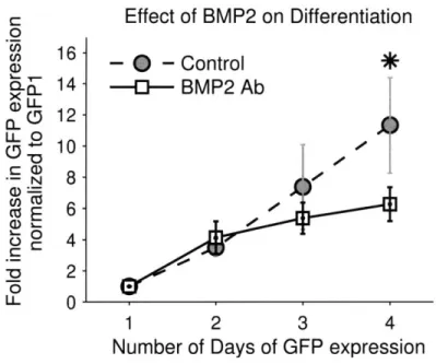
Figure 2-8: Effect of BMP2 on Differentiation... 47
Figure 2-9: Uniaxial Cyclic Stretch Inhibits Differentiation ... 48
Figure 3-1: Cardiomyocytes at Various Developmental Stages... 55
Figure 3-2: Fluorescent Sorting Results... 58
Figure 3-3: Schematics of Cell Seeding Conditions... 60
Figure 3-4: Cell Boundary Identification and Verification with N-Cadherin... 62
Figure 3-5: ESCDMs Embedded in Collagen I Hydrogel... 64
Figure 3-6: Representative Images of ESCDMs on Polystyrene... 65
Figure 3-7: Cell Adhesion to Overlaying Collagen Gel... 66
Figure 3-8: Effect of Collagen I on Aspect Ratio ... 67
Figure 3-9: Effect of Collagen I on Binucleation ... 68
Figure 3-10: Effect of Collagen I on Binucleation on Patterned Surfaces... 69
Figure 3-11: Cell Alignment with Microcontact Printing and Nanopattern Topography ... 70
Figure 3-12: Histogram of Cell Alignments ... 71
Figure 3-13: Effect of Surface Patterning on Elongation without the Addition of Collagen... 72 9
Figure 3-14: Elongation of ESCDMs with Soluble Collagen and Collagen Gel ... 73
Figure 3-15: Effect of Surface Patterning on Binucleation... 74
Figure 4-1: Particle Tracking to Investigate Synchronization... 83
Figure 4-2: Fluorescent Beads to Estimate Force Generation ... 84
Figure 4-3: Schematic Illustration of the Effect of Anisotropy on Contraction Direction... 85
Figure 4-4: In Phase Contraction of Cell Monolayer ... 87
Figure 4-5: M ovement of a Fluorescent Bead... 88
Figure 4-6: Schematic of Force Generation... 90
Figure 4-7: ESCDMs Contract along the Direction of Surface Patterning ... 91
Figure 4-8: Different Methods of Measuring Forces of Cardiomyocytes ... 95
Figure 5-1: Sarcomere organization of ESCDMs compared with adult cardiomyocytes... 102
Table of Tables
Table 1: Bioactuators powered by aggregates of cardiomyocytes in 2D or 3D ... 21 Table 2: Force measurements of cardiomyocytes ... 95 Table 3: Effect of Mechanical Stretch on Differentiation... 107
Chapter 1. Cardiomyocytes and Cardiogenesis
Research on cardiomyocytes has numerous important implications. Cardiovascular diseases are the leading cause of mortality in the world (1) and many fundamental mechanisms of such diseases remain elusive. From a basic science perspective, the highly-ordered hierarchy, from myosin-actin pair, sarcomere organization, myotube formations to the large myocyte sheet structures, calls attention to the fascinating underlying engineering principles in biology. In this chapter, we provide the background necessary to motivate this thesis work. It is structured to present information ranging from a biological standpoint to a more engineering application perspective, from understanding the structure of the native myocardium to in vitro robotic machines powered by cardiomyocytes.
1.1.
Cardiomyocytes in the Native Myocardium
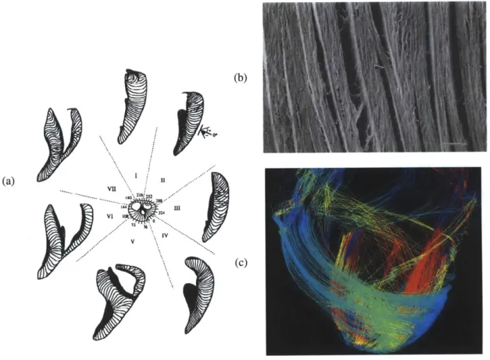
The heart is a chemical-mechanical-electrical biological organ. To ensure the accurate electrical conduction and subsequent contraction, Precise and intricate cell organization has to be maintained. By volume, cardiomyocytes make up 70% of the cell volume in the heart (2). The rest of the cells, such as endothelial cells and cardiac fibroblasts, are critical in adequate nutrient/waste transport and maintenance of the structural integrity by producing adequate extracellular matrix proteins (3,4). From tissue dissections and SEM images, it can be seen that cardiomyocytes (- 4 myocytes thick) are layered as sheets with specific orientations to produce the observed coordinated contractions (5). Helm et. al. used Diffusion Tensor Magnetic Resonance Imaging to illustrate the fiber orientations of a canine heart and found similar orientation results (Figure 1-1) (6). All these studies support the notion that aligned cardiomyocytes with specific orientations are essential in producing the precise twisting motion during each contraction (7,8).
(b)
(a)
V11
(c)
Figure 1-1: Cardiomyocyte Orientation in Native Myocardium
(a) and (b) are reproduced from Legrice et. al. (5). With SEM, they demonstrated high anisotropy in the left ventricle of a canine heart. Cardiomyocytes are arranged in sheets with 1-2 myocyte gaps in
between (Scale bar: 100pm). (c) Helm et. al obtained similar results for fiber orientation with Diffusion Tensor Magnetic Resonance Imaging (6).
In addition to cells, the extracellular matrix (ECM) is equally important. ECM not only acts as a supporting scaffold and a microenvironment for cell-cell interactions, but it also signals to the cells via integrins and focal adhesion complexes (9). The composition of the extracellular matrix is dynamic both temporally and spatially (10,11). Although the exact composition remains unknown, it has been shown that the collagen content increases over time and the activation of
aisi integrin, which primarily binds to collagen I, is necessary for forming the rod-shape phenotype of cardiomyocytes (12). Spatially, the composition and specific orientation of the
13
collagen matrix play a significant role in maintaining the orientation of the cells and accommodating for the mechanical strains during each contraction (4,13). Furthermore, the collagen fibril network is important in preventing cardiomyocytes from over stretching (14,15).
Ott et. al. decellularized mouse hearts with an extensive SDS washing procedure preserving the intricate ECM network comprised of collagen I, collagen III, laminin and fibronectin (Figure 1-2) (16). The shape of the decellularized heart remains intact suggesting the ECM network, rather than cellular constituents, is responsible for the structural integrity. Neonatal myocytes and endothelial cells were introduced into the decellularized tissues and the recellularized hearts were cultured with pulsatile medium perfusion and electrical stimulation mimicking the physiological condition. Even so, the left ventricle systolic pressure was less than 25% of a fetal heart and 2% of an adult heart (17). The limited pumping capability highlights the need for further optimizations of cell seeding protocol and culture conditions.
Figure 1-2: Decellularized Heart
A cadaveric heart was decellularized with SDS for 12 hours. The remaining network consisted of mostly collagen fibrils (16).
In summary, the heart is comprised of various cell types and extracellular matrix proteins. Cardiomyocytes, the cells responsible for contraction, are highly organized, layered as 2D sheets,
14
-and intimately surrounded by extracellular matrix, which is highly dynamic spatially -and temporally. All components are essential in maintaining the proper function of the heart. CVDs usually lead to the maladaptations of the extracellular matrix and loss of cellular functions.
1.2. Cardiovascular Disease and Therapeutic Options
To step back and examine the prevalence and significant disease burden of CVDs, one easily comes to the conclusion that better and more widely available treatments are urgently needed. A large portion of CVDs is attributed to myocardial infarction (MI) from coronary artery occlusions. MI results in rapid and massive cardiomyocyte death. On average, 1 billion cardiomyocytes undergo cell death in an MI (18). Effective replenishment of functional cardiomyocytes along with revascularization is the Holy Grail of numerous cell therapy strategies.
Current clinical treatments after a severe MI include surgical procedures, such as implantation of left ventricle assisting device (LVAD) or a heart transplant. Neither of them is ideal and both are very costly. As CVDs become a global issue, a major paradigm shift is necessary to repair the damaged tissue and restore its function. Cell-based therapy is a novel approach and has shown initial promises (19-23). In the following section, I will briefly summarize the current state-of-the-art on different cell-based therapies.
1.2.1. Muscle Based Treatment Options
Unlike post-mitotic cardiomyocytes, skeletal muscle cells and their progenitor cells proliferate. Myoblasts respond to injuries and differentiate into skeletal muscle cells. Researchers have attempted to replenish an infracted area with skeletal muscles or precursor cells with inconsistent successes. Taylor et. al. reported a beneficial effect of skeletal myoblast transplantation (24) while Menasche et. al. failed to exhibit statistically significant effects in a double-blind six months study (25). Although the mechanism of the transient benefits of myoblast transplantation remains unanswered, several challenges are more consistently reported. Massive cell death is reported within the first hour of implantation, and surviving muscle cells do not express cardiac
specific proteins (26). Skeletal muscle and myoblast treatments are further complicated with induced arrthymias and tachycardia (27). While the possibility of autologous transplantation makes this treatment option attractive, proper cell homing, integration, trans-differentiation and survival have proven to be challenging.
1.2.2. Adult Stem Cell and Other Progenitor Cell Based Treatment Options
Most adult stem cells are multipotent, able to self-renew and differentiate into several, but not all, cell lineages. Until recently, cardiogenesis with adult stem cells has focused on the potential transdifferentiation of mesenchymal stem cells (28,29). Cardiac stem cells and induced pluripotent stem cells are discovered within this past decade offering new treatment possibilities.
Adult progenitor cells such as bone marrow derived stem cells, mesenchymal stem cells (MSCs), and adipose stem cells have been studied for their potential in differentiating into cardiomyocytes. MSCs naturally differentiate into osteoblasts, chondroblasts and adipocytes when exposed to the appropriate stimuli. There have been some reports of transdifferentiation (30). However, it is unclear whether transdifferentiation actually occurred or the results were obscured by cell fusion events (31-33). While there is no consensus on MSC's capacity to transdifferentiate, the transient benefits of MSCs have been largely speculated to originate from the paracrine effects in increasing capillary density around the infarct zone or tissue mass to reduce the stress on the failing myocardial wall (34-36). Regardless of the exact mechanism, repeatable and long term improvements of cardiac function with MSC implantation remain elusive (37,38).
Since transdifferentiation is controversial, much attention has been paid to progenitor cells with the known capacity to differentiate into cardiomyocytes. Cardiac stem cells have been
discovered in the past decade and there has been a lot of interest in understanding and isolating cardiac stem cells for regeneration (39,40). In animal studies, there was wide variance on how these stem cells assisted in restoring cardiac function (41-44). The variability came largely from the differences in cell isolation and identification techniques. More importantly, it also came from the limited understanding of mechanisms of these cells in the myocardium. Much more fundamental understanding is required before cardiac stem cells can be fully utilized as a treatment option.
1.2.3. Embryonic Stem Cell Based Treatment Options
Cell therapy with embryonic stem cell derived cardiomyocytes (ESCDMs) has been investigated because their ability to self-renew and differentiate into all cell types. Unlike skeletal muscle precursors, ESCs have been successfully differentiated into cardiomyocytes with similar phenotypes and characteristics as fetal cardiomyocytes (45,46). ESC injection into the myocardium induces the formation of teratomas (18). Laflamme et. al. demonstrated electromechanical coupling of transplanted ESCDMs into the host myocardium at 4 weeks but a separate study showed no benefit of transplantation at 12 weeks (47,48). The conflicting and complex findings on ESCDMs illuminate the need for a fundamental mechanistic understanding and characterization of the cardiogenesis process of embryonic stem cells (49).
1.3. Utilization of the Contractile Function of Cardiomyocytes for Miniature Machines
In the previous sections, in vivo cellular and extracellular compositions were described. The therapeutic importance of stem cell derived cardiomyocytes as a new source of functional cardiomyocytes after injuries was also discussed. In this section, the focus is shifted and the potential to utilize cardiomyocytes as a source of contractile units is explored.
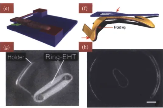
Neonatal cardiomyocytes, harvested within 2 days after birth, are the gold standard for the cell-powered miniature machines because, unlike adult cardiomyocytes, neonatal myocytes contract spontaneously and continuously and do not lose their phenotype in vitro. Tanaka et. al. seeded neonatal cardiomyocytes on the surface of a hollow PDMS sphere and on a thin PDMS sheet to devise spherical and linear pumps respectively (Figure 1-3 a&b) (50,51). For the hollow PDMS sphere, the contractile force generated by the myocyte sheets was strong enough to squeeze the sphere creating a pulsatile flow through the glass capillaries connected to the sphere. This device was extremely delicate and difficult to produce and while the objective was to create a heart-like flow chamber, it resembled little of the heart. Kim et. al. similarly seeded neonatal cardiomyocytes on 2D to construct a walker (Figure 1-3c) (52). The walker was fabricated with soft-lithography techniques and capable of propelling forward for over one week. This hybrid design, albeit sophisticated in fabrication, lacks real world applications. Feinberg et. al. induced the alignment of neonatal myocytes on a thin PDMS film microcontact printed with 20/2Opm line patterns of high (50ig/ml) and low (2.5pg/ml) concentrations of fibronectin. By cutting the monolayer sheet along the direction of cell alignment, the monolayer produced a forward motion and in contrast, when the rectangular monolayer sheet was cut at a 5-15' angle from the cell alignment, it resulted in a twisting motion (Figure 1-3d) (53). This study is one illustration in
taking advantage of the intrinsic cell responses to extracellular matrix protein variations to produce efficient bioactuators.
In addition to utilizing cardiomyocytes by seeding them on a 2D substrate, Xi et. al. combined material science to seed cells in innovative arrangements (Figure 1-3 e&f) (54). They used the thermoresponsive polymer, poly N-isopropylacylamide (PNIPAAM), with the Lower Critical Solution Temperature (LCST) at 32'C, below which PNIPAAM is hydrophilic. In room temperature, microstructures, coated with gold, were embedded in PNIPAAM hydrogel. Cells were subsequently seeded on the hydrogel with a preferred attachment to the gold surface. Upon cell adherence, the system was heated above LCST resulting in PNIPAAM becoming hydrophobic and the hydrogel collapsing leaving cells attached only to the microstructures.
Last category of machines mixed neonatal myocytes or ESCDMs in an extracellular matrix hydrogel (Figure 1-3 g&h) (55,56). Cell-gel mixture was polymerized in a toroid-shaped mold. Once it was polymerized, it was released and stretched to induce alignment. After an extended period of stretch (>7 days), the cell-gel band was able to spontaneously contract, albeit asynchronously.
In this section, we provided a summary of existing designs of micromachines powered by cardiomyocytes - either by seeding them in 2D, utilizing unique thermo-responsive materials or mixing with 3D hydrogel. The limitations and force output of each design are listed in Table 1. Specifically, many 2D experiments were terminated within 1 week and phenotypic changes were observed without further investigation (50). The pumps and the walker did not utilize the intrinsic behaviors of cardiomyocytes, such as cell alignment or synchronization. Instead, it deployed sophisticated fabrication techniques and used cardiomyocytes only as a miniature
actuator. This approach generally produces much lower force. In Chapter 4, we will explore how to harness the understanding of cardiomyocytes as a cell population into implementing microdevices.
Noatal
Myopytes70 pN* 11 days Difficult to fabricate
Require PDMS support Lack of feasible therapeutic applications
Small forces
Unaligned cell monolayer
60 pN Upuire PDMS support
2KPa 3 days Lack of feasible therapeutic applications
Short term experiments Q.51mN (Fmax)
Dif~uttg44Myceapurity
Toroid~o skalwts Mysologil relevance
Decellularized Neonatal 320Pa 8 days Small pressure
and recellularized myocytes heart (16)
Wtvetr~e Adult mnyocytt~s l 4Ka** Adult'
vivo
Fetal ventricle(57) Fetal myocytes 350mL/min per Fetal
kg lamb in
vivo
Table 1: Bioactuators powered by aggregates of cardiomyocytes in 2D or 3D
(55)
*: the forces are estimated as Stokes' drag force assuming the kinetic viscosity of medium to be 0.7x10⁻³ NsIm2; **: determined by estimating forces required for PDMS deflection ***: assuming
LVEDP-10mmHg and peak stress -120mmHg
Uh
()
F)
Figure 1-3: Microdevices Powered by Cardiomyocytes
(a,b) a spherical pump and a vertical pump produced by Tanaka et. al. (50,51) (c) PDMS-based walker developed Kim et. al. (52) (d) patterned neonatal myocyte sheets by Feinberg et. al. (53). (e,f) A myocyte cantilever and a walker developed by Xi. et. al. with PNIPAAm and microfabrication (54).
(g) three-dimensional engineered heart tissue (EHT) (55). (h) Guo et. al. developed similar 3D hydrogel-cell construct (56).
1.4. Induced Organization of Cardiomyocytes
One pertinent tissue engineering technique to control cell alignment is surface patterning. Cardiomyocytes and the underlying extracellular matrix network are both highly anisotropic in
vivo and to reproduce similar anisotropy in vitro, two techniques have been widely used -patterning with varying protein distributions or imposing direct topographical cues.
1.4.1. Protein-based Patterning
Microcontact printing technique was developed in the Whitesides and Ingber laboratories in the mid-90's (58) and since then, it has been widely used to control cell shapes and functions. Line patterns of alternating concentrations of ECM proteins are often used to produce aligned monolayers, as Feinberg et. al. did with fibronectin (Figure 1-4 a&b) (53). Variations in line widths (12.5pm/12.5pm lines vs. 25pm/25pm lines) have been tested and no statistically significant differences in the electrophysiological properties of neonatal myocytes was found by measuring the conduction velocities, wavelengths and action potential durations in the longitudinal and traverse directions (59). This study suggests that the exact dimensions of protein patterning, within a reasonable range, do not affect cell behaviors. Microcontact printing, unlike topographical cues, does not induce additional geometric constraints on cells which can complicate analyses. However, since many cells secrete their own extracellular matrix proteins, the efficacy of microcontact printing is likely to be transient.
1.4.2. Topographical Patterning
With the advancement of microfabrication techniques, nanometer-scale patterns can be made and this has been used for biological applications. Cells seeded on top of the nanotopography align along the direction of the patterns (Figure 1-4 c&d) (60). The exact mechanism of the alignment is still unclear but one speculation assumes the alignment is a result of integrins distributing
along the ridges. Kim et. al. recently proposed a different hypothesis suggesting that the effect of nanoridges and grooves is attributed to an increase of cell spread area and contraction-mediated stress (Figure 1-5a) (61). Cell spread area of neonatal myocytes were significantly larger on 800nm/800nm ridges/grooves surface compared with 400nm/400nm surface or unpatterned surface because cells seeded on 800nm/800nm surface extended and spread into the grooves while those on 400nm/400nm surface did not (Figure 1-5b&c). Despite the phenotypic differences, the conduction velocities of the two conditions were not statistically different. Furthermore, whether or not nanoscale topographical cues exist in vivo remains controversial. Nevertheless, nanoscale topography has the advantage over microcontact printing in providing the alignment cues over a long period of time.
Cell sensitivities to variations in nanotopography (periodicity, duty cycle and depth of the grooves) are unknown. Au et. al. demonstrated qualitatively clearer sarcomeres for neonatal myocytes seeded on a 1pm ridge/lpm groove surface compared with that on a 1pm ridge/4pm groove surface although the cause for this difference remains elusive (60). Additional studies are necessary to quantify the effects of nanotopography on cells.
Figure 1-4: Alignment of Neonatal Myocytes with Microcontact Printing and Nanoscale Topography (a,b) Cardiomyocytes aligned along 20pim/20pm line pattern of alternating concentrations of fibronectin (53). (c,d) Nanoscale topography was imposed on neonatal myocytes to induce alignment.
Fa.m
(a)
(b)
(c)
I
Conduction velocityFigure 1-5: Proposed Mechanism of Nanotopography
Kim et. al. suggested that cellular response was greatly impacted by the nanoscale topography (61). (a) a schematic of their hypothesis was illustrated. Areas of direct plasma membrane-substratum contact are highlighted in green and gap junctions are shown in red. (b,c) SEM image of neonatal
myocytes seeded on 800nm/800nm and 400/400nm ridge/groove patterns respectively. Scale bar: 200nm
tColl penetration I nto dhe grooves Cefi-subsatum adhesion Cell Con&acuon spreading mediatedsm as
I
TCx43
WCiil coupling 17, 9W,I i I
1.5. Applications of Hydrogel Scaffolds on Cardiomyocytes
In addition to surface patterning, another commonly-used tissue engineering technique is the incorporation of hydrogel scaffolds. The hydrogel can be naturally-present ECM components such as collagen I or basement membranes, or synthetic materials, such as polyacrylamide or self-assembly peptide gels. Several groups have used hydrogels to study cardiomyocyte properties in vitro. One common application is to use hydrogels as a cell culture supporting scaffold. Leung et. al. used a mixture of collagen I and MatrigelTM to study endothelial cell-neonatal myocyte co-culture and found the cell sheet to be electrically responsive (62). In our laboratory, we have also demonstrated that co-culture of endothelial cells and neonatal myocytes on a synthetic peptide gel promoted cardiomyocyte survival (63).
Another property of in vitro hydrogel scaffolds is the tunability of the substrate stiffness. Bhana et. al. cultured neonatal myocytes on polyacrylamide gels of different stiffnesses by varying cross-linking densities (64). They found that cardiomyocytes displayed the clearest striations and the highest contractile force development when cultured on a substrate with similar stiffness as the native myocardium.
In addition to providing the structural support, the composition of ECM hydrogel is critical to the activations of integrins for cell binding and further mechanotransduction responses. For example, EHTs developed by Zimmermann et. al. is comprised of a mixture of collagen I, Matrigelm, and chick serum which activates multiple integrins (55). The engagement and activation of multiple integrins, such as aip,, a3Pi, a15 i and a7pi, are necessary for the formation of a coherent
cardiomyocyte structure as neonatal myocytes and ESCDMs suspended in collagen I alone are unable to extend and form cell-cell contacts (65).
These are several examples of how in vitro tissue engineering tools can be used to characterize cell behavior. In Chapter 3, we will investigate the effect of collagen hydrogel on the phenotypic changes of embryonic stem cell derived cardiomyocytes.
1.6.
Conclusions
This chapter provides the necessary background to motivate this thesis work whose overarching aim is to obtain a better understanding of the cardiogenesis process in vitro. This is essential because of the increasing epidemic of cardiovascular diseases and the lack of viable and affordable options. Stem cell based therapies have been studied for over 25 years and no consistent success has been reported. Most in vivo studies are plagued with high complexities (e.g. cell survival, integration and safety) and experimental artifacts (e.g. cell fusions) (18,49). In
vitro characterizations are a powerful tool to systematically elucidate individual stimuli on the
cardiogenesis process.
To our knowledge, it has not been possible to produce mature cardiomyocytes from undifferentiated embryonic stem cells in vitro. Several key questions still remain to be answered: 1. how is differentiation affected by in vitro culture conditions? and 2. how do nascently differentiated cardiomyocytes develop more mature phenotypes? In the following two chapters, we separately addressed the two questions. First we examined how the differentiation process could be modulated with the changes in culture dimension and mechanical stimulations. Second, we extended the investigation to characterize the morphological changes of differentiated and isolated cardiomyocytes with the presence of collagen I and surface patterning and showed that these biophysical factors can influence the maturing phenotypes. Lastly, we explored the possibility to use these cells for non-medical applications. The findings of this thesis have direct implications on how differentiation and maturation can be augmented with the appropriate biophysical stimuli.
Chapter 2. Generation of Cardiomyocytes Derived from Murine Embryonic
Stem Cells and the Study of Differentiation in a Compliant Microfluidic
Platform*
* The majority of the work presented in the chapter has been submitted.
2.1. Introduction
In this chapter, the differentiation of embryonic stem cells into cardiomyocytes, a process termed cardiogenesis, was investigated. Studying cardiogenesis is important since embryonic stem cells have been considered as a therapeutic means for cardiovascular diseases. As described in the last chapter, there is limited understanding of how cardiogenesis is affected by biophysical stimulations. Here, cardiogenesis was investigated with a compliant microfluidic platform which allows for versatile cell seeding arrangements, optical observation access, long term cell viability, and programmable uniaxial cyclic stretch. Specifically, two environmental cues were examined with this platform - culture dimensions and uniaxial cyclic stretch. First, the differentiation process was enhanced in microfluidic devices compared with conventional well-plates, partially from the accumulation of cardiogenic factor, BMP2, in a confined space. Second, uniaxial cyclic stretch at 1Hz was found to have a negative impact on differentiation. This microfluidic platform builds upon an existing design and extends its capability to test cellular responses to mechanical strain. It provides capabilities not found in other systems for studying differentiation, such as seeding embryoid bodies in 2D or 3D in combination with cyclic strain. This study demonstrates that the restricted transport in a microfluidic system contributes to enhanced differentiation and may be a superior platform compared with conventional well plates. In addition to studying the effect of cyclic stretch on differentiation, this compliant platform can also be applied to investigate other biological mechanisms. The results in this study illuminate the importance of
biophysical cues on the differentiation process and provide guidance as to how these cues can be effectively used to improve the differentiation process.
2.2. Background
2.2.1. Use of Microfluidic Platform in in Vitro Cell Culture
Microfluidic devices (ptFDs) are excellent in vitro systems in which to study cell functions, build disease/organ models, and dissect mechanisms of specific stimulations in a systematic manner (66). Previous work from our laboratory has demonstrated that pFDs can be used to examine interactions of multiple cell types and effects of chemotaxis on angiogenesis and cancer cell migration (67,68). The versatile design allows for cell seeding arrangements in both 2D and 3D, application of shear stress or interstitial flow, and microscope access for continuous observation. In this study, a modified device, capable of imposing periodic uniaxial stretch without sacrificing the imaging capabilities, was developed to study the differentiation of embryonic stem cells (ESCs) into cardiomyocytes. With this platform, we were able to study how cyclic stretch affects the cardiogenesis process in a well-controlled microfluidic system.
Previous work has shown that murine cardiogenesis, involving the generation and manipulation of embryoid bodies (EBs), can be augmented both biochemically and biophysically. Biochemically, ascorbic acid, DMSO, retinoic acid, FGF and BMP2/4 are some of the growth factors that have been demonstrated to promote cardiogenesis and new cardiogenic chemicals are continuously being discovered (69-77). Biophysically, control of EB size, electromagnetic stimulation and mechanical strain have also been shown to enhance cardiac differentiation (78-81). In this study, we attempt to utilize a microfluidic system to impose biochemical and biophysical stimulations to EBs.
Microfluidic platforms have been shown to affect diffusion-dominated processes (82). Yu et. al. demonstrated that cell proliferation rate was dependent on the height of the microchannels, presumably due to an accumulation of secreted factors. By comparing microchannels to conventional cell culture well plates, higher proliferation rates have been observed for murine mammary gland cells and during murine embryo development (83,84). Existing literature suggests that the diffusion of growth factors enhances cell processes, such as proliferation, and augments cardiogenesis, so we hypothesize that differentiation will be enhanced in the confined
space of microfluidic devices.
2.2.2. Methods to Generate Embryoid Bodies (EBs)
EBs are aggregates of embryonic stem cells and are regarded as the gold standard of murine embryonic stem cell differentiation (85). EBs are formed in a low stress suspension culture. Different methods have been used to efficiently generate EBs of uniform sizes including hanging drops, seeding within microwells, rotating bioreactors and culturing within non-adherent
scaffolds. These methods are superior to monolayer culture or single cell suspension culture with higher EB yields and more homogenous EB sizes. Rotating bioreactors offer the possibility of large-scale production (86) but require additional equipments. Hanging drops method can be performed in any laboratory with cell culture facilities. Microwells and non-adherent scaffold
methods are ideal for designing EBs with specific sizes (78).
EBs develop three germ layers over time as observed in development. The differentiation lineage can be directed by exposing EBs to the appropriate set of stimuli. While EBs are necessary in the induction of differentiation, they pose unique challenges in transport and dissociation for further characterizations (87). Considerations regarding to various dissociation techniques are discussed in Section 3.3.1.
2.2.3. Effect of Mechanical Stretch on Cardiomyocytes in Vitro
The effect of mechanical stretch on differentiated myocytes in vitro has been studied in great detail. It has been demonstrated that cyclic stretch is required for neonatal myocytes suspended in hydrogel to form a coherent tissue construct and develop sarcomeres and cell-cell contacts (22). In vitro studies have also provided invaluable knowledge on how mechanical stress induces protein expressions and/or activations. Bullard et al. found that protein kinase C (PKC) phosphorylated differently depending on the direction of stretch, cyclic or static and the magnitude of stretch (88). Furthermore, Mansour et. al. suggested that PKCc and focal adhesion kinase (FAK) are necessary to restore the resting length of sarcomeres after uniaxial static strain (89). Mechanical stress has also been demonstrated to affect the production and distribution of junctional proteins such as connexin 43 and cadherins (90,91).
Mechanical stretch has also been proven to cause morphological changes in myocytes. One in
vitro study suggested that neonatal rat ventricular myocytes display adult myocyte characteristics
after two weeks of culture under mechanical stretch with an increased and better-organized sarcomere structure (92). Another study found in vitro culture of cardiac myocytes align along the direction of stress (88), a process that is apparently mediated by N-cadherin and Rac-1 (91,93). Yet, another study by Kada et. al. found that orientation of myocytes changes depending on the time course of stretch stimulation (94). All current data on protein expression/activation and cell morphology have unequivocally demonstrated the importance of mechanical stimulation on differentiated cardiac myocytes.
2.2.4. Effect of Mechanical Forces on Embryonic Stem Cell Differentiated Cardiomyocytes in Vitro
While the mechanotransduction pathways of differentiated cardiomyocytes have been extensively studied, the effect of mechanical stimulation on the differentiation of embryonic stem cells (ESCs) into cardiomyocytes and the mechanisms of action are less clear (95-97). Schmelter et. al. suggest that mechanical stretch activates the reactive oxygen species signaling pathway and thus enhances the differentiation of murine embryonic stem cells into cardiomyocytes (79). Opposite results indicating that stretch inhibits differentiation have also been shown, attributed to the activation of TGF-p/Activin/Nodal pathway (98). These conflicting results illustrate the need for further studies.
It is also important to note that current studies on EB cardiogenesis with stretch are limited to a two dimensional seeding condition. Three-dimensional environments, however, resemble more closely the native myocardial environment during development and myocardial infarct zones targeted for stem cell therapy (99-101). Therefore, a microfluidic system which allows EBs to be seeded in 3D and experience cyclic uniaxial stretch might provide valuable new insights into the differentiation of embryonic stem cells into cardiomyocytes, and might also elucidate other important cellular behaviors where mechanotransduction is implicated.
2.3. Materials and Methods
2.3.1. Maintenance of Murine Embryonic Stem Cells
Murine embryonic stem cells (mESC) expressing a cardiac specific a-MHC promoter that was tagged with green fluorescent protein (GFP) (line CGR8, kindly provided by RT Lee, Harvard Medical School) allowed direct observation of differentiation into cardiomyocytes. To maintain ESCs in an undifferentiated state, Glasgow Minimum Essential Medium (GMEM) (Invitrogen), supplemented with 1,000U/ml leukemia inhibitory factor (LIF, Sigma), 1mM Sodium Pyruvate (Invitrogen), 1x Non-Essential Amino Acid (Invitrogen), 15% Knockout Serum Replacement (Invitrogen), 25mM of HEPES, 10-4M
p-mercaptoethanol
(Sigma), and 1x Penicillin-Streptomycin (Invitrogen) was used. Cells were maintained in flasks coated with 0.1% gelatin in PBS. Cell confluency was tightly controlled not to exceed 70%. The detailed protocol can be found in Appendix I.2.3.2. Induction of Differentiation and Maintenance of Endothelial Cells
By removing LIF and creating a three-dimensional environment, mESCs spontaneously differentiated. The composition of the differentiation medium was identical to that of the maintaining medium except for the removal of LIF, the replacement of knockout serum by ESC Fetal Bovine Serum (Invitrogen), and the addition of 100pM of ascorbic acid (74).
A standard hanging drop technique was used to induce differentiation (102). Briefly, cell suspension solution was prepared at 10,000 cells/ml. 30pl drops were placed on the inside of a 100mm non-tissue culture treated Petri dish containing approximately 10ml of 1x PBS to prevent evaporation. Drops, containing small cell aggregates, were cultured for 2 days before being
collected with a 10ml pipette. These aggregates were then cultured in differentiation medium for 3 more days for embryoid body (EB) formation.
Human microvascular endothelial cells (hMVECs, Lonza) were cultured with complete EBM-2 (Lonza). Passages 4-7 were used for experiments with stretch stimulation.
2.3.3. Embryoid Body Culture Condition
Embryoid bodies (EBs) are aggregates of ESCs. For the 2D experiments, 5-8 EBs were seeded into pFD channels or onto 12-well plates coated with 0.1% gelatin. To seed EBs in 3D, stock collagen I solution derived from rat tail tendon (BD Biosciences) was mixed with 0.5N NaOH, lOx DMEM, water, medium containing 500 EBs/ml to produce a pH 7.4, 2mg/ml collagen I gel containing EBs. 10ptl of gel were used for each microfluidic device.
Specific device preparation protocol and gel filling techniques have been described previously (67). Briefly, pFDs were permanently bonded with plasma and coated with 0.1% gelatin. Then they were dried overnight in an 80'C oven to restore PDMS hydrophobicity. Channels were filled with differentiation medium after collagen gel had fully polymerized.
2.3.4. Microfluidic Device Design
Microfluidic devices (pFD) were used to study differentiation. pFDs have been designed in our laboratory to study various biological processes, such as angiogenesis, cell migration, and liver tissue engineering (67,68). The existing design consists of three fluid channels and two hydrogel scaffold regions (Figure 2-la). It has the following key advantages:
1. Cells can be cultured either on 2D or in 3D,
2. Devices are made with polydimethylsiloxane (PDMS), a cell-benign and transparent elastomer, and glass coverslips - allowing for cell live imaging, and
3. Devices allow for adequate nutrient, gas, and waste transport.
Modifications were required in order to apply uniaxial cyclic stretch without sacrificing the advantages of the existing design. These modifications included (Figure 2- lb&c),
1. Channel patterns were made on a 0.5-1mm thick PDMS layer, rather than 5-7mm, 2. Coverslips in the original design were replaced by a PDMS film of similar thickness, and
Rectangular, instead of circular, devices were used with additional clamp anchors.
(a) (C)
Figure 2-1: Microfluidic Design
The schematics of the microfluidic device: the original design (a), the updated flexible device (b) and the sample (c) of the final design which satisfied all the design criteria.
A spin coater was used to produce thin films of PDMS. The films had to be thin, pliable and at
the same time easy to handle with a tweezer. We calibrated the spin coater to determine the
relationship between rotational velocity and film thickness (Figure 2-2). In this study, PDMS film was produced with the spin coater at 500 rpm for 30 seconds.
PDMS Film Thickness 600 500 400 300 -y = 56522x-0.894 200 - R2= 0.9726 100 -0 ,I I I I 0 200 400 600 800 1000 1200 Rotational Velocity [rpm]
Figure 2-2: Calibration of PDMS film thickness as a function of spin coater rotational speed PDMS with 10:1 base to curing agent was used to calibrate the thickness. PDMS was spun for 10
seconds to spread over the surface and 30 seconds at the indicated velocity. 2.3.5. Stretch Application Apparatus
The actuator to apply cyclic stretch to the pFDs was chosen based on the following requirements:
" Max Travel Distance = (Maximal Strain, 20%) x (Total Length, 10mm) = 2mm
* Accuracy = (Min Travel Distance) x (Allowable Error) = (Min Strain, 5%) x (Total Length, 10mm) x (Allowable Error, 5%) = 12.5pm
" Max Velocity = Max Travel Distance x Max Frequency = 2mm x 10Hz = 20 mm/s
Parker MX80S precision linear motor (Irwin, PA) satisfied all the requirements. To prevent rust, the motor was enclosed in a stainless steel box. Parker MX 80S is capable of withstand a thrust load up to 123N, much higher than forces required to stretch 4 pFDs.
To connect pFDs to the linear motor, an in-house made clamp was used (Figure 2-3). The clamp could accommodate up to 4 pFDs for one set of experiments. One side of the clamp was firmly attached to the plates inside the incubator while the other side was connected to the linear motor.
(a)
Controller Incubator (95% humidity,
Controlle 370C, 5% CO -) Stepper Linear
Micro-Motor Precision fluidic Anchor Driver I Stage Devices
Power a
(b)
Commanded position vs. Time
E 1000 5 0 E X -00 0 1 2 4 -1000
(b)
Figure 2-3: Uniaxial Stretch Platform
Design of the stretch platform: (a) schematic diagram illustrates that the stretch apparatus was placed in the incubator; (b) displacement could be precisely controlled.
2.3.6. Other Stretch Application Considerations
The most commonly used and commercially available stretch platform is the Flexcell* system which imposes programmable equibiaxial cyclic or static strain. The system replaces the bottom of a conventional 6-well plate with a flexible where cells can be cultured. This setup requires a large amount of cells and also limits cell seeding to a 2D culture. Furthermore, while equibiaxial strain is applicable in some physiologically relevant settings, it is more appropriate to apply uniaxial stretch to mimic what cardiomnyocytes experience in the heart. Because of the limitations of the need for a large number of cells, cell seeding conditions and stretch directions, we decided
40 . ..... ....
to build an in-house system where a small amount of cells can be stretched uniaxially in a microfluidic condition.
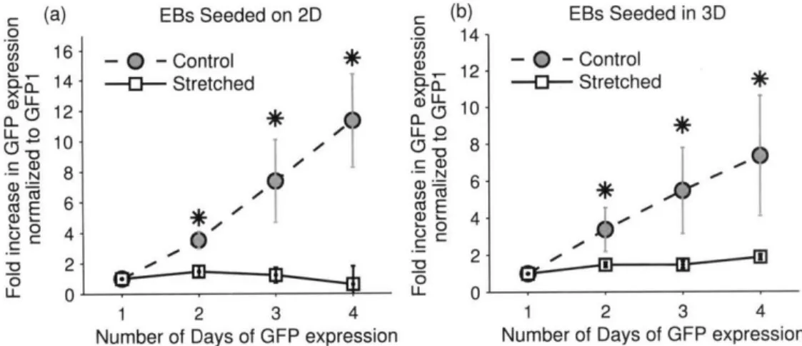
2.3.7. Image Analysis, Quantification and Statistical Analysis
Both phase contrast and fluorescent images (20x) were taken with a Nikon Eclipse TE300 Microscope with Open Imagem Software. Individual embryoid bodies (EBs) were tracked and observed daily for GFP expression with identical exposure settings. The first day that GFP expression could be observed was defined as GFP1 and subsequent days as GFP2, GFP3 and so on. Images were taken daily and analyzed with Matlab. Without any contrast enhancement, a GFP-positive pixel was defined to be brighter than 120 on a 256 gray scale image. Most differentiation occurred on the flat parts of the adherent EBs that have spread outwards so the errors due to measuring the 2D projection were considered to be small. Still, direct comparisons between the 2D and 3D experiments are subject to error.
All data are presented as mean ± SEM. The Student's t-test was used to identify statistical significance (p<0.05). At least 20 EBs were examined and more than 3 independent samples were used for each condition.
2.4. Results
2.4.1. Validation of Motorized Microfluidic Platform
The compliant tFDs were connected to a precision linear stage which could be programmed to translate at specific frequencies and magnitudes (Figure 2-3). Cells seeded in the p1FDs were stretched for 24 hours at 10% strain and 1Hz after they have fully adhered after 3 days.
Two different validation tests were initially conducted to ensure that cells actually experienced the imposed mechanical stimulation. First, we confirmed that the gel could withstand cyclic
stretch without fracturing or detaching from the PDMS walls. A 2mg/mi collagen gel was injected into the pFD, allowed to polymerize, then subjected to cyclic stretch of different strains. At 12% strain, collagen gel in the device remained well adhered to the PDMS walls of the pFDs (Figure 2-4a). However, at larger strain (22%), the gel was clearly observed to detach from the walls. All subsequent tests with EBs were performed with a maximum of 10% cyclic strain.
A second test was designed to ensure that the cells were capable of responding to the mechanical stimulus. For this purpose, human microvascular endothelial cells (hMVECs) were cultured on the microfluidic channel surface and 10% strain, 1Hz uniaxial cyclic stretch was imposed (Figure 2-4b). Under these conditions, endothelial cells have been well documented to align perpendicular to the direction of strain (103). Observed alignment was similar to what has been reported in literature, confirming that the pFD platform is capable of translating mechanical forces into cellular responses.
(a)
12% Strain, 1Hz 22%4 Strain, 1Hz
(b) " 'yd C Streti
1Hz, 10% strain, 24 hours
Figure 2-4: Validations of the Proper Functions of the Microfluidic Device and the Cyclic Stretch Platform
Three validations of proper functions of the motorized microfluidic system. (a) assessment of hydrogel detachment with cyclic stretch and gel remained adhered to the wall below 22% strain; (b)
hMVECs aligned perpendicular to 10%, 1Hz strain, as reported in the literature.
2.4.2. Differentiated Embryoid Bodies Exhibit Distinctly Different Morphologies in Microfluidic Environment vs. Conventional Well Plates
EBs were generated with a standard hanging drop assay with 2 days of cell aggregation in a droplet format and 3 days in suspension. Afterwards, EBs were transferred to gelatin-coated well plates or microfluidic devices. EBs adhered to the gelatin coated surfaces in both culturing environments. In well plates, EBs remained circular and spread out uniformly (Figure 2-5a), and
in the microfluidic system, some EBs self-organized into complex 3D structures after 6 days (Figure 2-5b).
(a) (b)
Figure 2-5: Morphological Differences of Embryoid Bodies
(a) When embryoid bodies were seeded in well plates, they remained circular and cells spread out uniformly from all directions. (b) When EBs were seeded in microfluidic devices, they rearranged to
form complex aggregate patterns. Scale bar: 500pm
2.4.3. Microfluidic Environment Enhances the Differentiation of Embryonic Stem Cells into Cardiomyocytes
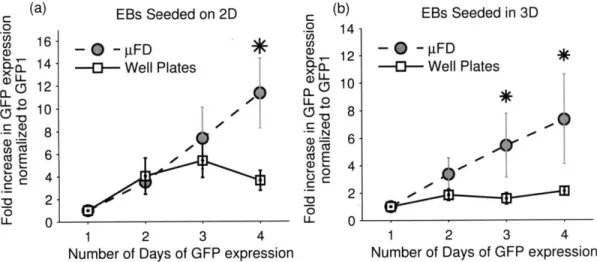
To examine the effects of confinement in the pFDs on cardiogenesis, compared with conventional culture plates, EBs were either seeded directly on the substrate - 2D - or embedded in a three-dimensional hydrogel - 3D (Figure 2-6a). GFP-positive and contracting cardiomyocytes were detected 48-72 hours after EBs were allowed to adhere to a substrate (Figure 2-6b). Compared with well plates, the rate of increase in GFP was higher in both 2D and 3D seeding conditions (Figure 2-7). When EBs were cultured on 2D, the increase in GFP expression persisted over time in pFDs while those on well plates reached a plateau after three days. A higher rate of differentiation was also observed in pFDs as opposed to culture plates when EBs were suspended in 2mg/ml collagen I hydrogel (Figure 2-7b).
Conventional
(b)
GFP1
Microfluidic Devices
Figure 2-6: Method of Differentiation and Representative Images of Differentiated Cardiomyocytes Procedure of differentiation and representative images of differentiated cardiomyocytes: (a) procedures of ESC differentiations, EB formations and seeding conditions (b) representative images