HAL Id: tel-01580957
https://tel.archives-ouvertes.fr/tel-01580957
Submitted on 4 Sep 2017HAL is a multi-disciplinary open access archive for the deposit and dissemination of sci-entific research documents, whether they are pub-lished or not. The documents may come from teaching and research institutions in France or abroad, or from public or private research centers.
L’archive ouverte pluridisciplinaire HAL, est destinée au dépôt et à la diffusion de documents scientifiques de niveau recherche, publiés ou non, émanant des établissements d’enseignement et de recherche français ou étrangers, des laboratoires publics ou privés.
Coordinating growth arrest and myogenesis in muscle
stem cells : a molecular and cellular analysis
Despoina Mademtzoglou
To cite this version:
Despoina Mademtzoglou. Coordinating growth arrest and myogenesis in muscle stem cells : a molecu-lar and cellumolecu-lar analysis. Neurobiology. Université Pierre et Marie Curie - Paris VI; Freie Universität (Berlin), 2016. English. �NNT : 2016PA066231�. �tel-01580957�
1
Coordinating growth arrest and myogenesis in muscle stem cells:
A molecular and cellular analysis
Defended on 02/09/2016 by
Despoina MADEMTZOGLOU
Jury
Prof. Miranda GROUNDS UWA, Perth (Australia) UPMC reviewer Dr. Pascal MAIRE Institut Cochin, Paris (France) UPMC reviewer Prof. Simone SPULER FU, Berlin (Germany) FU reviewer Prof. Fritz RATHJEN FU, Berlin (Germany) FU reviewer Prof. Frederic RELAIX UPEC, Créteil (France) Thesis director Dr. Delphine DUPREZ UPMC, Paris (France)
3
ACKNOWLEDGMENTS 6
LIST OF ABBREVIATIONS 12
ABSTRACT 13
INTRODUCTION 19
CHAPTER 1. SKELETAL MUSCLE DEVELOPMENT, GROWTH, AND REGENERATION 21
1.1 EMBRYONIC MYOGENESIS: FROM SOMITES TO THE FIRST MUSCLE MASSES 21
1.1.1SOMITOGENESIS: FORMATION OF MULTIPOTENT MESODERMAL STRUCTURES 21
1.1.2MYOTOME: THE FIRST SKELETAL MUSCLE 27
1.1.3MIGRATION OF MUSCLE PROGENITORS TO SUPPORT LIMB MYOGENESIS 29
1.2 GENETIC HIERARCHIES IN HEAD AND BODY MUSCULATURE ESTABLISHMENT 33
1.3 PAX PROTEINS AND BHLH MRFS PLAY A CENTRAL ROLE IN THE MYOGENIC PROGRAM 35
1.3.1PAX3 AND PAX7 AS UPSTREAM MYOGENIC REGULATORS 35
1.3.2MRFS PLAY A CENTRAL ROLE IN MYOGENIC DETERMINATION AND DIFFERENTIATION 39
1.4 FROM EMBRYONIC MYOGENIC DEVELOPMENT TO POSTNATAL MUSCLE 45
1.4.1EMBRYONIC AND FETAL WAVES OF MYOGENESIS 45
1.4.2POSTNATAL MUSCLE GROWTH 47
1.4.3ADULT MUSCLE: STRUCTURE & FUNCTION 47
1.5 SATELLITE CELLS: THE SKELETAL MUSCLE STEM CELLS 49
1.5.1ESTABLISHMENT DURING DEVELOPMENT 51
4
1.5.3ACQUISITION OF QUIESCENCE FOR FUNCTION PRESERVATION 53
1.5.4SATELLITE CELL NICHE 55
1.5.5SATELLITE CELLS IN THE CONTROL OF REGENERATION 55
1.5.6SATELLITE CELL HETEROGENEITY 67
1.5.7AGING EFFECT IN MUSCLE AND SATELLITE CELLS 69
CHAPTER 2. CELL CYCLE AND GROWTH ARREST IN SKELETAL MUSCLE AND BEYOND 71
2.1 CELL CYCLE OVERVIEW 72
2.2 CDK-CYCLIN COMPLEXES: CELL CYCLE PROGRESSION 73
2.3 THE POCKET PROTEIN- E2F NETWORK: DOWNSTREAM EFFECTORS OF CDK/CYCLINS 79
2.4 CDKIS: MAJOR NEGATIVE REGULATORS OF CDK-CYCLIN ACTIVITY 87
2.5 P57 – “KI P”LAYER IN CELL PHYSIOLOGY AND PATHOLOGY 97
CHAPTER 3. NOTCH SIGNALING PATHWAY: PLEIOTROPIC ROLE OF A MASTER CELL FATE REGULATOR IN
MYOGENESIS 103
RESULTS 109
AIMS AND HYPOTHESES 111
ANTAGONISTIC REGULATION OF p57KIP2 BY HES/HEY DOWNSTREAM OF NOTCH SIGNALING AND MUSCLE REGULATORY FACTORS REGULATES SKELETAL MUSCLE GROWTH ARREST 112
A p57 CONDITIONAL MUTANT ALLELE THAT ALLOWS TRACKING OF P57-EXPRESSING CELLS 126
DISTINCT REGULATION AND FUNCTION OF p21 AND p57 DURING MUSCLE STEM CELL ACTIVATION AND
5
CDKIS, MRFS AND NOTCH SIGNALING INTERPLAY IN CELL CYCLE EXIT DURING DEVELOPMENT 183
CONDITIONAL p57 ABLATION FOR POSTNATAL STUDIES 185
CDKIS IN THE CONTROL OF SATELLITE CELLS 187
ANNEX 193
Acknowledgments
6
ACKNOWLEDGMENTS
This co-tutelle Thesis was defended on 02/09/2016, in the presence of an international jury of distinguished scientists whom I would like to acknowledge. I wish to thank them for accepting to examine my work and for the helpful discussions to get a better understanding of the field and to improve my work.
Firstly I would like to thank my four reviewers Prof. Miranda Grounds, Dr. Pascal Maire, Prof. Simone
Spuler, and Prof. Fritz Rathjen for the time they spent to read my manuscript, write their reports, and
attend the defense, which included travelling from Australia and Germany.
A special thanks to Prof. M.Grounds, whom I only met late during my PhD, but through stimulating discussions, her passion for science, and her influential work, inspires me in my future career.
Merci à Dr. P.Maire for accepting to participate in my Jury. I much admire his scientific rigor since I first met him at MyoGrad’s summer school and I am grateful to receive his input on my work.
Prof. S.Spuler has followed me in the course of my Thesis in the Thesis Committee meetings and through MyoGrad, the PhD program she directs and in which I am enrolled. I would like to thank her for her continuous efforts to improve MyoGrad.
I would like to thank Prof. F.Rathjen for taking time from his busy schedule to review my work and come to France for the defense.
Dr. Delphine Duprez, as part of my Thesis Committee, helped me at critical points of my PhD to evaluate my work and define the next steps. I would like to thank her for her feedback and for redacting the Committee meeting reports. I would also like to thank her for hosting us in her lab last summer for a very insightful workshop on chick embryology.
Muchas gracias to Dr. Ezequiel Mendoza for coming to Paris to attend one more MyoGrad defense and for the interesting discussions that preceded the defense.
7
In the second part of the acknowledgments I would like to thank the people who directly or indirectly participated in this project and, more generally, in my formation as a researcher.
Following a … geographical (CréteilParisBerlinThessalonikiMadrid) order:
I would like to thank Prof. Fred Relaix for trusting me with a project irrelevant to my previous lab background and for the “fred request” to join his group. He gave me the chance to work on the kind of projects that I admired since the first conferences I attended! His expertise, insightful comments, and patience added considerably to my graduate experience. I’d also like to thank him for taking time out of his busy agenda whenever I needed his guidance. I appreciate his vast knowledge and his way to present ideas and results. De ce fait, un grand merci for encouraging me to present my work in meetings. Especially the Gordon’s conference just a few months after starting my project, was a great opportunity to get familiar with key players and recent advancements of the muscle field. Finally, his assistance in writing reports, his calm and pleasant character, his positive and supportive approach towards his team members have set an example for me.
Muchas gracias to Dr. Sonia Alonso-Martin for her continuous help throughout my thesis. Starting or completing my PhD would not be possible without the technical skills she taught me and the theoretical aspects she helped me grasp! I recognize how much effort and patience this takes and I understand por qué preferió evitar desempeñar el mismo cargo en mi español-aprendizaje. Even after my initial learning period, it was a great relief to know that she would be around whenever I had a question (or lacked a protocol or was threatened by deadlines). Her constant good mood and enthusiasm make it very enjoyable to be in the lab (and very difficult to get angry if you somehow get trapped in a black room, counting fibers, at 10 pm). Her input and encouragement were critical many times, as were her recognition and her caring. Equally important was that she decided to include me in her projects, giving me the chance to learn and participate in things ranging from bioinformatics to graft experiments. I consider her my scientific big sister and I feel very lucky to have had her valuable guidance all these years.
Acknowledgments
8
I would like to express my gratitude to Dr. Philippos Mourikis (Φίλιππο Μουρίκη, to be grammatically correct). His continuous support, motivation, and constructive comments were essential to develop my project, to make the best use of obtained data, and to find alternatives to failed experiments. With his immense knowledge and guidance he provided me with direction. I much appreciate his patience, encouragement, and availability when it comes to new&ignorant lab members. Κι ένα μεγάλο ευχαριστώ for involving me in different aspects of the academic life and for encouraging me to interact with the people that have the expertise on the questions I needed to answer.
After four years of interaction with Dr. Frederic Aurade, I can confidently tell him “aie nide iou” when it comes to any aspect of molecular biology! I would like to thank him for his help sofar and for all the clonings for the p57 project (and the upcoming support for his beloved nucleo-cytoplasmic trafficking?). His magical troubleshooting abilities and extensive knowledge set a (very welcome) safety pillow for our molecular problems. Finally, it is very important to know that there is somebody in the lab to share geeky jokes with.
Mèsi Bernie Drayton for the immense help to organize our mice, “resurrect” our lines in the new animal facilities, and follow all necessary trainings. And all this in a permanent good-humored state, (transcriptionally?) primed to sing! I would also like to thank Bernie for her help during stressful periods as well as her student, Thomas Marius, for performing much of my genotyping.
I would like to acknowledge the work of Bernie, Dr. Ted Chang, and Dr. Keren Bismuth for setting the pillar of my PhD project by generating the p57-floxed mice. I am very happy to continue this project with Dr. Matthew Borok. His enthousiasm, creativity, and aptitude to rapidly start working independently are of great value. I would like to thank him for his understanding for my recent overwhelmed program and for his help to avoid ceasing the project while I was writing.
I want to thank Dr. Marianne Gervais-Taurel, Marie Quetin, and Lina Gomez for their continuous efforts and interest in solving our single fiber problems (or share our grief over clusterless fibers). Un
9
grand merci for Marie, Zeynab Koumaiha, and Anikó Szegedi for their ideas and creativity to make such a great atmosphere in the lab and help enrich the writing experience.
I would also like to thank some past members of Fred’s group. Σ’ευχαριστώ Dr. Amalia Stantzou for all the things we shared: protocols, advice-to-freshman, χώσιμο για lab meeting presentations κάθε δυο βδομάδες το 2013, pineapple pizzas, and in general all the fun moments in and out of the lab! It was a pleasure to have a friend like you when starting! Merci Dr. Antoine Zalc, ありがとうございま した Dr. Shin Hayashi for your advice to handle embryos and develop the developmental part of my project. Merci Dr. Vanessa Ribes for your persistent advice to define “what is my question”.
As tutor and scientific expert of my Thesis Committee, Dr. Benedicte Chazaud and Dr. Shahragim
Tajbakhsh contributed to advancing my work. Particularly I would like to thank Shahragim for helpful
discussions and for giving me the chance to participate in a very informative and exceptionally well-prepared stem cell course that he co-organizes. I must add that I admire his way of thinking and his creative and enjoyable presentations.
Merci Dr. Bruno Cadot, Petra Gimpel, Jean-François Darrigrand for adopting me in your team until the new lab at Créteil was ready. It was very helpful to have a bench and to be in such a nice environment the last few stressful months of experiments. A special thanks to Bruno for giving me access to the super-microscopes that helped me see p57 in the cytoplasm for the first time! With his extensive knowledge and calm personality he is ready to help everyone at anytime. Vielen Dank Petra for the immense help with C2C12 culture and westerns, but also for serving as an example of how professional you can be even as a first year student. Also thanks for the psychological support, the funny atmosphere, all the german-greek exchange, and for helping me get started in Berlin!
Un clin d’oeil tout particulier à Petra and Dr. Audrey Der Vartanian for their help to communicate my results to the German and French audience. Un grand merci a Petra, Audrey, Valerie Vilmont, Joana
Esteves de Lima, and Matthew for all the nice moments in the lab (or in Paris in general). I admire
Acknowledgments
10
I also wish to acknowledge Bénédicte Hoareau from the Flow Cytometry Core CyPS as well as the animal care facilities at UPMC and CDTA, and especially Olivier Brégerie and Kim Nguyen for the excellent collaboration. I’d also like to thank Lila Bendameche, Susanne Wissler, and Michael Strehle for (very efficiently) taking a big administrative burden from our shoulders.
Prof. Carmen Birchmeier möchte ich ganz herzlich bedanken, for accepting me in her lab in Berlin and including me in her team’s projects. Ηer critical thinking, our discussions over the paper, and her stimulating questions incented me to widen my background. I enjoyed the interesting talks that she was organizing and the impressive meeting that her colleagues organized for her birthday, inviting speakers from all around the world!
Vielen Dank for Dr. Dominique Bröhl for giving me the chance to work by his side and for teaching me how to take 106 satellite cells from just a few mice. His calm and efficient way of working along with his amusing comments make him a very pleasant person to work with.
This list of people who helped me or forged me in the past few “lab years” would not be complete without Prof. Penelope Mavragani-Tsipidou and Dr. Paloma Morales. κ. Mavragani recruited me in her team for my dissertation back in 2008 and I am very grateful for having such a great first lab experience as well as for our interaction all these years. Her guidance and her personality were crucial in my early steps in research and I hope to always keep in mind the important lessons she taught me. Σας ευχαριστώ για την αμέριστη συμπεράσταση εντός κι εκτός εργαστηρίου και για την υποστήριξή σας όλα αυτά τα χρόνια! Muchas gracias to Paloma for accepting me in her lab as an Erasmus student, for giving me the first insights in cell culture, and for guiding me through writing the first paper! I am very happy to meet her every time I have the chance to go back to Madrid.
It’s very difficult to thank in just a few sentences Dr. Myrto Manolaki for her support. Her eagerness to solve problems, her μεράκι in science, το πηγαίο χιούμορ της gave her a great potential to help me approximate the post-doc universe. Even though our fields exclude us from a mutual Nobel prize or
11
Fields medal I still hope that we’ll get the chance to collaborate in one of her brilliant ideas to combine mathematics and life sciences.
Finally, I wish to thank my friends for tolerating cheering me up when the failed recombinations were striking. Especially Nestoras, Magda, Elma for the nice memories I have with them in Paris, Maria,
Matina για τις ξέγνοιαστες βόλτες μας to SugarAngel Thessaloniki, and Marietta for our expeditions
in Athens&Roma. Last but not least, I would like to thank my parents Prodromos and Anastasia for being supportive to my decisions and for giving me the strength to chase my dreams.
LIST OF ABBREVIATIONS
12
LIST OF ABBREVIATIONS
bHLH basic Helix-Loop-Helix BMP Bone Morphogenetic Protein BWS Beckwith-Wiedemann Syndrome CAK Cdk-Activating Kinase
CDK Cyclin-Dependent Kinase
CDKI Cyclin-Dependent Kinase Inhibitor DKO Double Knock-Out
DTA Diphtheria Toxin Fragment
DP Differentiation-regulated transcription factor-1 Polypeptide EMT Epithelial–Mesenchymal Transition
ECM ExtraCellular Matrix
FAP Fibro/Adipogenic Progenitor
Fucci Fluorescent Ubiquitynation-based Cell Cycle Inditator HGF Hepatocyte Growth Factor
KID Kinase Inhibitory Domain LIMK LIM domain Kinase
MRF Myogenic Regulatory Factor MyHC Myosin Heavy Chain
NICD Notch IntraCellular Domain NLS Nuclear Localization Signal
PDGFR Platelet-Derived Growth Factor Receptor PP Pocket Protein
PIC PW1+ interstitial cell Rb RetinoBlastoma protein SF Scatter Factor
SP Side Population
13
ABSTRACT
Tightly controlled growth arrest coordinates the equilibrium between cell proliferation and cell differentiation during embryonic tissue formation as well as in adulthood during stem cell-mediated tissue regeneration. Myogenic differentiation requires a coordinated course of tissue-specific gene expression and irreversible cell cycle exit. However, I contributed to showing -using genetic manipulation in the mouse embryo- that these processes can be uncoupled. During development, growth arrest in myogenic cells is mediated by the cyclin-dependent kinase inhibitors (CDKIs) p21 and p57, which act redundantly to promote cell cycle exit. We demonstrated that skeletal muscle progenitors require a direct interaction with the differentiating myoblasts via the Notch signaling pathway to maintain their pool. We also identified a muscle-specific regulatory element of p57 that directly receives the input of Myogenic Regulatory Factors (MRFs) and Notch downstream targets. During my Ph.D. I examined whether this regulatory mechanism is also involved in postnatal myogenesis.
Adult skeletal muscle has a remarkable regenerative capacity, involving a stem cell population, called satellite cells (SCs), located on close contact to the myofibers under the basal lamina. At the transition from juvenile to adult state (around 3-4 week during postnatal growth in mice) they enter a non-cycling, quiescent state. Upon injury the SCs rapidly get activated, expand by proliferation and provide differentiated progeny for muscle repair, while a subpopulation self-renews and re-enters quiescence, allowing the support of additional rounds of muscle damage. To understand the mechanisms regulating acquisition of quiescence, we explored the role of the aforementioned CDKIs in adult muscle. Although absent from quiescent SCs, they become rapidly expressed upon activation (even in proliferating myoblasts) and their levels remain high in the differentiating muscle cells. Strikingly, during the course of differentiation p57 translocated from the cytoplasm of activated myoblasts to the nuclei of differentiating cells. Since p57 deficient mice die at birth, we generated a conditional knock-out allele to perform functional studies at the postnatal stages. This new allele, in which the coding region can be removed by the loxP/Cre recombination system, also contains a β-galactosidase reporter allowing the identification of p57-expressing cells. We generated a complete
ABSTRACT
14
loss of function allele using a ubiquitously expressed Cre, and observed developmental and perinatal phenotypes similar to previously described germline knock-outs. Furthermore, we showed that the reporter inserted in the p57 conditional allele faithfully recapitulates p57 expression profile at embryonic and adult tissues. Conditional ablation of p57 in adult SCs resulted in reduced myogenic differentiation in primary myoblast culture. Similarly, p21-null myoblasts exhibited proliferation and differentiation defects in single myofiber cultures. In vivo regeneration studies with p21 mutants showed an early impact on the SC pool, while both SCs and muscle structure were re-established by the end of the regeneration process. My Ph.D. work suggests that p21 and p57 are at play during adult myogenesis and cell cycle exit, although via different mechanisms compared to the developmental scenario. They both work at the early steps of satellite cell activation but do not compensate for each other’s loss. Future studies will elucidate whether they lie genetically downstream of the MRFs and Notch targets during postnatal myogenesis.
RÉSUMÉ
Au cours du développement embryonnaire, tout comme chez l’adulte, la formation ainsi que la régénération des tissus nécessitent une régulation fine du cycle cellulaire afin de maintenir l’équilibre entre la prolifération et l’entrée en différenciation des cellules. La différenciation myogénique nécessite une coordination parfaite entre l’expression des gènes spécifiques au développement musculaire et la sortie irréversible du cycle cellulaire des cellules constitutives du tissu. Cependant, j’ai contribué à montrer que ces processus peuvent être découplés chez l’embryon via la génération de modèles murins génétiquement modifiés.
Au cours de la différenciation myogénique chez l’embryon, l’arrêt du cycle cellulaire est contrôlé par les inhibiteurs de kinases cyclines-dépendantes (CDKI) p21 et p57 qui présentent une activité redondante. Nous avons démontré que les cellules progénitrices du muscle squelettique requièrent une communication directe avec les myoblastes en différentiation via la voie de signalisation cellulaire Notch afin de maintenir cette population dans un état indifférenciée. De plus, nous avons mise en évidence la présence d’un élément de régulation spécifique du muscle dans la séquence du
15
gène p57 répondant aux facteurs myogéniques (Myogenic Regulatory Factors, MRFs) et aux gènes cibles de la voie de Notch. Dans la poursuite de ces travaux, j’ai ensuite étudié l’implication de ce mécanisme de régulation au cours de la myogenèse postnatale pendant mon travail de thèse.
Les cellules souches du muscle, ou cellules satellites, sont localisées sous la lame basale au contact des myofibres et confèrent une capacité de régénération remarquable au tissu musculaire. Chez la souris, au cours de la transition entre le stade juvénile et le stade adulte (c’est à dire 3-4 semaines après la naissance) les cellules satellites rentrent en état de quiescence. Sous l’action d’un stimuli externe ou lors d’une blessure musculaire, les cellules satellites s’activent et prolifèrent. Au cours de la régénération du muscle, une sous-population va se différencier pour réparer le muscle lésé alors qu’une partie de cette population va s’auto-renouveler afin de maintenir le stock de cellules souches quiescentes.
Dans le but de comprendre les mécanismes qui régissent l’entrée en quiescence de ces cellules, nous avons étudié le rôle de p21 et p57 et dans le muscle adulte. Bien que l’expression des gènes codants pour ces CDKIs ne soit pas détectée dans les cellules satellites quiescentes, ils sont rapidement détectés dès leur activation mais également dans les myoblastes et les cellules musculaires en différentiation où ils sont fortement exprimés. Au cours de la différenciation myogénique p57 est transloqué depuis le cytoplasme des myoblastes jusqu´au noyau des cellules en différenciation.
Chez la souris, l’ablation du gène p57 est létale à la naissance. Afin de pouvoir étudier le rôle fonctionnel de cette protéine après la naissance, nous avons généré un modèle murin présentant une modification génique conditionnelle permettant de muter le gène p57. Cette construction conditionnelle utilise le système de recombinaison Cre/LoxP qui permet d’exciser la séquence codante du gène p57. Elle contient également le gène rapporteur β-galactosidase afin de pouvoir identifier les cellules qui expriment p57. La perte totale de fonction générée par l’utilisation d’une Cre recombinase exprimée de manière ubiquitaire a permis de caractériser les phénotypes observés au cours du développement embryonnaire et au cours de la période périnatale. Les phénotypes observés sont identiques aux phénotypes décrits précédemment chez les souris présentant une perte
ABSTRACT
16
de fonction du gène p57. De plus, nous avons pu caractériser le profile d’expression de ce gène au cours du développement embryonnaire et dans les différents tissus chez l’adulte grâce au gène rapporteur.
Dans les cultures primaires de cellules satellites adultes, la délétion de p57 conduit à une diminution de la différenciation myogénique. De même, les cultures de fibres isolées issues des myoblastes mutants pour le gène p21, présentent des défauts de prolifération et de différenciation. In vivo, l’étude de régénération chez les mutants p21 montre une réduction précoce de la population satellitaire. Paradoxalement, la population des cellules souches du muscle ainsi que la structure du tissu musculaire sont entièrement reconstituées à la fin du processus de régénération.
Mon travail de thèse suggère que p21 et p57 jouent un rôle important dans la myogenèse et la régulation du cycle cellulaire. Ces protéines ont une action similaire qui est déterminante sur l’activation des cellules satellites et agissent de manière précoce sur ces dernières. Cependant, chez l’adulte leur activité semble distincte lorsqu’un des deux CDKIs est manquant contrairement au stade embryonnaire. Mes travaux ouvrent des perspectives nouvelles sur le rôle de p21 et p57 en aval de la voie de signalisation de Notch et des MRFs au cours de la myogenèse post-natale.
ABSTRAKT
Das Gleichgewicht zwischen Zellproliferation und Zelldifferenzierung wird während der embryonalen Gewebebildung sowie während der stammzellvermittelten Geweberegeneration im Adultstadium durch einen streng kontrollierten Wachstumsarrest koordiniert. Die myogene Differenzierung erfordert sowohl eine koordinierte Abfolge von gewebespezifischer Genexpression als auch einen irreversiblen Zellzyklusaustritt. Jedoch konnte ich durch genetische Manipulation von Mausembryos dazu beitragen, aufzuzeigen, dass diese beiden Prozesse voneinander entkoppelt werden können. Ein Wachstumsarrest myogener Zellen wird während der Entwicklung durch die Cyclin-abhängigen Kinaseinhibitoren (CDKIs) p21 und p57 vermittelt. Letztere wirken dabei redundant, um einen Zellzyklusaustritt zu fördern. Wir konnten bereits nachweisen, dass skelettale Muskelvorläuferzellen
17
eine direkte Interaktion mit differenzierenden Myoblasten über den Notch-Signalweg benötigen, um ihren Pool aufrecht zu erhalten. Des Weiteren haben wir ein muskelspezifisches regulatorisches Element von p57 identifiziert, das direkten Input von myogenen Regulationsfaktoren (MRFs) und Notch Stromabwärts-Zielen erhält. Im Rahmen meiner Doktorarbeit habe ich untersucht, ob dieser regulatorische Mechanismus auch in der postnatalen Myogenese involviert ist.
Der adulte skelettale Muskel hat eine bemerkenswerte regenerative Kapazität, die eine Stammzellpopulation, sogennante Satellitenzellen (SCs, engl. für "satellite cells") involviert. Diese befinden sich zwischen der Basallamina und der Muskelfaser. Beim Übergang zwischen juvenilen und adultem Stadium (zwischen 3-4 Wochen während des postnatalen Wachstums in Mäusen), gehen die Satellitenzellen in einen nicht-zyklischen Ruhezustand über. Satellitenzellen werden nach einer Verletzung schnell aktiviert, expandieren und stellen differenzierte Abkömmlinge für die Muskelreparatur zur Verfügung. Eine Subpopulation der Satellitenzellen erneuern sich selbst und gehen zurück in den Ruhezustand, so dass bei erneuter Muskelverletzung Unterstützung gewährleistet ist. Um die Mechanismen verstehen zu können, die den Übergang in den Ruhezustand regulieren, haben wir die Rolle der zuvor genannten CDKIs im adulten Muskel untersucht. Obwohl CDKIs in ruhenden Satellitenzellen abwesend sind, werden sie nach Aktivierung der Satellitenzellen schnell exprimiert, und ihr Expressionslevel bleibt in differenzierenden Muskelzellen aufrecht erhalten. Erstaunlicherweise transloziert p57 während des Differenzierungsprozesses vom Zytosplasma in den Zellkern. Da p57-defiziente Mäuse bei Geburt sterben, haben wir eine konditionelle Mausmutante generiert, um funktionale Studien im postnatalen Stadium durchführen zu können. Dieses neue Mausmodell hat ein modifiziertes p57-Allel, in dem die codierende Region von p57 durch loxP/Cre-Rekombination entfernt werden kann. Des Weiteren beinhaltet es auch das Reportgen β-Galactosidase, um p57-exprimierende Zellen identifizieren zu können. Wir haben durch eine ubiquitär exprimierte Cre-Rekombinase vollständige Knockout-Mäuse generiert und dabei entwicklungsorientierte und perinatale Phänotypen, ähnlich wie bei bereits beschriebenen Knockout-Mäusen, beobachtet. Des Weiteren konnten wir zeigen, dass der in das konditionelle p57-Allel eingefügte Reporter dem Expressionsprofil von p57 im embryonalen und adulten Gewebe entspricht. Konditionelle Ablation von p57 in Satellitenzellen resultierte in einer reduzierten myogenen
ABSTRACT
18
Differenzierung von primären Myoblastenkulturen. p21-defiziente-Myoblasten wiesen ähnliche Proliferation- und Differenzierungsdefekte in einzelnen Muskelfaser-Kulturen auf. In-vivo-Regenerationsstudien mit p21-Mutanten haben eine initiale Reduktion der Satellitenzellenanzahl gezeigt. Zum Ende des Regenerationsprozesses waren die Anzahl der Satellitenzellen sowie die Muskelstruktur jedoch re-etabliert. Meine Doktorarbeit lässt darauf schließen, dass p21 und p57 während der adulten Myogenese und des Zellzyklusaustritts eine Rolle spielen, jedoch unterscheidet sich der Mechanismus im Vergleich zum pränatalem Stadium. Beide fungieren bei frühzeitigen Schritten der Satellitenzellenaktivierung, aber kompensieren sich nicht für den gegenseitigen Verlust. Künftige Studien werden zeigen, ob p21 und p57 von MRFs und Notch während der postnatalen Myogenese reguliert werden.
19
INTRODUCTION
Chapter 2. Cell cycle and growth arrest in skeletal muscle and beyond
20 Figure 1.1. From somites to myotome.
Trunk and limb muscle derive from the paraxial mesoderm. The presomitic mesoderm will get progressively segmented into distinct, epithelially enclosed units, termed somites, which undergo several steps of specification and differentiation in a posterior-to-anterior orientation. As maturation proceeds they are compartmentalized into sclerotome and dermomyotome in response to signals from the adjacent structures. Cells from the dermomyotome migrate later to form the underlying myotome, the first differentiated skeletal muscle, as well as to distal myogenic sites, such as the developing limbs, diaphragm, and tongue.
21
Chapter 1. Skeletal muscle development, growth, and regeneration
1.1 Embryonic myogenesis: from somites to the first muscle masses
1.1.1 Somitogenesis: formation of multipotent mesodermal structures
Starting as a unicellular totipotent zygote the nascent organism develops into a multitude of differentiated, functionally interacting tissues and organs. During gastrulation, cells produced by consecutive divisions of the zygote and its descendants ingress through the blastopore/primitive streak to generate the three germ layers of the embryo, namely ectoderm, mesoderm and endoderm that will contribute all embryonic tissues. Skeletal muscle is of mesodermal origin and its emergence and organization is a complex process starting soon after gastrulation (E8 at mouse) [1].
Among the mesodermal compartments (axial, paraxial, intermediate, and lateral plate), the paraxial mesoderm gives rise to all the muscles of the limbs and ventral body [Christ & Ordahl, 1995], while branchiomeric and ocular muscles derive from cranial/pharyngeal and prechordal cranial mesoderm [Scaal & Christ, 2004; Buckingham & Mayeuf, 2012]. The paraxial mesoderm constitutes two longitudinal columns of mesoderm on each side of the neural tube/notochord [Pourquié, 2001]. It will undergo somitogenesis, meaning generation of somites, which are segmented epithelial units that develop stepwise to give rise to the ventral mesenchymal sclerotome and the dorsal epithelial dermomyotome (Fig. 1.1). Apart from the differences in origin of body (trunk, limb) and head musculature, distinct genetic networks operate in each of them [Tajbakhsh, 2009; Bismuth & Relaix, 2010] and diverse turnover has been reported [McLoon et al., 2004; Keefe et al., 2015; Pawlikowski et al., 2015]. Limb and trunk musculature are, in general, more extensively studied and of more relevance to my PhD work and, thus, will be on focus in this section.
Somitogenesis occurs in coordination with embryo extension along the antero-posterior axis. Within the presomitic mesoderm, cellular arrangements of prospective somites point to a segmental pattern prior to somitogenesis and in response to intrinsic signals [Pourquié, 2001]. During gastrulation, new
Chapter 2. Cell cycle and growth arrest in skeletal muscle and beyond
22 Table 1.1. Somitogenesis across species [Hubaud & Pourquié, 2014].
Organism Frequency of new pair addition
Final number of somite pairs zebrafish 25 minutes 33
mouse 2 hours 65
human 4-5 hours 38-44
Figure 1.2. The “clock and wavefront” model for periodic somite generation
A. Major molecular contributors to the oscillation (clock) and gradient (wavefront) components of the model. B. Schematic representation of the location of activity of the clock and wavefront. C. Oscillatory expression of c-hairy1 (in situ hybridization during formation of somites 15 to 17).
23
mesenchyme cells enter the paraxial mesoderm leading to the addition of bilaterally symmetrical somite pairs at the anterior end of the presomitic mesoderm [Christ & Ordahl, 1995]. Convergence-extension movements of this phase produce the future anterior somitic mesoderm, while later on the tail bud contributes caudal somites [Pourquié, 2001]. Somitogenesis is a stepwise procedure involving periodic formation, separation, epithelialization, specification, and differentiation (with additional Epithelial–Mesenchymal Transition (EMT) transitions to form the sclerotome and myotome) [Musumeci et al., 2015].
Somites emerge periodically in a species-specific frequency (Table 1.1). To explain this feature, a theoretical model, the “clock and wavefront” model, was proposed in the 1970s (Fig. 1.2) [Cooke & Zeeman, 1976]. The clock refers to intrinsic oscillator(s) that make(s) presomitic mesoderm fluctuate between permissive and non-permissive states of somite formation (Fig. 1.2B) [Kalcheim & Ben-Yair, 2005]. The wavefront of competence to generate somites lays at a defined position with regard to the tail bud (Fig. 1.2B) [Saga, 2012]. On a molecular level, the idea of the clock was supported by the discovery of cyclic waves of c-hairy1 linking it with somitogenesis (Fig. 1.2C) [Palmeirim et al., 1997]. This was followed by the identification of several other transcription factors (e.g. HES7, AXIN2) or enzymes (e.g. LFNG), mainly belonging to the Notch, FGF and Wnt pathways, which are periodically expressed at defined time intervals imitating the segmentation rounds (Fig. 1.2A) [Pourquié, 2011; Musumeci et al., 2015]. Notch, a key player of intercellular communication of neighboring cells, is further suggested to act by synchronizing the oscillations of individual cells [Horikawa et al., 2006]. The clock is mainly considered to act through the activation of the MESP transcription factors [Saga, 2012]. Different segment periodicities in the anterior and posterior presomitic mesoderm of zebrafish were revealed by recent live imaging experiments [Shih et al., 2015]. Another recent study challenged the role of the clock, counter-suggesting that cell-cell interactions drive somitogenesis [Dias et al., 2014]. However, they described somite-like structures [Dias et al., 2014] which were later criticized as being expected self-organizing differentiating derivatives, missing several somite characteristics [Hubaud & Pourquié, 2014]. The wavefront corresponds at the molecular level to a threshold of different signaling gradients, with Wnt and FGF signaling being highest at the posterior unsegmented paraxial mesoderm and retinoic acid following a counter-gradient (Fig. 1.2A) [Hubaud & Pourquié,
Chapter 2. Cell cycle and growth arrest in skeletal muscle and beyond
24 Figure 1.3. Dorsalizing and ventralizing signals involved in somite differentiation.
Left half simplistically focuses on the major dorsalizing (Wnt) and ventralizing (Shh) signals that act on the developing somites. Right half underlines the necessary activity of further factors for the final fine gradient driving the formation of dermomyotome in the dorsal somite and sclerotome in the ventral moiety.
25
2014; Mallo, 2016]. These gradients depend on mRNA decay or gradients of synthesizing and degrading enzymes, respectively [Aulehla & Pourquié, 2009]. Retinoic acid is also suggested to participate in synchronically generating left and right somites [Sirbu & Duester, 2006].
Once the new somite pair is signaled to be formed, it needs to undergo detachment and epithelialization. Separation of consecutive somites and formation of an acellular intersomitic border depend on the families of cadherins and ephrins and their receptors [Kalcheim & Ben-Yair, 2005]. Before complete detachment, mesenchymal-epithelial transition takes place, driven by the bHLH transcription factor PARAXIS and the GTPases CDC42 and RAC1 [Burgess et al., 1996; Nakaya et al., 2004]. Adhesion molecules (e.g. N-cadherin) and Fibronectin, an extracellular matrix protein were also found to participate in epithelialization [Duband et al., 1987; Linask et al., 1998]. Interestingly, in
Paraxis-null mice epithelial somites are substituted by mesenchymal blocks, uncoupling segmentation
and epithelialization [Burgess et al., 1996].
The next steps of somitogenesis involve specification and differentiation. Somite specification appears to depend on HOX genes, with maintenance of the HOX profile even after heterotopic transplantation [Nowicki & Burke, 2000]. Differentiation is largely attributed to interactions and molecular signals originating from the surrounding tissues. The ectoderm and notochord mediate dorsalization with signals such as Wnt proteins and Bone Morphogenetic Proteins (BMP), while the neural tube provides ventralizing signals, such as Sonic hedgehog homolog (Shh) or the BMP antagonist Noggin [Christ & Brand-Saberi, 2002; Scaal & Christ, 2004]. Further factors, such as SFRP2 or GAS1 antagonize Wnt and Shh, respectively, to prevent their long-range signaling and induce gradients of dorsalizing and ventralizing signals (Fig. 1.3) [Brent & Tabin, 2002].
Somites bud off the paraxial mesoderm at its rostral extremity, so that the caudalmost somite is the youngest or somite number I, according to the applied dynamic staging system with consecutive Roman numbers (Fig. 1.2B) [Scaal & Christ, 2004]. At early stages somites show developmental plasticity, as evidenced by single cell transplantation experiments [Kato & Gurdon, 1993]. Moreover, transplantations of groups of cells demonstrate a community effect [Gurdon et al., 1993]. Both cases
Chapter 2. Cell cycle and growth arrest in skeletal muscle and beyond
26 Figure 1.4. Myotome formation.
Myotome formation depends on cells migrating from the dermomyotome, initially from the lips (A) and subsequently from the central region (B).
27
underline the influence of the surrounding microenvironment. Up to somites III-IV, they are shaped as epithelial spheres enclosing mesenchymal cells. Later on, the adjacent structure provide the aforementioned ventralizing and dorsalizing signals to compartmentalize somites into sclerotome and dermomyotome, respectively [Christ & Brand-Saberi, 2002; Scaal & Christ, 2004]. The ventral moiety undergoes EMT to form the sclerotome, which is mainly the source of the cartilage and bone of vertebral column and ribs (axial skeleton), but also contributes tendons, and joints [Musumeci et al., 2015]. The dorsal dermomyotome remains epithelial and stretches to form a sheet that roofs sclerotome. At later steps and while it adapts its rectangular-like form, dermomyotome develops a central mesenchymal sheet adjacent to the ectoderm and four inwardly curved epithelial lips. Dermomyotome gives rise to the dermis of the back and skeletal muscle, with their precursors originating in asymmetric divisions at the dorsal-ventral axis during EMT of the central part (see section 1.1.2) [Ben-Yair & Kalcheim, 2005]. Somites are divided in epaxial and hypaxial domains, lying dorsally and ventrally, respectively, to the horizontal septum of the vertebrae. The epaxial compartment generates muscles of the back, while the hypaxial part is the source of the muscles of the ventral body and limbs [Birchmeier & Brohmann, 2000; Musumeci et al., 2015].
1.1.2 Myotome: the first skeletal muscle
As the somite matures, cells delaminate and migrate underneath to form a third compartment, called myotome (Fig. 1.4), which corresponds to the first differentiated skeletal muscle. The groups of Ordahl and Kalcheim came to contradictory results when trying to elucidate the starting points and movements of myotomal precursors [Brent & Tabin, 2002], but a more recent stepwise model described by Gros et al. [2004] helped resolve the controversy. According to these findings, at a first phase cells translocate from the dorso-medial lip to the myotome and once there, they elongate along the rostral-caudal axis. At a second phase, cells invade myotome starting from all four lips and elongate along the anterior-posterior axis. Elongation can be unidirectional (cells from rostral or caudal lip) or bidirectional (cells from dorso-medial or ventro-lateral lip).
Chapter 2. Cell cycle and growth arrest in skeletal muscle and beyond
28 Figure 1.5. Progenitor migration for limb myogenesis.
A) Major factors controlling the consecutive steps of limb myogenesis. B) At E10.5 in the mouse embryo, Pax3-LacZ progenitors have migrated to the forelimb (up) and begin to migrate to the hindlimb (down). C,E) Cxcr4 expression in limb at E10.5 mouse embryo. D,F) Sdf1 expression in limb at E10.5 mouse embryo. G-J) Lbx1 expression in somites and limbs of chick embryos at HH18 and HH22 stages. K-L) Limb muscles (black arrows) are absent from and somites are fused and truncated (red arrowheads) in Pax3IRESnLacZ/Sp E11.5 mouse embryos as opposed to Pax3IRESnLacZ/+ control embryos at the same stage. M-P) PAX3-expressing cells do not colonize the forelimbs of Splotch mutants at 30-33 somite stages. Q-R) Lack of muscle (identified with MyoD) in the forelimbs of c-Met mutant E11.5 mouse embryos, in contrast to controls. NC: notochord, NT: neural tube, SE: surface ectoderm.
Adapted from: Bober et al., 1994; Maina et al., 1996; Mennerich et al., 1998; Buckingham et al., 2003; Relaix et al., 2003; Vasyutina et al., 2005.
29
Once the primary myotome is formed, a second population of myogenic progenitor cells originating from the central dermomyotome is colonizing the underlying myotome, rendering its initial name “dermatome” erroneous (Fig. 1.4). At later stages of embryonic and fetal life, muscle growth was found to depend on progenitors originating from the central dermomyotome, rather than the lips (see session 1.2.1) [Ben-Yair & Kalcheim, 2005; Gros et al., 2005; Kassar-Duchossoy et al., 2005; Relaix et al., 2005]. Of note, embryonic and fetal progenitors are mitotically active and have not engaged to the myogenic program. They maintain their proliferative status in embryonic and fetal muscles of trunk and limbs throughout development. They depend on transcription factors of the PAX family and they contribute to the forming muscles as well as their associated stem cells, as discussed in the following sections.
1.1.3 Migration of muscle progenitors to support limb myogenesis
Distant sites of myogenesis, such as the developing limb, depend on long-range migration of progenitors from the hypaxial dermomyotome to the limb buds, where they proliferate and subsequently commit to the myogenic lineage and undergo differentiation into skeletal muscle (Fig.
1.5 A) [Buckingham et al., 2003].
The transcription factor LBX1 is considered a bona fide marker of long-range migrating muscle precursors [Dietrich et al., 1998]. Lbx1 starts to be expressed in the dispersing dermomyotomal lips, meaning prior to delamination. It then follows the migrating population (Fig. 1.5 G-J) and declines only when these progenitors arrive at the target sites and start to differentiate [Jagla et al., 1995; Dietrich et al., 1998; Mennerich et al., 1998]. In its absence, migratory precursors manage to form and delaminate but they display defective routing, demonstrating that LBX1 is critical for migration [Schäfer & Braun, 1999; Brohmann et al., 2000; Gross et al., 2000].
The migratory behavior of muscle progenitors is also controlled by PAX3, which is essential for the initiation of their migration. Splotch and other PAX3 mutant embryos show a number of developmental phenotypes in dorsal neural regions, neural crest cells and derivatives and muscle
Chapter 2. Cell cycle and growth arrest in skeletal muscle and beyond
31
tissues [Auerbach, 1954; Relaix et al., 2004]. Strikingly, PAX3-deficient embryos are devoid of myogenic migrating cells, leading to complete absence of muscular diaphragm, tongue and limb muscles (Fig. 1.5 K-P) [Mennerich et al., 1998; Relaix et al., 2004]. PAX3-expressing migrating progenitor cells also express LBX1 [Vasyutina et al., 2005].
Central in the genetic hierarchy controlling delamination and migration are the c-MET tyrosine kinase receptor - expressed by hypaxial muscle precursors - and its ligand scatter factor/ hepatocyte growth factor (SF/HGF) - lining the migratory route in the limb mesenchyme and other sites of migratory myogenesis [Birchmeier & Brohmann, 2000]. In their absence, migrating myogenic progenitors and, subsequently, muscle masses are missing from the limbs, tongue and diaphragm (Fig. 1.5 Q-R) [Bladt et al., 1995; Maina et al., 1996; Dietrich et al., 1999]. c-Met transcription depends on PAX3 [Epstein et al., 1996; Relaix et al., 2003]; c-Met expression as well as migratory progenitors and limb muscles are absent from Splotch embryos [Bober et al., 1994; Epstein et al., 1996; Yang et al., 1996; Tajbakhsh et al., 1997]. The c-Met promoter contains a PAX3 binding site [Epstein et al., 1996] and c-Met has been established as PAX3 target in vitro and in vivo [Epstein et al., 1996; Relaix et al., 2003]. Cxcr4 and Sdf1 constitute a further receptor-ligand pair affecting progenitor migration to the limbs [Vasyutina et al., 2005]. Similarly to c-MET and SF/HGF, CXCR4 receptor-expressing muscle progenitors are guided by a SDF1-paved route to the limb (Fig. 1.5 C-F). However, CXCR4/SDF1 seems to be required only for a subset of cells and to have a transient expression [Vasyutina et al., 2005].
Chapter 2. Cell cycle and growth arrest in skeletal muscle and beyond
32 Fig. 1.6. Distinct genetic hierarchies control trunk, limb, and head myogenesis.
33
1.2 Genetic hierarchies in head and body musculature establishment
A complex repertoire of transcription factor is crucial for the acquisition of the myogenic fate and skeletal muscle differentiation. Myogenic determination and differentiation rely on the Myogenic Regulatory Factors (MRFs), a family of basic helix-loop-helix (bHLH) transcription factors, including MYF5, MYOD, MRF4, and MYOGENIN. Upstream transcription factors act in the activation of MRFs as well as by regulating the proliferation and survival of progenitor cells. The upstream regulators differ between head/neck and trunk/limb muscles (Fig. 1.6) [Bismuth & Relaix, 2010; Braun & Gautel, 2011; Buckingham & Mayeuf, 2012].
In the body musculature PAX3/7 play a central role (see section 1.3), while a similar upstream role, linked to that of PAX3, was shown for the SIX homeodomain transcription factors and EYA cofactors [Buckingham & Rigby, 2014]. SIX1/4 or EYA1/2 deficient mice show a pronounced downregulation of MRFs and lacked limb and many trunk muscles [Grifone et al., 2005; Grifone et al., 2007]. Accordingly, SIX proteins were found to control Myf5 [Giordani et al., 2007], MyoD [Relaix et al., 2013] and Myogenin [Spitz et al., 1998] expression, the first two in synergy with PAX3.
Head muscle development follows a distinct program, not requiring PAX3/7 but depending on four transcription factors -MYOR, Capsulin, PITX2, and TBX1- acting on different head muscle groups. PAX3 is not expressed in mesodermal derivatives in the head, while PAX7 is expressed in some head muscles, but its absence does not cause any head muscle phenotype [Bismuth & Relaix, 2010]. MYOR and Capsulin are bHLH transcription factors that redundantly function in specifying masticatory muscles [Bismuth & Relaix, 2010]. PITX2 is central in the regulation of non-somitic myogenic progenitors, controlling the survival and differentiation of muscle progenitors from the first branchial arches as well as progenitors that will form extraocular muscles [Buckingham & Mayeuf, 2012]. Finally, TBX1 has been described as “genetically equivalent to PAX3 during branchial arch development”, as it is expressed in the mesodermal cores of branchial arches and it is involved in bilateral branchiomeric myogenesis [Bismuth & Relaix, 2010].
Chapter 2. Cell cycle and growth arrest in skeletal muscle and beyond
34 Table 1.2. Pax transcription factors structure and control of development [Buckingham & Relaix, 2007].
CNS: central nervous system, HD: homeodomain, OP: octapeptide, PD: paired domain.
Figure 1.7. Pax and MRF expression during development of trunk and limb muscles.
PAX3+ progenitors first appear in the presomitic mesoderm and remain present until late stages of somitogenesis. Pax7 starts to be expressed later and is restricted to the central dermomyotome. MRF+ myogenic cells are initially found in the forming myotome and the nascent muscle masses of ventral and dorsal limb areas. A: anterior, NC: notochord, NT: neural tube, P: posterior.
35
1.3 PAX proteins and bHLH MRFs play a central role in the myogenic program
My PhD work focuses on limb and trunk musculature and, thus, this section will cover general aspects of the function of PAX and MRFs in body musculature (Fig. 1.7) as well as their essential participation in embryonic myogenesis. Their role in postnatal growth and adult regeneration will be included in the session presenting satellite cells, which are the stem cells providing muscle precursors after birth.
1.3.1 PAX3 and PAX7 as upstream myogenic regulators
PAX proteins control the development of many lineages during embryogenesis (Table 1.2), with PAX3 and PAX7 acting as key regulators in the muscle lineage [Buckingham & Relaix, 2007]. In mammals, nine PAX proteins have been described, structurally characterized by a common paired box domain offering sequence-specific DNA binding. Some of them (including PAX3/7) also possess an octapeptide motif and an entire or truncated homeodomain (Table 1.2) [Buckingham & Relaix, 2007; Olguín & Pisconti, 2012]. Pax genes encode transcription factors and both PAX3 and PAX7 were shown to act as transcriptional activators in vivo [Relaix et al., 2003; Relaix et al., 2004] and orchestrate various biological aspects of myogenic progenitors and stem cells, including survival, proliferation, migration, self-renewal and triggering the myogenic program [Buckingham & Relaix, 2015]. Apart from their essential role in the muscle tissue, they are also important for neural crest derivatives and the central nervous system [Buckingham & Relaix, 2007].
As early as in the somite, compartmentalization and lineage specification are accompanied by alterations in the expression patterns of Pax genes [Christ & Ordahl, 1995]. PAX3 is mainly functioning during early embryonic myogenesis and gets downregulated in most muscles after birth, while PAX7 prevails in the post-natal growth phase as well as during adult muscle regeneration [Buckingham & Relaix, 2015]. Genetic replacement of PAX3 by PAX7 rescues most of the phenotypes of PAX3 mutants, but also shows that PAX7 cannot fully substitute PAX3 function in delamination, migration and proliferation of limb muscle progenitors (Fig. 1.8) [Relaix et al., 2004]. Furthermore, despite some overlapping functions of PAX3 and PAX7 in triggering the adult myogenic program, PAX7 has a distinct
Chapter 2. Cell cycle and growth arrest in skeletal muscle and beyond
36 Figure 1.8. PAX3 and PAX7 have only partially overlapping functions.
A-D) PAX7 cannot rescue limb defects of Pax3-mutant embryos, when knock-in in the Pax3 locus. E) In the absence of PAX7, PAX3 cannot replace its antiapoptotic function.
37
role in survival (Fig. 1.8) and cell cycle progression [Relaix et al., 2006]. Large scale analysis of PAX3 and PAX7 binding profiles revealed several factors that could account for these differences, such as a) differential binding affinities for paired (PAX3) versus homeobox (PAX7) motifs, b) PAX3 binding only a subset of PAX7 targets (~5K sites for PAX3 vs ~53K sites for PAX7, with ~3.5K common sites), c) PAX7 occupying in the adult sites bound by PAX3 in the embryo, d) unique PAX3 targets involved in embryonic myogenesis (i.e. enrichment in ontology terms of skeletal muscle morphogenesis and neural and epithelial tube formation) [Soleimani et al., 2012].
PAX3 and PAX7 expression begins early in the nascent myogenic lineage and their absence leads to a complete arrest of skeletal muscle development. Transcript and reporter analyses revealed that PAX3 expression initiates in the presomitic mesoderm prior to segmentation (around E8 in the mouse) and its expression is progressively confined to the dermomyotome covering the epaxial and hypaxial extremities, while PAX7 appears later (around E9 in the mouse) and is concentrated in the central dermomyotome [Murphy & Kardon, 2011]. In the limb, PAX3+ progenitors migrate to the limb buds, where they are transiently present from E10 to E12.5 [Bober et al., 1994], while PAX7 appears later (E11.5), in PAX3-expressing myogenic progenitors and persists until fetal/neonatal stages [Relaix et al., 2004]. Later on, PAX3 gets downregulated but PAX7 persists [Kassar-Duchossoy et al., 2005]. In adult muscles, PAX7 is a universal marker of satellite cells, the progenitor/stem cell population responsible for postnatal growth/regeneration (see section 1.3), while PAX3 is restricted to a subset of trunk and limb muscle satellite cells [Seale et al., 2000; Relaix et al., 2006; Calhabeu et al., 2013].
Importantly, in 2005 a PAX3/PAX7+ progenitor population was identified in both chick and mouse embryos as the major source of myogenic cells for the forming muscle fibers in the trunk and limb [Ben-Yair & Kalcheim 2005; Gros et al., 2005; Kassar-Duchossoy et al., 2005; Relaix et al., 2005]. They are maintained in a proliferative state and lack MRFs or other muscle-specific markers. Reporter- or quail-to-chick-based genetic tracing place these dermomyotome-derived progenitors in lineage continuum with the forming MRF+, post-mitotic cells that enter the myogenic program. Strikingly, satellite cells also derive from the same dermomyotome population once cells become embedded under the basal lamina at E18.5 acquiring the characteristic “satellite” position [Gros et al., 2005;
Chapter 2. Cell cycle and growth arrest in skeletal muscle and beyond
38 Figure 1.9. PAX3/7 muscle progenitor population.
a-i) Somite and trunk muscle defects (arrowheads) in Pax3 and Pax3/7 single and double mutants. j-o) Skeletal muscle loss in Pax3 and Pax3/7 mutants. q-r) Apoptosis in double but not single mutants. s-u) the PAX3/7 progenitor population. Dm: dermomyotome, Myo: myotome
39
Kassar-Duchossoy et al., 2005; Relaix et al., 2005]. PAX3/7 compound deficiency demonstrated their unequivocal role in the survival and specification of embryonic myogenic progenitors. Specifically, in the absence of PAX3/7, the progenitor cells either undergo cell death or fail to enter the myogenic program and get integrated to other tissues (Fig. 1.9) [Relaix et al., 2005]. PAX3 mutants, also known as splotch-mice (Auerbach, 1954), show defects in somitogenesis and segmentation, later affecting hypaxial trunk musculature. Furthermore, their migratory muscle progenitors are absent and they lack limb musculature (Fig. 1.9) [Bober et al., 1994; Tajbakhsh et al., 1997; Relaix et al., 2003; Relaix et al, 2004]. PAX7-null mice do not manifest any overt embryonic muscle phenotype and their trunk, limb, and facial muscles seem to develop normally [Mansouri et al., 1996; Relaix et al., 2004]. Embryonic lethality of splotch mice leaves PAX3 role uncharacterized in later stages; conversely, PAX7 exerts principal and indispensable functions at postnatal stages (see section 1.3).
1.3.2 MRFs play a central role in myogenic determination and differentiation
Skeletal muscle identity is conferred by the MRF family of transcription factors, which are expressed solely in skeletal muscle. In order to activate muscle-specific genes via direct binding to an E-box -a specific DNA sequence (CANNTG)-, MRFs heterodimerize with the ubiquitously expressed E proteins [Singh & Dilworth, 2013]. The MRF family consists of four members, MYOD [Davis et al., 1987], MYF5 [Braun et al., 1989], MRF4 [Rhodes & Konieczny, 1989], and MYOGENIN [Wright et al., 1989], which were originally identified by their ability to trigger conversion of non-muscle cell types into myogenic fate when ectopically expressed [Olson & Klein, 1994]. All four MRFs share a bHLH domain, mediating DNA binding as well as dimerization to form transcriptional complexes [Maroto et al., 2008]. The bHLH domain is characterized by ~80% amino acid identity among the four members, while limited sequence similarity is observed in the transcriptional activation domains, residing in the amino- and carboxyl-termini [Olson & Klein, 1994]. Target binding and expression profiling revealed shared targets between some members of the family and, in the case of MYOD and MYOGENIN, suggested a model whereby MYOD establishes an open chromatin structure at muscle-specific genes and MYOGENIN enhances transcription once chromatin is rendered accessible [Blais et al., 2005; Cao et al., 2006]. A further study implicated MYOD in chromatin loop dynamics regulation [Battistelli et al.,
Chapter 2. Cell cycle and growth arrest in skeletal muscle and beyond
40 Figure 1.10. MRF expression profile at early embryonic myogenesis.
a-d) MRF transcripts at somites of E9.75 mouse embryos. e-g) MYF5 (reporter-based) and MyoD/Mrf4 (transcript-based) expression at E10.5 mouse embryos. FL: forelimb, My: myotome.
41
2014], while MYOD and MYOGENIN targets include chromatin remodeling factors [Cao et al., 2006].
The specific expression of MRFs transcripts is initiated early during muscle development and follow distinct spatio-temporal patterns (Fig. 1.10) [summarized in Murphy & Kardon, 2011; Singh & Dilworth, 2013]. Myf5 is the first MRF expressed, with its transcripts being observed from E8 in the epaxial dermomyotome and showing declining levels from recently formed (caudal) to mature (rostral) somites [Ott et al., 1991]. Myf5 expression decreases from E14 onwards [Ott et al., 1991]. Of note, some PAX7+ cells do not express MYF5 and represent progenitors with slower proliferation and earlier exit from the cell cycle [Picard & Marcelle, 2013]. Myogenin is found from E8.5, accumulating in the most rostral somites and coinciding with differentiating muscle cells [Sassoon et al., 1989; Ott et al., 1991]. MYOD appears at E10.25-E10.5 [Sassoon et al., 1989; Kablar et al., 1997; Zabludoff et al., 1998]. In the limb, Myf5 is detected in forelimb and hindlimb from E10.5 and E11, respectively, and gets downregulated by E11.5 when MYOD and MYOGENIN accumulate [Sassoon et al., 1989; Ott et al., 1991; Kablar et al., 1997]. Mrf4 shows a biphasic pattern, with its transcripts appearing from E9 until E12 and then again from E16 onwards [Bober et al., 1991]. Adult myonuclei will maintain the expression of MRF4, which becomes the predominant MRF in adult muscle [Hinterberger et al., 1991; Gayraud-Morel et al., 2007].
Genetic ablation during embryonic and fetal myogenesis established MYF5, MYOD, and MRF4 as myogenic determination factors and MYOD, MYOGENIN, and MRF4 as myogenic differentiation factor [Murphy & Kardon, 2011]. MYF5-null mice form myotome with a 2-day delay [Braun et al., 1992; Tajbakhsh et al., 1997; Kassar-Duchossoy et al., 2004] and a MYF5-driven LacZ reporter revealed the presence of progenitors which activated Myf5 in MYF5-null mice, but remained multipotent, failed to localize correctly and eventually differentiated into non-muscle derivatives according to their local environment [Tajbakhsh et al., 1996]. Despite the delayed myotome initiation in MYF5-deficient embryos, the myogenic program gets rescued around E11.5 by the delayed activation of MYOD [Braun et al., 1994] and muscles of MYF5 mutants become structurally and functionally normal until birth (Figs. 1.11-1.12) [Braun et al., 1992; Tajbakhsh et al., 1997]. It has been proposed that MYF5-independent MYOD- expressing myoblasts sustain myogenesis in the absence of a distinct, MYF5-
Chapter 2. Cell cycle and growth arrest in skeletal muscle and beyond
42 Figure 1.11. Embryonic myogenesis in the absence of MyoD or Myf5.
Developing muscles marked by MD6.0-LacZ reveal subtle differences in the absence of either MyoD (B, E, H, K) or Myf5 (C, F, I, L) compared to age-matched control embryos (A, D, G, J).
Source: Kablar et al., 1997
Figure 1.12. Limb myogenesis in the absence of MyoD or Myf5.
Delayed muscle development in the limbs of MyoD mutant (B, E, H, K), but not Myf5 mutant embryos (C, F, I, L) compared to age-matched controls (A, D, G, J) as evidenced by MD6.0-LacZ expression. dpc: days post coitum Source: Kablar et al., 1997
43
dependent lineage [Haldar et al., 2008]. Conversely, mice lacking MYOD have morphologically normal muscles (albeit showing 2-day delayed differentiation in the limb [Kablar et al., 1997]) (Figs.
1.11-1.12) and maintain high levels of MYF5 [Rudnicki et al., 1992; Kablar et al., 1998]. In the absence of
both factors, newborns are completely devoid of skeletal muscles in both trunk and limbs [Rudnicki et al., 1993]. However, this effect appears to depend on compromised Mrf4 expression, since skeletal muscle manages to differentiate in MYF5/MYOD double knockouts with functional MRF4 [Kassar-Duchossoy et al., 2004]. It has been proposed that the proximity of Mrf4 and Myf5 (the former residing 8kb 5’ of the latter) likely account for cis-regulatory interactions [Olson et al., 1996], that are diversely affected in different MYF5 nulls. Thus, Mrf4 was also identified as a determination gene, while genetic manipulation of the three factors placed both MYF5 and MRF4 upstream of MyoD [Kassar-Duchossoy et al., 2004]. Specific ablation of Myf5-expressing cells using Myf5Cre; R26RDTA/+, suggested the presence of distinct Myf5-dependent and Myf5-independent MyoD-expressing myoblasts [Gensch et al., 2008; Haldar et al., 2008]. However, when Myf5-expressing cells were eliminated in a MyoD null background, no muscles were formed, indicating that the previously observed Myf5-independent myoblasts were in fact MYF5+ escaper cells [Comai et al., 2014].
Myogenic differentiation was found to depend on MYOGENIN, MYOD, and MRF4 [Murphy & Kardon, 2011]. MYOGENIN did not overlap with MYOD or MYF5 in specification of the myogenic lineage [Rawls et al., 1995]. However, its deficiency led to compromised muscle-specific gene expression and differentiation, including a generalized fusion defect so that mutant mice presented with severely reduced muscle masses associated with lethality at birth [Hasty et al., 1993; Nabeshima et al., 1993; Rawls et al., 1995; Rawls et al., 1998]. These defects are phenocopied in MYOD/MRF4 double knockout mice [Rawls et al., 1998], implying that either MYOGENIN or MYOD and MRF4 need to be present to drive differentiation. Finally, single MRF4 loss-of-function overall did not jeopardize muscle development [reviewed in Olson et al., 1996], although different strategies for Mrf4 disruption resulted in phenotypes as different as ranging from perinatal lethality to normal survival [Braun & Arnold, 1995; Patapoutian et al., 1995; Zhang et al., 1995], again likely due to interrelated cis-regulatory interactions with Myf5 [Olson et al., 1996].
Chapter 2. Cell cycle and growth arrest in skeletal muscle and beyond
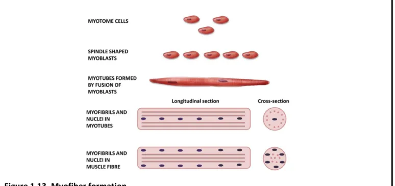
44 Figure 1.13. Myofiber formation.
Round myotomal cells undergo a step-wise differentiation and fusion procedure into long myotubes that further mature to myofibers with peripherally-positioned myonuclei.
Source: Musumeci et al., 2015
Fig. 1.14. Embryonic and fetal waves of myogenesis.