Can We Build Artificial Stem Cell Compartments?
The MIT Faculty has made this article openly available.
Please share
how this access benefits you. Your story matters.
Citation
Semino, Carlos E. “Can We Build Artificial Stem Cell
Compartments?” Journal of Biomedicine and Biotechnology 2003,
no. 3 (2003): 164–169. © 2003 Hindawi Publishing Corporation
As Published
http://dx.doi.org/10.1155/S1110724303208019
Publisher
Hindawi Publishing Corporation
Version
Final published version
Citable link
http://hdl.handle.net/1721.1/96133
Terms of Use
Creative Commons Attribution
©2003 Hindawi Publishing Corporation REVIEW ARTICLE
Can We Build Artificial Stem Cell Compartments?
Carlos E. Semino∗
Center for Biological Engineering and Biotechnology Process Engineering Center, Building 56-354, Massachusetts Institute of Technology, Cambridge, MA 02139, USA
Received 18 April 2002; revised 15 May 2002; accepted 21 May 2002
Animals carry stem cells throughout their entire life, from embryogenesis to senescence. Their function during development and adulthood consists basically of forming and sustaining functional tissues while maintaining a small self-renewing population. They reside in a complex three-dimensional environment consisting of other nearby cells extracellular matrix components, endogenous or exogenous soluble factors, and physical, structural, or mechanical properties of the tissues they inhabit. Can we artificially recreate tissue development such that stem cells can both self-renew and be instructed to mature properly? The main factors required to regulate the maintenance and differentiation of some types of stem cells are known. In addition, new bioengineered synthetic materials that mimic extracellular matrix components can be used as initial scaffolding for building stem cell microenvironments.
INTRODUCTION
Embryonic and adult stem cells share common char-acteristics but differ in certain intrinsic properties. Both have the capacity to differentiate into at least one func-tional cell type which defines them as uni-, multi-, or pluripotential. Instead, they differ in intrinsic cell division kinetics. For instance, during early embryonic develop-ment, the cellular environment constantly changes, and the maintenance of pluripotentiality is short and transi-tory. Embryonic stem cells possess high expansive capac-ity in order to increase rapidly the amount of pluripotent cells able to generate all different tissues of the embryo. On the other hand, adult stem cell behavior and environ-ment are paradoxically opposite. Adult stem cells stay as unchanged as possible in adult tissue niches while produc-ing progeny at lower dividproduc-ing capacity. In order to achieve these two distinct mechanisms of cell maintenance and function, tissues regulate intrinsic and extrinsic factors, creating niches. Here, I review some of the main com-ponents present in embryonic and two adult stem cells, bone marrow and the skin microenvironments as well as propose the use of new synthetic scaffolds to artificially recreate stem cell niches in vitro.
STEM CELLS AND NEW BIOCOMPATIBLE MATERIALS
How can we recreate in vitro niches for embryonic and adult stem cells? Due to knowledge of their natural environments, we can isolate them and maintain them as undifferentiated as possible. Although there is a consider-able amount of knowledge of the main factors required for stem cell maintenance and differentiation in vitro, many of the pieces are still missing. If we want to use stem cells
to obtain highly specialized and functional bioengineered tissues, we need to work with developmental paradigms where we use artificial materials that mimic stem cell compartments. If stem cells self-renew in such compart-ments, we have much better chances in instructing them for proper differentiation. How can we prepare three-dimensional environments that will provide an initial transient scaffold for stem cell homing in vitro? The main initial components we need are biocompatible, defined, and synthetic three-dimensional scaffold materials such as bioceramics, microfiber scale polymers, or nanofiber hy-drogels. These materials already exist: bioceramics such as hydroxyapatite (HA) [1]; the polylactic and polyglycolic acids (PLA and PGA) [2]; and self-assembling peptide hy-drogels [3]. Why are micro- or nanoscale fibers impor-tant? They provide the stem cells with an initial three-dimensional (3D) scaffold. In the first case, HA has suf-ficient rigidity to be used as artificial bone matrix. PLA or PGA polymers provide less rigid structures of medium mechanical strength with 50–100µm pore size to which cells adhere and grow on a pseudo 3D environment be-cause their pore dimensions are in the same order of mag-nitude of an average cell size. On the other hand, peptide hydrogels have poor mechanical strength but their pore size is in between 50–100 nm (1,000 times smaller), in which cells experience a truly 3D environment. They are free to grow, migrate, contact other cells, change in shape, and expose membrane receptors in a proper way. The hy-drogels are permeable to gases, metabolites, and macro-molecules. Like the natural extracellular matrix, these hy-drogels embed cells but do not entrap them. The advan-tage of having defined and artificial materials is immense. It provides an ideal opportunity to control conditions for stem cell maintenance, instruction, and differentiation.
2003:3 (2003) Can We Build Artificial Stem Cell Compartments? 165
The peptide hydrogel provides an additional advantage. It mimics the extracellular matrix (ECM) with very low signaling capacity suggesting that membrane receptor lig-ands (ie, small molecules, growth factors, cytokines, ex-tracellular matrix components or their protein domains) can be easily added to specifically decorate the environ-ment as desired. The process can be easily industrialized since the synthesis is controllable (the hydrogel consists of short peptides, 12–16 residues) and can easily be scaled up. The advantage of HA, CPC, PLA, and PGA lies in their mechanical resistance. Eventually, materials can be used together where physical or mechanical factors can be con-sidered instructive for stem cell differentiation.
EARLY DEVELOPMENT: A CHANGING ENVIRONMENT
The inner cell mass (ICM) and the origin of embryonic stem cells
In the mouse, cell division or cleavage starts 18 hours after fertilization producing an eight-cell embryo stage in which the cells are identical. During this stage, the embryo suffers from compaction so that all the cells adhere to each other. At the 32-cell stage, the first cell differentiation program is initiated; the cells on the periphery form the trophectoderm cell layer (TE), an epithelial monolayer of cells enclosing an internal group of cells, or the inner cell mass (ICM) (see Figure 1a). The external membrane of the TE contains a large amount of glycoproteins that play an important role in implantation and later on partici-pate in the formation of the placenta. The ICM is the first population of embryonic stem cells (ESCs), but their ex-istence is transitory since they will rapidly differentiate to form the tissues of the embryo and extraembryonic mem-branes, such as the allantois and the amnion. Embryonic stem cells isolated from the ICM have pluripotent capac-ity. After either transplantation or maintenance in certain culture conditions in vitro, they can give rise to derivative cells of each of the three primary germ layers—ectoderm, mesoderm, and endoderm—but not to an entire organ-ism. In addition, an intrinsic characteristic of ESCs is their capacity for symmetric self-renewal and expansion. When isolated and cultured in proper conditions, they can be ex-panded to about 1010undifferentiated cells.
Cell adhesion and the extracellular matrix
Cell adhesion plays an important role in keeping the ICM intact and is mediated mainly by cadherins and integrins. E-cadherin cell adhesion is regulated post-translationally via protein kinase C and other signal-ing molecules. It coordinates cellular allocation and spa-tial organization of the ICM in the blastocyst [4]. β1-integrins are required for normal morphogenesis and survival of the ICM. The interaction between the β1-integrins and extracellular matrix components occurs via binding to laminin that is secreted by endodermal cells of the ICM [5]. In addition, proteoglycans, including
embryoglycans composed of poly-N-acetyllactosamines, with molecular weight over 100 kd, are present on the sur-face of the compacted cells and play an important role in maintaining cell adhesion [6]. Embryoglycans carry a se-ries of developmentally regulated oligosaccharide struc-tures expressed in the embryonic ectoderm of early em-bryos, embryonic stem cells, and embryonic carcinoma cells [7]. The trisaccharide SSEA-1 (stage-specific embry-onic antigen-1), with the structure Gal(β1−4)[Fuc(α1−
3)]GlcNac, mediates cell adhesion in pre-implanted em-bryos [8]. Moreover, the embryoglycan-carrying SSEA-1 epitope might serve as a regulator in the signal trans-duction pathway of fibroblast growth factor-2 (FGF-2) and the high-affinity transmembrane receptor fibroblast growth factor-1 (FGFR-1) [9]. SSEA-1 acts as a recogni-tion molecule for FGF-2, playing a role in ligand-receptor dimerization and regulating the mitotic effect of FGF-2 in embryonic stem cells [9].
Oct-4 and LIF: prerequisites for ESCs pluripotentiality
The transcription factor Oct-4 is essential for stem cells originated from ICM because it regulates the expression of downstream genes involved in maintain-ing ICM pluripotential capacity such as FGF-4. Oct-4 is expressed in all cells during the cleavage stage and be-comes restricted to the ICM at the blastula stage [10]. In addition, the leukemia inhibitory factor (LIF) also con-trols the self-renewing capacity and undifferentiated state of ESCs in culture [11]. ESCs can be maintained indefi-nitely in culture with pluripotential capacity in the pres-ence of LIF. LIF is expressed in the TE and secreted into the ICM. LIF operates through heterodimerization of two different classes of cytokine receptors that are expressed in ICM cells, the low-affinity LIF receptor (LIF-R), and the IL-6 signal transducer gp130 [12]. After LIF-induced dimerization, several tyrosine residues in the cytoplasmic domain of gp130 are phosphorylated by JAK kinases. The phosphorylated domain of gp130 interacts with the SH2 domain of the transcription factor STAT3, and, as a con-sequence, STAT3 is activated. The activation of STAT3 is sufficient for ESC self-renewal and maintenance in vitro [13]. Nevertheless, in vivo there is no requirement for LIF, gp130, or STAT3, indicating that the expansion of the epiblast is also under the control of unknown par-allel signaling pathways which are not well known. This multilateral strategy demonstrates that the developmental program relies on alternative complementary pathways.
ADULTHOOD: THE CHALLENGE FOR MAINTENANCE
Fighting the changes
During animal development, cells differentiate and give rise to all tissues of the organism. Adult tissues main-tain regenerative capacity based on cermain-tain minimal pop-ulations of stem cells that share similar properties with embryonic stem cells: multipotency and self-renewing ca-pacity. For instance, adult tissues regulate cell mass and
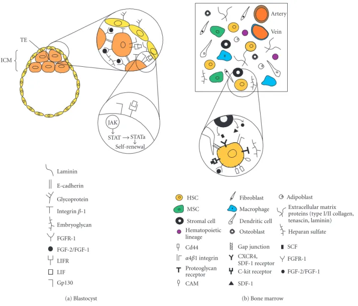
Laminin E-cadherin Glycoprotein Integrinβ-1 Embryoglycan FGFR-1 FGF-2/FGF-1 LIFR LIF Gp130 JAK STAT STATa Self-renewal TE ICM (a) Blastocyst HSC MSC Stromal cell Hematopoietic lineage Fibroblast Macrophage Dendritic cell Osteoblast Adipoblast Extracellular matrix proteins (type I/II collagen, tenascin, laminin) Heparan sulfate Cd44 α4β1 integrin Proteoglycan receptor CAM Gap junction CXCR4, SDF-1 receptor C-kit receptor SDF-1 SCF FGFR-1 FGF-2/FGF-1 Vein Artery (b) Bone marrow
Figure 1. Schematic representation of the main components present in the blastocyst and bone marrow microenvironment. (a) Preimplanted embryo (blastocyst) composed by the inner cell mass (ICM) and the trophectoderm cell layer (TE). Abbreviations: FGF2/FGF, fibroblast growth factor 2 and 1; FGFR-1, fibroblast growth factor receptor-1; LIF, leukemia inhibitory factor; LIFR, leukemia inhibitory factor receptor; JAK, JAK kinase; STAT, transcription factor STAT; STATa, transcription factor STAT activated. (b) Adult bone marrow composed by HSCs, hematopoietic stem cells; MSCs, mesenchymal stem cells; and other cellular components such as adipocytes, stromal cells, hematopoietic lineage, macrophages, dendritic cells, osteoblasts, and adipoblasts. In addition, it is composed also by extracellular matrix proteins such as types I and II collagen, tenascin, laminin, and the proteoglycan heparan sulphate. Abbreviations: SDF-1, stroma-derived factor-1; SCF, stem cell factor; CAM, cell adhesion molecule.
shape by a mechanism involving cell expansion based, in part, on stem cell self-renewal through asymmetric cell di-vision. This type of cell division produces two distinct cell daughters: a committed differentiated cell and another one identical to the original stem cell. In addition, adult tissues, unlike those in the early embryo, keep the mi-croenvironment around the stem cells unchanged as long as possible. Adult stem cells divide slowly in tissues, and in general, the actively dividing cells are the “transitory cells” that are already committed to a tissue type. However, in certain tissue injuries, such as skin wounds, it is clear that the surrounding progenitor cells or transitory cells
around the damaged tissue rapidly expand by changing their mitotic activity during healing. Intrinsic regulators, local chemical signals, and environmental factors govern this dramatic change in cell division and differentiation.
The bone marrow microenvironment
In the bone marrow, blood cells are constantly pro-duced from hematopoietic stem cells (HSCs) by a mech-anism of asymmetric cell divisions that generate a self-renewing HSC and a progenitor cell. These progeni-tors ultimately generate all the cells in the hematopoi-etic lineage: red and white cells, eosinophils, mass cells,
2003:3 (2003) Can We Build Artificial Stem Cell Compartments? 167
monocytes, neutrophils, T and B lymphocytes, and oth-ers [14] (see Figure 1b). But where do HSCs originate from? During embryogenesis, HSCs develop in the dor-sal aorta, the yolk sack, and the lateral plate mesenchyme. This population of HSCs then migrates to the primitive liver, which becomes the hematopoietic organ of the fe-tus. Later, before birth, HSCs migrate to the bone mar-row and stay there indefinitely. How do HSCs both regu-late their progeny and maintain themselves? Part of the secret is based on stroma that is composed of many cells (mainly macrophages, fibroblasts, and adipocytes), and on an intrinsic property of stem cells self-renewal. Stromal cells are in intimate contact with HSCs, work-ing as housekeepwork-ing cells by producwork-ing and maintainwork-ing the bone marrow microenvironment. These cells appar-ently interact physically with HSCs by gap junctions, in-tercellular channels that allow cytoplasmic exchange of small molecules between stromal cells and HSCs [15]. In addition, stromal cells create a three-dimensional en-vironment by secreting ECM components, such as type I–IV collagens, tenascin, laminin, and heparan sulfate (HS) [16, 17, 18]. Stromal cells are positive for vi-mentin, muscle actin, CD10, and Stro-1 and negative for CD45. HSC adhesion to stroma involves a variety of membrane recognition molecules, such asα4β1 integrins which bind to fibronectin; the membrane receptor CD44 which interacts with the glucosaminoglycan hyaluronic acid (HA) [16]; and proteoglycan receptors acting in con-cert with cell adhesion molecules (CAMs), which recog-nize heparan sulfate [17]. The large number of CAMs ex-pressed by the HSCs includes members of the sialomucin family such as CD34, members of the immonoglobu-lin family such as CD31 and CD50, and ligands for se-lectines. This last group belongs to a class of membrane receptors that especially recognize carbohydrate struc-tures [18]. Integrin-mediated interactions of HSCs with ECM have multiple functions including regulation of proliferation and survival in the bone marrow and ad-hesion. In addition, stromal cells produce a cytokine, stroma-derived factor-1 (SDF-1), which is a chemoat-tractant for CD34+ cells. CD34+ hematopoietic
progen-itors express the SDF-1 receptor CXCR4, which stimu-lates transendothelial migration of these cells in presence of SDF-1 [19]. The soluble membrane-associated stem cell factor (SCF) participates in stem cell maintenance and adhesion by interacting with the c-kit receptor on the HSC membrane. Other stromal factors include FGF-1 and FGF-2 which contribute (in addition to SDF-FGF-1) to HSC survival, homing, homeostasis, and proliferation [20].
The bone marrow microenvironment is even more complex. It contains not only HSCs and stroma but also another stem cell type, the mesenchymal stem cell (MSC); adherent macrophages; antigen-presenting cells, or den-dritic cells; endothelial cells; and mesenchymal origin cells such as osteoblasts and adipoblasts. The MSCs self-perpetuate as undifferentiated cells and also undergo dif-ferentiation to produce all the mesenchymal tissues inside
and outside the bone marrow, including their own mar-row stroma, bone, cartilage, tendon, fat, and muscle [21]. The MSCs have some characteristic cell surface markers, such as cytokine receptors (IL-1R, IL-3R, IL-4R, IL-6R, IL-7R), extracellular matrix receptors (I, ICAM-2, VCAM-1, ALCAM), hyaluronate receptors, integrins (α1, α2, α3, αA, αV, β1, β2, β3, β4), growth factor re-ceptors (BFGFR, PDGFR), and other rere-ceptors (Thy-1, IFNγR, TGFβR, TNFR) [22]. This basically indicates the complexity of the cellular and molecular interactions in which HSCs and MSCs are engaged. Interestingly, from the structural point of view, the bone marrow can be con-sidered as a soft tissue surrounded by hard tissue residing in the internal part of the large bones. In other words, it is not exposed to strong mechanical loading in the way that cartilage, tendon, and muscle are. This could be one im-portant factor in maintaining MSCs undifferentiated, and the exposure to mechanical forces in combination with specific microenvironment would dictate the di fferentia-tion into cartilage, tendon, or muscle tissues after MSCs colonize other tissues.
Skin and epidermal stem cells
The skin is of ectodermal origin, and is constantly be-ing renewed at a high rate. It is organized in four main layers. The innermost layer, a thin layer of cells called the basal layer, is covered with a thick layer of cells, the spinous layer. Above these layers is the granular layer, which is covered with dead cells, or stratum corneum (our external skin). The basal layer undergoes active mitosis, constantly producing cells that migrate to the upper lay-ers and terminally differentiate. The skin is a dynamic flow of cells from the inner basal layer to the surface. The epidermal stem cells, or high proliferative capacity ker-atinocytes, reside in the basal layer. Interestingly, the ad-herent properties of the epidermal stem cells and the high content of the extracellular matrix are what keep these cells attached to their microenvironment. They express surface integrins that adhere to collagen type IV (α2β1 receptor) and to fibronectin (α5β1 receptor), as well as low levels of the intercellular junction protein, E-cadherin [23]. This suggests that they interact strongly with the ex-tracellular matrix and poorly with each other. In addition, epidermal stem cells contain a high level of β-catenin, suggesting that Wnt/Frizzled signaling is involved in the regulation of cell proliferation/differentiation of epider-mal stem cells. Moreover, β-catenin has been shown to interact with E-cadherin, thus promoting cell adhesion. An excess of β-catenin will interact with other proteins such as Tcf, Groucho, SMAD4, CtBP, and CBP to form a transcription complex that interacts with DNA, regu-lating transcription of target genes such as CyclinD1 (cell cycle commitment) and c-MYC (exit from the stem cell compartment) [24, 25]. Exit of the stem cell compart-ment is produced by a decrease in the levels of integrins, thus reducing adhesion to the ECM. As a consequence, epidermal stem cells enter cell cycle arrest and terminal differentiation.
CONCLUSIONS
In summary, some examples of essential biomaterials and stem cell niches have been described. In this final part, our intention is to illustrate to the readers to some exam-ples of the combined use of two emerging disciplines, ma-terial science and stem cell technology. For instance, with the potential of building better stem cell environments, can we find conditions in which ESCs can be instructed in vitro to become an adult stem cell by controlling its mi-croenvironment? By recreating developmental programs in designed 3D environments, ECSs can experience a rational sequence of factors (growth factors, extracellular matrix components, mechanic stimulus) required to mimic embryonic development. In a similar way, HSCs can be seeded in hydrogels loaded with purified extracel-lular matrix components mimicking bone marrow. The maintenance and expansion capacity of long-term HSCs can be challenged into these new compartments as well as their capacity to generate blood components. It will be extremely important to artificially regenerate blood or some of their main components in vitro. Can mechanical inputs be considered as a part of stem cell instructive factors? For instance, tubes of PGA mimicking arteries and veins can be built, and their microscale pores can be filled with hydrogel, clonally derived stem cells, microen-vironment decorations, and flow frequencies applied. This will provide a similar tissue environment present during vertebrate circulatory system development. Simi-larly, for bone and cartilage, mesenchymal stem cells in the right microenvironment can be subjected to mechan-ical loading with a program that can recapitulate limb movement and mechanical strength during development. Finally, the skin area can also be explored in more detail. A basal layer-like structure can be easily obtained simply by applying collagens, laminins, and fibronectins onto a porous membrane where keratinocytes will attach, ac-tively divide, and migrate to an upper layer of specifically decorated hydrogel. The flow of cells from the inner to the upper layers can be reproduced as in normal tissues with the hope to obtain better artificial skin. The use of new designed biocompatible material in combination with clonally derived stem cells is an emerging discipline with unlimited potential for future reparative medicine.
ACKNOWLEDGMENTS
I would like to specially thank my colleagues at MIT, Shuguang Zhang, Alan Grodzinsky, John Kisiday, Kim Hamad, Colette Shen, and Christine Ko, for many gratify-ing discussions that helped me to write this work. I would also like to thank the directors of the Center for Biological Engineering (CBE) and Biotechnology Process Engineer-ing Center (BPEC) for their support.
REFERENCES
[1] Shikinami Y, Okuno M. Bioresorbable devices made of forged composites of hydroxyapatite (HA)
parti-cles and poly-L-lactide (PLLA): Part I. Basic charac-teristics. Biomaterials. 1999;20(9):859–877.
[2] Perrin DE, English JP. Polyglycolite and polylactide. In: Domb AJ, Kost J, Wiseman DM, eds. Handbook of Biodegradable Polymers. New York: Harwood Aca-demic Publishers; 1997:3–27.
[3] Holmes TC, de Lacalle S, Su X, Liu G, Rich A, Zhang S. Extensive neurite outgrowth and active synapse formation on self-assembling peptide scaf-folds. Proc Natl Acad Sci USA. 2000;97(12):6728– 6733.
[4] Fleming TP, Sheth B, Fesenko I. Cell adhesion in the preimplantation mammalian embryo and its role in trophectoderm differentiation and blasto-cyst morphogenesis. Front Biosci. 2001;6:D1000– D1007.
[5] Stephens LE, Sutherland AE, Klimanskaya IV, et al. Deletion of beta 1 integrins in mice results in in-ner cell mass failure and peri-implantation lethality. Genes Dev. 1995;9(15):1883–1895.
[6] Wight TN, Heinegard DK, Hascall VC. Proteogly-cans. Structure and function. In: Hay E, ed. Cell Bi-ology of the Extracellular Matrix. New York: Plenum Press; 1991:45–78.
[7] Muramatsu T. Early embryogenesis. In: Fukuda M, ed. Cell Surface Carbohydrates and Cell Development. Boca Raton, Florida: CRC Press; 1992:239–256. [8] Kojima N, Fenderson BA, Stroud MR, et al. Further
studies on cell adhesion based on Le(x)-Le(x) inter-action, with new approaches: embryoglycan aggre-gation of F9 teratocarcinoma cells, and adhesion of various tumour cells based on Le(x) expression. Gly-coconj J. 1994;11(3):238–248.
[9] Dvor´ak P, Hampl A, Jirmanov´a L, Pachol´ıkov´a J, Kusakabe M. Embryoglycan ectodomains regulate biological activity of FGF-2 to embryonic stem cells. J Cell Sci. 1998;111(pt 19):2945–2952.
[10] Palmieri SL, Peter W, Hess H, Scholer HR. Oct-4 transcription factor is differentially expressed in the mouse embryo during establishment of the first two extraembryonic cell lineages in-volved in implantation. Dev Biol. 1994;166(1):259– 267.
[11] Williams RL, Hilton DJ, Pease S, et al. Myeloid leukaemia inhibitory factor maintains the develop-mental potential of embryonic stem cells. Nature. 1988;336(6200):684–687.
[12] Gearing DP, Thut CJ, VandeBos T, et al. Leukemia inhibitory factor receptor is structurally related to the IL-6 signal transducer, gp130. EMBO J. 1991;10(10):2839–2848.
[13] Niwa H, Burdon T, Chambers I, Smith AG. Self-renewal of pluripotent embryonic stem cells is mediated via activation of STAT3. Genes Dev. 1998;12(13):2048–2060.
[14] Weissman IL. Stem cells: units of development, units of regeneration, and units in evolution. Cell. 2000;100(1):157–168.
2003:3 (2003) Can We Build Artificial Stem Cell Compartments? 169
[15] Montecino-Rodriguez E, Dorshkind K. Regulation of hematopoiesis by gap junction-mediated intercel-lular communication. J Leukoc Biol. 2001;70(3):341– 347.
[16] Clark BR, Keating A. Biology of bone marrow stroma. Ann N Y Acad Sci. 1995;770:70–78.
[17] Siczkowski M, Clarke D, Gordon MY. Binding of primitive hematopoietic progenitor cells to mar-row stromal cells involves heparan sulfate. Blood. 1992;80(4):912–919.
[18] Whetton AD, Spooncer E. Role of cytokines and ex-tracellular matrix in the regulation of haemopoietic stem cells. Curr Opin Cell Biol. 1998;10(6):721–726. [19] Mohle R, Bautz F, Rafii S, Moore MA, Brugger W,
Kanz L. The chemokine receptor CXCR-4 is ex-pressed on CD34+ hematopoietic progenitors and leukemic cells and mediates transendothelial migra-tion induced by stromal cell-derived factor-1. Blood. 1998;91(12):4523–4530.
[20] Whetton AD, Graham GJ. Homing and mobi-lization in the stem cell niche. Trends Cell Biol. 1999;9(6):233–238.
[21] Pittenger MF, Mackay AM, Beck SC, et al. Multilin-eage potential of adult human mesenchymal stem cells. Science 1999;284(5411):143–147.
[22] Pittenger MF, Marshak DR. Regenerative mesenchy-mal stem cells from adult bone marrow. In: Marshak DR, Gardner RL, Gottlieb D, eds. Stem Cells. Cold Spring Harbor, NY: Cold Spring Harbor Laboratory Press; 2001:349–373.
[23] Moles JP, Watt FM. The epidermal stem cell com-partment: variation in expression levels of E-cadherin and catenins within the basal layer of human epidermis. J Histochem Cytochem. 1997; 45(6):867–874.
[24] Tetsu O, McCormick F. Beta-catenin regulates ex-pression of cyclin D1 in colon carcinoma cells. Na-ture. 1999;398(6726):422–426.
[25] He TC, Sparks AB, Rago C, et al. Identification of c-MYC as a target of the APC pathway. Science. 1998;281(5382):1509–1512.