metabolic and virulence gene potential in gastric
versus enterohepatic Helicobacter species
The MIT Faculty has made this article openly available. Please share
how this access benefits you. Your story matters.
Citation
Mannion, Anthony et al. "Comparative genomics analysis to
differentiate metabolic and virulence gene potential in gastric
versus enterohepatic Helicobacter species." BMC Genomics 19
(November 2018): 830 © 2018 The Author(s)
As Published
https://doi.org/10.1186/s12864-018-5171-2
Publisher
Biomed Central Ltd
Version
Final published version
Citable link
http://hdl.handle.net/1721.1/119470
Terms of Use
Creative Commons Attribution
R E S E A R C H A R T I C L E
Open Access
Comparative genomics analysis to
differentiate metabolic and virulence gene
potential in gastric versus enterohepatic
Helicobacter species
Anthony Mannion
*, Zeli Shen and James G. Fox
Abstract
Background: The genus Helicobacter are gram-negative, microaerobic, flagellated, mucus-inhabiting bacteria associated with gastrointestinal inflammation and classified as gastric or enterohepatic Helicobacter species (EHS) according to host species and colonization niche. While there are over 30 official species, little is known about the physiology and pathogenic mechanisms of EHS, which account for most in the genus, as well as what genetic factors differentiate gastric versus EHS, given they inhabit different hosts and colonization niches. The objective of this study was to perform a whole-genus comparative analysis of over 100 gastric versus EHS genomes in order to identify genetic determinants that distinguish these Helicobacter species and provide insights about their evolution/ adaptation to different hosts, colonization niches, and mechanisms of virulence.
Results: Whole-genome phylogeny organized Helicobacter species according to their presumed gastric or EHS classification. Analysis of orthologs revealed substantial heterogeneity in physiological and virulence-related genes between gastric and EHS genomes. Metabolic reconstruction predicted that unlike gastric species, EHS appear asaccharolytic and dependent on amino/organic acids to fuel metabolism. Additionally, gastric species lack de novo biosynthetic pathways for several amino acids and purines found in EHS and instead rely on environmental uptake/ salvage pathways. Comparison of virulence factor genes between gastric and EHS genomes identified overlapping yet distinct profiles and included canonical cytotoxins, outer membrane proteins, secretion systems, and survival factors.
Conclusions: The major differences in predicted metabolic function suggest gastric species and EHS may have evolved for survival in the nutrient-rich stomach versus the nutrient-devoid environments, respectively. Contrasting virulence factor gene profiles indicate gastric species and EHS may utilize different pathogenic mechanisms to chronically infect hosts and cause inflammation and tissue damage. The findings from this study provide new insights into the genetic differences underlying gastric versus EHS and support the need for future experimental studies to characterize these pathogens.
Keywords: Gastric and enterohepatic Helicobacter species, Gastrointestinal pathogens, Phylogenetic classification, Metabolism, Virulence factor genes, Comparative genome analysis
* Correspondence:manniona@mit.edu
Division of Comparative Medicine, Massachusetts Institute of Technology, Cambridge, MA, USA
© The Author(s). 2018 Open Access This article is distributed under the terms of the Creative Commons Attribution 4.0 International License (http://creativecommons.org/licenses/by/4.0/), which permits unrestricted use, distribution, and reproduction in any medium, provided you give appropriate credit to the original author(s) and the source, provide a link to the Creative Commons license, and indicate if changes were made. The Creative Commons Public Domain Dedication waiver (http://creativecommons.org/publicdomain/zero/1.0/) applies to the data made available in this article, unless otherwise stated.
Background
Since Helicobacter pylori was discovered in 1982 as the cause of chronic gastritis and later established its role in peptic ulcers and stomach cancers [1,2], the genus Helico-bacterhas expanded to include multiple enterohepatic
Heli-cobacter species (EHS) that colonize and can induce
inflammation and cancer in the lower bowel, liver, and gall-bladder in susceptible hosts [3–5]. The genus now includes over 30 formally named species. These gram-negative, spiral-shaped bacterial species have been detected and iso-lated from the stomach, gastrointestinal tract, liver, and gallbladder in mammals, birds, and reptiles.
In general, Helicobacter species are associated with chronic inflammation and the development of cancer; however, they often colonize their hosts as pathobionts [3, 6]. Chronic infection by Helicobacter spp. in im-munocompetent hosts can cause subclinical disease that during immunocompromised states can manifest with overt clinical signs and pathology. Most Helicobacter spp.have been isolated from animal reservoirs with zoo-notic potential. Additionally, experimental and spontan-eous animal models have shown infection by EHS can induce gastrointestinal, hepatic, and biliary tract inflam-matory pathology and cancers [3]. Most tantalizing is the possibility that, analogous to H. pylori infection with gastric inflammation and cancer, EHS may instigate hu-man inflammatory bowel disease, colorectal cancer, and hepatobiliary disease [7, 8]. Nevertheless, most research has focused on H. pylori and related gastric species that have been isolated and cause disease in humans, leaving a void in our understanding of the mechanisms of colonization and virulence potential in EHS.
The advent of whole genome sequencing has rapidly enhanced the characterization of Helicobacter spp. In 1997, the first H. pylori genome was published [9], and today genomes from over 1,000 different strains are available. Bioinformatic analyses have provided invalu-able insights about the physiology and mechanisms of virulence of H. pylori. Later in 2003, the genome se-quence for the prototype EHS, H. hepaticus, was pub-lished [10]. Genomic comparison of H. pylori versus H.
hepaticus revealed considerable differences in gene
structure and content [10], suggesting that important distinctions underlie the contrasting colonization niches and pathogenic potentials of gastric versus EHS.
Given the different physiology and environmental con-ditions in the stomach versus the lower intestine, such as pH, nutrient digestion/availability, and the micro-biome, we have hypothesized that prominent genetic dif-ferences evolved between gastric versus EHS. By characterizing and comparing genomes of representative gastric versus EHS, we have identified features that dis-tinguish these different species, have provided a ration-ale for their adaptation to different colonization niches,
and have highlighted differences in virulence potential. To complement these efforts, we have also sequenced over 30 novel EHS genomes, thereby substantially in-creasing the number of EHS genomes available to the re-search community. Identifying these similarities and differences is critical for understanding the unique physiology and pathogenic potential of current and an-ticipated identification of additional Helicobacter spp., especially in the context of human infection and zoo-notic disease.
Results
Phylogenetic classification of gastric and enterohepatic Helicobacter species
Phylogenetic trees based on 16s rRNA genes sequences, pan-genome orthologous gene clusters, and average nu-cleotide identity (ANI) similarity were constructed for taxo-nomic organization of gastric and EHS. Interestingly, the phylogenetic organization of species differed between trees. Based on 16s rRNA gene sequences, gastric and EHS did not always cluster with other strains and/or species in their respective subgroupings (i.e., gastric or EHS) (Fig.1a). Pre-viously, it has been noted that phylogenetic organization of Helicobacter spp.based on 16s rRNA gene sequences is dis-cordant with phylogenies based on other genes (such as 23s rRNA or hsp60), isolation/colonization site, biochemical traits, or morphological characteristics [11]. However, pan-genome phylogenetic trees clearly differentiated gastric and EHS from each other (Fig.1b). Furthermore, EHS ap-peared to separate into 9 clades (Fig. 1b). Hierarchical clustering of ANI, an in silico surrogate to experimental
DNA-DNA hybridization [12–14], organized genomes
into a dendrogram that more closely resembled the whole-genome than the 16s rRNA phylogenetic tree (Fig.1c). An ANI threshold of≥95% appears appropriate for differentiat-ing gastric and EHS, although some H. pylori strain-strain comparisons had ANI values slightly less than 95% (Additional file 1: Table S1).
According to the phylogenetic trees, the genetic basis that determines host colonization appears more biased for anatomical niches (i.e., stomach versus lower intestine) ra-ther than assigned to a particular host species, recognizing that in some cases, particular Helicobacter spp. have been isolated in only select hosts. For example, Helicobacter spp. isolated from reptiles fall into three different clades. Likewise, even within clades in which all species were iso-lated from mammals, there are different hosts, such as clade 4, which includes rodent, non-human primates, and pigs. Interestingly, H. bilis strains have been isolated from human, rodent, pig, dog, and sheep sources and appear to diverge into branches irrespective of their host. However,
H. mustelae and H. enhydrae were notable outliers. H.
mustelaehas been traditionally classified as a gastric spe-cies because it can colonize and cause gastric disease in
ferrets [15], but its genetic profile appears more similar to EHS and consequently was classified as so in subsequent analyses. H. enhydrae is a novel species isolated from in-flamed gastric tissue of southern sea otters, a mustelid re-lated to ferrets [16], and was also considered an EHS in this study.
Next, we sought to identify and compare the genetic deter-minants that differentiate the physiological and pathogenic potential of gastric species versus EHS. This included study-ing how H. mustelae, a phylogenetically-classified EHS, colo-nizes the stomach.
Gastric and enterohepatic species have different genomic characteristics and gene annotations
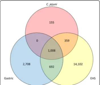
Comparison of gross genomic characteristics found that EHS in general have larger genome sizes with lower GC content and encode more protein coding sequences than gastric species (see Additional file 2). EHS are physically larger than gastric species which may accommodate for their larger genomes and more putative gene products. A total of 19,024 orthogroups were identified in the genomes of gastric, EHS, and Campylobacter jejuni, a close relative to the Helicobacter genus (Fig. 2). 1,008 of these were common to gastric, EHS, and C. jejuni genomes, while 692 were shared between just gastric and EHS genomes. 2,708 and 14,102 orthogroups were unique to gastric and EHS genomes, respectively, indicating substantial genetic
diversity and heterogeneity between EHS and gastric species.
An overwhelming majority of the gastric- or EHS-specific orthogroups were hypothetical proteins (Additional file 1: Tables S2, S3). These hypothetical annotations were pre-dicted to contain autotransportator, transmembrane, signal peptides, intracellular signaling, protein binding and/or unknown protein domains according to InterProScan analysis. Notable unique genes identified in the gastric clade included outer membrane proteins, D-amino acid dehydro-genase involved in D-phenylalanine metabolism, and lipid A 1-phosphatase (lpxE) functioning in lipid A modification during lipopolysaccharide (LPS) biosynthesis. Between EHS clades, hypothetical proteins were also mainly different. However, notable clade-specific genes include: a predicted oxidoreductase for secondary bile acid metabolism unique to clades 1; a hypothetical protein with an aspartic-type endo-peptidase activity domain unique to clade 2; and a carbon monoxide dehydrogenase for carbon fixation and energy production from carbon monoxide unique to clade 3.
Phylogenetic organization by 16s and 23s rRNA gene similarity [11] and more recently whole-genome analysis [17] by other groups have shown that H. mustelae more closely groups with EHS than gastric species. To further study this, we identified the orthogroups shared or unique to H. mustelae compared with other EHS and gastric species (Additional file1: Table S4). Of the 2,356 orthogroups identified in H. mustelae, 269 were unique to only H. mustelae, 179 were shared with EHS but no gastric species and only 15 orthogroups were shared between gastric species but not EHS, reinforcing that H. mustelae has more genetic similarity with EHS than gastric species. Al-most all of the 269 orthogroups unique to H. mustelae encoded for hypothetical proteins aside from annotations for a membrane-fusion protein, signal-transduction regulatory protein flgR, penicillin-binding protein, and four different pu-tative autotransporter protein genes. Of the orthogroups shared between H. mustelae and gastric species, this in-cluded several outer membrane proteins and membrane-associated transporters. Interestingly, H. mustelae along with the gastric species H. acinonychis, H. cetorum, and H. felis harbor two different urease ureA genes that belong to
differ-ent orthogroups (ureA: OG0001420 versus ureA2:
OG0004327). Unlike ureA, ureA2 was not contained within the ureBIEFGH gene cluster, but instead was flanked by the ureB2 gene suggesting expression of a complete urease en-zyme (ureA2B2). Previously, ureA2B2 has been shown to form an enzymatically active urease that is expressed only
(See figure on previous page.)
Fig. 1 a) 16s rRNA gene and b) pan-genome phylogenetic trees both differentiated gastric and EHS genomes. The pan-genome tree more accurately organized EHS into clades consistent with known phenotypic and genetic similarities (e.g., size, morphology, biochemical traits). c) Heatmap and hierarchal clustering of ANI values. Genomes clustered into gastric versus EHS clades based on ANI similarity that resembled the pan-genome tree. See supplement for table with ANI values (Additional file1: Table S1).
Fig. 2 Three-way Venn diagram showing number of shared and unique orthogroups for gastric, EHS, and C. jejuni genomes
under nickel-restricted conditions [18]. The presence of two different urease gene systems in H. mustelae may enable this EHS-like organism to colonize the ferret stomach. Other gastric species and EHS genomes did not encode the ureA2B2operon; however, the presence and role of urease genes in gastric and EHS is discussed in a subsequent section.
To infer the potential physiological and pathogenic signifi-cance of the genetic heterogeneity between gastric species and EHS, genomes were analyzed by KAAS to assign protein coding sequences into functional classifications and meta-bolic pathways (i.e., KEGG pathways). Hierarchical clustering of KEGG pathway profiles organized genomes into gastric versus EHS designation, indicating specific metabolic func-tions/pathways differentiate these types (Fig. 3a, Additional file1: Table S5). EHS genomes were enriched in genes func-tioning in de novo amino acid biosynthesis and metabolism of 2-oxocarboxylic acid (e.g., pyruvate and oxaloacetate) as well as ABC transporters (Fig.3b). Gastric species genomes had more genes functioning in bacterial chemotaxis, carbon metabolism, the pentose phosphate pathway, and folate bio-synthesis (Fig.3b). Gastric species were also highly enriched for“Epithelial cell signaling in Helicobacter pylori infection,” which includes the virulence factors genes vacuolating cyto-toxin and the cag type-IV secretion system (cag-T4SS); this is discussed in more detail later.
Gastric and enterohepatic species have different metabolic potentials
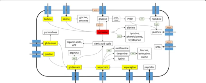
The above differences were further explored via reconstruc-tion and comparison of metabolic pathways between 76 EHS and gastric genomes representative of all species and strains isolated from different host species (Additional file1: Table S6). Metabolic reconstruction predicted that EHS cannot up-take or metabolize glucose or other simple sugars and in-stead are dependent on amino/organic acids to fuel metabolism. Gastric species are able to utilize glucose as well as amino/organic acids, but lack de novo biosynthetic path-ways for several amino acids and purines commonly found in EHS. Gastric instead may rely on environmental uptake/ salvage pathways to acquire these molecules. For all Helicobacter spp., pyruvate appeared to be the central metab-olite linking the networks for carbohydrates, amino acids, and nucleotides metabolism as well as the precursors for energy production (Fig. 4, Additional file 3: Figure S3, Additional file4: Figure S4, Additional file5: Figure S5). The differences predicted for carbohydrate, amino acid, and nucleotide metabolism between gastric and EHS genomes are discussed below as well as summarized in Fig. 4 and Tables 1, 2, 3.
Carbohydrate metabolism
All gastric species (except H. heilmannii ASB1 and H. suis) and only two EHS (H. mustelae and H. apodemus) have
glucose permease genes for uptake of environmental glucose (Table1). Unlike other prototypical enteric bacterial species like Escherichia coli, all Helicobacter spp. do not have a func-tional glycolysis pathway to metabolize glucose into pyruvate because they lack phosphofructokinase (Additional file 1: Tables S2 and S7), which is a rate limiting step catalyzing the formation of fructose 1,6-bisphosphate from fructose-6-phosphate. Alternatively, these gastric species and select EHS likely metabolize glucose into pyruvate via the Entner-Dou-doroff (ED) pathway (Table1). Thus, EHS appear to be asac-charolytic like C. jejuni, which also lacks a glucose uptake and metabolism pathway [19, 20]. While C. jejuni is also considered to be asaccharolytic [19, 20], some rare strains harbor genetic loci to uptake and convert glucose into pyru-vate via the ED pathway [21,22] or uptake and metabolize the simple sugar fucose into pyruvate and lactaldehyde [23–25]. Lactaldehyde can be subsequently converted into pyruvate [23–25]. The alterative glucose metabolism locus was not identified in any EHS genomes, but fucose metab-olism loci were identified in four EHS (H. anseris and the novel reptile isolates Helicobacter sp. 11S03491–1, Helico-bacter sp.11S02629–2, and Helicobacter sp. 13S00401–1) (Table1), indicating select EHS may be able to utilize al-ternative carbohydrate sources.
All Helicobacter spp. require carbohydrates as critical intermediate metabolites and for macromolecules (e.g., LPS and glycosylated surface proteins). The carbohydrate pre-cursors for these requirements likely arise via converting pyruvate into glucose via the gluconeogenesis pathway, which is complete in all Helicobacter spp. Consequently, the asaccharolytic nature of EHS suggests a reliance on or-ganic and amino acids to fuel gluconeogenesis and satisfy their carbon, nitrogen, and energy demands.
Organic and amino acid metabolism
All gastric and EHS genomes have lactate permease and lactate dehydrogenase genes which enable the uptake and conversion of lactate into pyruvate (Table1). Lactate can support H. pylori growth in vitro as the sole carbon source, and mutation of the L-lactate dehydrogenase gene both ablated this growth in vitro as well as in vivo stomach colonization [26,27]. In the stomach and lower intestine, lactate can arise as a byproduct from host and microbial metabolism [28, 29]. Additionally, a majority of EHS and a few gastric species also encoded short chain fatty acid transporters and the enzymatic pathways to metabolize propionate to pyruvate and succinate (Table 1). In the lower intestine, propionate and similar short chain fatty acids are byproducts of microbial me-tabolism of indigestible dietary carbohydrates [29] and can be catabolized as a carbon/energy source for some bacteria like Salmonella spp [30,31].
Serine, proline, glutamine, glutamate, asparagine, and aspartate appear to be important carbon and nitrogen
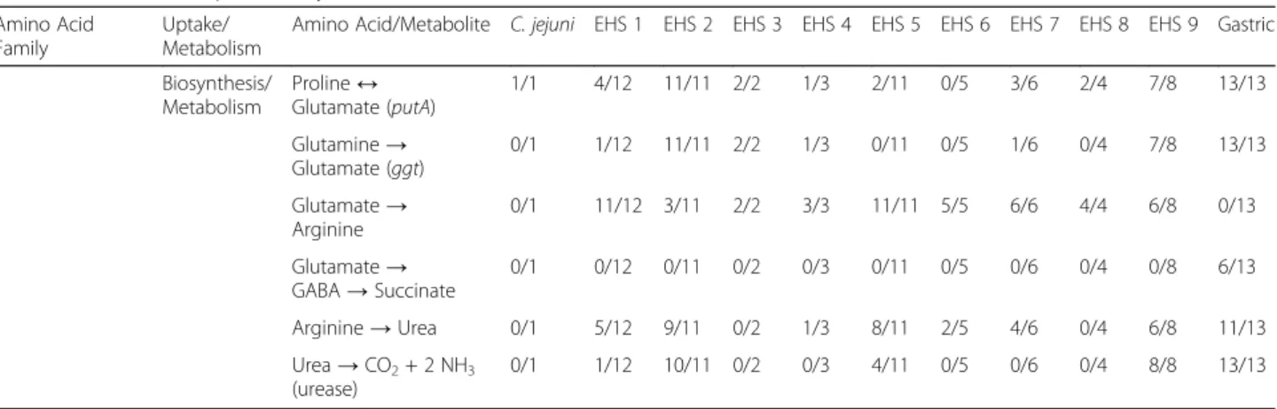
sources for all Helicobacter spp. genomes because they can be directly imported and serve as precursors for de novo synthesis pathways (Fig. 4, Additional file 3: Figure S3, Additional file4: Figure S4). Nearly all Helicobacter spp. en-code genes for a serine transporter and sdaA allowing the transport and conversion of serine to pyruvate (Table2and Additional file3: Figure S3, Additional file4: Figure S4). In C. jejuni, disruption of serine transport or sdaA significantly impaired its growth in vitro and colonization of chickens [32]. This suggests that serine may also have a significant contribution in Helicobacter spp. metabolism, especially for EHS that appear to lack the metabolic mechanisms to pro-duce pyruvate. Additionally, glutamate and aspartate can enter and fuel the citric acid cycle (Fig.4, Additional file6: Figure S2). Proline and glutamine can be converted to glu-tamate via the putA and ggt genes, respectively, in gastric species, but only in select EHS clades (Table2). Nearly all EHS but no gastric genomes contain the de novo synthesis pathway to produce arginine from glutamate (Table 2).
Conversely, arginine transport is almost exclusively
encoded in gastric, but not EHS genomes (Table 2).
Asparagine and glutamate can be metabolized to aspartate by the aspB and ansB genes in all almost all Helicobacter spp.genomes (Table2). Aspartate is the precursor in the de novo biosynthesis pathways for methionine, lysine, threo-nine, and the branched amino acids valine, leucine, and
isoleucine (Fig.4, Additional file 4: Figure S4). Gastric ge-nomes lack the pathways for methionine, valine, leucine, and isoleucine synthesis, while EHS in general encode these pathways (Table2).
de novo synthesis pathways for serine, glycine, cyst-eine, tryptophan, tyrosine, lysine, and threonine appear to be present in most gastric and EHS (Table 2). Gastric species can synthesize alanine from pyruvate, while only
select EHS clades have this capability (Table 2).
Conversely, phenylalanine and histidine de novo synthe-sis are absent in gastric but present in EHS (Table 2). Interestingly, not all Helicobacter spp. genomes compen-sate for missing de novo amino acid synthesis pathways with specific transporters. For example, gastric genomes lack transporters and the de novo synthesis pathway for branched amino acids. Both gastric and EHS may over-come these deficiencies by importing and metabolizing small peptides supported by the presence of peptidases and transporters for this function (Additional file 1: Tables S2, S7).
Citric acid cycle
The citric acid cycle (CAC) metabolizes pyruvate to oxalo-acetate to reduce NAD+ and FAD, which are later used to generate ATP via the electron transport chain. CAC inter-mediates are also extracted to serve as precursors for the
(See figure on previous page.)
Fig. 3 a) Heatmap and hierarchal clustering of KEGG metabolic and functional pathways according to relative gene abundance per genome. Gastric and EHS genomes clustered into distinct clades from each other, and enterohepatic genomes clustered more closely with C. jejuni. See supplement for table heatmap data (Additional file1: Table S5). b) Top 10 enriched KEGG metabolic and functional pathways in gastric versus EHS genomes
Fig. 4 Graphic summary of major metabolic pathways in Helicobacter spp. Transporters and pathways enriched in/unique to gastric and EHS genomes are indicated. Also annotated are pyruvate (red box), the central metabolite in Helicobacter spp. metabolism, and other critical nutrients (yellow boxes) that are precursors for biosynthetic pathways and energy production. See Additional file6: Figure S2, Additional file3: Figure S3, Additional file4: Figure S4 for expanded metabolic reconstruction diagrams
de novo synthesis of several amino acids, nucleotides, and fatty acids (Additional file 3: Figure S3). The CAC for all Helicobacter spp., except for EHS clades 2 and 3, are incom-plete because the gene for succinyl-CoA synthetase is miss-ing (Additional file 1: Tables S2, S7). Therefore, the CAC appears to divide into an oxidative (C6) branch from citrate to succinyl-CoA and a reductive (C4) branch from oxaloace-tate to succinate. Similar to C. jejuni, only the EHS clades 2
and 3 have complete “aerobic-like” CAC because they
encode succinyl-CoA synthetase. In general, almost all Helicobacter spp. genomes encode C4-dicarboxylate trans-porters that allow uptake and replenishing of succinate, fu-marate, and malate into the CAC, while only select Helicobacter spp. genomes encode transporters for citrate and alpha-ketoglutarate (Table1).
Nucleotide metabolism
Glutamine is the precursor for pyrimidine de novo biosyn-thesis, and along with glutamine transporters, gastric and EHS are capable of de novo pyrimidine synthesis (Fig.4, Additional file 4: Figure S4, Additional file 5: Figure S5). Conversely, phosphoribosyl pyrophosphate (PRPP) and histidine are the precursors for de novo purine biosyn-thesis (Fig.4, Additional file6: Figure S2, Additional file4: Figure S4). While all Helicobacter spp. genomes can synthesize PRPP from glucose via the non-oxidative branch of the pentose phosphate pathway, only EHS ge-nomes encode the pathways to metabolize PRPP into his-tidine and then into purines (Table3). Gastric species, as result of this deficiency, harbor a salvage pathway that in-cludes transporters and enzymes for uptake and metabolic interconversion of purines (Table 3). Previous in silico predictions have noted that H. pylori cannot biosynthesize the purine precursor inositol monophosphate (IMP), and
it has been experimentally shown that H. pylori growth in vitro is dependent on the salvage of purines from the en-vironment [33]. Select EHS clades also harbor this purine salvaging mechanism.
Interestingly, EHS clade 8, which includes H. brantae, H. cholecystus, H. pametensis, and H. enhydrae, lacked many of the features conserved in most EHS genomes. For example, these genomes were missing 5/8 enzymes in the CAC (Additional file1: Tables S2, S7). This is the only EHS clade to rely on purine salvage instead of de novo synthesis from histidine as well (Table 3). Other generally conserved amino acid transporters and metab-olism pathways for cysteine, tryptophan, phenylalanine, tyrosine, methionine, valine, leucine, and isoleucine present in EHS were also not found in this clade (Table 2). EHS clade 8 seems to be an outlier that is likely compensated by novel genes and metabolic path-ways not readily apparent in our analysis.
Virulence factor profiles of enterohepatic species differ compared to gastric species
Both gastric species and EHS are frequently associated with gastrointestinal inflammation and cancer. While numerous pathogenic mechanisms have been identified and characterized in H. pylori, these are less understood for other gastric species and EHS. Additionally, it is largely unknown if gastric species versus EHS have con-served similarities and differences in virulence factor gene profiles. To identify and compare potential viru-lence factors profiles between gastric and EHS genomes, a BLASTP analysis was performed using the VICTORs and VFDB virulence factor databases and known Helico-bacter spp. virulence factors genes described in the lit-erature. Gastric genomes on average contained 492.4 ±
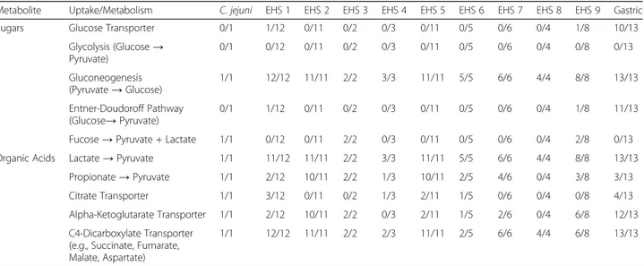
Table 1 Uptake and Metabolism of Sugars and Organic Acids in Gastric versus EHS Genomesa
Metabolite Uptake/Metabolism C. jejuni EHS 1 EHS 2 EHS 3 EHS 4 EHS 5 EHS 6 EHS 7 EHS 8 EHS 9 Gastric
Sugars Glucose Transporter 0/1 1/12 0/11 0/2 0/3 0/11 0/5 0/6 0/4 1/8 10/13
Glycolysis (Glucose→ Pyruvate) 0/1 0/12 0/11 0/2 0/3 0/11 0/5 0/6 0/4 0/8 0/13 Gluconeogenesis (Pyruvate→ Glucose) 1/1 12/12 11/11 2/2 3/3 11/11 5/5 6/6 4/4 8/8 13/13 Entner-Doudoroff Pathway (Glucose→ Pyruvate) 0/1 1/12 0/11 0/2 0/3 0/11 0/5 0/6 0/4 1/8 11/13
Fucose→ Pyruvate + Lactate 1/1 0/12 0/11 2/2 0/3 0/11 0/5 0/6 0/4 2/8 0/13
Organic Acids Lactate→ Pyruvate 1/1 11/12 11/11 2/2 3/3 11/11 5/5 6/6 4/4 8/8 13/13
Propionate→ Pyruvate 1/1 2/12 10/11 2/2 1/3 10/11 2/5 4/6 0/4 3/8 3/13
Citrate Transporter 1/1 3/12 0/11 0/2 1/3 2/11 1/5 0/6 0/4 0/8 4/13
Alpha-Ketoglutarate Transporter 1/1 2/12 10/11 2/2 0/3 2/11 1/5 2/6 0/4 6/8 12/13
C4-Dicarboxylate Transporter (e.g., Succinate, Fumarate, Malate, Aspartate)
1/1 12/12 11/11 2/2 2/3 11/11 2/5 6/6 4/4 6/8 13/13
a
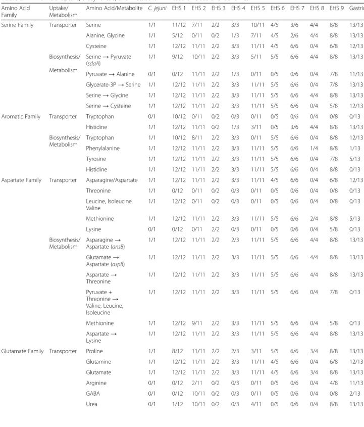
Table 2 Amino Acid Uptake, Biosynthesis, and Metabolism in Gastric versus EHS Genomesa
Amino Acid Family
Uptake/ Metabolism
Amino Acid/Metabolite C. jejuni EHS 1 EHS 2 EHS 3 EHS 4 EHS 5 EHS 6 EHS 7 EHS 8 EHS 9 Gastric
Serine Family Transporter Serine 1/1 11/12 7/11 2/2 3/3 10/11 4/5 3/6 4/4 8/8 13/13
Alanine, Glycine 1/1 5/12 0/11 0/2 1/3 7/11 4/5 2/6 4/4 8/8 13/13 Cysteine 1/1 12/12 11/11 2/2 3/3 11/11 4/5 6/6 0/4 6/8 12/13 Biosynthesis/ Metabolism Serine→ Pyruvate (sdaA) 1/1 9/12 10/11 2/2 3/3 5/11 5/5 6/6 4/4 8/8 13/13 Pyruvate→ Alanine 0/1 0/12 11/11 2/2 1/3 0/11 0/5 0/6 0/4 7/8 11/13 Glycerate-3P→ Serine 1/1 12/12 11/11 2/2 3/3 11/11 5/5 6/6 0/4 7/8 13/13 Serine→ Glycine 1/1 12/12 11/11 2/2 3/3 11/11 5/5 6/6 4/4 8/8 13/13 Serine→ Cysteine 1/1 12/12 11/11 2/2 3/3 11/11 5/5 6/6 0/4 5/8 12/13
Aromatic Family Transporter Tryptophan 0/1 10/12 0/11 0/2 0/3 0/11 0/5 0/6 0/4 0/8 0/13
Histidine 1/1 12/12 11/11 0/2 1/3 3/11 0/5 3/6 4/4 8/8 13/13 Biosynthesis/ Metabolism Tryptophan 1/1 10/12 8/11 2/2 3/3 0/11 5/5 6/6 0/4 8/8 12/13 Phenylalanine 1/1 12/12 11/11 2/2 3/3 11/11 5/5 6/6 1/4 8/8 1/13 Tyrosine 1/1 12/12 11/11 2/2 3/3 11/11 5/5 6/6 0/4 7/8 5/13 Histidine 1/1 12/12 11/11 2/2 3/3 11/11 5/5 6/6 0/4 8/8 0/13
Aspartate Family Transporter Asparagine/Aspartate 1/1 12/12 11/11 2/2 3/3 11/11 4/5 6/6 0/4 6/8 12/13
Threonine 1/1 0/12 0/11 0/2 0/3 0/11 0/5 0/6 0/4 0/8 0/13 Leucine, Isoleucine, Valine 1/1 12/12 0/11 0/2 0/3 0/11 0/5 0/6 0/4 0/8 0/13 Methionine 1/1 12/12 11/11 2/2 3/3 11/11 5/5 6/6 2/4 8/8 5/13 Lysine 0/1 0/12 0/11 2/2 0/3 0/11 0/5 0/6 0/4 5/8 0/13 Biosynthesis/ Metabolism Asparagine→ Aspartate (ansB) 1/1 12/12 11/11 2/2 2/3 11/11 5/5 6/6 4/4 8/8 13/13 Glutamate→ Aspartate (aspB) 1/1 12/12 11/11 2/2 3/3 11/11 5/5 6/6 4/4 8/8 13/13 Aspartate→ Threonine 1/1 12/12 11/11 2/2 3/3 11/11 5/5 6/6 4/4 8/8 13/13 Pyruvate + Threonine→ Valine, Leucine, Isoleucine 1/1 12/12 11/11 2/2 3/3 11/11 5/5 6/6 0/4 7/8 0/13 Methionine 1/1 12/12 9/11 2/2 3/3 11/11 5/5 6/6 0/4 5/8 0/13 Aspartate→ Lysine 1/1 12/12 11/11 2/2 3/3 11/11 5/5 6/6 4/4 8/8 13/13
Glutamate Family Transporter Proline 1/1 8/12 11/11 2/2 2/3 3/11 5/5 6/6 3/4 8/8 13/13
Glutamine 1/1 12/12 11/11 2/2 3/3 11/11 4/5 6/6 0/4 6/8 12/13
Glutamate 1/1 12/12 11/11 2/2 3/3 11/11 4/5 6/6 3/4 8/8 13/13
Arginine 0/1 0/12 2/11 0/2 0/3 0/11 0/5 0/6 0/4 4/8 11/13
GABA 0/1 0/12 10/11 0/2 0/3 0/11 0/5 0/6 0/4 0/8 2/13
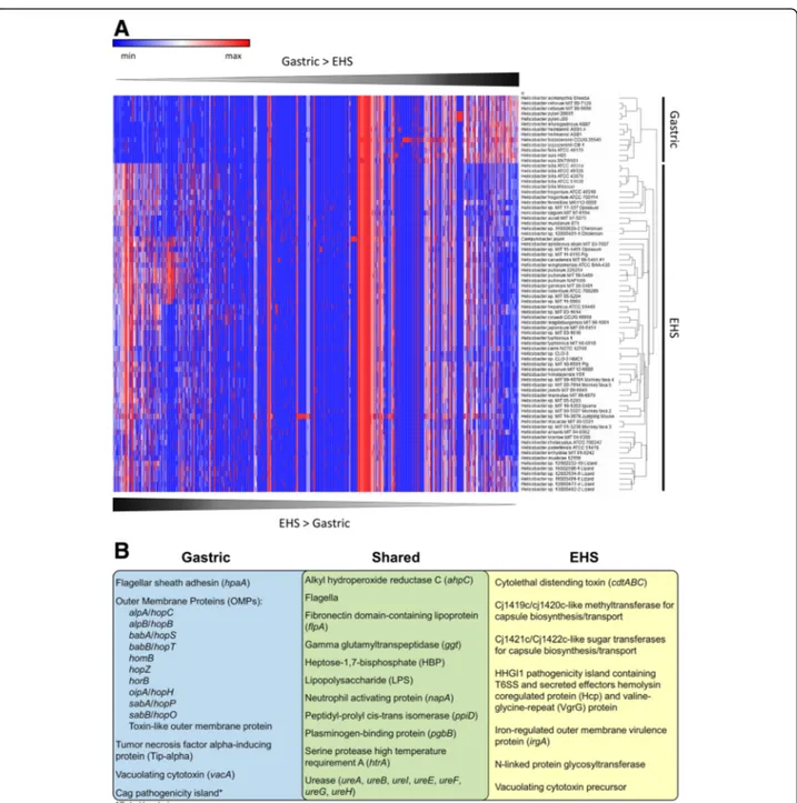
33.7 homologous virulence factor genes compared to 534.0 ± 44.9 genes in EHS genomes and 603 genes in C. jejuni (Additional file 1: Table S8). Numerous virulence factor homologs were shared in all Helicobacter spp. ge-nomes; however, hierarchical clustering indicated gastric and EHS have different overall virulence factor profiles (Fig. 5a, Additional file 1: Table S9). Notable virulence factors shared and different between gastric and EHS
ge-nomes are summarized in Fig. 5b and discussed below.
An expanded description of these genes can be found in the Additional file7results/discussion section.
Cytotoxins
H. pylori expresses vacuolating cytotoxin A (VacA), a
pore-forming cytotoxin that produces vacuoles in gastric epithelial cells that results in apoptosis and can trigger inflammation events [34–36]. It has also been shown to inhibit T cell activation and proliferation allowing im-mune evasion and colonization persistence. H. cetorum and H. acinonychis were the only other gastric species beside H. pylori to encode vacA genes (Additional file1: Table S8). Select EHS also encode homologs to vacuolat-ing cytotoxin precursor annotations originally described in H. canadensis MIT 98–5491 [37] (Additional file 1: Table S8). It is unknown if any of these vacA-like genes in gastric and EHS have virulence properties. The only known cytotoxin for EHS is cytolethal distending toxin (CDT), a double-stranded DNA nuclease that has been shown experimentally to promote pro-inflammatory
pathology and induce pro-carcinogenic DNA damage in the intestine by infection with H. hepaticus and other se-lect EHS [38–41]. Genes for cdt were detected in 53/81 EHS genomes analyzed, but not in any gastric species (Additional file1: Table S6).
Secretion systems
The cag pathogenicity island in H. pylori is composed of a type-IV secretion system (T4SS) that injects the cytotoxin-associated gene A (CagA) effector into host cells to exert a variety of cytotoxic effects on cell junc-tion integrity, proliferajunc-tion, morphology, and dysregula-tion of intracellular signaling cascades [35]. Patients with H. pylori strains harboring the cag pathogenicity island have a significantly greater risk of developing gastric cancer [35]. H. pylori strains also encode three other T4SS gene islands called ComB, Tfs3, and Tfs4 which function in DNA uptake/transfer from the environment and/or secretion of virulence factors that promote gas-tric inflammation [42–45]. A BLAST analysis identified that nearly all gastric and EHS genomes contained ho-mologs for virB, virD, and other T4SS components orga-nized in genetic loci (Additional file 1: Table S10). ComB, Tfs3, or Tfs4 system fragments with sequence identities > 80% were detected in the genomes of H. cetorum, H. suis, and H. acinonychis (Additional file 1: Table S10), which has also been previously described by
Delahay and co-workers [46]. However, for most
ge-nomes, hits often had sequence identities < 50% to cag, Table 2 Amino Acid Uptake, Biosynthesis, and Metabolism in Gastric versus EHS Genomesa(Continued)
Amino Acid Family
Uptake/ Metabolism
Amino Acid/Metabolite C. jejuni EHS 1 EHS 2 EHS 3 EHS 4 EHS 5 EHS 6 EHS 7 EHS 8 EHS 9 Gastric Biosynthesis/ Metabolism Proline↔ Glutamate (putA) 1/1 4/12 11/11 2/2 1/3 2/11 0/5 3/6 2/4 7/8 13/13 Glutamine→ Glutamate (ggt) 0/1 1/12 11/11 2/2 1/3 0/11 0/5 1/6 0/4 7/8 13/13 Glutamate→ Arginine 0/1 11/12 3/11 2/2 3/3 11/11 5/5 6/6 4/4 6/8 0/13 Glutamate→ GABA→ Succinate 0/1 0/12 0/11 0/2 0/3 0/11 0/5 0/6 0/4 0/8 6/13 Arginine→ Urea 0/1 5/12 9/11 0/2 1/3 8/11 2/5 4/6 0/4 6/8 11/13 Urea→ CO2+ 2 NH3 (urease) 0/1 1/12 10/11 0/2 0/3 4/11 0/5 0/6 0/4 8/8 13/13 a
Number of genomes positive for uptake/metabolism per clade
Table 3 de novo Biosynthesis and Salvage of Purines in Gastric versus EHS Genomesa
Uptake/Metabolism C. jejuni EHS 1 EHS 2 EHS 3 EHS 4 EHS 5 EHS 6 EHS 7 EHS 8 EHS 9 Gastric
Histidine Transporter 1/1 12/12 11/11 0/2 1/3 3/11 0/5 3/6 4/4 8/8 13/13
PRPP→ Histidine 1/1 12/12 11/11 2/2 3/3 11/11 5/5 6/6 0/4 8/8 0/13
Purine Transport/Salvage 1/1 4/12 0/11 2/2 0/3 0/11 0/5 0/6 4/4 8/8 13/13
de novo Purine Biosynthesis 1/1 12/12 11/11 2/2 3/3 11/11 5/5 6/6 4/4 8/8 3/13
a
comB, tfs3, or tfs4 genes (Additional file1: Tables S8 and S10), making it difficult to conclude whether these ho-mologs clearly are representative of the T4SS found in H. pylori. As shown by Fischer and co-workers, T4SS genes from different systems in H. pylori genomes (e.g., Tfs3 vs. Tfs4) can have low to high sequence similarity, but should still be considered distinct from each other [47]. The loci identified in gastric and EHS genomes
herein may encode novel T4SS that are assembled and function differently compared to those known for H. pyl-ori. Whether these putative T4SS represent DNA-uptake systems or have virulence properties requires future ex-perimental studies.
H. hepaticus harbors the HHGI1 pathogenicity island
that contains a contiguous 11 gene cluster for type VI secretion system components (HH_0242-HH_0252) and
Fig. 5 a) Heatmap and hierarchal clustering of virulence factor genes according to relative abundance per genome. Gastric and EHS genomes clustered into distinct clades from each other, and enterohepatic genomes clustered more closely with C. jejuni. See supplement for table heatmap data (Additional file1: Table S9). b) Venn diagram of representative virulence factor genes shared and unique between gastric and EHS genomes
includes the secreted effectors called hemolysin co-regulated protein (Hcp) and valine-glycine-repeat (VgrG) protein [10, 48, 49]. H. hepaticus strains lacking this loci induce less severe hepatic and lower intestinal inflammatory pathology [48–50]. H. bilis, H. cinaedi, H. fennelliae, H. hepaticus, H. japonicum, some strains of H. pullorum, H. saguini, Helicobacter sp. MIT 03–1614,
Helicobacter sp. MIT 05–5294, Helicobacter sp. MIT
11–5569, H. didelphidarum MIT 17–337 Opossum,
Helicobacter sp. MIT 14–3879 Jumping Mouse, and H.
trogontum, but no gastric genomes, also appeared to harbor homologous loci for this type VI secretion system and effector proteins (Additional file1: Table S11).
Membrane-associated factors
Gastric species were enriched in homologs for the hor, hom, and hop gene families of outer membrane proteins (OMPs) [51] (Additional file 1: Tables S8, S12). In H. pyloriand other gastric species, these OMPs function as adhesins to the stomach mucosal and epithelial surface and are highly susceptible to mutations that facilitate rapid adaptation and colonization in the gastric environ-ment. Homologs to the prototypical H. pylori hor, hom, and hop gene families of OMPs genes were only identi-fied in 6 EHS genomes. H. saguini contained two hypo-thetical proteins with autotransporter beta-domains (InterProScan domain: IPR005546) and homology to the toxin-like outer membrane protein gene from H. pylori [52] (Additional file 1: Table S8, S12). Helicobacter sp. 13S00482–2 Lizard, Helicobacter sp. MIT 10–6591 Pig,
Helicobacter sp. 12S02232–10 Lizard and encode hopZ
homologs, while horB homologs were detected in
Helico-bacter sp. 12S02634–8 Lizard and Helicobacter sp.
13S00477–4 Lizard.
Aside from these OMPs, all EHS genomes (except Helico-bacter sp. 11S02629–2) contained several genes homolo-gous to virulent adhesins/OMPs, such as iron-regulated outer membrane virulence protein (irgA) from other enteric pathogens including Campylobacter spp., Salmonella enter-ica, and Escherichia coli (Additional file 1: Table S8, S13). Fibronectin domain-containing lipoprotein (flpA) genes were conserved in all gastric and EHS genomes, indicating the potential to adhere to fibronectin in the extracellular matrix of the intestinal epithelium [53–55]. Furthermore, additional novel outer membrane proteins were identified in all gastric and EHS genomes by screening outer mem-brane/hypothetical annotations for outer membrane Inter-ProScan domains (Additional file1: Table S14). On average, outer membrane-associated virulence factor genes were twice as frequent in gastric species compared to the EHS and C. jejuni genomes (gastric: 15.5 ± 8.3; EHS: 8.2 ± 5.4; C. jejuni: 9 genes per genome).
All EHS genomes contained methyltransferases and sugar transferases genes not found in any gastric
genomes. These genes are homologous to the C. jejuni cj1419c/cj1420c and cj1421c/cj1422c genes (Additional file 1: Table S8) that are putatively involved in the bio-synthesis and transport of the extracellular capsule, which has been described to have virulence properties relating to resistance killing by complement proteins in vitro as well as the activation of pro-inflammatory cyto-kine expression and colonization persistence in vivo [56–58]. Also, EHS but not gastric genomes encoded an N-linked protein glycosyltransferase homologous to gen-eral protein glycosylation (pgl) genes found in C. jejuni. These glycosyltransferases mediate conjugation of simple sugar complexes to amino acids residues on extracellular proteins yielding glycans that enable adherence and in-vasion of host epithelial cells in vitro and in vivo [59]. Glycosylation may also protect extracellular proteins from degradation by host proteases, also promoting sur-vival [60]. Previously, it has been reported that a func-tional N-Linked protein glycosylation system exists in the EHS H. pullorum [61]. Thus, EHS may have different mechanisms for forming and decorating their extracellu-lar surfaces compared to gastric species.
Lipopolysaccharide
Lipopolysaccharide (LPS) is a component of the outer cell membrane that facilitates host colonization and modulates immune responses. Biosynthesis of the lipid A, core oligosaccharide, and O-antigen domains of LPS by H. pylori has only been partially elucidated, but in general follows that of other gram-negative bacteria [62]. Little is known about LPS biosynthesis and structure in other gastric species and EHS. The pathway for lipid A biosynthesis appears to be conserved among gastric and EHS genomes (Additional file 1: Table S15). Interest-ingly, all gastric and EHS are missing a homolog for lpxM, the last enzyme in lipid A biosynthesis pathway, while EHS clade 2 and 3 are also missing a homolog for lpxL, the penultimate enzyme. This suggests that alter-native, unidentified genes compensate for lpxM and lpxL function in Helicobacter spp.
LPS by gram-negative pathogens are typically potent induces of immune responses; however, H. pylori consti-tutively modifies its lipid A to dramatically suppress LPS immune reactivity. Modification of lipid A for H. pylori includes removal of phosphates groups by lpxE and lpxF, addition of a phosphoethanolamine (PEtN) moiety by eptA, removal of keto-deoxyoctulosonate (Kdo) sugar residue by Kdo hydrolase, and removal of 3-O-linked acyl chains by lpxR [62]. All of these modification en-zymes are also found in the H. acinonychis and H.
cetorum strains, whereas other gastric and EHS
genomes, except Helicobacter sp. MIT 10–6591 Pig,
Helicobacter sp. MIT 16–1353 Iguana, H. canadensis
least one homolog of these genes (Additional file 1: Table S15).
The core oligosaccharide is synthesized by a series of glycosyltransferases. Only a few glycosyltransferases have been defined in H. pylori, while the remaining are un-identified. Several glycosyltransferases are conserved in all gastric and EHS genomes, including the rfac and rfaF heptosyltransferase genes (Additional file 1: Table S15). However, InterProScan analysis found that gastric and EHS genomes are enriched in different profiles of novel glycosyltransferases genes that suggests the synthesis and structure of the core oligosaccharide differs among
Helicobacter spp. (Additional file 1: Table S8). The
O-antigen of H. pylori is composed of saccharides yield-ing Lewis antigens with structural similarity to host cell surface antigens. As a result, H. pylori can mimic host Lewis antigens thereby providing a mechanism of immune evasion or production of destructive auto-antibodies against the host [62]. Homologs for N-acetylglucosamine transferase (rfaJ), galactose transferase, and alpha-1,3-fucosyltransferase (futA), or alpha-1,2-fucosyltransferase (futC) responsible for the syn-thesis of the O-antigen were identified in all gastric and EHS genomes (Additional file 1: Table S15). All genomes from clade 1 (except H. apodemus), H. cholecystus, H. macacae, Helicobacter sp. 12S02634–8 Lizard, and Helicobacter sp.
MIT 01–3238 Monkey taxa 3 lacked homologs for all of
these genes (Additional file 1: Table S15). Novel glycosyl-transferases identified in the Helicobacter genomes may also contribute to the biosynthesis and modification of the O-antigen as well.
Discussion
In this whole-genus comparative study, we have identi-fied genetic features that differentiate gastric and entero-hepatic Helicobacter spp. on the basis of phylogenetics, genomic characteristics, metabolic pathways, and viru-lence factors genes. The results from these analyses pro-vide new insight into the conserved and contrasting physiological and pathogenic mechanisms that have evolved in gastric species versus EHS to colonize and potentially cause pathology in their respective host intes-tinal environments.
We found that whole-genome phylogenetics more ef-fectively organized Helicobacter genomes as belonging to either gastric or enterohepatic clades than the com-monly used 16s rRNA sequence comparison. Our whole-genome phylogenetic tree resembled those con-structed by Gilbert and co-workers [63] as well as by
Smet and co-workers [17]. Unlike Gilbert and
co-workers’ tree, we considered H. mustelae and the 6 novel lizards Helicobacter spp. isolates as EHS and not gastric species. Smet and co-workers also classified H. mustelae as an EHS, but did not include H. anseris, an EHS isolated from the feces of the geese [64], nor the 6
novel lizards Helicobacter spp. in their analysis. The 6 novel lizard Helicobacter spp. isolates were cultured from cloacal swabs [63], so it is unclear if these organ-isms genuinely colonize the stomach, lower intestinal tract, or both. While H. mustelae is known to colonize and cause gastric disease in ferrets, this organism is commonly detected in the feces of juvenile ferrets, sug-gesting it could transit out of the stomach and poten-tially colonize the lower intestinal tract [15, 65]. Furthermore, phylogenetic organization by 16s and 23s rRNA gene similarity has found that H. mustelae more closely clusters with EHS than gastric species [11]. The lipid profile of H. mustelae is also enriched in hexa-decanoic fatty acids, which has been described to be a characteristic of EHS and rare for gastric Helicobacter spp. [66]. Concordant with these results, we found that
H. mustelae shared substantially more genes with EHS
than gastric genomes, although genes unique to H.
mus-telae not found in other EHS, like ureA2B2, may
con-tribute to its prominent gastric colonization.
According to our whole-genome phylogenetic tree, EHS could be divided into nine different clades. Individ-ual clades did not cluster EHS according to host species, suggesting that evolution for the lower intestinal tract may have occurred independent of host species. Like-wise, the orthogroups different between EHS clades mainly comprised hypothetical proteins, which obfus-cates interpretation of what genetic factors delineates different EHS clades. Nevertheless, the whole-genome phylogenetic tree and drastically contrasting repertoire of orthogroups between gastric and EHS genomes indi-cates that a core set of genetic characteristics have evolved to dictate whether a Helicobacter spp. will colonize and cause pathology at the stomach or lower intestinal tract. Species like H. mustelae may have a blend of these genetic determinants to allow colonization at both sites. There-fore, we performed metabolic reconstruction and analysis for virulence factor genes to understand the potential physiological and pathogenic mechanisms that differenti-ate gastric and enterohepatic species.
Our metabolic reconstruction predicted that gastric and enterohepatic species have fundamentally different requirements for carbohydrate, amino acid, and nucleo-tide substrates to fuel metabolism. Compared to their gastric counterparts, EHS appear to have restricted sources for carbohydrates and nitrogen sources. Most striking is the inability of EHS to utilize simple sugars like glucose and subsequent reliance on amino and or-ganic acids to fuel metabolism. Glutamine/glutamate, as-paragine/aspartate, serine, and proline appear to be the most critical amino acids that fuel EHS metabolism be-cause they can be readily acquired from the environment and enter metabolic pathways rapidly as pyruvate or dir-ectly into the CAC. As a result, the metabolic triangle of
pyruvate-phosphoenolpyruvate-oxaloacetate connecting
the CAC with gluconeogenesis (see Additional file 6:
Figure S2, Additional file 3: Figure S3) appear particu-larly important in the metabolism of Helicobacter spp., especially EHS because it is the only mechanism for pro-ducing carbohydrates from amino and organic acids via gluconeogenesis. Additionally, EHS are enriched in de novo biosynthesis pathways for several amino acids as well as purines that are absent in gastric species. The predicted inability of gastric species to synthesize the amino acids histidine, leucine, methionine, phenylalan-ine, and valine agrees with experimental evidence show-ing H. pylori requires media supplemented with these amino acids for in vitro grow in the absence of serum [67]. Unexpectedly, transporters are not always present in gastric and EHS to compensate for de novo biosyn-thetic deficiencies, suggesting novel transporters and/or metabolic pathways may be present in their respective genomes.
Evolutionary adaptation for survival in the stomach versus the large intestine may have influenced the pre-dicted metabolic differences we detected between gastric and EHS genomes. This includes the drastically different anatomy, physiology, and possibly tissue-centric micro-biomes of the gastric compartment versus the intestinal tract. The stomach and proximal small intestine are more acidic than the lower intestine and are the primary sites for nutrient digestion and absorption [68]. In the lower intestine, indigestible carbohydrates and proteins predominate and are primarily processed by the resident microbiota [68]. Bacterial species colonizing the stomach and small intestine (e.g., H. pylori and Lactobacillus reu-teri, respectively) often have smaller size genomes with a restricted number of biosynthetic pathway genes because
they occupy a nutrient rich environments [69].
Con-versely, we observed that EHS, like other bacterial spe-cies that colonize the lower intestine, typically have larger size genomes and more diverse biosynthetic path-way genes, likely enabling them to adapt and utilize a variety of available nutrients [69]. Consequently, the lar-ger genomes of EHS may endow them with increased metabolic flexibility compared to gastric species because EHS must be more resourceful to survive.
From our analysis, we observed that gastric species are enriched in methyl-accepting chemotaxis proteins and two-component signaling proteins compared to EHS. An abundance of these chemotaxis genes may give gastric species an advantage within the stomach to respond to environment stimuli with flagellated movement away from acidic pH levels and towards necessary nutrient gradients close to gastric epithelia. EHS, despite similar mucous-colonizing ability, likely have less access to basic nutrient molecules (e.g., simple sugar and free amino acids) than gastric species and therefore may have
acquired de novo biosynthetic pathways, such as amino acids, to enable survival.
Microbiota differences in the stomach versus lower in-testinal tract should also be considered as candidates that have influenced Helicobacter evolution. Aside from gastric Helicobacter spp., the stomach does not have a complex microbiota unlike the lower bowel, which sug-gests reduced competition for colonization niches and nutrients. In comparison, the lower intestine is abun-dantly colonized by diversity of bacteria, fungi, and
vi-ruses, all of which may be competing for the
colonization niches and nutrients needed by EHS for survival. However, our metabolic analysis also found that microbiome-derived nutrients like lactate and propion-ate may also benefit EHS and suggests that cooperative relationships could be essential for EHS colonization and pathogenicity within the intestinal tract. One study found that germfree mice infected with H. hepaticus only develop significant typhlocolitis if co-infected with L. reuteri[70]. Another study observed that H. hepaticu-s-induced intestinal pathology is exacerbated or attenu-ated depending on the composition of the murine microbiome [71]. It would be informative to further study how interactions with intestinal microorganisms influence the physiology and pathogenic potential of EHS.
It is important to appreciate that many EHS colonize not only the large intestine (e.g., cecum and colon), but have been detected in the gall bladder, biliary tract, and liver as well. In order to survive in this diversity of intes-tinal and extraintesintes-tinal niches, EHS may have experi-enced an evolutionary pressure to acquire/maintain a repertoire of flexible metabolic pathways. This may have been unnecessary for gastric species given their re-stricted niche. For some strains of C. jejuni, it has been shown that tropism and successful colonization of the
intestinal tract versus the liver is dictated by
γ-glutamyltranspeptidase (GGT) and a secreted isoform of asparaginase (AnsB), respectively [72].
GGT is an outer membrane-associated enzyme that metabolizes host glutamine into glutamate and ammo-nia, which are then imported by the bacterial cell as pre-cursors to fuel metabolic needs. ggt expression is necessary for colonization persistence and the develop-ment of inflammation-induced pathology by H. pylori [73], H. suis [74], and C. jejuni [75]. We detected ggt ho-mologs in all gastric species as well as select EHS
ge-nomes (see Table 2, Additional file 1: Table S6).
Previously, it has been reported that H. bilis (EHS clade 2) contains two different ggt gene annotations: one that is enzymatically active and a second that has mutations in conserved functional regions rendering it inactive [76]. Interestingly, all EHS genomes from clade 2 also contain two different ggt gene annotations for the active gene and its putatively inactive paralog. GGT activity by
Helicobacter and Campylobacter spp. has also been shown to yield an anti-proliferative effect to rapidly div-iding intestinal epithelial and immune cells due to deple-tion of host glutamine reserves [76–80]. Therefore, ggt genes may be important factor that promotes intestinal tissue tropism and pathogenic potential in certain EHS.
AnsB catalyzes the conversation of asparagine to as-partate. While nearly all Helicobacter spp. have a homo-log for ansB, only select gastric and EHS genomes encoded an isoform with predicted signal-peptide re-quired for secretion (Additional file 1: Table S6). In C. jejuni, secreted AnsB, but the not cytoplasmic isoform, was required for significant liver tissue tropism [72]. EHS often isolated from the liver, like H. hepaticus and H. bilis, were found to encode the secreted ansB gene. Based on our predictions from the metabolic reconstruc-tion, glutamine and asparagine are key precursors of car-bon and nitrogen for EHS, and acquisition of these nutrients by GGT and AnsB may facilitate their entero-hepatic tropism.
Urease plays an essential role in stomach colonization by metabolizing urea into ammonia in order neutralize stom-ach acid needed to permit survival in the gastric compart-ment. Isogenic mutants of gastric species like H. pylori [81] or H. mustelae [82] lacking urease cannot establish persist-ent stomach colonization. As expected, urea uptake and urease genes were identified in all gastric genomes. Interest-ingly, species from several EHS clades (see Table 2) also harbored the urease genetic loci. Unexpectedly, H. enhy-drae, the novel species isolated from the stomachs of south-ern sea otters, lacks urease genes and activity [16]. Urease activity by these select EHS may facilitate survival in the acidic gastrointestinal environment during transmission to new hosts. However, urease may also produce ammonia for nitrogen assimilation rather than acid neutralization in the intestine (pH ~ 6.1) and liver (pH ~ 7.4) [83]. Experimen-tally, it has been shown that urease deficiency in isogenic H. hepaticus mutants does not impair cecal colonization, but does prevent hepatic colonization and consequently at-tenuates hepatic pathology compared to the wild-type strain [83]. Additionally, urease-expressing EHS (e.g., H. hepaticusand H. bilis) but not urease-negative species (e.g., H. cinaedior H. rodentium) are capable of inducing choles-terol gallstones and associated hepatobiliary inflammatory pathology in mice [84]. Likewise, only urease-expressing EHS can precipitate calcium in vitro [85]. Thus, urease ex-pression by EHS may promote hepatobiliary colonization and pathology.
Aside from metabolic differences, we found that gastric Helicobacter spp. and EHS encode a wide diversity of viru-lence genes ranging from adhesins, cytotoxins, and sur-vival factors. Some virulence genes are shared among all Helicobacter spp., while other contribute to the unique virulence profiles that differentiate gastric and EHS from
each other. Thus, fundamental differences appear to exist in the mechanisms by which gastric and EHS are capable of eliciting pathogenic infection in their hosts. For ex-ample, while flagella subunits and assembly genes were conserved in all Helicobacter spp. genomes, gastric ge-nomes encode flagellar sheath adhesin (hpaA), a protein that protects flagellin subunits from depolymerization in low pH environments [86]. The presence of hpaA in gas-tric but not EHS genomes indicates that virulence factor genes have also undergone evolutionary pressures to fit their colonization niche. Previously, it has been reported that O-antigen structure for H. pylori also varies depend-ing on the pH of in vitro culture [62]. Therefore, the pH difference in the colonization niche preference of gastric versus EHS may also affect their O-antigen structure. In agreement with our genetic analysis suggesting differences in LPS biosynthesis, phenotypic characterizations of LPS have found substantial structural and immune-reactive heterogeneity among gastric species and EHS [87] and emphasize the need to further experimentally validate the pathogenic significance of LPS in Helicobacter spp.
Interestingly, some virulence genes identified also have overlapping metabolic functions. For example, GGT is im-portant for glutamate acquisition and colonization, while simultaneously yielding deleterious effects to the host. These genes have been coined “nutritional virulence fac-tors” in C. jejuni and other pathogens [88,89] and may be an important new source for understanding the pathogenic determinants in Helicobacter spp. Identifying and character-izing if metabolically-associated genes have virulence prop-erties would not only enhance understanding of how Helicobacter spp. maintain colonization in their different sites, but may also elucidate mechanisms utilized by these organisms to induce inflammatory pathology and cancers.
Several proteomics studies have indicated that gastric and enterohepatic Helicobacter spp. express different protein profiles. Fowsantear and co-workers showed there are significantly different proteomic profiles be-tween several representative gastric and enterohepatic Helicobacter spp.[90]. Interestingly, these authors noted that H. felis, a gastric species isolated from felines, grouped more closely with other EHS, and H. mustelae did not cluster with H. pylori or other EHS. Another study by Kornilovs’ka and co-workers analyzing surface protein profiles of H. pylori and representative EHS found distinct differences among the Helicobacter spp. [91]. Antisera collected from rabbits immunized with sonicate from these Helicobacter spp. identified several surface proteins capable of inducing immunogenic host responses. A comparison of OMPs between H. bilis strains isolated from mice, dogs, rats, and gerbils to H. pylori found H. bilis strains have similar OMP profiles among each other, but were different compared to H. pylori [92]. These OMPs were also capable of inducing
immunogenic host responses. Lastly, Hynes and co-workers showed that the proteomic profiles of H. pylori and several representative EHS were not only distinct but also changed differently among species when challenged by bile stress in vitro [93, 94]. A limitation of the afore-mentioned studies is that whole-genome sequences were not available for all species/species at the time of their analysis, thereby impairing cross-validation of differen-tially expressed proteins using mass spectrometry-based protein identification methods. In our study, while gen-omic annotation do not unequivocally confirm differences in biological function between gastric and enterohepatic Helicobacter spp., the findings and datasets we present will facilitate further studies (such as transcriptomics, proteo-mics, metaboloproteo-mics, and other phenotypic experiments) needed to validate our predictions regarding Helicobacter spp.physiology and pathogenic mechanisms.
Conclusion
This genus-wide comparative analysis determined that gastric Helicobacter spp. and EHS can be differentiated on the basis distinct genetic features. Phylogenetic classi-fication by whole-genome and ANI was found to be a more effective way to taxonomically identify and classify gastric and EHS and was more accurate than traditional 16s rRNA gene sequences. This is important because it provided a means by which the novel EHS included in this study could be differentiated as novel species or new strains of existing species. Furthermore, metabolic reconstruction revealed key differences in the uptake, biosynthesis, and metabolism of carbohydrates, amino acids, and nucleotides between gastric and EHS. These findings have enhanced our insights into how these or-ganisms may have evolved and adapted to colonize their respective niches. Lastly, gastric species and EHS have overlapping as well as distinct virulence factor profiles. In addition to the canonical factors, and novel virulence gene homologs were identified in both gastric and EHS, thereby increasing the repertoire of possible virulence mechanisms. Most importantly, the findings from this study provide new opportunities in the future to experi-mentally probe how these metabolic and virulence genes affect colonization and pathogenicity of gastric species and EHS using in vitro and in vivo models.
Methods
Genome sequencing, assembly, and gene annotation
Helicobacter spp. were grown on trypticase soy agar
plate with 5% sheep blood (Remel Laboratories, Lenexa, KS). The plates were incubated at 37 °C under micro-aerobic conditions in a vented jar containing N2, H2, and
CO2(80:10:10) for 48 h. Bacteria pellets were collected
for isolation of genomic DNA using the MasterPure Complete DNA and RNA Purification Kit (Epicentre,
Madison, WI) following the manufacturer’s protocol for bacterial cell samples. DNA libraries were prepared by the Sequencing Core at the Forsyth Institute (Cam-bridge, MA) using NextraXT for sequencing of 2 × 150 or 2 × 250 paired-end reads by Illumina MiSeq. Raw se-quence reads were decontaminated of adapter sese-quences
and quality trimmed to a Phred quality score (Q)≥ 10
using BBDuk from the BBMap package version 36.99 (http://sourceforge.net/projects/bbmap/). Decontami-nated reads were then de novo assembled into contigs with SPAdes hosted by the PATRIC server (Pathosystems Resource Integration Center) [95,96].
Several genomes previously sequenced by our lab [97– 100] were re-assembled by first performing decontamin-ation and quality trimming on raw sequencing reads using BBDuk followed by de novo contigs assembly with SPAdes, as described above. All publically available gas-tric (only four representative H. pylori genomes) and EHS genomes were downloaded from the National Center for Biotechnology Information database (NCBI) (on 9/1/2017). In total 110 genomes were included in the analysis, representing 17 gastric speceis and 45 EHS (Additional file1: Table S1). The genome for Campylobacter jejunisubsp. jejuni NCTC 11168 = ATCC 700819 was also downloaded and included in the analysis as a closely related outgroup. All genomes were annotated with RAST on the PATRIC server for consistency in sub-sequent analyses.
Bioinformatic analyses
16s rRNA phylogenetic trees were constructed using MegAlign from the Lasergene software package (DNAStar Inc., Madison, WI) by aligning full length 16s rRNA gene sequences with ClustalW followed by phylogenetic tree construction using the neighbor-joining method. The Bac-terial Pan Genome Analysis (BPGA) tool was used to identify orthologous gene clusters with USEARCH at 50% identify threshold for subsequent pan-genome phylogen-etic tree making by the neighbor-joining method [101]. OrthoANI-usearch was used to calculate average nucleo-tide identity (ANI) between genomes in order to
differen-tiate species at a 95% similarity threshold [102].
Orthofinder was used to identify orthologous genes and assign them into orthogroups [103]. KAAS (KEGG Auto-matic Annotation Server) was used to assign KO (KEGG Orthology) annotations for metabolic reconstruction and functional predictions (parameters: program: GHOSTX; method: SBH; GENES data set: hsa, dme, ath, sce, pfa, eco, sty, hin, pae, nme, hpy, rpr, mlo, bsu, sau, lla, spn, cac, mge, mtu, ctr, bbu, syn, aae, mja, afu, pho, ape, hpj, hhe, hac, hms, cje, wsu) [104]. BLAST2GO [105] and Inter-ProScan 5 [106] were used to further predict and validate the domains and functions of protein gene annotations. Virulence factor genes were identified by BLASTP analysis
of genomes against the VICTORs [107] and VFDB [108] virulence factor databases as well as against known Helico-bacter spp.virulence factors genes described in the litera-ture. BLASTP parameters were set at sequence identity ≥25%, sequence coverage ≥75%, and E-value ≤10e-10.
Data was organized and analyzed using Python 2.7.14 (https://www.python.org/) and Pandas v0.22.0 (https:// pandas.pydata.org/). Heatmaps and hierarchical clustering of data was performed using Morpheus (Broad Institute,
Cambridge, MA; https://software.broadinstitute.org/
morpheus/). Phylogenetic trees and dendrograms were created with FigTree v1.4.3 (http://tree.bio.ed.ac.uk/ software/figtree/).
Additional files
Additional file 1:Table S1. Average Nucleotide Identity. Table S2. Genome Annotations. Table S3. Gastric and EHS Clade Orthogroups. Table S4. H. mustelae Orthogroups. Table S5. KEGG Heatmap. Table S6. Genome Metadata and Characteristics. Table S7. Metabolism Pathways per Genome. Table S8. Virulence Factor BLAST Results. Table S9. Virulence Factor Heatmap. Table S10. H. pylori T4SS. Table S11 H. hepaticus T6SS. Table S12. H. pylori Outer Membrane Proteins. Table S13. Other Outer Membrane Proteins. Table S14. Novel Membrane-Associated Proteins. Table S15. LPS Biosynthesis. (XLSX 147858 kb)
Additional file 2:Supplementary Results/Discussion Section (Additional file7: Figure S1). (DOCX 47 kb)
Additional file 3:Figure S3. Expanded diagram of carbohydrate, amino acids, and nucleotide metabolic pathways reconstructed in Helicobacter genomes. Nutrients that can be imported from the environment to fuel metabolism are labeled with blue boxes. The metabolic triangle between phosphoenolpyruvate (PEP), pyruvate, and oxaloacetate (red boxes) links the Entner-Doudoroff (ED) pathway and gluconeogenesis with the citric acid cycle (CAC; contained within pink box). Selected enzymes are indicated in circles with solid arrows showing their reactions. Dashed arrows indicate multi-enzyme reactions to different biosynthetic pathways (green boxes). Abbreviations: 4-aminobutyrate-2-oxoglutarate transaminase (puuE), acetate kinase (ackA), acetyl-coenzyme A synthetase (Acs), aldehyde dehydrogenase A (Ald), asparaginase (AnsB), aspartase (AspA), aspartate aminotransferase (AspB), Entner-Doudoroff (ED) pathway, gamma-aminobutyric acid (GABA), gamma-glutamyltranspeptidase (GGT), glutamate decarboxylase (GAD), lactate dehydrogenase (LDH), non-oxidative pentose phosphate pathway (PPP), phosphoribosyl pyrophosphate (PRPP), phosphotransacetylase (pta), proline dehydrogenase (PutA), serine dehydratase (SdaA), succinate-semialdehyde dehydrogenase (gabD), phos-phoenolpyruvate (PEP). (JPG 383 kb)
Additional file 4:Figure S4. Expanded diagram of amino acids biosynthesis pathways reconstructed in Helicobacter genomes. Enzymes are labeled in boxes with their enzyme code (E.C.) or gene abbreviations and solid arrows showing their reactions. Dashed arrows indicate multi-enzyme reactions to different biosynthetic pathways. Abbreviations: alanine (Ala), arginine (Arg), asparagine (Asn), aspartic acid (Asp), cysteine (Cys), glutamic acid (Glu), glutamine (Gln), glycine (Gly), histidine (His), isoleucine (Ile), leucine (Leu), lysine (Lys), methionine (Met), phenylalanine (Phe), proline (Pro), serine (Ser), threonine (Thr), tryptophan (Trp), tyrosine (Tyr), valine (Val), non-oxidative pentose phosphate pathway (PPP), asparaginase (AnsB), aspartate aminotransferase (AspB), gamma-glutamyltranspeptidase (GGT), proline dehydrogenase (PutA), serine dehydratase (SdaA), 5-Aminoimidazole-4-carboxamide ribonucleotide (AICAR), phosphoribosyl pyrophosphate (PRPP), erythrose 4-phosphate (E4P), glycerate-3P (3PG), phosphoenolpyruvate (PEP), pyruvate (Pyr), oxaloacetate (Oxa). (JPG 334 kb)
Additional file 5:Figure S5. Expanded diagram of purine (left) and pyrimidine (right) nucleotide biosynthesis pathways reconstructed in
Helicobacter genomes. Enzymes are labeled in boxes with their enzyme code (E.C.) and solid arrows showing their reactions. Dashed arrows indicate multi-enzyme reactions to different biosynthetic pathways. Ab-breviations: phosphoenolpyruvate (PEP), pyruvate (Pyr), glucose-6-phosphate (G6P), phosphoribosyl pyroglucose-6-phosphate (PRPP), glutamine (Gln), uridine triphosphate (UTP), cytidine triphosphate (CTP), inosine monopho-sphate (IMP) adenine monophomonopho-sphate (AMP), xanthine monophomonopho-sphate (XMP), guanine monophosphate (GMP), guanine (Gua), xanthine (Xan), hypoxanthine (Hyx), adenine (Ade). (JPG 148 kb)
Additional file 6:Figure S2. Plot of genome sizes (average ± standard deviation) for EHS, gastric, and C. jejuni genomes. Red dashed line indicates average gastric genome size for comparison. (JPG 305 kb)
Additional file 7:Figure S1. A) Genome sizes were plotted against the number of annotated protein coding sequences (CDS) and GC content. For all Helicobacter genomes, a linear relationship existed for genome size versus number of annotated protein CDS (R2= 0.8939). Three EHS genomes (H. muridarum ST1, H. pametensis ATCC 51478, and H. cholecystus ATCC 700242) and one gastric genome (H. bizzozeronii CCUG 35545) appeared to be outliers and are indicated in the graph. Diamonds (◆), gastric genomes; circles (●) EHS genomes; cross (X), C. jejuni genome. B) Genome sizes were plotted against the additive size of all protein CDS, RNA genes, and non-coding gene sequences. Linear relationships existed for genome size versus size of protein CDS (R2= 0.9782) and non-coding gene sequences (R2= 0.8057), but not for RNA gene sequences (R2= 0.0076). (ZIP 458 kb)
Acknowledgements
AM greatly acknowledges Greg Miller, PhD from Northeastern University as well as his thesis committee for their thoughtful insights and encouraging discussions. Funding
This work was supported by the following NIH grants given to J.G.F.: T32-OD010978, P30-ES002109, P01-CA028848, and R35-CA210088.
Availability of data and materials
All data generated or analyzed during this study are included in this published article and its supplementary information files or are available from the corresponding author on reasonable request.
Authors’ contributions
AM performed DNA isolation, analyzed and interpreted genome data, and wrote the manuscript. ZS performed bacterial culture, interpreted data, and reviewed the manuscript. ZS and JGF interpreted data, and reviewed the manuscript. All authors read and approved the final manuscript. Authors’ information
This work represents part of AM’s PhD thesis dissertation. Ethics approval and consent to participate
Not applicable. Consent for publication Not applicable. Competing interests
The authors declare that they have no competing interests.
Publisher’s Note
Springer Nature remains neutral with regard to jurisdictional claims in published maps and institutional affiliations.
Received: 24 May 2018 Accepted: 15 October 2018
References
1. Warren JR, Marshall B. Unidentified curved bacilli on gastric epithelium in active chronic gastritis. Lancet. 1983;1(8336):1273–5.
2. Marshall BJ. Helicobacter pylori in peptic ulcer: have Koch's postulates been fulfilled? Ann Med. 1995;27(5):565–8.