HAL Id: hal-01129772
https://hal.archives-ouvertes.fr/hal-01129772
Submitted on 28 May 2020
HAL is a multi-disciplinary open access archive for the deposit and dissemination of sci-entific research documents, whether they are pub-lished or not. The documents may come from teaching and research institutions in France or abroad, or from public or private research centers.
L’archive ouverte pluridisciplinaire HAL, est destinée au dépôt et à la diffusion de documents scientifiques de niveau recherche, publiés ou non, émanant des établissements d’enseignement et de recherche français ou étrangers, des laboratoires publics ou privés.
epithelium
Emilie Viennois, Teresa Esposito, Julie Dufour, Aurélien Pommier, Stéphane
Fabre, Jean-Louis Kemeny, Laurent Guy, Laurent Morel, Jean-Marc A.
Lobaccaro, Silvère Baron
To cite this version:
Emilie Viennois, Teresa Esposito, Julie Dufour, Aurélien Pommier, Stéphane Fabre, et al.. Lxrα regulates the androgen response in prostate epithelium. Endocrinology, Endocrine Society, 2012, 153 (7), pp.3211-3223. �10.1210/en.2011-1996�. �hal-01129772�
Lxr
␣ Regulates the Androgen Response in Prostate
Epithelium
Emilie Viennois, Teresa Esposito, Julie Dufour, Aurélien Pommier, Stephane Fabre, Jean-Louis Kemeny, Laurent Guy, Laurent Morel, Jean-Marc Lobaccaro, and Silvère Baron
Department of Génétique Reproduction et Développement (E.V., T.E., J.D., A.P., L.M., J.-M.L., S.B.), Clermont Université, Université Blaise Pascal, and Centre de Recherche en Nutrition Humaine
d’Auvergne (E.V., T.E., J.D., A.P., L.M., J.-M.L., S.B.), F-63000 Clermont-Ferrand, France; Centre National de la Recherche Scientifique, Unité Mixte de Recherche 6293 (E.V., T.E., J.D., A.P., L.M., J.-M.L., S.B.), and Institut National de la Santé et de la Recherche Médicale, Unité 1103 (E.V., T.E., J.D., A.P., L.M., J.-M.L., S.B.), GReD, F-63177 Aubiere, France; Dipartimento di Medicina Sperimentale (T.E.), Laboratorio di Biologia Molecolare, Section F. Bottazzi, Second University of Naples, F-80138 Naples, Italy; Department of Physiologie de la Reproduction et des Comportements (S.F.), Unité Mixte de Recherche 6175 Institut National de la Recherche Agronomique-Centre National de la Recherche Scientifique-Université de Tours-Haras Nationaux, F-37380 Nouzilly, France; and Service de Pathologie (J.-L.K.) and Service d’Urologie (L.G.), Hôpital Gabriel Montpied, F-63003 Clermont-Ferrand, France
Benign prostatic hyperplasia is a nonmalignant enlargement of the prostate that commonly occurs in older men. We show that liver X receptor (Lxr)-␣ knockout mice (lxr␣⫺/⫺) develop ventral pros-tate hypertrophy, correlating with an overaccumulation of secreted proteins in prostatic ducts and an alteration of vesicular trafficking in epithelial cells. In the fluid of the lxr␣⫺/⫺prostates, spermine binding protein is highly accumulated and shows a 3000-fold increase of its mRNA. This overex-pression is mediated by androgen hypersensitivity in lxr␣⫺/⫺mice, restricted to the ventral prostate. Generation of chimeric recombinant prostates demonstrates that Lxr␣ is involved in the estab-lishment of the epithelial-mesenchymal interactions in the mouse prostate. Altogether these re-sults point out the crucial role of Lxr␣ in the homeostasis of the ventral prostate and suggest lxr␣⫺/⫺ mice may be a good model to investigate the molecular mechanisms of benign prostatic hyperplasia. (Endocrinology 153: 3211–3223, 2012)
B
enign prostate hyperplasia (BPH) is a common disor-der that affects 50% of 60-yr-old men (1) character-ized by lower urinary tract disorders having severe effects on the quality of life. Three main forms of BPH have been described: glandular, fibrous, and muscular. Pharmaco-logical treatment of BPH relies on two types of medica-tions: 5␣-reductase inhibitors, such as finasteride, which inhibit the conversion of testosterone into dihydrotestos-terone (DHT) (2), and␣-blockers, such as prazosin, which block the ␣-adrenergic receptor in the smooth muscle helping to relax prostate-associated muscle fibers (3). In addition to pharmacological treatment, transurethral prostate resection is the reference surgical procedure.Androgens are important in prostate embryonic devel-opment as well as in the adult prostate (4), and androgen-mediated signaling plays a central role in the etiology of prostatic hypertrophy. Androgen receptor (AR; NR3C4) belongs to the nuclear hormone receptor superfamily and is activated by DHT or testosterone (5, 6). Interestingly, inhibition of testosterone conversion into DHT, the major active androgen within the prostate (7), is one of the most effective pharmacological treatments of BPH (2). Cunha (8) has extensively described the role of mesenchymal-epithelial interactions in normal and pathological prostate development as well as adult prostate homeostasis.
Com-ISSN Print 0013-7227 ISSN Online 1945-7170 Printed in U.S.A.
Copyright © 2012 by The Endocrine Society
doi: 10.1210/en.2011-1996 Received November 16, 2011. Accepted April 5, 2012. First Published Online April 30, 2012
Abbreviations: AR, Androgen receptor; BPH, benign prostate hyperplasia; DHT, dihydrotes-tosterone; EEA1, Early endosome antigen 1; IP, immunoprecipitation; LXR, liver X receptor; MEF, mouse embryonic fibroblast; MPE, mouse prostate epithelial; PMSF, phenylmethyl-sulfonyl fluoride; SBP, spermine binding protein; UGE, urogenital epithelia; UGM, uro-genital mesenchyme; VP, ventral prostate; WT, wild type.
binations were made between mesenchymal and epithelial tissue derived from normal embryos or mice with the mu-tation Tfm (9) (testicular feminized) in AR, which inhibits its function and makes animals insensitive to androgens. Different combinations of urogenital mesenchyme (UGM) and urogenital epithelia (UGE) demonstrated that mesen-chymal-epithelial interactions were necessary for prostate development. Moreover, AR signaling in epithelial cells is not sufficient for the morphological development of the prostate, whereas mesenchymal AR is necessary and suf-ficient. In addition, these experiments demonstrate the ex-istence of paracrine factors synthesized by mesenchymal cells in response to androgens that regulate the function and survival of epithelial cells.
Recent studies identified liver X receptors (LXR) as factors involved in prostate physiology (reviewed in Ref. 10). LXR␣ (NR1H3) and LXR (NR1H2), two members of the nuclear receptor superfamily, are bound by oxidized forms of cholesterol known as oxysterols. Activated LXR stimulate expression of target genes involved in lipid me-tabolism (11, 12). Interestingly, LXR ligands such as syn-thetic T0901317 have antiproliferative effects on the pros-tate cancer cell line LNCaP (13). We have previously shown that LXR activation also leads to LNCaP cell death by apoptosis as well as inhibition of tumor growth in xeno-graft models (14). Moreover, LXR activity can be down-regulated by AR in LNCaP cells at the promoter level (15). This regulation implies the involvement of the N-terminal domain of AR. Conversely, constitutive activation of Lxr␣ in the liver activates androgen catabolism in mice (16).
Kim et al. (17) demonstrated that Lxr␣-deficient mice were characterized by several BPH-like features such as dilated prostatic ducts, hyperproliferative epithelium, and hypertrophic stroma. The authors suggested that this phe-notype resulted from stromal compartment alterations but did not provide any mechanism to explain the BPH phenotype. Moreover, knowing the crucial role of andro-gens in prostate homeostasis, we hypothesized that this phenotype was in part due to alterations of androgen sig-naling. Neither the specific role of each compartment in phenotype establishment nor the specific role of the an-drogenic pathway has previously been investigated.
The aim of this study was to understand how Lxr␣ could be involved in prostate physiology and whether Lxr␣ could interfere with androgen signaling in vivo, which could account for the BPH-like phenotype in mice defective for this nuclear receptor.
Materials and Methods
Animal care and animal experiments procedure
Lxr-knockout mice [lxr␣⫺/⫺, lxr⫺/⫺, and lxr␣⫺/⫺ (18)]
and wild-type (WT) mice were maintained on a mixed strain
background (C57BL/6:129Sv) and housed in a temperature-con-trolled room with 12-h light, 12-h dark cycles. They were fed ad
libitum with water and Global-diet 2016S (Harlan, Gannat,
France). Eight- to 12-month-old mice were anesthetized by ket-amine/xylasine; blood was collected by cardiac puncture, where-upon animals were killed by cervical dislocation and organs har-vested. Some mice were surgically castrated at 6 months of age. Three weeks after the surgical procedure, castrated mice received two daily im injections of 75g/kg testosterone propionate for 1 wk (Sigma-Aldrich; L’Isle d’Abeau, France) to allow the tate to regenerate. Animals were then killed and the ventral pros-tate (VP) lobes were collected for various analyses. For antian-drogen experiments, 6-month-old mice were daily gavaged with the antiandrogen bicalutamide (12 mg/d/kg, Casodex; Astra-Zeneca, Rueil-Malmaison, France) or with vehicle methyl-cel-lulose. All the chemicals were from Sigma-Aldrich unless other-wise indicated. All mouse experiments were performed in agreement with the local ethic committee (no. CE26-11).
Anatomy and pathology analyses
VP lobes were collected, fixed in 4% PFA, and embedded in paraffin. Sections were stained with hematoxylin/eosin or Mas-son’s trichrome and analyzed with an Axioplan 2 microscope (Carl Zeiss Vision GmbH, LePecq, France). For electron micros-copy, samples were fixed in 2% glutaraldehyde-0.5% parafor-maldehyde in cacodylate buffer at 4 C for 24 h. Fixed VP were subsequently postfixed for 1.5 h in buffered osmium tetraoxide at 4 C and embedded in Epon Araldite (Delta Microscopies, Ayguesvives, France). Ultrathin sections was stained with uranyl acetate and observed with a Hitachi H-7650 transmission electron microscope (Hitachi Elexience, Verrières-le-Buisson, France). Use of human samples was approved by the local ethical committee. Subjects received counseling and provided written consent for the study.
Mouse prostate epithelial cell establishment
The culture procedure was derived from methods developed for mouse vas deferens epithelial cells by Manin et al. (19). Briefly, mouse prostate epithelial (MPE) cells were harvested from the VP lobes of 20- to 30-d-old lxr␣⫺/⫺or WT mice and
transferred onto cell culture insets (BD Falcon TM, Fontenay-sous-Bois, France) coated with a thin layer of extracellular ma-trix gel (Sigma Aldrich) and cultured in complete medium [DMEM/F12 (50:50; Invitrogen, Oslo, Norway) supplemented with 0.5% fetal bovine serum (Biowest, Nuaillé, France), cholera toxin (10 ng/ml), epidermal growth factor (5 ng/ml), gentamycin (100 g/ml), insulin (5 g/ml), transferrin (10 g/ml),L
-glu-tamine (2 mM), HEPES (20 mM), ethanolamine (0.6 g/ml),
cAMP (25g/ml), selenium (17.3 ng/ml), and hydrocortisone (10 nM)].
Cell immunofluorescence and lysosomal labeling
MPE were fixed in 4% paraformaldehyde and permeabilized in PBS Triton X-100 0.1%. Detection were performed using antirabbit EEA1 (Abcam, Paris, France) and antimouse tubulin (BD Transduction Laboratories, Le Pont de Claix, France) an-tibodies and revealed with Alexa 488-conjugated antirabbit and Alexa 555-conjugated antimouse immunoglobulins (Invitro-gen). For lysosomal analysis, MPE were incubated in minimal medium containing 50 mMof lysotrackerRed (Invitrogen).
Cell culture and transient transfection
Mouse embryonic fibroblast (MEF) were transfected 24 h after seeding with 1g of the luciferase reporter construct ARE-TK-LUC (20) in combination with 500 ng or 1g of pSG5-hAR using Lipofectamine 2000 (Sigma-Aldrich). After transfection, cells were starved for 12 h in a basal medium without growth factors and were then cultured in DMEM in the absence or the presence of 1 nMDHT (Sigma-Aldrich) for 24 h. Luciferase
ac-tivity was measured using luciferase assay kit (Promega, Char-bonnières-les-Bains, France).
Hormone measurement
Plasma testosterone was extracted with ethyl acetate-cyclo-hexane as previously described (21) and measured by RIA. The limit of detection of the testosterone assay was 6 pg/tube, and the intraassay coefficient of variation was less than 12%. The anti-serum cross-reacted with 5␣-dihydrotestosterone (65%), 5-di-hydrotestosterone (49.5%), androstenedione (0.7%), and less so with other steroids (⬍0.1%).
Intraprostatic DHT was quantified using an enzymatic im-munoassay kit from Diagnostics Biochem Canada Inc. (London, Canada) (22). Briefly, ventral prostate lobes were homogenized with tissue lyser (QIAGEN, Les Ulis, France) in a solution of PBS-0.1 mg/ml BSA. DHT concentration in the homogenate was determined according to the manufacturer’s instructions.
Quantitative PCR
mRNA were extracted using the NucleoSpin RNA II kit (Ma-cherey Nagel EURL, Hoerdt, France). cDNA was synthesized with 200 U of Moloney murine leukemia virus-reverse transcrip-tase (Promega), 5 pmol of random primers (Promega), 40 U RNAsin (Promega), and 2.5 mMdeoxynucleotide triphosphate.
Quantitative PCR was performed on a Mastercycler ep Realplex (Eppendorf, LePecq, France) using MESA GREEN quantitative PCR masterMix Plus for SYBR (Eurogentec, Angers, France). Sequences of the primers used are listed in Supplemental Fig. 1, published on The Endocrine Society’s Journals Online web site at http://endo.endojournals.org.
Coomassie blue gel and liquid chromatography and tandem mass spectrometry analysis
Proteins were extracted using HEPES 20 mM, NaCl 0.42M,
MgCl21.5 mM, EDTA 0.2 mM, and Nonidet P-40 1%
supple-mented with phenylmethylsulfonyl fluoride (PMSF) 1 mM
(Sigma-Aldrich), protease inhibitor (Complete 1X; Roche Mo-lecular Biochemicals, Meylan, France), NaF 0.1 mM, and
Na2VO30.1 mM(Sigma-Aldrich). Total proteins were loaded on
Mini-PROTEAN TGX 4 –15% precast gels (Bio-Rad Labora-tories, Marnes la Coquette, France), and gels were stained with Coomassie brilliant blue G-250 (Bio-Rad Laboratories). Protein bands were excised, destained, and submitted to tryptic diges-tion, as previously described (23). Briefly, positive ion matrix-assisted laser desorption ionization mass spectra were recorded in the reflectron mode of a matrix-assisted laser desorption ion-ization-time of flight-mass spectrometry (Voyager DE-Pro; Ap-plied Biosystems, Carlsbad, CA). The Mus musculus Swissprot database was queried using Mascot software. The following pa-rameters were considered for the searches: a maximum ion mass tolerance of⫾25 ppm, partial oxidation of methionine, and one maximum miss cleavage.
Western blot analysis
Total proteins were subjected to denaturing SDS-PAGE and transferred to nitrocellulose Hybond-ECL membrane (GE Healthcare Life Sciences, Velizy-Villacoublay, France). Detec-tions were performed using antibodies raised against -actin (Sigma-Aldrich), AR (PG21; Millipore, Euromedex, Mundol-sheim, France), or pan-prostate secretions (a kind gift from Dr. C. Abate-Shen, Department of Medicine, Columbia University Medical Center, New York, NY) and revealed with peroxidase-conjugated antirabbit IgG (P.A.R.I.S, Compiègne, France) using a Western Lightning System kit (PerkinElmer, Villebon sur Yvette, France).
Chromatin immunoprecipitation
Ventral prostates were harvested and homogenized in 200l of cell lysis buffer (5 mM1,4-piperazine diethane sulfonic acid
PIPES, 85 mMKCl, 0.5% Nonidet P-40) supplemented with
PMSF 1 mM, and protease inhibitors one time. After
centrifu-gation chromatin complexes were fixed by 1% formaldehyde/ PBS for 15 min at room temperature. Fixation was stopped by the addition of glycine (125 mMfinal). After centrifugation, pellets
were washed twice in PBS supplemented with 1 mMPMSF and
protease inhibitors. Nuclei were then lysed 45 min on ice in nucleus lysis buffer [50 mMTris-HCl (pH 8.0), 10 mMEDTA,
1% sodium dodecyl sulfate], and chromatin was sheared by son-ication. Chromatin was then precleared 2 h at 4 C in 500l immunoprecipitation (IP) buffer [0.01% sodium dodecyl sulfate, 1.1% Triton X-100, 1.2 mMEDTA, 16.7 mMTris-HCl (pH 8.1)
and 167 mMNaCl] containing 30l of Dynabeads Protein A
(Invitrogen). The beads were subsequently discarded with Mag-naRack (Invitrogen) and the sample was split in two identical fractions. Immunoprecipitation was performed overnight at 4 C with 5g of negative control IgG (Diagenode, Liège, Belgium) or specific anti-AR antibody (Millipore). Beads were washed six times in cold IP buffer and elutions were performed according to Chelex protocol (Bio-Rad Laboratories). Before PCR, chromatin samples were further purified using Qiaquick PCR purification columns (QIAGEN) and eluted in 30l of water. PCR was per-formed on 2l of eluted chromatin using GoTaq (Promega).
PCR was performed with the following primers: fkbp5, 5⬘-ACCCCCATTTTAATCGGAGAAC-3⬘ and 5⬘-TTTTGAAG
AGCACAGAACACCCT-3⬘;sbp,5⬘-GCCCCTACTGACCCAG
TATAGC-3⬘ and 5⬘-GAACTTTGTTTTCTGCTTATCCCT
CAG-3⬘; and pbsn, 5⬘-ATACTAAATGACACAATGTCAA
TG-3⬘ and 5⬘-CCCCAACACATTTGTTATTCTC-3⬘. The
targeted androgen-responsive element-containing sequences for the sbp and fkbp5 promoters were designed as previously de-scribed (24, 25).
Urogenital sinus dissection and subrenal prostate regeneration
Urogenital sinuses were collected from embryonic d 16.5 em-bryos and dissected into UGE and UGM as previously described (26). Briefly, dissected tissues were carefully digested with 10% trypsin at 4 C for 60 min and subsequent digestion with deoxy-ribonuclease (10 mg/ml; Roche). After 5 min, digestion was stopped with dissecting media (DMEM supplemented with 10% fetal bovine serum, penicillin-streptomycin, and glutamine; In-vitrogen). The mesenchyme (UGM) was separated from the ep-ithelium (UGE). Mesenchymes and epithelia were mixed in type
1 collagen prepared extemporaneously (collagen, NaOH 1N; BD
Biosciences). Four recombinations were generated: UGEWT/
UGMWT; UGElxr␣⫺/⫺/UGMlxr␣⫺/⫺; UGEWT/UGMlxr␣⫺/⫺; and
UGElxr␣⫺/⫺/UGMWT. After collagen polymerization at 37 C, the
recombinants were cultured at 37 C, 5% CO2in dissecting media
supplemented with 10⫺7MDHT (Sigma-Aldrich) for 24 h before
grafting under the kidney capsule in anesthetized male nude mice (Charles River, L’Arbresle, France) (26, 27). After kidney
repo-sitioning, mice were sutured. The grafts were harvested 8 wk after surgery. All mouse experiments were performed in agreement with the local ethic committee (no. CE21-11).
Statistical analysis
Values are expressed as means ⫾SEM.
Statistical comparisons were performed us-ing a two-tailed Student’s t test. A P⬍ 0.05 was considered statistically significant.
Results
Mice lacking Lxr␣ develop
BPH-like features associated with abnormal epithelial secretory activity
VP were obtained from 12-month-old WT, lxr␣⫺/⫺, lxr⫺/⫺, and
lxr␣⫺/⫺mice. Lobe weights (Fig. 1A) and sizes (Fig. 1B) were significantly higher in lxr⫺/⫺ compared with WT mice. Lxr␣⫺/⫺ mice had the most prominent phenotype with a 2.7-fold weight increase compared with WT (vs. 2-fold for lxr⫺/⫺and lxr␣⫺/⫺mice). Therefore, most of the subsequent ex-periments were carried out using the
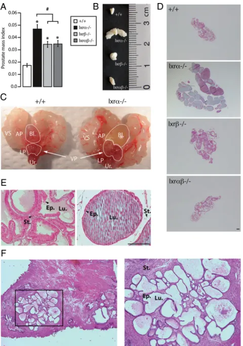
lxr␣⫺/⫺genotype. Macroscopic analy-sis (Fig. 1C) showed that lxr␣⫺/⫺mice had urine-filled bladders, a sign of uri-nary flow obstruction usually observed in BPH patients. Histological analysis showed that prostatic ducts were aber-rantly dilated (Fig. 1, D and E) and filled up with large amounts of secre-tion fluid, which could account for the increase in VP weight. Interestingly, this phenotype was restricted to VP (Supplemental Fig. 2A). These histolog-ical features are similar to dilated glands observed in some BPH patients (Fig. 1F). However, no evidence of fi-brous nodule formation was found in our cohort as previously described (17). Altogether these observations suggested that the enlarged VP phenotype could result from the deregulation of epi-thelial secretion activity. To further evaluate a potential secretory phenotype, VP tissue sections were analyzed by electron microscopy. These experiments showed larger se-cretion vesicles (Fig. 2, D and E) filled with a filamentous content (Fig. 2F) in the cytoplasm of lxr␣⫺/⫺VP cells
com-FIG. 1. lxr␣⫺/⫺mice develop prostate hypertrophy. A, VP weight normalized to body weight
of 12-month-old WT, lxr␣⫺/⫺, lxr⫺/⫺, and lxr␣⫺/⫺mice. *, P⬍ 0.05 compared with the WT
mice; #, P⬍ 0.05 compared with lxr␣⫺/⫺mice. B, Macroscopic observation of VP lobes after
necropsy (size in centimeters). Both VP weight and size are increased in lxr␣⫺/⫺mice
compared with WT, lxr⫺/⫺, and lxr␣⫺/⫺mice (n⫽ 17–26 for each genotype). C,
Macroscopic urogenital tract pictures of lxr␣⫺/⫺compared with WT mice. Lxr␣⫺/⫺mice
develop a bladder enlargement with urine accumulation. D and E, Masson trichrome staining of WT, lxr␣⫺/⫺, lxr⫺/⫺, and lxr␣⫺/⫺VP, at the age of 8 months. Bars, 200m. F,
Hematoxylin-eosin staining of human prostate that exhibits the duct enlargement frequently observed in BPH patients. Bl, Bladder; LP, lateral prostate; VS, seminal vesicle; AP, anterior prostate; Ur, urethra; Ep, epithelium; Lu, lumen; St, stroma.
pared with WT (Fig. 2, A–C). Interestingly, this phenotype was not observed in lxr⫺/⫺VP (data not shown), even though the LXR isoform is expressed (Supplemental Fig. 2B). There is no compensation of lxr isoform expression in each genotype (Supplemental Fig. 2C). Altogether these data suggested that there were an abnormal vesicle traf-ficking and vesicle structures in the VP of lxr␣⫺/⫺mice.
Vesicular trafficking is altered in epithelial cells derived from lxr␣⫺/⫺ventral prostate
To investigate the intrinsic role of Lxr␣ in VP epithe-lium, vesicular trafficking was analyzed in MPE cells de-rived from the VP of lxr␣⫺/⫺or WT mice. Expression of Early endosome antigen 1 (EEA1), a protein that binds phospholipid vesicles and is involved in endosomal traf-ficking, was analyzed. Immunolabeling of tubulin was used to assess cellular morphology and all trafficking ap-paratus integrity. We found that endosomal vesicles were smaller in lxr␣⫺/⫺MPE compared with WT (Fig. 3, Ac and Af). Lysosome biogenesis is tightly linked to vesicle traf-ficking (28). Therefore, we analyzed the effect of Lxr␣ ablation on lysosome structure by incubation of MPE cells with Lysotracker (Invitrogen). These experiments showed that lysosomes were smaller and less abundant in lxr␣⫺/⫺
vs. WT MPE (Fig. 3, Ai and Al). Taken together, these
observations show that the absence of Lxr␣ in ventral prostate epithelial cells results in abnormal vesicle traf-ficking and reduced lysosome biogenesis. Next we sought to ascertain whether the VP of mice lacking lxr␣ and/or
lxr exhibited deregulated expression of genes involved in
cell trafficking (Fig. 3B). We observed that syngr, lamp1, golgb1, and rab27b expressions were up-regulated in
lxr␣⫺/⫺ mice and synpr expression was down-regulated in lxr␣⫺/⫺, lxr⫺/⫺, and lxr␣⫺/⫺mice (Fig. 3C). Altogether these results demonstrated that Lxr␣ and Lxr are required for a normal traf-ficking and secretory machinery in prostatic epithelium.
Lxr␣⫺/⫺ventral prostate exhibits an overaccumulation of secreted spermine binding protein (SBP) in the prostatic fluid
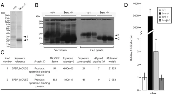
To decipher the molecular mecha-nisms leading to the phenotype ob-served in VP from lxr␣⫺/⫺mice, protein accumulation profiles were analyzed by Western blotting followed by protein identification by mass spectrometry. Coomassie blue staining showed that Lxr␣ ablation resulted in multiple al-terations in overall protein content (Fig. 4A). These ob-servations were confirmed by Western blot using an an-tiserum directed toward the whole secretory content of mouse prostatic fluid (29) in isolated secretions and in cell lysates from WT and lxr␣⫺/⫺mice (Fig. 4B). Both exper-iments showed strong accumulation of a 30-kDa protein in WT samples (band 1). This signal was absent from
lxr␣⫺/⫺samples. However, these samples were character-ized by the strong accumulation of a 22/25 kDa protein (band 2) (Fig. 4B). Surprisingly, mass spectrometry anal-ysis showed that both bands contained the same protein identified as SBP (Fig. 4C). The molecular weight discrep-ancy could result from differential posttranslational mod-ifications. Indeed, SBP is known to be a highly glycosy-lated protein, which can be detected at multiple molecular weights (30). We further investigated the mechanisms of SBP deregulation by analyzing sbp expression using quan-titative RT-PCR. This showed that sbp mRNA accumu-lation (Fig. 4D) was increased 3000-fold in lxr␣⫺/⫺VP, suggesting that Lxr␣ ablation affects Sbp gene transcrip-tion. It is also noteworthy that sbp expression presents a discrete deregulation in lxr⫺/⫺mice and no alteration in
lxr␣⫺/⫺mice (Fig. 4D). Given that lxr␣⫺/⫺mice exhibit an increased enlargement of VP lobe compared with
lxr⫺/⫺and lxr␣⫺/⫺mice and that SBP overaccumula-tion is observed only in lxr␣⫺/⫺prostatic fluid (data not shown), we can conclude that sbp gene deregulation plays a central role in the prostate phenotype of lxr␣⫺/⫺mice.
FIG. 2. Lxr␣-deficient mice present an abnormal epithelial secretory activity. Ultrathin sections of VP from WT (A–C) or lxr␣⫺/⫺(D–F) mice were made and analyzed by electron
microscopy to observe the ultrastructural organization of the cells within the cytoplasm.
White arrowheads indicate secretory vesicles. These are bigger and present filamentous
content in cytoplasm of lxr␣⫺/⫺epithelial cells. Ep, Epithelium; Lu, lumen; BM, basal
Sbp over accumulation in lxr␣⫺/⫺mice is mediated by androgens
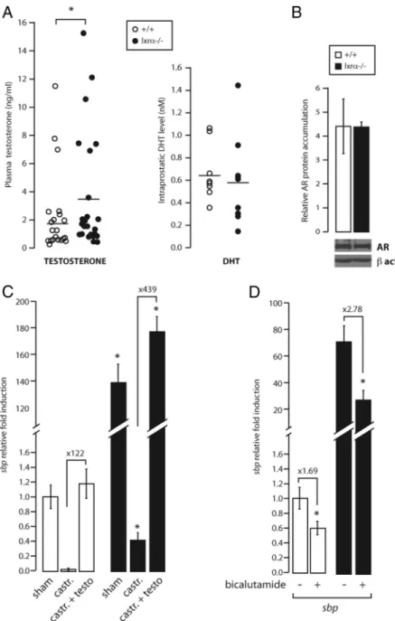
SBP is the most abundant protein within the prostatic fluid and its accumulation is tightly regulated by andro-gens (24, 31). To investigate whether the higher accumu-lation of SBP in the VP of lxr␣⫺/⫺ mice resulted from increased levels of androgens, the plasma testosterone level was evaluated. As shown in Fig. 5A, plasma testos-terone was significantly increased by 2-fold in Lxr ␣-lack-ing mice compared with WT. The increased circulat␣-lack-ing testosterone level can be explained by the increase of sts
(steroid sulfatase), a mRNA-encoding enzyme that con-verts sulfoned androgens into active metabolites in both the liver and VP. In contrast, sult2a1 (sulfotransferase 2a1) was undetectable in the VP and its expression was unaltered in the liver (Supplemental Fig. 3) (16). Even though testosterone was higher in lxr␣⫺/⫺mice, the con-centration of DHT, the active androgen in the prostate, was not significantly altered by Lxr␣ ablation (Fig. 5A). Likewise, AR protein accumulation was not altered in the VP of lxr␣⫺/⫺ mice (Fig. 5B). We thus concluded that increased ligand production or receptor expression was
FIG. 3. Vesicular trafficking is altered by Lxr ablation. A, WT and lxr␣⫺/⫺MPE were immunostained using anti-EEA1 (a and d) and antitubulin (b and e) antibodies. The EEA1 labeling demonstrated that endosomal vesicles were smaller in lxr␣⫺/⫺vs. WT. Lysotracker analysis (g, i, j, and l;
Invitrogen) was performed in MPE cells. Cell nuclei were stained with Hoechst. Lysotracker analysis showed that lysosomes were smaller and less abundant in lxr␣⫺/⫺vs. WT. Bar, 20m. B, Schematic representation of the main proteins involved in secretion machinery in epithelial cells. Eea1,
Early endosome antigen 1; Synpr, synaptoporin; Syngr, synaptogyrin; Lamp1, lysosomal-associated membrane protein 1; Golgb1, golgin B1; Rab27b, RAS oncogene family. C, mRNA relative accumulation levels of synpr1, syngr, lamp1, golgb1, and rab27b was measured in 9- to 12-month-old animals by quantitative PCR and normalized using 18s (n⫽ 7–10). *, P ⬍ 0.05, **, P ⬍ 0.01 compared with WT.
unlikely to account for the huge increase in sbp expression resulting from Lxr␣ ablation.
We then analyzed whether the increase in sbp expres-sion in lxr␣⫺/⫺VP was directly dependent on androgens by performing castration and testosterone complementa-tion experiments (Supplemental Fig. 4). As expected, cas-tration abolished sbp accumulation in the VP of WT mice (Fig. 5C, white bars). The same drastic decrease was ob-served in lxr␣⫺/⫺mice, although the reduction was not as pronounced as in WT mice. Interestingly, testosterone treatment restored sbp expression in both WT and lxr␣⫺/⫺ castrated-mice (Fig. 5C, black bars), confirming that sbp expression was regulated by androgens in both genotypes. Careful examination of these data showed that sbp accu-mulation was much higher after testosterone propionate treatment in lxr␣⫺/⫺mice (439-fold induction) compared with WT (122-fold induction). Furthermore, pharmaco-logical inhibition of AR by the antiandrogen bicalutamide (Fig. 5D) resulted in decreased accumulation of sbp tran-script both in WT (1.69-fold inhibition) and lxr␣⫺/⫺mice (2.78-fold inhibition). However, sbp accumulation was still higher in lxr␣⫺/⫺than in WT VP after bicalutamide treatment. Castration, testosterone supplementation, and bicalutamide treatment were validated by histology anal-ysis and prostate weight measurement (Supplemental Fig. 4). Altogether these data show that even though andro-gens are clearly involved in the regulation of sbp
expres-sion in VP of lxr␣⫺/⫺mice, these mice still express higher amounts of sbp upon total androgen depletion (castra-tion) or when AR is blocked (bicalutamide). We thus con-cluded that sbp accumulation per se was hypersensitive to androgens in lxr␣⫺/⫺mice.
Basal sbp accumulation was significantly higher in castrated lxr␣⫺/⫺than in WT mice (Fig. 5C). Some Lxr target genes show increased expression in Lxr-knock-out mice in the absence of oxysterol stimulation such as
star in the adrenal gland (18). This suggested that sbp
could be a bona fide Lxr␣ target gene. To test this hy-pothesis, WT mice were gavaged short-term with T0901317, a synthetic LXR agonist. Neither alteration of sbp level nor other androgen target genes such as
fkbp5 and pbsn were seen in T0901317-gavaged mice
(Supplemental Fig. 5), ruling out a direct regulation of
sbp expression by Lxr␣. These observations suggest
that Lxr␣ indirectly affects basal sbp expression, result-ing in increased androgen sensitivity.
Androgen hypersensitivity in lxr␣⫺/⫺mice targets specific genes and is restricted to the VP
Next, we sought to determine whether the abnormal androgen response was VP specific or was also present in other tissues. Protein accumulation profiles were analyzed in several androgen-dependent tissues of the genital tract: dorsolateral and anterior prostates, epididymis, testis, vas
FIG. 4. Lxr␣⫺/⫺ventral prostate exhibits an overaccumulation of the secreted protein SBP. A, Secretion protein lysates of WT and lxr␣⫺/⫺ventral
prostates were resolved using 4 –15% SDS-PAGE migration and the gel was stained with Coomassie Blue. B, Western blot analysis using
antiprostate secretory protein immune serum was performed on samples of whole-prostate protein extracts or on prostate secretion only, from WT or lxr␣⫺/⫺mice. C, The protein spots (arrows 1 and 2 in A) were excised from the gel (A) and analyzed by mass spectrometry, and SBP protein was
identified and found to be highly accumulated in secretions from lxr␣⫺/⫺VP compared with WT. D, mRNA relative accumulation levels of sbp were
measured in 9- to 12-month WT, lxr␣⫺/⫺, lxr⫺/⫺, and lx␣r⫺/⫺animals by quantitative PCR and normalized using 18s. Sbp expression was
3000-fold higher in lxr␣⫺/⫺VP compared with WT and is 3-fold higher in lxr⫺/⫺compared with WT. Sbp transcript accumulation remains unchanged in
deferens, and seminal vesicles. There was no clear differ-ence between the migration profiles of WT and lxr␣⫺/⫺ samples (Fig. 6A). This provided evidence that the
abnor-mal response to androgens was restricted to the VP. We next investigated whether sbp was the only androgen-regulated gene to have its expression altered in the VP by an-alyzing the expression of several androgen-regulated genes by quantitative RT-PCR. These included svs2 (seminal vesicle secre-tion-2) (32), spp1 (secretory prostatic pro-tein-1) (33), fkbp5 (fk506 binding protein prostate-5) (25), acpp (acid phosphatase, prostate protein) (34), calR (calreticulin) (35), and pbsn (probasin) (36). Analysis of the PCR data allowed stratification of the genes into distinct categories (Fig. 6B): genes with increased basal expression (svs2 and
spp1); genes with unaltered expression
(fkbp5, acpp and calR); and pbsn whose basal accumulation was significantly de-creased by Lxr␣ ablation.
To gain insight into the molecular mech-anisms accounting for these discrepancies, AR recruitment on the promoters of these genes was analyzed by in vivo chromatin immunoprecipitation. Surprisingly, the re-cruitment of AR to androgen-responsive el-ement sequences of sbp, pbsn, and fkbp5 promoters was unaltered by ablation of Lxr␣ (Fig. 6, C and D, and Supplemental Fig. 6). The similar recruitment of AR on target promoters in both WT and knockout VP suggested that Lxr␣ could act through an indirect route to modulate intrinsic AR transcriptional activity.
To test this hypothesis, WT and lxr␣⫺/⫺ MEF were transfected with the androgen-sensitive construct AREtk-Luc in the pres-ence or abspres-ence of an AR expression vector (Fig. 6E). As expected, Lxr␣ was present and functional in WT MEF cells (data not shown). Treatment with DHT in the ab-sence of AR transfection induced a moder-ate increase in activity of the androgen-sen-sitive luciferase reporter construct (1.6 fold) in WT MEF. This mild induction was not found in lxr␣⫺/⫺MEF. As expected, andro-gen-responsiveness of the construct was fur-ther increased to 12-fold after AR transfec-tion in WT cells. Surprisingly, there was no alteration of androgen responsiveness in
lxr␣⫺/⫺MEF upon AR transfection. This suggested that Lxr␣ did not directly alter intrinsic AR transcriptional efficiency.
FIG. 5. Sbp expression deregulation in lxr␣⫺/⫺mice is mediated by androgens. A, Testosterone concentrations were measured in the plasma of WT and lxr␣⫺/⫺mice.
Circulating testosterone was significantly increased in lxr␣⫺/⫺(n⫽ 25) compared with
WT (n⫽ 25) mice. The active androgen metabolite, DHT, was measured in the VP of WT and lxr␣⫺/⫺mice. DHT levels were unchanged in the ventral prostate (n⫽ 25). B, Basal
AR accumulation determined by Western blot. As observed, this accumulation is unchanged in lxr␣⫺/⫺VP. *, P⬍ 0.05. C, Sbp accumulation in 5- to 6-month-old WT or
lxr␣⫺/⫺castrated (castr.) mice. Three weeks after castration, castrated mice were injected with 75g/kg of testosterone (testo) twice a day for 1 wk. Castration caused a large decrease of sbp in WT and lxr␣⫺/⫺mice. Testosterone injection led to a larger
recovery of sbp expression in the VP of lxr␣⫺/⫺compared with WT mice, suggesting an
androgen hypersensitivity in lxr␣⫺/⫺(n⫽ 7). *, P ⬍ 0.05 compared with sham WT mice.
D, Five- to 6-month-old WT and lxr␣⫺/⫺mice were treated with bicalutamide at a daily
oral dose of 12 mg/kg for 21 d. Bicalutamide caused a 2.78-fold decrease of androgen-dependent sbp expression in lxr␣⫺/⫺mice (1.69-fold decreased in WT mice) (n⫽ 7–9).
FIG. 6. Androgen hypersensitivity in lxr␣⫺/⫺mice targets specific genes and is restricted to the VP. A, Whole-protein extracts from the dorsolateral and anterior prostate, epididymis, vas deferens, testis, and seminal vesicles were migrated in a 4 –15% polyacrylamide gel and stained with Coomassie blue. The sbp accumulation is lobe specific and is not found in the other male genital tract tissues. B, mRNA relative accumulation levels of svs2, spp1, fkbp5, acpp, calR, and pbsn were measured by quantitative PCR and normalized with 18s in the VP of intact WT mice. Some of them have the same accumulation profile as sbp, and others are down-regulated or remain unchanged in the VP (n⫽ 7–10). *, P ⬍ 0.05 compared with the WT animals. C, Schematic representation of the androgen-responsive element regulatory sites on sbp, fkbp5, and pbsn promoter sequences. The figure shows the amplified regions. Arrows represent primer localization around the amplified regions. D, Anti-AR or anti-IgG chromatin immunoprecipitation was performed on the VP of WT and lxr␣⫺/⫺mice. The AR specifically binds the regulatory element of
androgen-regulated genes (sbp, pbsn, and fkbp5). Chromatin enrichment was quantified by quantitative PCR (n⫽ 6–8 analyzed for three independent experiments). Lack of Lxr␣ does not modify AR recruitment on regulating regions. E, Lxr␣⫺/⫺and WT MEF cells were transfected with
the luciferase reporter construct ARE-tk-LUC in combination with pSG5-hAR and treated or not with 10⫺9MDHT (means⫾SEM). *, P⬍ 0.05 compared with the respective excipient incubated cells. DLP, Dorsolateral prostate; AP, anterior prostate; epid, epididymes; VD, vas deferens; SV, seminal vesicle.
Lxr␣ coordinates stroma/epithelium functions to control the androgen-dependent secretory activity of the ventral prostate in mice
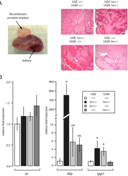
Androgen action on the prostate is the result of a com-plex paracrine network between stromal cells and epithe-lium (8). Integration of androgen signal is, in part, sup-ported by the stromal compartment which is necessary for epithelium maintenance and survival (8). To investigate whether stromal/epithelial interactions could be involved in the development of enlarged VP ducts and increased accumulation of SBP, we generated chimeric recombi-nant prostates derived from embryonic WT or lxr␣⫺/⫺
UGM and WT or lxr␣⫺/⫺UGE. After recom-bination, the four UGE/UGM combinations (UGEWT/UGMWT; UGElxr␣⫺/⫺/UGMlxr␣⫺/⫺; UGEWT/UGMlxr␣⫺/⫺; UGElxr␣⫺/⫺/UGMWT)
were grafted under the kidney capsule of nude mice (24). Eight weeks after grafting, the four types of recombinants had grown and presented a similar gross morphology characterized by differentiated prostatic lobes with enlarged tubules filled by accu-mulated secretions (Fig. 7A). Ar transcript accumulation was not altered in the differ-ent genotypes combinations. Sbp mRNA accumulation was strongly increased (767-fold accumulation) in the UGElxr␣⫺/⫺/ UGMlxr␣⫺/⫺ compared with the UGEWT/
UGMWTrecombinants (Fig. 7B). This showed that this phenotype was intrinsically pros-tatic because the recombinants were grafted in WT nude mice.
Interestingly, Sbp accumulation, the marker of the BPH-like phenotype, was not significantly altered when the mutant UGM was combined with the WT UGE or when the mutant UGE was combined with the WT UGM (Fig. 7B). This demonstrated that
sbp deregulation originates from combined
stromal and epithelial lxr␣⫺/⫺ablation. In contrast, Spp1 expression was deregulated when Lxr␣ was deleted in the epithelium alone or in combination with the mesen-chyme. However, in the mesenchyme alone, Lxr␣ ablation had no effect on Spp1 expres-sion. We therefore concluded that Lxr␣ played a physiological role in both the stroma and epithelium. We further showed that the contribution of one or the other compartment to the phenotype was gene specific.
Discussion
In this report we show that a BPH-like phenotype of
lxr␣⫺/⫺mice is characterized by increased secretory ac-tivity of the epithelium. Our work using UGE/UGM re-combinations provides evidence that Lxr␣ is involved both at the stromal and the epithelial levels. Indeed, an-drogen-regulated gene expression is deregulated alterna-tively by lxr␣ ablation in both compartments. Using
lxr␣⫺/⫺mice, we found that neither androgen levels in prostate, nor AR recruitment in targeted-sequences was
FIG. 7. The deregulation of androgen-dependent genes is dependent on both the
stromal and epithelial compartments in lxr␣⫺/⫺mice. A, UGE and UGM were dissected
from WT or lxr␣⫺/⫺embryos. Different combinations were performed (see text for more
details). The different explants were grafted under the kidney capsule of nude mice. After 8 wk of growth, recombinant prostates (left panel) were dissected. Histological analysis (hematoxylin-eosin staining) was performed on the four different combinations (right panel). Bar, 200m. B, Accumulations of ar, sbp, and spp1 mRNA were measured by quantitative PCR and the fold induction between each recombinant condition was represented (n⫽ 3–7). *, P ⬍ 0.05 compared with the UGEWT/UGMWTcondition.
altered. It can thus be concluded that the observed dereg-ulation of androgen signaling in prostate results from a complex paracrine network between the epithelium and stroma.
Lxr␣ and Lxr play an important role in prostate ep-ithelium homeostasis in other lobes, specifically when the mice are challenged with a high-cholesterol diet (Pommier
et al., submitted data). As already mentioned, the human
and murine prostates are architecturally different. Never-theless, the gene expression pattern of the peripheral and central zones in human is closely related to the murine dorsolateral and ventral lobes, respectively (37). These observations highlight that the molecular signature of re-gionalization in the prostate is an important process con-served between the two species. Given that each lobe har-bors specific features, it could be hypothesized that lxr␣ ablation results in a different phenotype according the prostate lobes in vivo. Consistent with our findings,
lxr␣⫺/⫺mice have been described to recapitulate “several BPH-like features” according to Kim et al. (17). These authors reported fibrous nodules, abnormal stroma growth, and lesions in the muscular compartment (17), whereas we mainly reported an epithelial phenotype. This apparent discrepancy could be due to the fact that the
lxr␣⫺/⫺strains were not similarly engineered (18, 38).
The main function of the prostate epithelium is the pro-duction and the secretion of proteins that compose pros-tatic fluid. This secretion activity is tightly regulated in
vivo by androgens that orchestrate the entire male genital
tract capacity. A possible connection between LXR and AR has been previously suggested. DHT or synthetic an-drogen R1881 treatments result in decreased abca1 accu-mulation in LNCaP cells (39), indicating that LXR target genes are sensitive to androgen stimulation. Krycer and Brown (15) showed that LXR were indeed required for the
abca1 down-regulation in response to R1881 treatment.
The association between the expression of LXR target genes and androgen sensitivity has also been described in xenograft models that recapitulate pharmaco-resistant prostate cancer (40). In these tumors, fas, srebp1c, abca1, and cyp-27 gene expressions decrease during androgen insensitivity evolution. Interestingly, another partner of retinoid X receptor, the pregnane X receptor (NR1I2) has been demonstrated to inhibit androgen-dependent prolif-eration of LAPC-4 cells (41). This raises the question whether LXR and pregnane X receptor could act through a similar molecular mechanism.
Given that lxr␣ ablation resulted in an aberrant pro-duction of androgen-regulated secretory proteins in the prostate, we investigated how Lxr␣ could interfere with androgen signaling in the epithelium. Indeed, transgenic mice that overexpressed a dominant-positive construct of Lxr␣ specifically targeted in liver (42) exhibit an inhibi-tion of androgen-dependent prostate regeneration after castration (16), indi-cating that Lxr activation impacts an-drogenic responsive tissues. Hepatic Lxr␣ activation leads to decreased cir-culating testosterone levels by regulat-ing genes such as sult2a1 and sts in-volved in androgen catabolism. In peripheral tissue, Lxr␣ controls andro-gens bioavailability through sts expres-sion, which encodes the steroid sulfa-tase that desulfonates androgens to convert them into active metabolites. These data could explain the increase in testosterone levels observed in the plasma of lxr␣⫺/⫺mice. Nevertheless, no modification of DHT accumulation or androgen receptor activity in andro-gen-regulated gene promoters was seen in the VP of lxr␣⫺/⫺mice. Our findings indicate that the mechanism by which Lxr␣ regulates the response of
andro-FIG. 8. Model for the physiological roles of the Lxr␣ in the ventral prostate and possible interactions with the androgen-regulated pathway. Schematic representation of Lxr␣ action in the stromal and epithelial compartments. Differential androgen-targeted genes regulation mechanisms by Lxr␣ are represented (sbp and spp1). Sbp expression is dependent of Lxr␣ in both the epithelium and stroma, suggesting that paracrine factors are involved. On the contrary, spp1 expression is strictly dependent on lxr␣ expression in the epithelium. Indeed,
spp1 expression is similar to WT recombinant in the absence of Lxr␣ in the stromal
recombinant prostates. These observations underline the multiple connections involved in the cross talk between LXR and AR.
gen-regulated genes results from a complex network. This could involve epithelial factors, AR cofactors, and/or paracrine interaction between the different cell compart-ments of the prostate.
Consistent with this hypothesis, androgen-regulated gene expression exhibits different profiles in lxr␣⫺/⫺ mouse prostate. Although sbp accumulation increases in mice lacking Lxr␣, calr remains unchanged and pbsn de-creases. These observations strongly support that several regulatory processes are involved. We schematized the pu-tative role of paracrine interactions between epithelial and stromal cells in Fig. 8. Prostate mesenchymal-epithelial interactions have a preponderant role in normal and path-ological prostate development as well as in adult prostate homeostasis (8). The role of AR has extensively been de-veloped in the literature (8). Here we identify Lxr␣ as a new actor that mediates epithelial physiology through its activity in both stroma and epithelium. Indeed, the ab-sence of Lxr␣ in both prostate stroma and epithelium is necessary to develop prostate hypertrophy. Lxr␣ also me-diates androgen signaling, as demonstrated by the numer-ous androgen-responsive genes dysregulated when Lxr␣ is missing. Indeed, normal spp1 gene expression needs Lxr␣ in epithelium, whereas the normal response of sbp to an-drogens by epithelium is dependent on Lxr␣ in both epi-thelial (directly) and stromal (indirectly) cells. The regu-lation of paracrine signals from the mesenchyme by lxr␣ might be one molecular mechanism.
Altogether these results demonstrate that Lxr␣ acts as a key modulator of the cross talk between the stromal and epithelial compartment, which is essential for the integra-tion of androgen signaling in the prostate and its effect on the epithelium. Finally, identification of the set of genes targeted by Lxr␣ specifically in the prostatic ventral lobe in mice could be informative in understanding the BPH etiology in humans.
Acknowledgments
We thank Dr. M. Thomsen (Institute of Cancer Research, Lon-don, UK) for his excellent advices on prostate regeneration; Dr. M. Manin (GReD) for her helpful comments for the MPE cell culture; J. P. Saru and A. De Haze for molecular biology technical assistance; C. Puchol and S. Plantade for animal facilities. Dr. P. Val (GReD) and Dr. S. Ingersoll (Georgia State University, At-lanta GA) for critically reading the manuscript; and the members of the Chester’s laboratory for assistance in animal dissections and discussions; C. Szczepaniak and C. Blavignac (CICS plat-form, Clermont University) for their scientific and technical as-sistance in electron microscopy; Dr. B. Viguès (LMGE, Cler-mont-Ferrand) for helpful discussion on electron microscopy. Pan-prostate secretion antibody was a kind gift from Dr. C.
Abate-Shen (Department of Medicine, Columbia University Medical Center, New York, NY). Pathology analyses have been done on the Anip@th facility platform.
Address all correspondence and requests for reprints to: Génétique Reproduction et Développement, Unité Mixte de Re-cherche, Centre National de la Recherche Scientifique 6293, Clermont Université, Institut National de la Santé et de la Re-cherche Médicale Unité 1103, 24 Avenue des Landais, BP80026, 63171 Aubière Cedex, France. E-mail: silvere.baron@univ-bpclermont.fr.
This work was supported by the Association de Recherche sur les Tumeurs Prostatiques, Ligue Contre le Cancer (Comité Al-lier), Fondation pour la Recherche Médicale, Fondation BNP-Paribas and the Association de Recherche Contre le Cancer, Nouveau Chercheur Auvergne research grants (to S.B.). E.V. was supported by the Région Auvergne and Fond Européen de De-veloppement Régional and grants from the Association de Re-cherche Contre le Cancer. J.D. is supportd by a Ministe`re de l’Education Nationale, de la Recherche et de la Technologie grant. T.E. was the recipient of an European Community Action Scheme for the Mobility of University Students (ERASMUS) ex-change grant.
Disclosure Summary: E.V., T.E., J.D., A.P., S.F., J.-L.K., L.G., L.M., J.-M.L., and S.B. have nothing to declare. E.V., J.D., L.M., J.-M.L., and S.B. are employed by the Université Blaise Pascal. A.P. was employed by the Université Blaise Pascal and now by AstraZeneca. T.E. is employed by the University of Naples. S.F. is employed by the Institut National de la Recherche Agronomique.
References
1. Madersbacher S, Haidinger G, Temml C, Schmidbauer CP 1998 Prevalence of lower urinary tract symptoms in Austria as assessed by an open survey of 2,096 men. Eur Urol 34:136 –141
2. Gormley GJ, Stoner E, Bruskewitz RC, Imperato-McGinley J, Walsh
PC, McConnell JD, Andriole GL, Geller J, Bracken BR, Tenover JS
1992 The effect of finasteride in men with benign prostatic hyper-plasia. The Finasteride Study Group. N Engl J Med 327:1185–1191 3. Lepor H 1990 Role of␣-adrenergic blockers in the treatment of
benign prostatic hyperplasia. Prostate Suppl 3:75– 84
4. Huggins C, Clark PJ 1940 Quantitative studies of prostatic secre-tion: II. The effect of castration and of estrogen injection on the normal and on the hyperplastic prostate glands of dogs. J Exp Med 72:747–762
5. Gelmann EP 2002 Molecular biology of the androgen receptor. J Clin Oncol 20:3001–3015
6. Heinlein CA, Chang C 2002 Androgen receptor (AR) coregulators: an overview. Endocr Rev 23:175–200
7. Bruchovsky N, Wilson JD 1968 The intranuclear binding of testos-terone and 5-␣-androstan-17--ol-3-one by rat prostate. J Biol Chem 243:5953–5960
8. Cunha GR 1973 The role of androgens in the epithelio-mesenchymal interactions involved in prostatic morphogenesis in embryonic mice. Anat Rec 175:87–96
9. Cunha GR, Chung LW 1981 Stromal-epithelial interactions—I. In-duction of prostatic phenotype in urothelium of testicular feminized (Tfm/y) mice. J Steroid Biochem 14:1317–1324
10. El-Hajjaji FZ, Oumeddour A, Pommier AJ, Ouvrier A, Viennois E,
Dufour J, Caira F, Drevet JR, Volle DH, Baron S, Saez F, Lobaccaro JM 2011 Liver X receptors, lipids and their reproductive secrets in
the male. Biochim Biophys Acta 1812:974 –981
11. Tontonoz P, Mangelsdorf DJ 2003 Liver X receptor signaling path-ways in cardiovascular disease. Mol Endocrinol 17:985–993 12. Volle DH, Lobaccaro JM 2007 Role of the nuclear receptors for
oxysterols LXRs in steroidogenic tissues: beyond the “foie gras,” the steroids and sex? Mol Cell Endocrinol 265–266:183–189 13. Fukuchi J, Kokontis JM, Hiipakka RA, Chuu CP, Liao S 2004
An-tiproliferative effect of liver X receptor agonists on LNCaP human prostate cancer cells. Cancer Res 64:7686 –7689
14. Pommier AJ, Alves G, Viennois E, Bernard S, Communal Y, Sion B,
Marceau G, Damon C, Mouzat K, Caira F, Baron S, Lobaccaro JM
2010 Liver X receptor activation downregulates AKT survival sig-naling in lipid rafts and induces apoptosis of prostate cancer cells. Oncogene 29:2712–2723
15. Krycer JR, Brown AJ 2011 Cross-talk between the androgen recep-tor and the liver x receprecep-tor: implications for cholesterol homeostasis. J Biol Chem 286:20637–205647
16. Lee JH, Gong H, Khadem S, Lu Y, Gao X, Li S, Zhang J, Xie W 2008 Androgen deprivation by activating the liver X receptor. Endocri-nology 149:3778 –3788
17. Kim HJ, Andersson LC, Bouton D, Warner M, Gustafsson JA 2009 Stromal growth and epithelial cell proliferation in ventral prostates of liver X receptor knockout mice. Proc Natl Acad Sci USA 106: 558 –563
18. Cummins CL, Volle DH, Zhang Y, McDonald JG, Sion B,
Lefran-çois-Martinez AM, Caira F, Veyssière G, Mangelsdorf DJ, Lobac-caro JM 2006 Liver X receptors regulate adrenal cholesterol
bal-ance. J Clin Invest 116:1902–1912
19. Manin M, Veyssiere G, Cheyvialle D, Chevalier M, Lecher P, Jean
C 1992 In vitro androgenic induction of a major protein in epithelial
cell subcultures from mouse vas deferens. Endocrinology 131: 2378 –2386
20. Lobaccaro JM, Poujol N, Térouanne B, Georget V, Fabre S,
Lum-broso S, Sultan C 1999 Transcriptional interferences between
nor-mal or mutant androgen receptors and the activator protein 1— dis-section of the androgen receptor functional domains. Endocrinology 140:350 –357
21. Monniaux D, Clemente N, Touzé JL, Belville C, Rico C, Bontoux
M, Picard JY, Fabre S 2008 Intrafollicular steroids and
anti-mulle-rian hormone during normal and cystic ovaanti-mulle-rian follicular develop-ment in the cow. Biol Reprod 79:387–396
22. Torres JM, Ortega E 2003 Differential regulation of steroid 5 ␣-reductase isozymes expression by androgens in the adult rat brain. FASEB J 17:1428 –1433
23. Nadaud I, Girousse C, Debiton C, Chambon C, Bouzidi MF, Martre
P, Branlard G 2010 Proteomic and morphological analysis of early
stages of wheat grain development. Proteomics 10:2901–2910 24. Sun Q, Yu X, Degraff DJ, Matusik RJ 2009 Upstream stimulatory
factor 2, a novel FoxA1-interacting protein, is involved in prostate-specific gene expression. Mol Endocrinol 23:2038 –2047 25. Magee JA, Chang LW, Stormo GD, Milbrandt J 2006 Direct,
an-drogen receptor-mediated regulation of the FKBP5 gene via a distal enhancer element. Endocrinology 147:590 –598
26. Lukacs RU, Goldstein AS, Lawson DA, Cheng D, Witte ON 2010
Isolation, cultivation and characterization of adult murine prostate stem cells. Nat Protoc 5:702–713
27. Cunha GR, Lung B 1978 The possible influence of temporal factors in androgenic responsiveness of urogenital tissue recombinants from wild-type and androgen-insensitive (Tfm) mice. J Exp Zool 205: 181–193
28. Hirota Y, Kuronita T, Fujita H, Tanaka Y 2007 A role for Rab5 activity in the biogenesis of endosomal and lysosomal compart-ments. Biochem Biophys Res Commun 364:40 – 47
29. Economides KD, Capecchi MR 2003 Hoxb13 is required for normal differentiation and secretory function of the ventral prostate. De-velopment 130:2061–2069
30. Chaurand P, DaGue BB, Ma S, Kasper S, Caprioli RM 2001 Strain-based sequence variations and structure analysis of murine prostate specific spermine binding protein using mass spectrometry. Bio-chemistry 40:9725–9733
31. Fujimoto N, Akimoto Y, Suzuki T, Kitamura S, Ohta S 2006 Iden-tification of prostatic-secreted proteins in mice by mass spectromet-ric analysis and evaluation of lobe-specific and androgen-dependent mRNA expression. J Endocrinol 190:793– 803
32. Kawano N, Yoshida M 2007 Semen-coagulating protein, SVS2, in mouse seminal plasma controls sperm fertility. Biol Reprod 76:353– 361
33. Seenundun S, Robaire B 2007 Time-dependent rescue of gene ex-pression by androgens in the mouse proximal caput epididymidis-1 cell line after androgen withdrawal. Endocrinology 148:173–188 34. Sharief FS, Mohler JL, Sharief Y, Li SS 1994 Expression of human
prostatic acid phosphatase and prostate specific antigen genes in neoplastic and benign tissues. Biochem Mol Biol Int 33:567–574 35. Zhu N, Pewitt EB, Cai X, Cohn EB, Lang S, Chen R, Wang Z 1998
Calreticulin: an intracellular Ca⫹⫹-binding protein abundantly ex-pressed and regulated by androgen in prostatic epithelial cells. En-docrinology 139:4337– 4344
36. Johnson MA, Hernandez I, Wei Y, Greenberg N 2000 Isolation and characterization of mouse probasin: an androgen-regulated protein specifically expressed in the differentiated prostate. Prostate 43: 255–262
37. Berquin IM, Min Y, Wu R, Wu H, Chen YQ 2005 Expression sig-nature of the mouse prostate. J Biol Chem 280:36442–36451 38. Alberti S, Schuster G, Parini P, Feltkamp D, Diczfalusy U, Rudling
M, Angelin B, Björkhem I, Pettersson S, Gustafsson JA 2001
He-patic cholesterol metabolism and resistance to dietary cholesterol in LXR-deficient mice. J Clin Invest 107:565–573
39. Fukuchi J, Hiipakka RA, Kokontis JM, Hsu S, Ko AL, Fitzgerald
ML, Liao S 2004 Androgenic suppression of ATP-binding cassette
transporter A1 expression in LNCaP human prostate cancer cells. Cancer Res 64:7682–7685
40. Chuu CP, Hiipakka RA, Kokontis JM, Fukuchi J, Chen RY, Liao S 2006 Inhibition of tumor growth and progression of LNCaP pros-tate cancer cells in athymic mice by androgen and liver X receptor agonist. Cancer Res 66:6482– 6486
41. Zhang B, Cheng Q, Ou Z, Lee JH, Xu M, Kochhar U, Ren S, Huang
M, Pflug BR, Xie W 2010 Pregnane X receptor as a therapeutic
target to inhibit androgen activity. Endocrinology 151:5721–5729 42. Uppal H, Saini SP, Moschetta A, Mu Y, Zhou J, Gong H, Zhai Y,
Ren S, Michalopoulos GK, Mangelsdorf DJ, Xie W 2007 Activation
of LXRs prevents bile acid toxicity and cholestasis in female mice. Hepatology 45:422– 432