HAL Id: hal-03078871
https://hal.archives-ouvertes.fr/hal-03078871
Submitted on 16 Dec 2020HAL is a multi-disciplinary open access archive for the deposit and dissemination of sci-entific research documents, whether they are pub-lished or not. The documents may come from teaching and research institutions in France or abroad, or from public or private research centers.
L’archive ouverte pluridisciplinaire HAL, est destinée au dépôt et à la diffusion de documents scientifiques de niveau recherche, publiés ou non, émanant des établissements d’enseignement et de recherche français ou étrangers, des laboratoires publics ou privés.
Characterizing single sinonasal squamous cell carcinoma
using dielectrophoresis and electrorotation
Thao Mai, Sakina Bensalem, Bénédicte Thiebot, Philippe Manivet, Juan
Pelta, Bruno Le Pioufle
To cite this version:
Thao Mai, Sakina Bensalem, Bénédicte Thiebot, Philippe Manivet, Juan Pelta, et al.. Characterizing single sinonasal squamous cell carcinoma using dielectrophoresis and electrorotation. MicroTAS, Oct 2020, on-line, France. �hal-03078871�
CHARACTERIZING SINGLE SINONASAL SQUAMOUS CELL CARCINOMA
USING DI-ELECTROPHORESIS AND ELECTROROTATION
Thao N.P. Mai
1*, Sakina Bensalem
1*, Bénédicte Thiebot
2, Philippe Manivet
3,Juan Pelta
2, and
Bruno Le Pioufle
11
Institut d’Alembert (IDA), Ecole Normale Supérieure Paris-Saclay, France
2Université Paris-Saclay, Université d’Evry, LAMBE, France
3
Biobank Lariboisière, Platform of Biopathology and Innovative Technologies in Health, Lariboisière
Hospital, INSERM 1141, University of Paris, Paris 10, France.
ABSTRACT
We show the capture and analysis single cells, in particular human sinonasal squamous cell carcinomas (SCC), by the combination of di-electrophoresis (DEP) force and electrorotation, within a microfluidic device. A set of 4 planar polynomial electrodes was employed to perform nDEP trapping of two lines tumor cell with different invasivity, named NC5 and NC7. Once captured at the center of the electrodes set, electrorotation was served to extract the rotational speed’s spectra. From the spectra, their electrophysiological properties can be estimated.
KEYWORDS: Sinonasal squamous cell carcinomas, di-electrophoresis, electrorotation, single cell analysis INTRODUCTION
Cancer cells can be found in various forms, with different physical properties at different stages of malignancy. Metastasis is the main cause of cancer patient death, physical biomarkers discovery is a challenge to develop diagnostic tools. In this context, single cell approach by di-electrophoresis (DEP) and electrorotation, which are electrokinetic phenomena, have demonstrated a potential for tumor cell characterization in lab-on-chip platforms [1]. We investigate and compare two cancer cells at different degrees of malignancy of sinonasal squamous cell carcinomas. The rotational speed’s spectra are different for these cells. This spectra indicates the cell dielectric characteristics, such as membrane capacitance, membrane permittivity, cytoplasm conductance and cytoplasm permittivity [1, 2].
THEORY
Di-electrophoresis (DEP) is a force affecting on a polarizable particle in a non-uniform static electric field. DEP drives the particles to move toward high (positive phenomenon) or low electric field area (negative phenomenon) depending on the relative polarizability of the cell and the suspending medium. The time-averaged DEP force is expressed as
ܨாൌ ʹߨݎଷ ߝߝܴ݁ሾ݂ெሺ߱ሻሿܧଶ (1) where ris the radius of cell, ߝand ߝare respectively the relative permittivityof the vacuum and the medium, ܴ݁ሾ݂ெሺ߱ሻሿ is the real part of the Clausius-Mossotti factor, and ܧ is the electric field.
In the case when the cell is suspended in the rotating electric field, the rotational speed is defined as
ȳ
ோை்ሺఠሻൌ െ
ߝ
ߝ
ܧ
ଶ
ʹߟ
ܫ݉ሾ݂
ெሺ߱ሻሿ
(2)
where ߟ is the dynamic permittivity of the medium and ܫ݉ሾ݂ெሺ߱ሻሿ is the imaginary part of the Clausius-Mossotti factor.
EXPERIMENT
The two cell lines NC5 and NC7 were cultured in RPMI 1640 (with GlutaMAX + 25mM HEPES) medium, supplemented with 10% of fetal bovine serum and 5% of penicillin. For the experiment, cells were collected by centrifugation, rinsed three times and resuspended in a low conductivity medium (ߪ ൌ ͳͲͲ݉ܵȀ݉).
The 4 planar polynomial shape electrodes with a tip-tip spacing of 75μm was fabricated on a quartz wafer by conventional photolithography technique, which generates DEP force to trap cancer cells, then rotates them by electrorotation force. A thin layer of SU8-2025 was built on the quartz as an open chamber with 25μm high. A cell suspension of 30μL was pipetted on the device surface and a coverslip was gently put on to close the chamber.
A voltage with 5V peak-to-peak amplitude is applied on the electrode array, at the frequency 500kHz to trap cells by negative DEP force in the center of the electrodes set. The rotational signal is induced on the set of four voltages, each has 90° phase-shift with the neighbour electrodes, at a fixed 2V amplitude and over a frequency range from 37kHz to 25MHz.
RESULTS AND DISCUSSION
When the cell is submitted in the non-uniform electric field (5Vpp and 500kHz), it is captured in the center of
the 4 electrodes which is the lowest electric field region by nDEP. After trapping, an electrorotation signal is served to determine the rotational spectra.
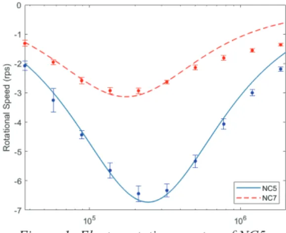
Curve fitting between the experimental and theoretical spectra was then performed by the Genetic algorithm in Matlab. The spectra of NC5 and NC7 were indicated in Figure 1. At 210kHz, both NC5 and NC7 cells achieve the maximum speed. However, we observe that NC5 (6.44rps) rotates faster than NC7 (2.93rps).
The electrophysiological properties of NC5, NC7, such as permittivity and conductivity of the membrane and cytoplasm, are estimated from the spectra and noted in Table 1.
Table 1: Estimated the electrophysiological parameters of cell
ોܕ܍ܕ(S/m) ઽܕ܍ܕ ો܋ܡܜܗ(S/m) ઽ܋ܡܜܗ NC5 2.932×10⁻⁴ 29.929 0.094 75.248
NC7 2.229×10⁻⁴ 29.758 0.045 37.021 CONCLUSION
We characterize the two cancer cells by di-electrophoresis and electrorotation techniques. When the cell is more invasive, it rotates more slowly in the rotating electric field. Furthermore, from the electrorotation spectra, we extract the conductivity and permittivity of the membrane and cytoplasm of the trapped cell.
ACKNOWLEDGEMENTS
This research was supported by ANR EPSILOMICS (ANR17-CE09-0044-02).
REFERENCES
[1] Trainito, Claudia I et al. “Characterization of sequentially-staged cancer cells using electrorotation.” PloS one vol. 14,9 e0222289. 19 Sep. 2019, doi:10.1371/journal.pone.0222289.
[2] Yang, J et al. “Dielectric properties of human leukocyte subpopulations determined by electrorotation as a cell separation criterion.” Biophysical journal vol. 76,6 (1999): 3307-14. doi:10.1016/S0006-3495(99)77483-7.
CONTACT
*Thao N.P. Mai; [email protected] *S. Bensalem; [email protected]
Figure 1: Electrorotation spectra of NC5 and NC7.