Publisher’s version / Version de l'éditeur: Biochemical Journal, 2018-11-19
READ THESE TERMS AND CONDITIONS CAREFULLY BEFORE USING THIS WEBSITE.
https://nrc-publications.canada.ca/eng/copyright
Vous avez des questions? Nous pouvons vous aider. Pour communiquer directement avec un auteur, consultez la première page de la revue dans laquelle son article a été publié afin de trouver ses coordonnées. Si vous n’arrivez pas à les repérer, communiquez avec nous à PublicationsArchive-ArchivesPublications@nrc-cnrc.gc.ca.
Questions? Contact the NRC Publications Archive team at
PublicationsArchive-ArchivesPublications@nrc-cnrc.gc.ca. If you wish to email the authors directly, please see the first page of the publication for their contact information.
NRC Publications Archive
Archives des publications du CNRC
This publication could be one of several versions: author’s original, accepted manuscript or the publisher’s version. / La version de cette publication peut être l’une des suivantes : la version prépublication de l’auteur, la version acceptée du manuscrit ou la version de l’éditeur.
For the publisher’s version, please access the DOI link below./ Pour consulter la version de l’éditeur, utilisez le lien DOI ci-dessous.
https://doi.org/10.1042/BCJ20180795
Access and use of this website and the material on it are subject to the Terms and Conditions set forth at Camelid single-domain antibodies raised by DNA immunization are potent inhibitors of EGFR signaling
Rossotti, Martin A.; Henry, Kevin A.; Van Faassen, Henk; Tanha, Jamshid; Callaghan, Deborah; Hussack, Greg; Arbabi-Ghahroudi, Mehdi; Mackenzie, C. Roger
https://publications-cnrc.canada.ca/fra/droits
L’accès à ce site Web et l’utilisation de son contenu sont assujettis aux conditions présentées dans le site LISEZ CES CONDITIONS ATTENTIVEMENT AVANT D’UTILISER CE SITE WEB.
NRC Publications Record / Notice d'Archives des publications de CNRC:
https://nrc-publications.canada.ca/eng/view/object/?id=7137e524-ba4b-422f-ad3c-d288f0c5a9a5 https://publications-cnrc.canada.ca/fra/voir/objet/?id=7137e524-ba4b-422f-ad3c-d288f0c5a9a5
Camelid single-domain antibodies raised by DNA immunization are potent inhibitors of EGFR signaling
2 3
Martin A. Rossotti1a, Kevin A. Henry1a, Henk van Faassen1, Jamshid Tanha1,2, Deborah 4
Callaghan1, Greg Hussack1, Mehdi Arbabi-Ghahroudi1,3 and C. Roger MacKenzie1* 5
6
1Human Health Therapeutics Research Centre, National Research Council Canada, 7
100 Sussex Drive, Ottawa, Ontario, Canada, K1A 0R6
8
2Department of Biochemistry, Microbiology and Immunology, University of Ottawa, 451 9
Smyth Road, Ottawa, Ontario, Canada, K1H 8M5
10
3Department of Biology, Carleton University, 1125 Colonel By Drive, Ottawa, Ontario, 11 Canada, K1S 5B6 12 aEqual contribution 13 14
*Address correspondence to:
15
C. Roger MacKenzie, Ph.D.
16
Human Health Therapeutics Research Centre, National Research Council Canada,
17
100 Sussex Drive, Ottawa, ON, Canada, K1A 0R6
18 Tel +1-613-990-0833 Fax +1-613-952-9092 19 roger.mackenzie@nrc-cnrc.gc.ca 20 21
Abstract word count: 213 Manuscript word count: 4,019
22 23
ACCEPTED MANUSCRIPT
10.1042/BCJ20180795
. Please cite using the DOI 10.1042/BCJ20180795
http://dx.doi.org/ up-to-date version is available at
encouraged to use the Version of Record that, when published, will replace this version. The most this is an Accepted Manuscript, not the final Version of Record. You are
: Biochemical Journal
Abstract
24
Upregulation of epidermal growth factor receptor (EGFR) is a hallmark of many
25
solid tumors, and inhibition of EGFR signaling by small molecules and antibodies has
26
clear clinical benefit. Here, we report the isolation and functional characterization of
27
novel camelid single-domain antibodies (sdAbs or VHHs) directed against human
28
EGFR. The source of these VHHs was a llama immunized with cDNA encoding human 29
EGFR ectodomain alone (no protein or cell boost), which is notable in that genetic
30
immunization of large, outbred animals is generally poorly effective. The VHHs targeted 31
multiple sites on the receptor’s surface with high affinity (KD range: 1–40 nM), including
32
one epitope overlapping that of cetuximab, several epitopes conserved in the
33
cynomolgus EGFR orthologue, and at least one epitope conserved in the mouse EGFR
34
orthologue. Interestingly, despite their generation against human EGFR expressed
35
from cDNA by llama cells in vivo (presumably in native conformation), the VHHs 36
exhibited wide epitope-dependent variation in their apparent affinities for native EGFR
37
displayed on tumor cell lines. As fusions to human IgG1 Fc, one of the VHH-Fcs 38
inhibited EGFR signaling induced by EGF binding with a potency similar to that of
39
cetuximab (IC50: ~30 nM). Thus, DNA immunization elicited high-affinity, functional 40
sdAbs that were vastly superior to those previously isolated by our group through
41
protein immunization.
42 43
Keywords: Antibody, single-domain antibody, VHH, EGFR, cancer, DNA immunization 44
Introduction
45
The epidermal growth factor receptor (EGFR) is a receptor tyrosine kinase that is
46
overexpressed and constitutively activated in up to 80% of solid cancers [1]. Following
47
the development of small-molecule inhibitors, naked antibodies (Abs) against EGFR,
48
exemplified by cetuximab, have shown clinical benefit in treating colorectal [2] and head
49
and neck cancers [3], and other EGFR Abs are under investigation in other indications
50
and as Ab-drug conjugates. Camelid single-domain antibodies (sdAbs or VHHs) have
51
previously been isolated against EGFR using protein immunization [4, 5] and whole-cell
52
immunization [6, 7], and biparatopic molecules with improved potency have been
53
constructed from these sdAb building blocks [8]. One advantage of sdAb-based
54
biologics for cancer therapy is that they tend to penetrate solid tumors better than
full-55
length IgGs [9].
56
DNA immunization of large outbred animals is generally recognized as
57
inconsistent and poorly effective in eliciting humoral immune responses [10]. The
58
mechanisms underlying this difficulty are thought to involve low rates of plasmid uptake
59
and antigen expression, potentially relating to concentration effects in peripheral tissue.
60
Six studies have examined DNA immunization of camelid species. In a study published
61
in 2016, Peyrassol et al. [11] immunized four llamas with DNA encoding the G
protein-62
coupled receptor (GPCR) ChemR23 by intradermal injection of plasmid DNA using a
63
Dermojet® device, then boosted the animals with ChemR23-expressing Dubca cells;
64
they observed ChemR23-specific Abs in the sera of only one of four llamas, and were
65
able to isolate antagonistic sdAbs with apparent affinities for ChemR23-expressing cells
66
of around 130–160 nM from phage display libraries constructed using the peripheral
cell repertoire of this animal. In a subsequent 2018 study, Peyrassol et al. [12]
68
immunized two llamas with DNA encoding the GPCR VPAC1, again by intradermal
69
injection of plasmid DNA followed by boosting with VPAC1-expressing CHO cells; they
70
did not describe the seroconversion of these animals, but were able to isolate sdAbs
71
with nM and µM apparent affinities for VPAC1-expressing cells from peripheral
72
repertoires. Van der Woning et al. [13] immunized four llamas with DNA encoding
73
glucagon receptor (GCGR) using intradermal injection followed by in vivo
74
electroporation, then boosted with GCGR-expressing Dubca cells. The authors claimed
75
that weak serum titers against GCGR were present in all four animals, although the
76
experiment was missing key controls (preimmune and irrelevant immune sera; irrelevant
77
antigens). Koch-Nolte et al. [14], Danquah et al. [15] and Fumey et al. [16] immunized
78
llamas with DNA encoding ART2.2, P2X7 and CD38, respectively, by biolistic
79
transfection using a Helios® gene gun system followed by boosting with either
80
recombinant protein or antigen-expressing cells. The off-rates of the anti-CD38 sdAbs
81
were consistent with binding affinities in the low-nM range. However, in these latter
82
three studies, neither serum analyses over the course of immunization nor per-animal
83
success rates were disclosed.
84
Here, we tested the hypothesis that high-affinity and functional camelid sdAbs
85
could be produced against a model antigen, EGFR, using DNA immunization alone. We
86
describe the comprehensive in vitro characterization of a panel of sdAbs generated in
87
this manner, which are dramatically superior to those we previously isolated using
88
protein immunization [4].
89 90
Experimental
91
Antibodies and reagents 92
Recombinant 6×His-tagged human EGFRvIII was produced by transient
93
transfection of HEK293-6E cells as previously described [17] and purified by
94
immobilized metal affinity chromatography (IMAC) followed by a final size exclusion
95
chromatography polishing step to remove aggregates. Recombinant 6×His-tagged
96
human EGFR ectodomain was from Genscript (Cat. No. Z03194; Piscataway, NJ),
97
recombinant in vivo biotinylated 6×His-tagged human EGFRvIII ectodomain was from
98
ACROBiosystems (Cat. No. EGR-H82E0; Newark, DE) and recombinant streptavidin
99
was from Thermo Fisher Scientific (Waltham, MA). Human EGFR-Fc fusion protein was
100
from Genscript (Cat. No. Z03381) and rhesus and mouse EGFR-Fc fusion proteins
101
were from Sino Biological (Cat. Nos. 90317-K02H and 51091-M02H; Beijing, China).
102
Horseradish peroxidase (HRP)-conjugated goat polyclonal anti-llama IgG was from
103
Cedarlane Labs (Cat. No. A160-100P; Burlington, Canada), HRP-conjugated goat
104
polyclonal anti-human IgG was from Sigma-Aldrich (St. Louis, MO) and
3,3’,5,5’-105
tetramethylbenzidine (TMB) substrate was from Mandel Scientific (Guelph, Canada).
106
Bovine serum albumin (BSA) and Tween-20 were from Sigma-Aldrich and all cell
107
culture reagents were from Thermo Fisher. Mouse monoclonal anti-c-Myc IgG was from
108
Santa Cruz Biotechnology (clone 9E10, Cat. No. sc-40; Dallas, TX), allophycocyanin
109
(APC)-conjugated goat polyclonal anti-mouse IgG was from Thermo Fisher (Cat. No.
110
A865), Alexa Fluor® 488 (AF488)-conjugated donkey anti-human IgG was from
111
Jackson ImmunoResearch (Cat. No. 709546098; West Grove, PA) and R-phycoerythrin
112
(PE)-conjugated streptavidin was from Thermo Fisher (Cat. No. S866). Erlotinib was
from Sigma-Aldrich, recombinant human epidermal growth factor (EGF) and mouse
114
monoclonal Ab against β-actin were from Genscript (Cat. Nos. Z00333 and A00702),
115
rabbit polyclonal Ab against phospho-EGFR (Tyr1068) was from Cell Signaling
116
Technology (Cat. No. 2234; Danvers, MA), mouse monoclonal Ab against human EGFR
117
was a generous gift from Anne Marcil (National Research Council Canada, Montréal,
118
Canada), and cetuximab was a generous gift from Yves Durocher (National Research
119
Council Canada, Montréal, Canada). HRP-conjugated donkey polyclonal anti-mouse
120
IgG was from Jackson ImmunoResearch (Cat. No. 715036150) and HRP-conjugated
121
goat polyclonal anti-rabbit IgG was from Cedarlane Labs (Cat. No. CLCC43007).
122
SuperSignalTM West Pico PLUS chemiluminescent substrate was from Thermo Fisher.
123
Llama immunization 124
A male llama (Lama glama) was immunized by biolistic transfection using a
125
Helios® gene gun system (Bio-Rad, Hercules, CA) followed by intradermal injection
126
using a DERMOJET device (AKRA DERMOJET, Pau, France). Two pTT5 vectors [17]
127
encoding either soluble human EGFRvIII (UniProt P00533: residues 1–29/Gly/297–645)
128
or membrane-tethered human EGFRvIII (UniProt P00533: residues 1–29/Gly/297–668)
129
were purified from overnight cultures of Escherichia coli DH5α cells using a QIAGEN®
130
Plasmid Maxi Kit (QIAGEN, Hilden, Germany). Briefly, 50 mg of gold particles were
131
coated with 100 µL of 0.05 M spermidine, then vortexed and sonicated. An equimolar
132
mixture of both pTT5 vectors (50 µg each; 100 µg total DNA in 100 µL ultrapure water)
133
was added to the spermidine-coated gold particles, then 100 µL of 1 M CaCl2 was 134
added dropwise to the mixture. After incubating for 10 min at room temperature, the
135
gold particles were pelleted in a microfuge, washed three times with 100% ethanol, and
resuspended in 6 mL of 100% ethanol containing 0.05 mg/mL polyvinylpyrrolidone. The
137
DNA-gold solution was dried onto the inner walls of two 30-inch lengths of gold-coat
138
tubing under nitrogen flow, then the tubing was cut into 0.5-inch lengths.
139
The llama was immunized six times (weeks 0, 2, 4, 6, 9 and 12) by biolistic
140
transfection; each immunization consisted of 12 bombardments administered at 600 PSI
141
to shaved sites on the neck and hind limb (10 µg total DNA per immunization).
142
Thereafter, four additional immunizations (weeks 16, 20, 24 and 28) were administered
143
by intradermal injection of 1 mg (1 mg/mL) of DNA using a DERMOJET device. Serum
144
titration ELISA was conducted as described previously [18, 19] and binding was
145
detected using HRP-conjugated polyclonal goat anti-llama IgG. Experiments involving
146
animals were conducted using protocols approved by the National Research Council
147
Canada Animal Care Committee and in accordance with the guidelines set out in the
148
OMAFRA Animals for Research Act, R.S.O. 1990, c. A.22.
149
Construction and panning of phage-displayed VHH library
150
A phage-displayed VHH library was constructed from the peripheral blood 151
lymphocytes of the immunized llama as described previously [18-20]. Briefly, peripheral
152
blood mononuclear cells were purified by density gradient centrifugation from blood
153
obtained 5 days following the third and the final DERMOJET immunizations. Total RNA
154
was extracted from ~5×10⁷ cells from each time point using a PureLinkTM RNA Mini Kit 155
(Thermo Fisher) and cDNA was reverse transcribed using qScript® cDNA supermix
156
containing random hexamer and olido(dT) primers (Quanta Biosciences, Gaithersburg,
157
MD). VHH genes were amplified using semi-nested PCR and cloned into the phagemid 158
vector pMED1 [20]; the final library had a size of 3×10⁷ independent transformants and
an insert rate of approximately 75%. Phage particles were rescued from the library
160
using M13K07 helper phage and panned against microplate-adsorbed human EGFRvIII
161
for three rounds with trimethylamine elution as described previously [18-20]. A second
162
independent library selection was carried out in the same manner except that the target
163
was streptavidin-captured biotinylated EGFRvIII.
164
Expression of VHHs and VHH-Fc fusions
165
VHH DNA sequences were cloned into the pSJF2H expression vector and 166
monomeric VHHs tagged C-terminally with c-Myc and 6×His were purified from the 167
periplasm of E. coli TG1 cells by IMAC as previously described [18-20]. In addition, in
168
vivo biotinylated monomeric VHHs were produced by co-transformation of E. coli BL21 169
(DE3) cells with two vectors encoding (i) VHHs C-terminally tagged with a biotin 170
acceptor peptide and 6×His and (ii) the biotin ligase BirA and purified by IMAC [21].
171
Bivalent VHH-human IgG1 Fc fusions were produced by transient transfection of 172
HEK293-6E cells followed by protein A affinity chromatography as previously described
173
[17, 22]. Heterodimeric biparatopic VHH-Fc fusions were produced by co-transfection of 174
HEK293-6E cells with two pTT5 vectors encoding (i) NRsdAb032-Fc tagged
C-175
terminally with 6×His and (ii) a second untagged VHH-Fc. The heterodimeric Ab was 176
purified by sequential protein A affinity chromatography and IMAC and eluted using a
177
linear 0→0.5 M imidazole gradient over 20 column volumes to separate species bearing
178
one or two 6×His tags. VHHs and VHH-Fcs were dialyzed against or buffer-exchanged 179
into phosphate-buffered saline (PBS), pH 7.4.
180
ELISA and EGF-competition ELISA 181
Wells of NUNC® MaxiSorpTM microtiter plates (Thermo Fisher) were coated
182
overnight at 4°C with 2 µg/mL streptavidin in 100 µL of PBS, pH 7.4. The wells were
183
blocked with 200 µL of PBS containing 1% (w/v) BSA for 1 h at 37°C, then biotinylated
184
VHHs (10 µg/mL in 100 µL of PBS containing 1% BSA and 0.1% (v/v) Tween-20) were 185
captured for 30 min at room temperature. The wells were washed 5× with PBS
186
containing 0.1% Tween-20 and then incubated with human EGFR-Fc (500 ng/mL in 100
187
µL of PBS containing 1% BSA and 0.05% Tween-20) in the presence or absence of
188
EGF (17 µg/mL) for 1 h at room temperature. The wells were washed 5× again and
189
incubated with HRP-conjugated goat anti-human IgG (1 µg/mL in 100 µL of PBS
190
containing 1% BSA and 0.05% Tween-20) for 1 h at room temperature. After a final
191
wash (5× with PBS containing 0.1% Tween-20), the wells were developed with TMB
192
substrate, stopped with 1 M H2SO4 and the absorbance at 450 nm was measured using 193
a MultiskanTM FC photometer (Thermo Fisher).
194
Surface plasmon resonance 195
Prior to surface plasmon resonance (SPR) analyses, monomeric VHHs were 196
purified by preparative size exclusion chromatography using a SuperdexTM 75 10/300
197
GL column (GE Healthcare, Piscataway, NJ) connected to an ÄKTA FPLC protein
198
purification system (GE Healthcare). In the first SPR experiment, multi-cycle kinetic
199
analyses were performed on a BiacoreTM 3000 instrument (GE Healthcare) at 25°C in 200
HBS-EP buffer (10 mM HEPES, pH 7.4, containing 150 mM NaCl, 3 mM EDTA, and
201
0.005% (w/v) surfactant P20). Approximately 1304–2158 and 741 resonance units
202
(RUs), respectively, of recombinant human EGFR and EGFRvIII ectodomains were
203
immobilized on a CM5 sensor chip (GE Healthcare) in 10 mM acetate buffer, pH 4.5,
using an amine coupling kit (GE Healthcare). An ethanolamine-blocked flow cell served
205
as the reference. Monomeric VHHs at concentrations ranging from 0.1 nM – 100 nM 206
were injected over the EGFR and EGFRvIII surfaces in HBS-EP buffer at a flow rate of
207
20 µL/min. For NRC-sdAb032 only, the VHH (212 RUs) was immobilized on a CM5 208
sensor chip by amine coupling and 0.5 nM – 50 nM recombinant human EGFR
209
ectodomain was injected over the VHH surface in HBS-EP buffer at a flow rate of 20 210
µL/min. Contact times were 180–300 s and dissociation times were 300–600 s. The
211
EGFR, EGFRvIII and NRC-sdAb032 surfaces were regenerated using a 10-s pulse of
212
10 mM glycine, pH 1.5.
213
In the second SPR experiment, single-cycle kinetic analyses were performed on
214
a BiacoreTM T200 instrument (GE Healthcare) at 25°C in HBS-EP buffer. Approximately
215
618, 1051 and 600 RUs of human, rhesus and mouse EGFR-Fc, respectively, were
216
immobilized on three flow cells of a Series S Sensor Chip CM5 (GE Healthcare) in 10
217
mM acetate buffer, pH 4.5, using an amine coupling kit. An ethanolamine-blocked flow
218
cell served as the reference. Monomeric VHHs at concentrations ranging from 0.6 nM – 219
50 nM were injected over the EGFR surfaces in HBS-EP buffer at a flow rate of 40
220
µL/min. The contact time was 180 s and the dissociation time was 600 s. The EGFR
221
surfaces were regenerated using a 10-s pulse of 10 mM glycine, pH 1.5.
222
Epitope binning experiments were performed essentially as described above on
223
a BiacoreTM 3000 instrument, except that 3383 RUs of human EGFR-Fc were
224
immobilized on a CM5 sensor chip. A single VHH (or cetuximab) at a concentration 225
equivalent to 25× KD was injected at 40 µL/min with a contact time of 150 s to saturate 226
the EGFR surface. The second injection consisted of the same VHH (or cetuximab) in 227
the presence of 25× the KDconcentrationof a second VHH. All data were analyzed by 228
fitting to a 1:1 interaction model using BIAevaluation 4.1 software (GE Healthcare).
229
Flow cytometry and mirrorball® assays 230
MDA-MB-468 and MCF7 cells were cultured at 37°C in a humidified 5% CO2 231
atmosphere in T75 flasks containing RPMI-1640 medium supplemented with 10% (v/v)
232
FBS, 2 mM glutamine, 100 U/mL penicillin, 100 µg/mL streptomycin and 250 ng/mL
233
amphotericin B. For flow cytometry experiments, cells were grown to 70–80%
234
confluency, dissociated using trypsin-EDTA solution, washed in PBS and then
235
resuspended in PBS containing 1% BSA. Approximately 1×10⁵ cells were stained 236
sequentially on ice for 30 min with: (i) 20 µg/mL of each VHH, (ii) 5 µg/mL of mouse anti-237
c-Myc IgG, and (iii) 5 µg/mL of APC-conjugated goat anti-mouse IgG. The cells were
238
washed with PBS in between each staining step, and after a final wash, data were
239
acquired on a BD FACSCantoTM instrument (BD Biosciences, San Jose, CA).
240
For mirrorball® experiments, cells were dissociated from flasks using Accutase® 241
solution, washed in Hank’s Balanced Salt Solution (HBSS) and then approximately
242
5,000 cells in growth medium were plated in each rat tail collagen-coated well of a
243
NuncTM MicroWell 96-well optical bottom plate (Thermo Fisher). After incubating at
244
37°C/5% CO2 for 24 h, the cells were washed with HBSS and Abs (biotinylated VHHs, 245
VHH-Fcs or cetuximab, serially diluted in live cell imaging buffer (LCIB) containing 1% 246
BSA) were added to wells for 2 h at 4°C. The cells were washed with LCIB and
247
secondary detection reagents (40 µg/mL PE-conjugated streptavidin or 30 µg/mL
248
AF488-conjugated donkey anti-human IgG in LCIB containing 1% BSA) were added as
249
appropriate to each well for 1 h at 4°C. The cells were washed with LCIB and stained
with 1 µM DRAQ5TM for 10 min at 4°C. After a final wash with LCIB, data were acquired
251
on a mirrorball® microplate cytometer (TTP Labtech, Melbourn, UK) and analyzed using
252
Cellista software (TTP Labtech).
253
EGFR phosphorylation assay 254
Approximately 2×10⁵ MDA-MB-468 cells were seeded in wells of 12-well tissue
255
culture plates and then starved in serum-free RPMI-1640 medium overnight. The next
256
day, the medium was replaced with RPMI-1640 containing 1% BSA and various
257
concentrations of erlotinib or Abs (VHH-Fcs or cetuximab). After 30 min at 37°C, EGF 258
was added to a final concentration of 25 nM and incubated for a further 15 min. The
259
cells were cooled immediately on ice, washed twice with PBS and then scraped in 100
260
µL Laemmli buffer. Cell lysates (5 µL) were electrophoresed on 4–20% Mini-Protean®
261
TGXTM precast gels (Bio-Rad Laboratories, Hercules, CA) and transferred to
262
polyvinylidene fluoride membranes using the semi-dry method. Western blotting was
263
performed as previously described [6]. Briefly, membranes were blocked overnight at
264
4°C with 2% BSA in PBS, then sequentially incubated for 1 h at room temperature with:
265
(i) primary Abs (anti-phospho-EGFR, anti-EGFR or anti-β-actin, all diluted 1:1,500 in
266
PBS containing 1% BSA and 0.1% Tween-20) and (ii) secondary Abs (HRP-conjugated
267
donkey anti-mouse IgG or goat anti-rabbit IgG, diluted 1:3,000 in PBS containing 1%
268
BSA and 0.1% Tween-20). The membranes were washed extensively with PBS
269
containing 0.1% Tween-20 following incubations with primary and secondary Abs. The
270
blots were developed using enhanced chemiluminescence and imaged using a
271
Molecular Imager® Gel DocTM XR+ System (Bio-Rad Laboratories). Band densitometry
272
analysis was conducted using ImageJ version 1.52.
VHH humanization
274
VHHs were humanized by alignment with human IGHV3-30*01 and IGHJ1-1*01 275
amino acid sequences. For each VHH, three humanized variants were designed 276
representing a spectrum of increasing homology to the human germline: (i) variant H1,
277
in which all FR sequences were reverted to the human consensus excepting residues
278
located within five positions of a FR-CDR boundary; (ii) variant H2, in which all FR
279
sequences were reverted to the human consensus excepting residues located within
280
two positions of a FR-CDR boundary; and (iii) variant H3, in which all FR sequences
281
were fully human. CDR residues as well as FR2 positions 42 and 52 (IMGT numbering)
282
were left unaltered in all variants.
283
Results and Discussion
285
Llama DNA immunization using gene gun and DERMOJET 286
We immunized a llama with DNA encoding human EGFRvIII six times every 2–3
287
weeks by biolistic transfection using a gene gun. No polyclonal serum Ab response was
288
evident in the animal following the six gene gun immunizations, but boosting three times
289
by intradermal injection using a DERMOJET device elicited serum Abs against
290
recombinant EGFR with a half-maximal titer of approximately 1:5,000 (Fig. 1). Binding
291
of polyclonal serum Abs to EGFR and EGFRvIII was roughly equivalent and was not
292
improved by further boosting.
293
VHHs elicited by DNA immunization targeted five unique EGFR epitopes including an
294
epitope overlapping that of cetuximab 295
A phage-displayed VHH library was constructed from the peripheral blood 296
lymphocytes of the immunized llama and VHHs were isolated by panning against either 297
plate-adsorbed EGFRvIII or streptavidin-captured biotinylated EGFRvIII. Ten unique
298
VHHs falling into three sequence families were identified in the panning against plate-299
adsorbed EGFRvIII (NRC-sdAb021 – NRC-sdAb030; Supplementary Table S1); all of
300
these VHHs as well as two others (NRC-sdAb032 and NRC-sdAb033) were identified by 301
panning against streptavidin-captured biotinylated EGFRvIII. The VHHs had monovalent 302
binding affinities for recombinant human EGFR, ranging from 1 nM – 40 nM as
303
measured by SPR, and, despite immunization with DNA encoding EGFRvIII, all showed
304
identical binding to EGFR and EGFRvIII (Fig. 2A, Table 1). In contrast with a previous
305
report, we measured the EGFR-binding affinity of EG2 (a VHH raised by immunization 306
with recombinant EGFRvIII ectodomain) as approximately 15–20 nM, not 55 nM [4]. The
VHHs showed a variety of cross-species reactivity patterns (Table 2 and 308
Supplementary Fig. S1): EG2 VHH bound only human EGFR, NRC-sdAb022-family 309
VHHs and NRC-sdAb032 bound human and rhesus EGFR with similar affinity, and 310
NRC-sdAb029-family VHHs and NRC-sdAb033 bound human, rhesus and mouse EGFR 311
with similar affinity. Surprisingly, NRC-sdAb028 bound human and mouse EGFR with
312
similar affinity but did not react with rhesus EGFR. SPR epitope binning co-injection
313
experiments showed that: (i) EG2 VHH recognized a distinct epitope present only on 314
human EGFR; (ii) NRC-sdAb029-family VHHs recognized a distinct epitope conserved 315
across human, rhesus and mouse EGFR; (iii) NRC-sdAb022-family VHHs and NRC-316
sdAb028 recognized highly overlapping but distinct epitopes (conserved across human
317
and rhesus EGFR and human, rhesus and mouse EGFR, respectively) while
NRC-318
sdAb033 recognized a partially-overlapping but highly conserved epitope; and (iv)
NRC-319
sdAb032 recognized an epitope partially overlapping that of cetuximab (Fig. 2B and
320
Supplementary Fig. S2). NRC-sdAb032 shared further similarities with cetuximab, in
321
that both Abs cross-reacted with rhesus but not mouse EGFR, neither Ab bound well in
322
SPR to immobilized EGFR but did bind EGFR in solution (data not shown), and both
323
Abs competed with EGF for EGFR binding (Fig. 2C).
324
Despite their broadly similar monovalent affinities for recombinant EGFR (1 nM –
325
40 nM), the VHHs showed significant variability in their ability to recognize EGFR-326
positive tumor cell lines. Flow cytometry showed that four of five epitope bins targeted
327
by the VHH monomers were accessible on native cell-surface EGFR; neither binding of 328
NRC-sdAb029 nor, surprisingly, binding of EG2 to MDA-MB-468 cells was detectable at
329
single concentration (20 µg/mL, equivalent to approximately 1.3 µM) in this assay (Fig.
3A). Titration of the VHH-Fc fusions against adherent MDA-MB-468 cells in mirrorball® 331
microplate cytometry assay revealed very weak binding of EG2-Fc (EC50: 279 nM), 332
moderate binding of NRC-sdAb022-Fc, NRC-sdAb028-Fc and NRC-sdAb033-Fc
333
(EC50s: 67 nM, 86 nM and 49 nM, respectively) and strong binding of NRC-sdAb032 334
(EC50: 0.5 nM, similar to cetuximab) (Fig. 3B). Similar binding patterns were observed 335
for in vivo biotinylated VHH monomers (data not shown), and no binding of either the 336
VHHs or VHH-Fcs was observed to EGFR-low MCF7 cells (Fig. 3C). Binding by 337
biparatopic heterodimeric VHH/VHH-Fcs combining NRC-sdAb032 with each of the other 338
possible VHHs was similar to the parental homodimeric NRC-sdAb032-Fc (Fig. 3D), 339
suggesting that monovalent interaction of the NRC-sdAb032 VHH arm with EGFR drove 340
the majority of binding in this assay. Thus, almost all of the VHHs raised by DNA 341
immunization recognized native EGFR on tumor cells better than EG2 VHH, and one 342
VHH-Fc (NRC-sdAb032-Fc) had an EC50 1–2 logs lower than previously reported VHHs 343
(in bivalent form) raised by recombinant protein immunization [4, 6].
344
NRC-sdAb032, a VHH elicited by DNA immunization, inhibited EGFR signaling with
345
potency similar to cetuximab 346
We tested the ability of all of the VHH-Fcs showing significant binding to EGFR-347
positive tumor cells (sdAb022-Fc, sdAb028-Fc, sdAb032-Fc and
NRC-348
sdAb033-Fc), as well as cetuximab and EG2-Fc as a historical control, to inhibit
EGF-349
induced EGFR phosphorylation. At a single concentration of 500 nM, NRC-sdAb032-Fc
350
was the only VHH-Fc showing significant inhibition of EGFR signaling (Fig 4A, B). 351
Reduction of EGFR phosphorylation in the presence of NRC-sdAb032-Fc was
dose-352
dependent, with an IC50 between 25–50 nM, similar to that of cetuximab (Fig. 4C, D).
No improvement in potency was achieved by biparatopic heterodimeric VHH/VHH-Fcs 354
combining NRC-sdAb032 with each other VHH, although there appeared to be a general 355
advantage of molecules bearing two EGFR-binding arms (other than EG2) over
356
VHH/VHH-Fcs bearing a single EGFR-binding arm (Fig. 4E, F). 357
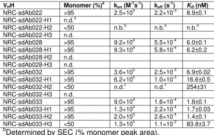
Humanization of VHHs
358
With the aim of using these VHHs in therapeutic applications, the sequences of 359
the four VHHs showing significant binding to EGFR-positive tumor cells (NRC-sdAb022, 360
NRC-sdAb028, NRC-sdAb032 and NRC-sdAb033) were humanized with reference to
361
human IGHV3-30*01 and IGHJ1*01 germline genes. This process yielded at least one
362
humanized variant with unimpaired solubility and EGFR-binding affinity for three of the
363
four parent VHHs (NRC-sdAb028-H1, NRC-sdAb032-H1, NRC-sdAb033-H2). The 364
framework regions of these humanized variants bore 89–94% sequence identity with
365
human IGHV3-30*01, with no or minimal impact on the biophysical properties and
366
EGFR-binding affinities of the resulting VHHs (Table 3 and Supplementary Table S2). 367
Conclusions and implications 368
The VHHs isolated and characterized here were generated by immunization of a 369
llama with DNA alone (no protein or cell boost), using biolistic transfection (gene gun)
370
followed by intradermal injection (DERMOJET). Many of the VHHs had high affinities for 371
recombinant human EGFR, cross-reacted with rhesus and/or mouse EGFR, and
372
recognized native cell-surface EGFR on tumor cell lines; in all of these respects, the
373
VHHs described here are dramatically superior to those previously isolated by our group 374
using recombinant protein immunization [4]. One VHH, NRC-sdAb032, showed clear 375
functional activity as an inhibitor of EGFR signaling, and any of the humanized VHHs 376
may have other therapeutic applications. NRC-sdAb032 was isolated by panning on
377
streptavidin-captured EGFR but not on passively-adsorbed EGFR, and its epitope
378
appears to be present on native EGFR and recombinant EGFR ectodomain in solution
379
but not on the same ectodomain when it is passively adsorbed to microtiter plates or
380
amine coupled to sensor chips. This epitope, which overlaps the cetuximab epitope and
381
the EGF-binding site in EGFR domain III, is apparently poorly available for binding by
382
NRC-sdAb032 or cetuximab in ELISAs against directly-adsorbed EGFR or in SPR
383
experiments in which these antibodies are flowed over amine-coupled EGFR surfaces;
384
by contrast, both NRC-sdAb032 and cetuximab bound EGFR+ tumor cells with lower 385
EC50s than other VHHs with similar monovalent binding affinities for recombinant EGFR. 386
We speculate that the NRC-sdAb032 epitope, though easily destroyed, contributes to
387
this VHH’s superior recognition of native EGFR displayed on the tumor cell surface. 388
Other groups have had similar experiences using streptavidin-captured antigens as
389
‘bait’ during panning of phage-displayed VHH libraries [23]. 390
We are currently investigating whether the DNA immunization schedule
391
presented here is consistent and generalizable to other targets. Our preliminary findings
392
have indicated that up to five intradermal injections alone were unable to elicit a
393
polyclonal serum Ab response against EGFR in several other llamas, and thus one
394
possibility is that either a prolonged immunization schedule or a combination of two
395
immunization routes (particle bombardment; intradermal injection) may be necessary to
396
trigger seroconversion. These hypotheses are under investigation (Mehdi
Arbabi-397
Ghahroudi, in preparation), although evaluating the impact of immunization strategies in
the context of the significant heterogeneity expected in large outbred animals is a major 399 challenge. 400 Acknowledgements 401
We gratefully acknowledge the excellent technical help of Yonghong Guan, Hong
402
Tong-Sevinc, Qingling Yang, and Shalini Raphael. This work was supported by funding
403
from the National Research Council Canada.
404 405
Author Contributions
406
KAH, CRM and MAG designed the immunization study. KAH performed serology
407
and built the phage-displayed VHH library. KAH and MAR isolated VHHs by panning. 408
HvF and GH conducted surface plasmon resonance experiments. DC performed
409
mirrorball® experiments. MAR produced VHH-Fc fusions and performed western blotting 410
experiments to assess EGFR phosphorylation status. KAH and GH made the figures
411
and KAH wrote the paper. JT and MAG proofread the text. All authors approved the
412 final manuscript. 413 414 Additional Information 415
Competing Financial Interests 416
None to declare.
References
418
1 Herbst, R. S. and Shin, D. M. (2002) Monoclonal antibodies to target epidermal
419
growth factor receptor-positive tumors: a new paradigm for cancer therapy. Cancer. 94,
420
1593-1611
421
2 Jonker, D. J., O'Callaghan, C. J., Karapetis, C. S., Zalcberg, J. R., Tu, D., Au, H.
422
J., Berry, S. R., Krahn, M., Price, T., Simes, R. J., Tebbutt, N. C., van Hazel, G.,
423
Wierzbicki, R., Langer, C. and Moore, M. J. (2007) Cetuximab for the treatment of
424
colorectal cancer. N Engl J Med. 357, 2040-2048
425
3 Bonner, J. A., Harari, P. M., Giralt, J., Azarnia, N., Shin, D. M., Cohen, R. B.,
426
Jones, C. U., Sur, R., Raben, D., Jassem, J., Ove, R., Kies, M. S., Baselga, J.,
427
Youssoufian, H., Amellal, N., Rowinsky, E. K. and Ang, K. K. (2006) Radiotherapy plus
428
cetuximab for squamous-cell carcinoma of the head and neck. N Engl J Med. 354,
567-429
578
430
4 Bell, A., Wang, Z. J., Arbabi-Ghahroudi, M., Chang, T. A., Durocher, Y., Trojahn,
431
U., Baardsnes, J., Jaramillo, M. L., Li, S., Baral, T. N., O'Connor-McCourt, M.,
432
Mackenzie, R. and Zhang, J. (2010) Differential tumor-targeting abilities of three
single-433
domain antibody formats. Cancer Lett. 289, 81-90
434
5 Gottlin, E. B., Xiangrong, G., Pegram, C., Cannedy, A., Campa, M. J. and Patz,
435
E. F., Jr. (2009) Isolation of novel EGFR-specific VHH domains. J Biomol Screen. 14,
436
77-85
437
6 Roovers, R. C., Laeremans, T., Huang, L., De Taeye, S., Verkleij, A. J., Revets,
438
H., de Haard, H. J. and van Bergen en Henegouwen, P. M. (2007) Efficient inhibition of
EGFR signaling and of tumour growth by antagonistic anti-EFGR Nanobodies. Cancer
440
Immunol Immunother. 56, 303-317
441
7 Salema, V., Manas, C., Cerdan, L., Pinero-Lambea, C., Marin, E., Roovers, R.
442
C., Van Bergen En Henegouwen, P. M. and Fernandez, L. A. (2016) High affinity
443
nanobodies against human epidermal growth factor receptor selected on cells by E. coli
444
display. MAbs. 8, 1286-1301
445
8 Roovers, R. C., Vosjan, M. J., Laeremans, T., el Khoulati, R., de Bruin, R. C.,
446
Ferguson, K. M., Verkleij, A. J., van Dongen, G. A. and van Bergen en Henegouwen, P.
447
M. (2011) A biparatopic anti-EGFR nanobody efficiently inhibits solid tumour growth. Int
448
J Cancer. 129, 2013-2024
449
9 Niu, G., Murad, Y. M., Gao, H., Hu, S., Guo, N., Jacobson, O., Nguyen, T. D.,
450
Zhang, J. and Chen, X. (2012) Molecular targeting of CEACAM6 using antibody probes
451
of different sizes. J Control Release. 161, 18-24
452
10 Babiuk, L. A., Pontarollo, R., Babiuk, S., Loehr, B. and van Drunen Littel-van den
453
Hurk, S. (2003) Induction of immune responses by DNA vaccines in large animals.
454
Vaccine. 21, 649-658
455
11 Peyrassol, X., Laeremans, T., Gouwy, M., Lahura, V., Debulpaep, M., Van
456
Damme, J., Steyaert, J., Parmentier, M. and Langer, I. (2016) Development by Genetic
457
Immunization of Monovalent Antibodies (Nanobodies) Behaving as Antagonists of the
458
Human ChemR23 Receptor. J Immunol. 196, 2893-2901
459
12 Peyrassol, X., Laeremans, T., Lahura, V., Debulpaep, M., El Hassan, H.,
460
Steyaert, J., Parmentier, M. and Langer, I. (2018) Development by Genetic
461
Immunization of Monovalent Antibodies Against Human Vasoactive Intestinal Peptide
Receptor 1 (VPAC1), New Innovative, and Versatile Tools to Study VPAC1 Receptor
463
Function. Front Endocrinol (Lausanne). 9, 153
464
13 van der Woning, B., De Boeck, G., Blanchetot, C., Bobkov, V., Klarenbeek, A.,
465
Saunders, M., Waelbroeck, M., Laeremans, T., Steyaert, J., Hultberg, A. and De Haard,
466
H. (2016) DNA immunization combined with scFv phage display identifies antagonistic
467
GCGR specific antibodies and reveals new epitopes on the small extracellular loops.
468
MAbs. 8, 1126-1135
469
14 Koch-Nolte, F., Reyelt, J., Schossow, B., Schwarz, N., Scheuplein, F.,
470
Rothenburg, S., Haag, F., Alzogaray, V., Cauerhff, A. and Goldbaum, F. A. (2007)
471
Single domain antibodies from llama effectively and specifically block T cell
ecto-ADP-472
ribosyltransferase ART2.2 in vivo. FASEB J. 21, 3490-3498
473
15 Danquah, W., Meyer-Schwesinger, C., Rissiek, B., Pinto, C., Serracant-Prat, A.,
474
Amadi, M., Iacenda, D., Knop, J. H., Hammel, A., Bergmann, P., Schwarz, N.,
475
Assuncao, J., Rotthier, W., Haag, F., Tolosa, E., Bannas, P., Boue-Grabot, E., Magnus,
476
T., Laeremans, T., Stortelers, C. and Koch-Nolte, F. (2016) Nanobodies that block
477
gating of the P2X7 ion channel ameliorate inflammation. Sci Transl Med. 8, 366ra162
478
16 Fumey, W., Koenigsdorf, J., Kunick, V., Menzel, S., Schutze, K., Unger, M.,
479
Schriewer, L., Haag, F., Adam, G., Oberle, A., Binder, M., Fliegert, R., Guse, A., Zhao,
480
Y. J., Cheung Lee, H., Malavasi, F., Goldbaum, F., van Hegelsom, R., Stortelers, C.,
481
Bannas, P. and Koch-Nolte, F. (2017) Nanobodies effectively modulate the enzymatic
482
activity of CD38 and allow specific imaging of CD38(+) tumors in mouse models in vivo.
483
Sci Rep. 7, 14289
17 Durocher, Y., Perret, S. and Kamen, A. (2002) High-level and high-throughput
485
recombinant protein production by transient transfection of suspension-growing human
486
293-EBNA1 cells. Nucleic Acids Res. 30, E9
487
18 Henry, K. A., Hussack, G., Collins, C., Zwaagstra, J. C., Tanha, J. and
488
MacKenzie, C. R. (2016) Isolation of TGF-beta-neutralizing single-domain antibodies of
489
predetermined epitope specificity using next-generation DNA sequencing. Protein Eng
490
Des Sel. 29, 439-443
491
19 Henry, K. A., Tanha, J. and Hussack, G. (2015) Identification of cross-reactive
492
single-domain antibodies against serum albumin using next-generation DNA
493
sequencing. Protein Eng Des Sel. 28, 379-383
494
20 Baral, T. N., MacKenzie, R. and Arbabi Ghahroudi, M. (2013) Single-domain
495
antibodies and their utility. Curr Protoc Immunol. 103, Unit 2 17
496
21 Rossotti, M. A., Pirez, M., Gonzalez-Techera, A., Cui, Y., Bever, C. S., Lee, K.
497
S., Morisseau, C., Leizagoyen, C., Gee, S., Hammock, B. D. and Gonzalez-Sapienza,
498
G. (2015) Method for Sorting and Pairwise Selection of Nanobodies for the
499
Development of Highly Sensitive Sandwich Immunoassays. Anal Chem. 87,
11907-500
11914
501
22 Zhang, J., Liu, X., Bell, A., To, R., Baral, T. N., Azizi, A., Li, J., Cass, B. and
502
Durocher, Y. (2009) Transient expression and purification of chimeric heavy chain
503
antibodies. Protein Expr Purif. 65, 77-82
504
23 Pardon, E., Laeremans, T., Triest, S., Rasmussen, S. G., Wohlkonig, A., Ruf, A.,
505
Muyldermans, S., Hol, W. G., Kobilka, B. K. and Steyaert, J. (2014) A general protocol
506
for the generation of Nanobodies for structural biology. Nat Protoc. 9, 674-693
24 Hussack, G., Arbabi-Ghahroudi, M., van Faassen, H., Songer, J. G., Ng, K. K.,
508
MacKenzie, R. and Tanha, J. (2011) Neutralization of Clostridium difficile toxin A with
509
single-domain antibodies targeting the cell receptor binding domain. J Biol Chem. 286,
510 8961-8976 511 512 513 514
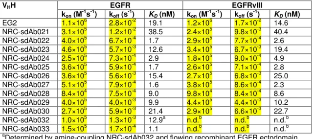
Table 1. Monovalent affinities of VHHs for recombinant human EGFR and EGFRvIII 515 extracellular domains (pH 7.4, 25°C) 516 VHH EGFR EGFRvIII kon (M-1s-1) koff (s-1) KD (nM) kon (M-1s-1) koff (s-1) KD (nM) EG2 1.1×10⁶ 2.8×10⁻² 19.1 1.2×10⁶ 1.7×10⁻² 14.6 NRC-sdAb021 3.1×10⁵ 1.2×10⁻² 38.5 2.4×10⁵ 9.8×10⁻³ 40.4 NRC-sdAb022 4.0×10⁵ 6.7×10⁻⁴ 1.7 2.9×10⁵ 7.7×10⁻⁴ 2.6 NRC-sdAb023 4.6×10⁵ 5.7×10⁻³ 12.6 3.4×10⁵ 6.7×10⁻³ 19.4 NRC-sdAb024 2.5×10⁵ 7.3×10⁻⁴ 2.9 1.8×10⁵ 9.0×10⁻⁴ 4.9 NRC-sdAb025 3.6×10⁵ 5.9×10⁻⁴ 1.7 2.6×10⁵ 7.1×10⁻⁴ 2.8 NRC-sdAb026 3.6×10⁵ 5.6×10⁻³ 15.4 2.7×10⁵ 6.8×10⁻³ 25.0 NRC-sdAb027 5.1×10⁵ 7.9×10⁻⁴ 1.6 3.8×10⁵ 8.6×10⁻⁴ 2.3 NRC-sdAb028 8.4×10⁴ 7.5×10⁻⁴ 9.0 9.8×10⁴ 8.4×10⁻⁴ 8.6 NRC-sdAb029 4.0×10⁵ 4.0×10⁻³ 9.9 4.4×10⁵ 4.4×10⁻³ 10.2 NRC-sdAb030 2.7×10⁵ 5.9×10⁻³ 21.4 2.9×10⁵ 6.6×10⁻³ 22.7 NRC-sdAb032 1.0×10⁵ 1.3×10⁻³ 12.9a n.d.b n.d.b n.d.b NRC-sdAb033 1.5×10⁵ 1.7×10⁻⁴ 1.1 n.d.b n.d.b n.d.b
aDetermined by amine-coupling NRC-sdAb032 and flowing recombinant EGFR ectodomain
517
bNo difference was observed in ELISA binding to human EGFR or EGFRvIII (data not shown)
518
n.d., not determined
519 520
Table 2. Monovalent affinities and kinetics of the interaction between VHHs and
521
human, rhesus and mouse EGFR-Fc (pH 7.4, 25°C).
522
VHH Human EGFR-Fc Rhesus EGFR-Fc Mouse EGFR-Fc
kon (M -1 s-1) koff (s -1 ) KD (nM) kon (M -1 s-1) koff (s -1 ) KD (nM) kon (M -1 s-1) koff (s -1 ) KD (nM) EG2 8.5×10⁵ 8.5×10⁻³ 11.1±0.2 - - - - - - NRC-sdAb022 2.5×10⁵ 2.2×10⁻³ 8.9±0.1 8.5×10⁴ 1.7×10⁻³ 19.6±0.7 - - - NRC-sdAb028 9.2×10⁴ 5.5×10⁻⁴ 6.0±0.1 - - - 6.5×10⁵ 7.5×10⁻³ 11.6±0.3 NRC-sdAb029 5.3×10⁵ 4.5×10⁻³ 8.5±0.1 5.5×10⁵ 6.4×10⁻³ 11.8±0.3 5.1×10⁵ 6.3×10⁻³ 12.5±1.2 NRC-sdAb032 3.6×10⁵ 2.5×10⁻³ 6.9±0.02 4.8×10⁵ 3.9×10⁻³ 8.1±0.1 - - - NRC-sdAb033 9.0×10⁴ 1.6×10⁻⁴ 1.8±0.1 1.1×10⁵ 1.1×10⁻⁴ 1.0±0.1 9.2×10⁴ 2.8×10⁻⁴ 3.1±0.2 (-) indicates no binding 523
KD values are expressed as the means ± standard deviations of three independent experiments
524 525 526
Table 3. Aggregation propensities and monovalent affinities of humanized VHHs for 527 human EGFR-Fc. 528 VHH Monomer (%)a kon (M-1s-1) koff (s-1) KD (nM) NRC-sdAb022 >95 2.5×10⁵ 2.2×10⁻³ 8.9±0.1 NRC-sdAb022-H1 n.d.a NRC-sdAb022-H2 <50 n.b.b n.b.b n.b.b NRC-sdAb022-H3 n.d. NRC-sdAb028 >95 9.2×10⁴ 5.5×10⁻⁴ 6.0±0.1 NRC-sdAb028-H1 >95 9.3×10⁴ 5.8×10⁻⁴ 6.2±0.2 NRC-sdAb028-H2 n.d. NRC-sdAb028-H3 n.d. NRC-sdAb032 >95 3.6×10⁵ 2.5×10⁻³ 6.9±0.02 NRC-sdAb032-H1 >95 6.2×10⁵ 1.0×10⁻² 16.6±0.5 NRC-sdAb032-H2 <50 n.d.c n.d.c 254±31 NRC-sdAb032-H3 n.d. NRC-sdAb033 >95 9.0×10⁴ 1.6×10⁻⁴ 1.8±0.1 NRC-sdAb033-H1 >95 1.3×10⁵ 2.2×10⁻⁴ 1.7±0.03 NRC-sdAb033-H2 >95 2.0×10⁵ 2.6×10⁻⁴ 1.4±0.1 NRC-sdAb033-H3 <50 1.3×10⁵ 1.1×10⁻² 83.8±3.7
bDetermined by SEC (% monomer peak area).
529
bn.d., not determined due to low or no expression.
530
cn.b., no binding of the purified monomeric sdAb to human EGFR-Fc could be detected.
531
dn.d., not determined because steady-state K
D was calculated.
Figure Legends
533
534
Figure 1. Serum titration ELISA of the immunized llama against recombinant human
535
EGFR and human EGFRvIII ectodomains. Wells of NUNC® MaxiSorpTM microtiter plates 536
were coated overnight at 4°C with 100 ng of EGFR or EGFRvIII in 35 µL of PBS. The
537
wells were blocked with 200 µL of PBS containing 2% skim milk for 1 h at 37°C, then
538
sera (serially diluted in PBS containing 1% BSA and 0.1% Tween-20) were added to
539
wells for 2 h at room temperature. The wells were washed 5× with PBS containing 0.1%
540
Tween-20, incubated for 1 h with HRP-conjugated goat anti-llama IgG (diluted 1:5,000
541
in PBS containing 1% BSA and 0.1% Tween-20), washed 5× again and then developed
542
with TMB substrate.
543
Figure 2. Binding of VHHs to EGFR by SPR and ELISA. (A) SPR sensorgrams showing 544
single-cycle kinetic analysis of VHH binding to human EGFR-Fc. Recombinant EGFR-Fc 545
was immobilized on a Series S Sensor Chip CM5 using amine coupling, then the
546
indicated VHH was flowed over the surface at concentrations ranging from 0.6 nM – 50 547
nM (NRC-sdAb022, 1.5 nM – 25 nM; NRC-sdAb028, 1.5 nM – 25 nM; NRC-sdAb029,
548
1.5 nM – 25 nM; NRC-sdAb032, 3 nM – 50 nM; NRC-sdAb033, 0.6 nM – 10 nM; EG2, 3
549
nM – 50 nM). Black lines show data and red lines show fits. (B) Summary of epitope
550
binning by SPR. The colors of the circles indicate the cross-species conservation of the
551
epitope (red, human EGFR only; blue, human and rhesus EGFR; yellow, human and
552
mouse EGFR; green, human, rhesus and mouse EGFR). (C) Competitive ELISA
553
showing binding of VHHs to EGFR in the presence or absence of EGF. 554
555
Figure 3. Binding of VHHs to EGFR-positive MDA-MB-431 cells and EGFR-low MCF7 556
cells. (A) Binding of VHH monomers (20 µg/mL) to EGFR-positive MDA-MB-431 cells by 557
flow cytometry detected using anti-c-Myc and AF647-labeled anti-mouse antibodies. (B,
558
C) Binding of serially diluted VHH-Fcs to EGFR-positive MDA-MB-431 cells (B) and 559
EGFR-low MCF7 cells (C) by mirrorball® microplate cytometry detecting using
AF488-560
labeled anti-human Fc antibody. EC50s were determined by curve fitting in Graphpad 561
Prism using the equation for one-site specific binding with Hill slope. (D) Binding of
562
serially diluted biparatopic VHH-Fcs by mirrorball® microplate cytometry detecting using 563
AF488-labeled anti-human Fc antibody. EC50s were determined by curve fitting in 564
Graphpad Prism using the equation for one-site specific binding with Hill slope.
565
566
Figure 4. Inhibition of EGF-induced EGFR phosphorylation in MDA-MB-468 cells by
567
VHH-Fcs. (A) Western blotting of phosphorylated EGFR (Tyr1068), total EGFR and β-568
actin in serum-starved MDA-MB-468 cells treated with EGF in the presence or absence
569
of the indicated inhibitor (all at 500 nM). (B) Densitometry analysis of bands in A. (C)
570
Western blotting of phosphorylated EGFR (Tyr1068), bulk EGFR and β-actin in
serum-571
starved MDA-MB-468 cells treated with EGF in the presence or absence of the
572
indicated concentration of inhibitor. (D) Densitometry analysis of bands in C. The
573
concentrations used are as shown in C. A20.1-Fc is an irrelevant VHH-Fc against 574
Clostridium difficile toxin A [24] included as a negative control. (E) Western blotting of 575
phosphorylated EGFR (Tyr1068), total EGFR and β-actin in serum-starved
468 cells treated with EGF in the presence or absence of the indicated inhibitor (all at 5
577
nM). (F) Densitometry analysis of bands in E.
Supplementary Material
Camelid single-domain antibodies raised by DNA immunization are potent inhibitors of EGFR signaling
Martin A. Rossotti1a, Kevin A. Henry1a, Henk van Faassen1, Jamshid Tanha1,2, Deborah
Callaghan1, Greg Hussack1, Mehdi Arbabi-Ghahroudi1,3 and C. Roger MacKenzie1*
1Human Health Therapeutics Research Centre, National Research Council Canada,
100 Sussex Drive, Ottawa, Ontario, Canada, K1A 0R6
2Department of Biochemistry, Microbiology and Immunology, University of Ottawa, 451
Smyth Road, Ottawa, Ontario, Canada, K1H 8M5
3Department of Biology, Carleton University, 1125 Colonel By Drive, Ottawa, Ontario,
Canada, K1S 5B6
aEqual contribution
*Address correspondence to: C. Roger MacKenzie, Ph.D.
Human Health Therapeutics Research Centre, National Research Council Canada, 100 Sussex Drive, Ottawa, ON, Canada, K1A 0R6
Tel +1-613-990-0833 Fax +1-613-952-9092
S
Supplementary Figure S1. Binding of VHHs elicited by DNA immunization to human, rhesus and mouse EGFR-Fc in ELISA. Wells of Nunc® MaxiSorpTM microtiter plates were coated with 100 ng of each EGFR-Fc in 35 μL PBS overnight at 4°C. The next day, wells were blocked with 200 μL of 2% (w/v) skim milk in PBS for 1 h at 37°C. Antibodies were serially diluted in PBS containing 1% (w/v) BSA and 0.1% (v/v) Tween-20 and added to wells for 2 h at room temperature. The wells were washed 5× with PBS containing 0.1% Tween-20, incubated for 1 h with HRP-conjugated rabbit anti-6×His antibody (Cedarlane Labs), washed 5× again and then developed with TMB substrate as described in the main text.
0.1 1 10 100 1000 10000 0.0 0.5 1.0 1.5 NRC-sdAb022 [VHH] (ng/mL) OD (45 0 nm ) human EGFR-Fc rhesus EGFR-Fc mouse EGFR-Fc 0.1 1 10 100 1000 10000 0.0 0.5 1.0 1.5 NRC-sdAb028 [VHH] (ng/mL) OD ( 45 0 nm ) human EGFR-Fc rhesus EGFR-Fc mouse EGFR-Fc 0.1 1 10 100 1000 10000 0.0 0.5 1.0 1.5 NRC-sdAb032 [VHH] (ng/mL) OD ( 45 0 nm ) human EGFR-Fc rhesus EGFR-Fc mouse EGFR-Fc 0.1 1 10 100 1000 10000 0.0 0.5 1.0 1.5 NRC-sdAb033 [VHH] (ng/mL) OD ( 45 0 nm ) human EGFR-Fc rhesus EGFR-Fc mouse EGFR-Fc
E
EG2 + NRC--ssdAb032 NNRC--ssdAb032 ++ EG2 EEG2 & NRC--ssdAb032
E
EG2 + NRC--ssdAb033 NNRC--ssdAb033 + EG2 EEG2 & NRC--ssdAb033
E
EG2 + NRC--ssdAb028 NNRC--ssdAb028 + EG2 EEG2 & NRC--ssdAb028
E
EG2 + NRC--ssdAb0222 NNRC--ssdAb0222 ++ EG2 EEG2 & NRC--ssdAb022
E
E
EG2 + ccetuximab ccetuximab ++ EG2 EEG2 & cetuximab
N
NRC--ssdAb028 + NRC--ssdAb022 NNRC--ssdAb022 + NRC--ssdAb028 NNRC--ssdAb028 & NRC--ssdAb022
N
NRC--ssdAb028 ++ NRC--ssdAb029 NNRC--ssdAAb029 ++ NRC--ssdAb028 NNRC--ssdAb028 & NRC--ssdAb029
N
NRC--ssdAb028 ++ cetuximab ccetuximab ++ NRC--ssdAb028 NNRC--ssdAb028 & cetuximab
N
N
NRC--ssdAb032 ++ NRC--ssdAb033 NNRC--ssdAb033 ++ NRC--ssdAb0032 NNRC--ssdAb032 & NRC--ssdAb033
N
NRC--ssdAb032 ++ NRC--ssdAb028 NNRC--ssdAb028 ++ NRC--ssdAb0032 NNRC--ssdAb032 & NRC--ssdAb028
N
NRC--ssdAb032 ++ NRC--ssdAb022 NNRC--ssdAb022 ++ NRC--ssdAb0032 NNRC--ssdAb032 & NRC--ssdAb022
N
NRC--ssdAb032 ++ cetuximab ccetuximab ++ NRC--ssdAb0032 NNRC--ssdAb032 & cetuximab
N
N
NRC--ssdAb033 ++ NRC--ssdAb022 NNRC--ssdAb022 ++ NRC--ssdAb0033 NNRC--ssdAb033 & NRC--ssdAb022
N
NRC--ssdAb033 ++ NRC--ssdAb029 NNRC--ssdAb029 ++ NRC--ssdAb0033 NNRC--ssdAb033 & NRC--ssdAb029
N
NRC--ssdAb033 ++ cetuximab ccetuximab ++ NRC--ssdAb0033 NNRC--ssdAb033 & cetuximab
N
NRC--ssdAb022 ++ NRC--ssdAb029 NNRC--ssdAb029 ++ NRC--ssdAb0022 NNRC--ssdAb022 & NRC--ssdAb029
N
S
Supplementary Figure S2. Sensorgrams for all epitope binning SPR co-injection experiments. One representative of each VHH family elicited by DNA immunization was selected (sdAb022, NRC-sdAb028, NRC-sdAb029, NRC-sdAb032, NRC-sdAb033) along with EG2 VHH and cetuximab. In the first injection, the EGFR surface was saturated using a concentration equivalent to 25× KD of the first antibody, then in the second injection, the first antibody was supplied in the presence of a second antibody (also 25× KD) to determine whether additional binding occurred. All possible permutations of antibodies were tested as described in the main text.
Supplementary Table S1. Sequence characteristics of anti-EGFR VHHs raised by
llama DNA immunization.
Family VHH CDR-H3 Length (aa)a
- EG2 18 1 NRC-sdAb021 8 NRC-sdAb022 8 NRC-sdAb023 8 NRC-sdAb024 8 NRC-sdAb025 8 NRC-sdAb026 8 NRC-sdAb027 8 2 NRC-sdAb028 10 3 NRC-sdAb029 17 NRC-sdAb030 17 4 NRC-sdAb032 12 5 NRC-sdAb033 11 aIMGT definition
Supplementary Table S2. Sequence characteristics of humanized anti-EGFR VHHs.
VHH No. of amino acid
changes with respect to parent
Overall amino acid identity with human IGHV3-30 (%)
Framework region amino acid identity with human IGHV3-30 (%)a NRC-sdAb022 0 71.3 77.5 NRC-sdAb022-H1 12 83.0 91.3 NRC-sdAb022-H2 14 85.1 93.8 NRC-sdAb022-H3 17 88.3 97.5 NRC-sdAb028 0 72.6 76.3 NRC-sdAb028-H1 10 82.1 88.8 NRC-sdAb028-H2 13 85.3 92.5 NRC-sdAb028-H3 17 89.5 97.5 NRC-sdAb032 0 67.4 75.0 NRC-sdAb032-H1 13 80.0 91.3 NRC-sdAb032-H2 16 83.2 95.0 NRC-sdAb032-H3 18 85.3 97.5 NRC-sdAb033 0 67.4 75.0 NRC-sdAb033-H1 12 79.0 90.0 NRC-sdAb033-H2 15 82.1 93.8 NRC-sdAb033-H3 18 85.3 97.5