R E S E A R C H A R T I C L E
Open Access
Raised serum levels of IGFBP-1 and IGFBP-2
in idiopathic pulmonary fibrosis
J. Guiot
1*, B. Bondue
2, M. Henket
1, J. L. Corhay
1and R. Louis
1Abstract
Background: Idiopathic pulmonary fibrosis (IPF) is a chronic lung disorder of unknown origin, which ultimately leads to death. Several growth factors such as IGFs (insulin-like-growth factor) and IGFBPs (insulin like growth factor binding proteins) seem to take part to the pathogenesis. We evaluated IGFs and IGFBPs in serum from patients with IPF and healthy subjects including 24 untreated IPF and 26 IPF receiving anti-fibrotic therapy and to compare them with healthy subjects.
Methods: Serum of 50 idiopathic pulmonary fibrosis and 55 healthy subjects (HS) were analysed by ELISA for IGFs and IGFBPs, TGF-β and KL-6, the latter being tested as positive control in IPF.
Results: Serum levels of IGFBP-1 and IGFBP-2 and KL-6 were significantly higher in the IPF group than in the healthy subjects (p < 0.05, p < 0.001 and p < 0.0001 respectively) while the picture was inversed regarding IGFs. By contrast there was no significant difference between the groups with respect to TGF-β. IGFBP-2 was significantly reduced in the patients with specific anti-fibrotic therapy pirfenidone and nintedanib compared to untreated patients (p < 0.05) but still significantly elevated in comparison to HS (p < 0.001).
Conclusion: Serum IGFBP-1 and−2 are increased in idiopathic pulmonary fibrosis and IGFBP-2 may be reduced by anti-fibrosing therapy. IGFBPs may be promising biomarkers in IPF.
Keywords: Idiopathic pulmonary fibrosis, Pulmonary fibrosis, Insuline-like growth factors, Insulin like growth factor binding proteins
Background
Idiopathic pulmonary fibrosis (IPF) is a complex diagno-sis and pathology requiring a multidisciplinary approach. This fibrotic disease has a poor prognosis and requires a specific and early appropriate therapy [1–3]. In this con-text several biomarkers (such as surfactant protein A or SP-A, the Krebs von den Lungen 6 or KL-6, Ig A and periostin [4–8] were studied without much success to be discriminant as early diagnostic biomarkers to identify IPF out of different interstitial lung diseases (ILDs). Nevertheless when taken together they provided convin-cing evidence that changes in blood proteins (KL-6, SP-A, MMP-7, CCL-18, among others) or cells (fibrocytes and T-cell subpopulations) are indicative in IPF and may somewhat predict outcome of the disease [9].
The transforming growth factor beta (TGF-β) has long been known to be involved in the pathophysiology of the IPF [1]. TGF-β is a stimulus for pulmonary fibrogenesis by its activity in the control of the remodelling of the extracellular matrix. TGF-β is also known to be involved in the fibroblast activity.
Besides TGF-β, there has been recently a growing
interest for the axis IGFs/IGFBPs in fibrosing process. A recently published study has shown a link between the production of TGF-β and the production of insulin-like growth factor binding protein-2 (IGFBP-2) [10] known to be related to IPF [11] within myofibroblasts cells de-rived from the lung. Moreover, IGFBP-2 was found at significantly higher level in the bronchoalveolar lavage of children with interstitial lung disease [12] relative to healthy subjects.
IGFBP-2 is also overexpressed in response to a deterior-ation of the lung parenchyma [13–15]. This overexpression is mainly perinuclear and proves to be a potential factor of
* Correspondence:J.Guiot@chu.ulg.ac.be
1Pneumology Department, CHU Liège, Domaine universitaire du Sart-Tilman,
B35, B4000 Liège, Belgium
Full list of author information is available at the end of the article
© 2016 Guiot et al. Open Access This article is distributed under the terms of the Creative Commons Attribution 4.0 International License (http://creativecommons.org/licenses/by/4.0/), which permits unrestricted use, distribution, and reproduction in any medium, provided you give appropriate credit to the original author(s) and the source, provide a link to the Creative Commons license, and indicate if changes were made. The Creative Commons Public Domain Dedication waiver (http://creativecommons.org/publicdomain/zero/1.0/) applies to the data made available in this article, unless otherwise stated.
fibroblast proliferation [16–19]. The IGFBP-2 protein is part of a group (IGFBPs) known to be involved in the regu-lation of insulin-like growth factor (IGF). IGF-1 and IGF-2 play an important role in the growth, differentiation and cellular metabolism [20, 21]. Signalling pathway of IGF con-sisted of two isoforms of IGF (−1 and −2), two types of receptors, six binding proteins with high affinity for IGFs (IGFBPs) and four binding proteins with low affinity for IGFs (or related protein IGFBP: IGFBPrp) [11, 22, 23]. The IGFBPs bind IGFs and can increase their half-life, alter their function (in potentiating or inhibiting it), or facilitate their passage to the target tissues [20, 24]. Activity of IGFBP-2 is controlled on one hand by its secretion and on the other hand by its proteolysis. Proteolysis of IGFBP-2 is made by several groups of proteolytic enzymes such as pappalysines, kallikreins, metalloproteases or the plasmin [25].
There has been recent pharmacological progress in the treatment of IPF. Drugs such as pirfenidone and ninte-danib have been shown to slow down the decline in lung volume that accompanies the lung fibrosing process [2].
We focused our study on the serum measurement of several growth factors including IGFs and IGFBPs in order to identify a potential new biomarker in IPF and sought to determine whether their levels might be influ-enced by recently developed anti-fibrotic therapy. Methods
Subject characteristics
We prospectively recruited patients from our ambulatory care policlinic at CHU Liege and Erasme University Hospital of Brussels. The patients were divided into 2 groups. The first group was the group of patients with untreated IPF (n = 24). The second group is a group of treated IPF patients with a specific therapy (n = 26). The diagnosis of (definite) IPF was made according to the international recommendations of the ATS [1] using the respiratory function test, HRCT scan (prob-able UIP pattern), bronchoalveolar lavage (when avail-able), as well as the clinical history of the patient. We excluded all other causes of interstitial lung disease (such as asbestosis, hypersensitivity pneumonitis, pneu-monia associated with connective tissue disease or toxic pneumonitis). We combined the different results for the diagnosis. All cases were discussed in a multidiscip-linary group about interstitial lung diseases composed of a pulmonologist, a specialist in pulmonary rehabilita-tion, a rheumatologist, a radiologist, a pathologist and a specialist in occupational medicine. Sixteen patients underwent a surgical biopsy and seven patients trans-bronchial cryobiopsies. Twenty-six patients benefit from a specific treatment of IPF (pirfenidone (n = 17) or nintedanib (n = 9)) Fig. 1. We also recruited healthy subjects by advertisement in our policlinic waiting room. They all denied any respiratory disease and had
normal spirometric values with FEV1 > 80 % predicted and FEV1/FVC ratio > 70 %.
The protocol was approved by the ethics committee of CHU of Liège, and all subjects gave written consent before their enrollment (Belgian number : B707201422832 ; ref : 2014/302).
Peripheral blood puncture
Venous blood was collected in Vacutainer tubes from an antecubital site immediately when controls and patients were included in the study. Blood cell values included white blood cell count, the differential leukocyte count, fibrinogen and C-reactive protein (CRP) levels were de-termined by the routine hospital laboratory.
Biomarkers measurements in serum
We analysed several biomarkers assumed to be critical growth factors in blood: TGF-β, IGF-1, IGF-2, IGFBP-1, IGFBP-2 and IGFBP-3 were measured by a specific en-zyme immunoassay with a commercial kit (TGF-β, IGF-1, IGFBP-IGF-1, IGFBP-2, IGFBP-3: Duoset®, R&D systems, Minneapolis; IGF-2 : Mediagnost, Reutlingen, Germany; KL-6 level was measured by a specific enzyme immuno-assay with a commercial kit (Lumipulse G KL-6 Fujirebio Europe).
Pulmonary function tests
We performed lung function tests in both routine respira-tory laborarespira-tory of CHU Liège and Erasme University hos-pital. All spirometric tests performed for this study were measured using the pneumotachograph JaegerMasterlab system (Erich Jaeger GmbH, Wuzburg, Germany). The forced expiratory volume in one second (FEV1) and forced vital capacity (FVC) were measured in accordance with the recommendations of the European Respiratory Society (ERS) [26]. The results were expressed in millilitre and percent predicted. The Tiffeneau index or FEV1/FVC was expressed in percent. The total lung capacity (TLC) was measured by body plethysmography according to ERS recommendations (Erich Jaeger GmbH, Wuzburg, Germany). The diffusion capacity of CO (DLCO) and the report DLCO/AV were measured by the single-breath carbon monoxide gas transfer method and expressed as percent predicted (Sensor Medics 2400 He/CO Analyzer System, Bilthoven, Netherlands).
Statistical analysis
Demographic and functional data were expressed as mean ± standard deviation (SD). The biomarkers levels were expressed as median (min-max). When the data showed normal distribution, they were compared with a one-way ANOVA, followed by Tukey-Kramer’s post-hoc testing. When the data did not show a normal distribu-tion, they were compared with the Kruskal-Wallis test
followed by Dunn's post-hoc testing. Correlations be-tween variables were performed using Spearman’s
rank correlation test. A p < 0.05 was considered as
significant. Results
Subject demographic functional and blood characteristics
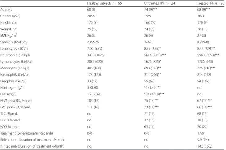
The demographic, functional and treatment characteris-tics of the subjects are given in Table 1. The average age of IPF patients was slightly higher (74 ± 9 years for un-treated patients versus 68 ± 9 years for un-treated patients) with a male dominance. Spirometric values were moder-ately lowered and comparable in both the treated and untreated IPF while DLCO was sharply reduced in both IPF groups.
There was a significant increase in leukocyte count of untreated and treated IPF patients (p < 0.0001). IPF patients also displayed neutrophil (treated IPF : p < 0.0001; untreated IPF : p < 0.001), monocyte (treated IPF :p < 0.0001; untreated IPF : p < 0.0001) and eosinophil counts (treated IPF : p > 0.05; untreated IPF : p < 0.001) compared to HS (Table 1). By contrast, circulating lymphocyte count was reduced in untreated IPF compared to HS. There was an increase in fibrinogen and CRP levels in untreated IPF compare to HS (Table 1).
Serum growth factors
The results of the serum biochemical markers are listed in the Table 2. There was a striking increase in IGFBP-1
and −2 in the group of untreated IPF patients compared
to healthy subjects (respectively p < 0.05, p < 0.0001) (Figs. 2–3), while untreated IPF showed a significant de-crease in serum IGF-1 (p < 0.05), IGF-2 (p < 0.01) and IGFBP-3 (p < 0.01). IGFBP-2 levels were lower in those patients receiving anti-fibrotic therapy but still signifi-cantly elevated compared to HS (p < 0.001) (Fig. 3).
IGFBP-2 seems to be significantly lowered by specific IPF therapy irrespective of the molecule used as anti-fibrotic treatment (median range of 189 (110–231) ng/ml in the group treated by nintedanib (n = 9) vs 155 (126–192) ng/ml in those receiving pirfenidone (n = 17), p > 0.05).
Similarly to IGFBP-2, KL-6 was increased in both IPF groups compared to HS (p < 0.0001) (Fig. 3). By contrast there was no significant difference between the groups regarding the levels of TGF-β.
We also calculated the serum molar ratio of IGFs:IGFBPs known as reflecting the real IGFs activity (Table 2). Serum ratios of IGF-1:IGFBP-1 (p < 0.01), IGF-1:IGFBP-2 (p < 0.0001), IGF-2:IGFBP-1 (p < 0.05) and IGF-2:IGFBP-2 (p < 0.0001) were significantly lower in the not treated IPF group than in healthy subjects (Table 2).
Relationship between growth factors and lung function
There was an inverse relationship between spirometric values and TGF-β in healthy subjects (FVC % pred R = −0.50, p < 0.05 and FEV1 % pred R = −0.48, p < 0.05) and untreated IPF (FEV1 % predR = −0.54, p < 0.05) and an inverse relationship between IGFBP-1 and DLCO in treated IPF (R = −0.52, p < 0.05). We didn’t find any correl-ation between pulmonary function tests and IGFBP-2 and other biomarkers assessed in our study (Additional file 1: Table S1).
Discussion
Our study shows for the first time that IPF features a marked increase in serum IGFBP-1 and IGFBP-2. In addition IGFBP-2 levels are attenuated in those patients receiving anti-fibrotic treatment even if the serum levels remained higher in than those measured in healthy subjects.
Table 2 Serum biomarkers
Healthy subjects Untreated IPF Treated IPF
IGF-1 (ng/ml) 31 (7–67) 20 (9–102)* 25 (11–57)
IGF-2 (ng/ml) 710 (401–2232) 590 (210–1027)* 586 (282–920)**
IGFBP-1 (ng/ml) 11 (0–180) 22 (4–110)* 9 (1–107)
IGFBP-2 (ng/ml) 94 (34–211) 206 (113–317)*** 153 (32–291)** °
IGFBP-3 (ng/ml) 2132 (1207–4059) 1536 (534–2556)* 2032 (893–3505)
Molar ratio IGF-1 : IGFBP-1 12 (1–314) 3,5 (0,3–36,3)* 9 (1–152)
Molar ratio IGF-1 : IGFBP-2 1,8 (0,2–6,2) 0,4 (0–4,3)*** 0,7 (0,3–5,2)*
Molar ratio IGF-1 : IGFBP-3 0,06 (0,02–0,12) 0,05 (0,03–0,25) 0,05 (0,03–0,1)
Molar ratio IGF-2 : IGFBP-1 247 (20–6735) 110 (11–769)* 259 (20–2429)
Molar ratio IGF-2 : IGFBP-2 40 (10–202) 14 (5–28)*** 19 (7–89)**
Molar ratio IGF-2 : IGFBP-3 1,4 (0,8-5,1) 1,6 (0,6-2,2) 1,2 (0,9-1,8)
TGF-β (ng/ml) 11 (3–21) 11 (5–22) 9 (4–17)
KL-6 (ng/ml) 262 (83–596) 1050 (314–4951)*** 889 (359–6168)***
Data are expressed as median (min-max)
*p < 0.05 **p < 0.001 ***p < 0.0001 compared to healthy subjects °p < 0.05 °°p < 0.001 °°°p < 0.0001 compared to untreated IPF
Table 1 Patients demographic, functional, treatment and blood characteristics
Healthy subjects n = 55 Untreated IPF n = 24 Treated IPF n = 26
Age, yrs 60 (8) 74 (9)*** 68 (9)*** Gender (M/F) 28/27 19/5 16/3 Height, cm 170 (8) 168 (10) 170 (9) Weight, Kg 75 (12) 74 (16) 78 (11) BMI, Kg/m2 26 (3) 26 (4) 27 (3) Smokers (NS/FS/S) 23/22/6 3/8/6 (6/19/0) Leucocytes x103/μl 7.00 (5.39) 8.35 (2.35)* 8.42 (2.91)** Neutrophils (Cell/μl) 3450 (1025) 5614 (2113)*** 5960 (3053)*** Lymphocytes (Cell/μl) 2085 (620) 1676 (825)* 1786 (643) Monocytes (Cell/μl) 486 (160) 698 (325)** 725 (218)*** Eosinophils (Cell/μl) 173 (125) 314 (266)** 214 (128) Basophils (Cell/μl) 33 (17) 55 (67) 94 (187) Fibrinogen (g/l) 3 (0.80) a4 (1.40)*** nd CRP (mg/l) 1.9 (2.89) a30 (37.89)*** nd
FEV1 post-BD, %pred. 105 (12) 75 (14)*** 67 (13)***
FVC post-BD, %pred. 111 (16) 73 (14)*** 66 (16)***
TLC, %pred. nd 71 (19) 68 (15)
DLCO %pred. nd 37 (11) 38 (13)
KCO %pred. nd 63 (16) 70 (20)
Treatment (pirfenidone/nintedanib) 0/0 0/0 17/9
Pirfenidone (duration of treatment -Month) nd nd 9.9 (7.4)
Nintedanib (duration of treatment -Month) nd nd 14.3 (15.8)
nd not determined
Data are expressed as mean (SD)
Non smoker (NS), former smoker (FS), smoker (S)
*p < 0.05 **p < 0.001 ***p < 0.0001 compared to healthy subjects
a
Previous studies focusing on IGFBP-2 in fibrosis showed an increase of IGFBP-2 in the bronchoalveolar lavage [12] and in the lung tissue of patients with interstitial lung disease (ILD) without focusing on IPF. IGFBP-2 was shown to be overexpressed in case of lung epithelial dam-age [12–15, 27] and its expression was found to be reduced, together with that of TGF-β, by cyclosporine in vitro [10]. The assay of IGFBP-2 in the serum of patients with idio-pathic pulmonary fibrosis had never been done before. In our current study IGFBP-2 was significantly higher in the serum of patients with IPF compared to healthy subjects. Of great interest, serum IGFBP-2 decreases with specific anti-fibrotic treatments without returning to normal values however. It supports the idea that IGFBP-2 may play a role in the fibrotic process. As our patients were not treated with corticoids we can here discard any possible impact of corticosteroids on IGFBP levels [28].
Reinforcing the potential role of IGFBPs in IPF, IGFBP-1 was also specifically increased, but to a lesser extent, in IPF compared to HS. The increase of KL-6 in IPF versus healthy subjects is confirmatory of previous findings [29, 30] and validates our IPF patient cohort. In sharp contrast to what we found with IGFBP, serum IGF-1
and−2 were not found to be decreased in IPF in
com-parison to HS which results in a decrease ratio IGF:IGFBP in patients with IPF. IGFs are very potent growth factors [20] but, to the best of our knowledge, had not been stud-ied in human lung fibrosis so far. In an animal model IGF-1 was shown to stimulate differentiation of fibroblast into myofibrolast [31], one of the effector of pulmonary fibrosis [32]. Intuitively we might have expected a dir-ect relationship between IGFs and disease severity. However our study didn’t show any correlation between IGF-:IGFBP ratio and functional impairment including forced vital capacity and total lung diffusing capacity. The rise of serum IGFBPs might be seen as a spill over of the
lung fibrotic process since previous studies showed raised levels in BAL from patients with ILD [12]. How IGFBP may favour lung fibrosis remains unclear. One potential explanation is IGFBP-2 can bind to the lung extracellular matrix [33] and thus can favour the IGF activity by in-creasing its local availability, which results in an increase of cellular response to IGF [34]. This trapping mechanism would explain why IGFs themselves were found to be re-duced in serum from our patients. Alternatively we cannot rule out the fact that high IGFBP-2 in IPF may actually re-flect a protective feed-back mechanism to limit the disease progression by neutralizing IGFs [35]. On the same line there is an example where IGFPB-2 is playing a protective role against anarchic cell proliferation. In non-small cell lung carcinoma, in vitro studies indicate that both soluble
HS IPF Treated IPF 0 100 200 300 400 P= 0.034 P<0.00001 P= 0.0004 l m/ g n 2-P B F GI HS IPF Treated IPF 10 100 1000 10000 P<0.0001 P<0.0001 l m/ g n 6-L K
Fig. 3 Serum IGFBP-2 and KL-6 concentrations: Comparison between treated IPF, untreated IPF and healthy subjects. HS = healthy subjects; IPF = idiopathic pulmonary fibrosis
HS IPF 0 100 200 300 400 P<0.0001 l m/ g n 2-P B F GI
Fig. 2 Serum IGFBP-2 concentration: Comparison between IPF and healthy subjects. HS = healthy subjects (n = 55); IPF = idiopathic pulmonary fibrosis (n = 50: treated and untreated)
and membrane-associated IGFBP-2 competes with IGF receptors for ligand binding and, thus, are likely to be im-portant determinants of IGF responsiveness [36]. Interest-ingly IGF-I and IGFBP-3 are lowered in untreated IPF patients in comparison to healthy subjects. This decrease is similar to what it has been shown in ARDS, cystic fibro-sis and COPD [37]. Moreover these authors suggest that there may be an inverse association between circulating levels of IGF-1 and IGFBP-3 and lung compartments levels where they are upregulated suggesting a different lung and blood regulation under these circumstances.
In our study serum levels of TGF-β were similar between the groups confirming the results described by a previous study [38]. Of course it does not mean that these molecules did not play a major role within the lung. Indeed TGF-β is known to take part to the lung fibrosing process and was shown to be overexpressed in lung tissue of patients with IPF [39].
Conclusion
We conclude that serum IGFBP-1 and IGFBP-2 are in-creased in patients with idiopathic pulmonary fibrosis in comparison to healthy subjects. Moreover, the raised IGFBP-2 level is attenuated by anti-fibrotic treatment. The prognostic value of these new biomarkers warrants further longitudinal studies.
Additional file
Additional file 1: Table S1. Correlations between blood biomarkers and pulmonary function test. (DOCX 173 kb)
Abbreviations
ATS:American thoracic society; AV: Alveolar ventilation; CCL-18: chemokine (C-C motif) ligand 18; COPD: chronic obstructive pulmonary disease; DLCO: diffusion capacity of CO in the lung; FEV1: forced expiratory volume in one second; FVC: forced vital capacity; HRCT: high resolution computed tomography; IGF: insulin-like growth factor; IGFBP: insulin-like growth factor binding protein; IGFBP-rp: insulin-like growth factor binding protein related protein; ILD: interstitial lung disease; IPF: idiopathic pulmonary fibrosis; KL-6: Krebs von den Lungen-6; MMP: matrix metalloproteinase; NSIP: non specific idiopathic pulmonary fibrosis; SD: standard deviation; A: surfactant protein-A; SP-D: surfactant protein-D; TGF-β: transforming growth factor beta; TLC: total lung capacity; YKL-40: Chitinase-3-like-1, human cartilage glycoprotein-39. Acknowledgment
We thank Georgitha Amand for the collection of datas.
We thank the Leon Fredericq ULG foundation for their financial support. Availability of data and materials
The data supporting our results are presented within the article (and its Additional file 1: Table S1).
Authors’ contribution
RL and JLC for their involvement in conception, hypothesis, and design of the study. MH for the acquisition and analysis of samples. BB for his help in the recruitment. All authors read and approved the final manuscript. Competing interests
The authors declare that they have no competing interests.
Consent for publication Not applicable.
Ethics approval and consent to participate
The protocol was approved by the ethics committee “hospitalo-facultaire” of Liège, and all subjects gave written consent before their enrolment (Belgian number : B707201422832 ; ref : 2014/302).
Author details
1
Pneumology Department, CHU Liège, Domaine universitaire du Sart-Tilman, B35, B4000 Liège, Belgium.2Pneumology Department, Erasme University
Hospital, Université Libre de Bruxelles, Route de Lennik, 808, B1070 Brussels, Belgium.
Received: 26 January 2016 Accepted: 16 May 2016
References
1. Ganesh Raghu HRC, Egan JJ, Martinez FJ, Juergen B, Brown KK, Colby TV, Jean-François C, Flaherty KR, Lasky JA, Lynch DA, Ryu JH, Swigris JJ, Wells AU, Julio A, Demosthenes B, Carlos C, Ulrich C, Masahito E, Hansell DM, Takeshi J, Dong Soon K, King TE Jr, Yasuhiro K, Jeffrey M, Müller NL, Nicholson AG, Luca R, Moisés S, Dudden RF, Griss BS, Protzko SL, Schünemann HJ. An Official ATS/ERS/JRS/ALAT Statement: Idiopathic Pulmonary Fibrosis: Evidence-based Guidelines for Diagnosis and Management. Am J Respir Crit Care Med. 2011;183(6):788–824. 2. Wilson KC, Raghu G. The 2015 guidelines for idiopathic pulmonary fibrosis:
an important chapter in the evolution of the management of patients with IPF. Eur Respir J. 2015;46(4):883–6.
3. Spagnolo P, Tonelli R, Cocconcelli E, Stefani A, Richeldi L. Idiopathic pulmonary fibrosis: diagnostic pitfalls and therapeutic challenges. Multidiscip Respir Med. 2012;7(1):42.
4. Ohshimo S, Ishikawa N, Horimasu Y, Hattori N, Hirohashi N, Tanigawa K, et al. Baseline KL-6 predicts increased risk for acute exacerbation of idiopathic pulmonary fibrosis. Respir Med. 2014;108(7):1031–9.
5. Samukawa T, Hamada T, Uto H, Yanagi M, Tsukuya G, Nosaki T, et al. The elevation of serum napsin A in idiopathic pulmonary fibrosis, compared with KL-6, surfactant protein-A and surfactant protein-D. BMC Pulm Med. 2012;12:55.
6. Ten Klooster L, van Moorsel CH, Kwakkel-van Erp JM, van Velzen-Blad H, Grutters JC. IgA in serum: An old acquaintance as a new prognostic biomarker in Idiopathic Pulmonary Fibrosis. Clin Exp Immunol. 2015;181(2):357–61. 7. Tajiri M, Okamoto M, Fujimoto K, Johkoh T, Ono J, Tominaga M, et al. Serum
level of periostin can predict long-term outcome of idiopathic pulmonary fibrosis. Respir Investig. 2015;53(2):73–81.
8. Ley B, Brown KK, Collard HR. Molecular biomarkers in idiopathic pulmonary fibrosis. Am J Physiol Lung Cell Mol Physiol. 2014;307(9):L681–91. 9. Zhang Y, Kaminski N. Biomarkers in idiopathic pulmonary fibrosis. Curr Opin
Pulm Med. 2012;18(5):441–6.
10. Hirota N, Ito T, Miyazaki S, Ebina M, Homma S. Gene expression profiling of lung myofibroblasts reveals the anti-fibrotic effects of cyclosporine. Tohoku J Exp Med. 2014;233(4):283–93.
11. Ruan W, Ying K. Abnormal expression of IGF-binding proteins, an initiating event in idiopathic pulmonary fibrosis? Pathol Res Pract. 2010;206(8):537–43. 12. Chadelat K, Boule M, Corroyer S, Fauroux B, Delaisi B, Tournier G, et al.
Expression of insulin-like growth factors and their binding proteins by bronchoalveolar cells from children with and without interstitial lung disease. Eur Respir J. 1998;11(6):1329–36.
13. Mouhieddine OB, Cazals V, Maitre B, Le Bouc Y, Chadelat K, Clement A. Insulin-like growth factor-II (IGF-II), type 2 IGF receptor, and IGF-binding protein-2 gene expression in rat lung alveolar epithelial cells: relation to proliferation. Endocrinology. 1994;135(1):83–91.
14. Cazals V, Mouhieddine B, Maitre B, Le Bouc Y, Chadelat K, Brody JS, et al. Insulin-like growth factors, their binding proteins, and transforming growth factor-beta 1 in oxidant-arrested lung alveolar epithelial cells. J Biol Chem. 1994;269(19):14111–7.
15. Mouhieddine OB, Cazals V, Kuto E, Le Bouc Y, Clement A. Glucocorticoid-induced growth arrest of lung alveolar epithelial cells is associated with increased production of insulin-like growth factor binding protein-2. Endocrinology. 1996;137(1):287–95.
16. Duan C. Specifying the cellular responses to IGF signals: roles of IGF-binding proteins. J Endocrinol. 2002;175(1):41–54.
17. Perks CM, Newcomb PV, Norman MR, Holly JM. Effect of insulin-like growth factor binding protein-1 on integrin signalling and the induction of apoptosis in human breast cancer cells. J Mol Endocrinol. 1999;22(2):141–50. 18. Jones JI, Gockerman A, Busby Jr WH, Wright G, Clemmons DR. Insulin-like
growth factor binding protein 1 stimulates cell migration and binds to the alpha 5 beta 1 integrin by means of its Arg-Gly-Asp sequence. Proc Natl Acad Sci U S A. 1993;90(22):10553–7.
19. Firth SM, Baxter RC. Cellular actions of the insulin-like growth factor binding proteins. Endocr Rev. 2002;23(6):824–54.
20. Duan C, Xu Q. Roles of insulin-like growth factor (IGF) binding proteins in regulating IGF actions. Gen Comp Endocrinol. 2005;142(1–2):44–52. 21. Yau SW, Azar WJ, Sabin MA, Werther GA, Russo VC. IGFBP-2 - taking the lead
in growth, metabolism and cancer. J Cell Commun Signal. 2015;9(2):125–42. 22. Baxter RC, Binoux M, Clemmons DR, Conover C, Drop SL, Holly JM, et al.
Recommendations for nomenclature of the insulin-like growth factor binding protein (IGFBP) superfamily. Growth Horm IGF Res. 1998;8(3):273–4. 23. Kim HS, Nagalla SR, Oh Y, Wilson E, Roberts Jr CT, Rosenfeld RG. Identification
of a family of low-affinity insulin-like growth factor binding proteins (IGFBPs): characterization of connective tissue growth factor as a member of the IGFBP superfamily. Proc Natl Acad Sci U S A. 1997;94(24):12981–6.
24. Shimasaki S, Ling N. Identification and molecular characterization of insulin-like growth factor binding proteins (IGFBP-1, -2, -3, -4, -5 and -6). Prog Growth Factor Res. 1991;3(4):243–66.
25. Pickard A, McCance DJ. IGF-Binding Protein 2 - Oncogene or Tumor Suppressor? Front Endocrinol. 2015;6:25.
26. Miller MR, Crapo R, Hankinson J, Brusasco V, Burgos F, Casaburi R, et al. General considerations for lung function testing. Eur Respir J. 2005;26(1):153–61. 27. Besnard V, Corroyer S, Trugnan G, Chadelat K, Nabeyrat E, Cazals V, et al.
Distinct patterns of insulin-like growth factor binding protein (IGFBP)-2 and IGFBP-3 expression in oxidant exposed lung epithelial cells. Biochim Biophys Acta. 2001;1538(1):47–58.
28. Miell JP, Taylor AM, Jones J, Holly JM, Gaillard RC, Pralong FP, et al. The effects of dexamethasone treatment on immunoreactive and bioactive insulin-like growth factors (IGFs) and IGF-binding proteins in normal male volunteers. J Endocrinol. 1993;136(3):525–33.
29. Furuhashi K, Suda T, Nakamura Y, Inui N, Hashimoto D, Miwa S, et al. Increased expression of YKL-40, a chitinase-like protein, in serum and lung of patients with idiopathic pulmonary fibrosis. Respir Med. 2010;104(8):1204–10. 30. Korthagen NM, van Moorsel CH, Barlo NP, Ruven HJ, Kruit A, Heron M, et al.
Serum and BALF YKL-40 levels are predictors of survival in idiopathic pulmonary fibrosis. Respir Med. 2011;105(1):106–13.
31. Hung CF, Rohani MG, Lee SS, Chen P, Schnapp LM. Role of IGF-1 pathway in lung fibroblast activation. Respir Res. 2013;14:102.
32. Bagnato G, Harari S. Cellular interactions in the pathogenesis of interstitial lung diseases. Eur Respir Rev. 2015;24(135):102–14.
33. Russo VC, Azar WJ, Yau SW, Sabin MA, Werther GA. IGFBP-2: The dark horse in metabolism and cancer. Cytokine Growth Factor Rev. 2014;26(3):329–46. 34. Kelley KM, Oh Y, Gargosky SE, Gucev Z, Matsumoto T, Hwa V, et al. Insulin-like growth factor-binding proteins (IGFBPs) and their regulatory dynamics. Int J Biochem Cell Biol. 1996;28(6):619–37.
35. Jones JI, Clemmons DR. Insulin-like growth factors and their binding proteins: biological actions. Endocr Rev. 1995;16(1):3–34.
36. Reeve JG, Morgan J, Schwander J, Bleehen NM. Role for membrane and secreted insulin-like growth factor-binding protein-2 in the regulation of insulin-like growth factor action in lung tumors. Cancer Res. 1993;53(19):4680–5.
37. Ahasic AM, Zhai R, Su L, Zhao Y, Aronis KN, Thompson BT, et al. IGF1 and IGFBP3 in acute respiratory distress syndrome. Eur J Endocrinol. 2012;166(1):121–9. 38. Alhamad EH, Cal JG, Shakoor Z, Almogren A, AlBoukai AA. Cytokine gene
polymorphisms and serum cytokine levels in patients with idiopathic pulmonary fibrosis. BMC Med Genet. 2013;14:66.
39. Khalil N, Greenberg AH. The role of TGF-beta in pulmonary fibrosis. Ciba Found Symp. 1991;157:194–207. discussion −11.
• We accept pre-submission inquiries
• Our selector tool helps you to find the most relevant journal
• We provide round the clock customer support
• Convenient online submission
• Thorough peer review
• Inclusion in PubMed and all major indexing services
• Maximum visibility for your research Submit your manuscript at
www.biomedcentral.com/submit