HAL Id: hal-01911531
https://hal.archives-ouvertes.fr/hal-01911531
Submitted on 20 Jan 2020
HAL is a multi-disciplinary open access
archive for the deposit and dissemination of sci-entific research documents, whether they are pub-lished or not. The documents may come from teaching and research institutions in France or abroad, or from public or private research centers.
L’archive ouverte pluridisciplinaire HAL, est destinée au dépôt et à la diffusion de documents scientifiques de niveau recherche, publiés ou non, émanant des établissements d’enseignement et de recherche français ou étrangers, des laboratoires publics ou privés.
Transcription at Intergenic Regions by PXD to NTAIL
Binding Strength
Louis-Marie Bloyet, Joanna Brunel, Marion Dosnon, Véronique Hamon, Jenny
Erales, Antoine Gruet, Carine Lazert, Christophe Bignon, Philippe Roche,
Sonia Longhi, et al.
To cite this version:
Louis-Marie Bloyet, Joanna Brunel, Marion Dosnon, Véronique Hamon, Jenny Erales, et al.. Modula-tion of Re-initiaModula-tion of Measles Virus TranscripModula-tion at Intergenic Regions by PXD to NTAIL Binding Strength. PLoS Pathogens, Public Library of Science, 2016, 12 (12), pp.e1006058. �10.1371/jour-nal.ppat.1006058�. �hal-01911531�
Modulation of Re-initiation of Measles Virus
Transcription at Intergenic Regions by P
XD
to
N
TAIL
Binding Strength
Louis-Marie Bloyet1,2,3,4,5☯, Joanna Brunel1,2,3,4,5☯, Marion Dosnon6,7☯, Ve´ronique Hamon8,9,10, Jenny Erales6,7, Antoine Gruet6,7, Carine Lazert1,2,3,4,5, Christophe Bignon6,7, Philippe Roche8,9,10, Sonia Longhi6,7☯*, Denis Gerlier1,2,3,4,5☯*
1 CIRI, International Center for Infectiology Research, Universite´ de Lyon, Lyon, France, 2 INSERM, U1111,
Lyon, France, 3 Ecole Normale Supe´rieure de Lyon, Lyon, France, 4 Universite´ Claude Bernard Lyon 1, Centre International de Recherche en Infectiologie, Lyon, France, 5 CNRS, UMR5308, Lyon, France, 6 Aix-Marseille University, Architecture et Fonction des Macromole´cules Biologiques (AFMB) UMR 7257, Aix-Marseille, France, 7 CNRS, AFMB UMR 7257, Marseille, France, 8 Aix Marseille University, Institut Paoli-Calmettes, Centre de Recherche en Cance´rologie de Marseille (CRCM), Marseille, France, 9 CNRS, CRCM UMR 7258, Marseille, France, 10 INSERM, CRCM U1068, Marseille, France
☯These authors contributed equally to this work.
*Sonia.Longhi@afmb.univ-mrs.fr(SL);denis.gerlier@inserm.fr(DG)
Abstract
Measles virus (MeV) and all Paramyxoviridae members rely on a complex polymerase machinery to ensure viral transcription and replication. Their polymerase associates the phosphoprotein (P) and the L protein that is endowed with all necessary enzymatic activi-ties. To be processive, the polymerase uses as template a nucleocapsid made of genomic RNA entirely wrapped into a continuous oligomer of the nucleoprotein (N). The polymerase enters the nucleocapsid at the 3’end of the genome where are located the promoters for transcription and replication. Transcription of the six genes occurs sequentially. This implies ending and re-initiating mRNA synthesis at each intergenic region (IGR). We explored here to which extent the binding of the X domain of P (XD) to the C-terminal region of the N pro-tein (NTAIL) is involved in maintaining the P/L complex anchored to the nucleocapsid
tem-plate during the sequential transcription. Amino acid substitutions introduced in the XD-binding site on NTAILresulted in a wide range of binding affinities as determined by
combin-ing protein complementation assays in E. coli and human cells and isothermal titration calo-rimetry. Molecular dynamics simulations revealed that XD binding to NTAILinvolves a
complex network of hydrogen bonds, the disruption of which by two individual amino acid substitutions markedly reduced the binding affinity. Using a newly designed, highly sensitive dual-luciferase reporter minigenome assay, the efficiency of re-initiation through the five measles virus IGRs was found to correlate with NTAIL/XD KD. Correlatively, P transcript
accumulation rate and F/N transcript ratios from recombinant viruses expressing N variants were also found to correlate with the NTAILto XD binding strength. Altogether, our data
sup-port a key role for XD binding to NTAILin maintaining proper anchor of the P/L complex
thereby ensuring transcription re-initiation at each intergenic region.
a11111
OPEN ACCESS
Citation: Bloyet L-M, Brunel J, Dosnon M, Hamon
V, Erales J, Gruet A, et al. (2016) Modulation of Re-initiation of Measles Virus Transcription at Intergenic Regions by PXDto NTAILBinding
Strength. PLoS Pathog 12(12): e1006058. doi:10.1371/journal.ppat.1006058
Editor: Roberto Cattaneo, Mayo Clinic, UNITED
STATES
Received: July 13, 2016 Accepted: November 12, 2016 Published: December 9, 2016
Copyright:© 2016 Bloyet et al. This is an open access article distributed under the terms of the
Creative Commons Attribution License, which permits unrestricted use, distribution, and reproduction in any medium, provided the original author and source are credited.
Data Availability Statement: All relevant data are
within the paper and its Supporting Information files.
Funding: This work was carried out with the
financial support of the Agence Nationale de la Recherche, specific programs "Physico-Chimie du Vivant" (ANR-08-PCVI-0020-01), and “ASTRID” (ANR-11-ASTR-003-01). JE and LMB were supported by fellowships from the Fondation pour la Recherche Me´dicale (FRM). MD was supported by a PhD fellowship from the French Ministry of National Education, Research and Technology. The
Author Summary
Three proteins, the polymerase L, the phosphoprotein P and the nucleoprotein N, inter-play to ensure transcription and replication of measles virus, a member of the Paramyxo-viridae family. A regular array of nucleoprotein shields the viral genomic RNA. The
resulting nucleocapsid constitutes the template of RNA synthesis used by the polymerase complex made of L and P, with the latter ensuring L anchoring onto the nucleocapsid. We herein report a correlation between the binding affinity of the C-terminal X domain of P (XD) and the intrinsically disordered C-terminal tail of N (NTAIL), the ability to reinitiate the transcription at the intergenic regions and the accumulation rate of viral transcripts from recombinant viruses. We therefore propose that the NTAIL/XD interaction contrib-utes to maintaining the polymerase complex anchored onto the nucleocapsid while end-ing the upstream transcript and re-initiatend-ing the downstream transcript at every
intergenic region. As such, the NTAIL/XD interaction strength must be controlled so as to keep the viral transcription gradient within an optimal efficiency window. The conserva-tion of this mode of interacconserva-tion between the viral P and N proteins in many members of theParamyxoviridae family reflects one of the major evolution constraints to which their
polymerase machinery is subjected.
Introduction
Measles virus (MeV), a member of theMorbillivirus genus, belongs to the Paramyxoviridae
family of theMononegavirales order [1]. These viruses possess a non-segmented RNA genome of negative polarity that is encapsidated by the nucleoprotein (N) to form a helical nucleocap-sid. Not only does N protect viral RNA from degradation and/or formation of viral dsRNA, but it also renders the latter competent for transcription and replication. Indeed, the viral poly-merase cannot processively transcribe nor replicate RNA unless the viral genome is encapsi-dated by the N protein within a helical nucleocapsid [2,3]. Transcription and replication are ensured by the RNA-dependent RNA polymerase complex made of the large protein (L) and the phosphoprotein (P), with P serving as an essential tethering factor between L and the nucleocapsid. The complex made of RNA and of the N, P and L proteins constitutes the repli-cation machinery. In order to perform messenger RNA synthesis, the polymerase has not only to bind to the 3’ transcription promoter, but also to re-initiate the transcription of downstream genes upon crossing each intergenic region (IGR). Following polyadenylation, which serves as gene end (GE) signal, the polymerase proceeds over three nucleotides (3’-GAA-5’ or 3’-GCA-5’) without transcribing them and then restarts transcription upon recognition of a down-stream gene start (GS) signal.
Within infected cells, N is found in a soluble, monomeric form (referred to as N0) and in a nucleocapsid assembled form [4]. Following synthesis, the N protein requires chaperoning by the P protein so as to be maintained in a soluble and monomeric form. The P N-terminal region (PNT) binds to the neosynthesized N protein thereby simultaneously preventing its ille-gitimate self-assembly and yielding a soluble N0P complex the structure of which have been characterised for MeV [5] as well as for four other members of theMononegavirales order
[6,7,8,9]. N0P is used as the substrate for the encapsidation of the nascent genomic RNA chain during replication [10], (see also [4,11,12,13] for reviews on transcription and replication). In its assembled homopolymeric form or nucleocapsid, N also makes complexes with either iso-lated P or P bound to L, with all these interactions being essential for RNA synthesis by the funders had no role in study design, data collection
and analysis, decision to publish, or preparation of the manuscript.
Competing Interests: The authors have declared
viral polymerase [14,15,16]. Throughout theMononegavirales order, P and P+L binding to the
nucleocapsid is mediated by interaction of the C-terminal region of P with either the C-termi-nal tail of N (Paramyxoviridae members), or to the N-terminal globular moiety (or core) of N
(see [11,17] for review).
The MeV N protein consists of a structured N-terminal moiety (NCORE, aa 1–400), and a C-terminal domain (NTAIL, aa 401–525) [18,19] that is intrinsically disordered, i.e. it lacks highly populated secondary and tertiary structure under physiological conditions of pH and salinity in the absence of a partner (for a recent review on intrinsically disordered proteins see [20]). While NCOREcontains all the regions necessary for self-assembly and RNA-binding [10,21,22] and a binding site for anα-MoRE located at the N terminus of the P protein, NTAILis responsi-ble for interaction with the C-terminal X domain (XD, aa 459–507) of P [11,18,21,23,24,25,26, 27] (Fig 1A).
Fig 1. Binding affinity of the NTAIL variants as evaluated by gfp- and glu-PCA. (a) Representation of N and P proteins. (b) Relative binding
strength between NTAILvariants and XD as assessed by gfp-PCA. (c) Correlation between binding strengths as inferred from gfp-PCA and as
inferred from glu-PCA. (d) Correlation between binding strengths as obtained from monomeric glu-PCA and from multimeric (i.e. PMD-XDagainst N)
glu-PCA. Shown are means and standard deviations (SD) from three independent experiments performed in triplicate. The color code used for symbols is the same in panel b-d. The color code for N variants adopted in panel (b) will be used throughout all figures.
NTAILbinding to XD triggersα-helical folding within a molecular recognition element [28,29] ofα-helical nature (α-MoRE, aa 486–502) located within one (Box2, aa 489–506) out of three conserved NTAILregions [18,24,27,30,31,32,33,34,35,36,37]. XD-inducedα-helical folding of NTAILis not a feature unique to MeV, being also conserved within the Paramyxoviri-dae family [38,39,40,41,42,43]. XD consists of a tripleα-helical bundle [27,34,44], and binding to theα-MoRE leads to a pseudo-four-helix arrangement that mainly relies on hydrophobic contacts [18,27,44]. Theα-MoRE of NTAILis partly preconfigured as anα-helix prior to bind-ing to XD [31,32,35,37,45] and adopts an equilibrium between a fully unfolded form and four partly helical conformers [37]. In spite of this partial pre-configuration, NTAILfolds according to a “folding after binding mechanism” [45,46]. Previous mutational studies showed that Box2 is poorly evolvable in terms of its binding abilities towards XD, in that amino acid substitu-tions therein introduced lead to a dramatic drop in the binding strength, as judged from a protein complementation assay (PCA) based on split-GFP reassembly (gfp-PCA) [47]. In par-ticular, substitutions within the N-terminal region of Box2 (aa 489–493) and at position 497 were found to lead to the most dramatic drops in the interaction strength [47].
In the context of the viral nucleocapsid, NTAILpoints towards the interior of the latter and then extrafiltrates through the interstitial space between NCOREmoieties, with the first 50 residues (aa 401–450) being conformationally restricted due to their location between successive turns of the nucleocapsid [37]. The NTAILregion spanning residues 451–525 and encompassing theα-MoRE is, by contrast, exposed at the surface of the viral nucleocapsid and thus accessible to the viral polymerase. Binding of XD to NTAILhas been proposed to ensure and/or contribute to the recruitment of the viral P/L polymerase complex onto the nucleocapsid template. However, its precise function has remained enigmatic so far with reports of apparent conflicting observations. From the analysis of four NTAILvariants it was concluded that the accumulation rate of primary transcripts is rather insensitive to a drop in the apparent XD to NTAILaffinity [26], while an XD variant showing a 1.7 times stronger interaction with NTAILwas associated with a 1.7-fold reduction in the accumula-tion rate of viral transcripts [48]. Furthermore, deletions studies of NTAILhave indicated that the interaction between XD and NTAILmay be dispensable for transcription and repli-cation [49].
In the present work, we further investigate the molecular mechanisms by which substitu-tions in critical posisubstitu-tions of NTAILpreviously identified by a random approach [47] affect the viral polymerase activity. We did so by combining biochemical studies and molecular dynam-ics (MD) simulations on one hand with functional studies that made use of minigenomes and recombinant viruses on the other hand. Results identify positions 491 and 497 as the most crit-ical in terms of both binding affinities and functional impact. In addition, thanks to the avail-ability of a newly conceived minigenome made of two luciferase reporter genes, with the second one being conditionally expressed via RNA edition of its transcript by the viral poly-merase, we could quantify the efficiency of transcription re-initiation after polymerase scan-ning through each of the five IGRs of MeV genome and on an elongated un-transcribed IGR (UTIGR). A low NTAIL-XD affinity was found to be associated to a reduced ability of N to sup-port expression of luciferase from the second gene. Furthermore, in infected cells, the accumu-lation rate of primary transcripts and transcript ratios were found to correlate with the equilibrium dissociation constant (KD) of the NTAIL/XD pair. Altogether obtained data argue for a key role of the NTAIL/XD interaction in transcription re-initiation at each intergenic region.
Results
Binding strengths of N
TAILvariants from two protein complementation
assays
In a previous random mutagenesis study that made use of a PCA based on split-GFP re-assem-bly (gfp-PCA) [47], we identified NTAILvariants that either decrease or increase the interaction strength towards XD [47]. Variant MX208, which bears the D437V, P485L and L524R substi-tutions that are all located outside theα-MoRE, is an example of the latter group. We previ-ously reported the generation and assessment of binding properties by gfp-PCA of six single-site variants (R489Q, R490S, S491L, A492T, D493G and R497G) bearing each a unique substi-tution within theα-MoRE [47]. Here, we additionally designed and generated the MXSF vari-ant, which bears D437V, R439S, P456S and P485L substitutions that are all found in variants displaying an increased fluorescence [47]. Gfp-PCA inE. coli showed that the binding strength
of these variants towards XD is scattered over a wide range, with the S491L and R497G variants showing the lowest interaction and with variant MXSF displaying interaction strength only moderately higher thanwt NTAIL(Fig 1B). Incidentally, this latter finding indicates that the effects of the substitutions are not cumulative.
We then sought at assessing to which extent results afforded by the split-GFP assay inE. coli
cells reflect NTAIL/XD binding occurring in the natural host cells of MeV. To this end, the interaction between XD and NTAILvariants was measured using the split-luciferase reassembly assay [50]. This technique is based on the same principle as the split-GFP reassembly assay. The reporter (i.e.Gaussia princeps luciferase) and the measured parameter (luminescence) are
however different, and the assay is performed in human cells. Moreover, contrary to the split-GFP reassembly assay where reporter reassembly is irreversible, in the split-luciferase assay (glu-PCA), association of the two luciferase fragments is reversible. As such, while the mea-sured parameter in the former assay is dominated by thekon, the measured parameter in the
latter assay does reflect the equilibrium between akonand akoffand hence a true KD. A signifi-cant correlation was obtained between the two PCA methods (Fig 1C), a finding that provides additional support for the significance of the observed differences in binding strength among variants. Furthermore, a significant correlation was also observed when comparing binding strengths as obtained using monomeric constructs (i.e. NTAIL/XD) and binding strengths obtained using their natural multimeric counterparts, i.e. P multimerization domain (PMD)-XD (P303-507) and full-length mutated N protein constructs (Fig 1D). The rationale for using P multimeric constructs devoid of the N-terminal region (PNT) was to eliminate the binding site to NCORElocated within PNTand involved in P chaperoning of N protein to form N0P complexes [5] (seeFig 1Afor depicting scheme). Importantly, all N variants accumulated in cells in similar amounts (S1 Fig) indicating that variations in the level of reconstituted Gaussia
luciferase likely reflects variations in NTAILto XD binding strength.
Generation, purification and characterization of the single-site Box2
N
TAILvariants
In order to characterize Box2 variants (Fig 2A) at the biochemical level, we expressed and purified sixα-MoRE variants of NTAILas N-terminally hexahistidine tagged proteins. All NTAILvariants were purified to homogeneity from the soluble fraction of the bacterial lysate through immobilized metal affinity chromatography (IMAC) followed by size exclusion chro-matography (SEC) (Fig 2B). The identity of all purified proteins were checked and confirmed by mass spectrometry. Even if their molecular mass is ~16 kDa, they all migrate on a denatur-ing gel with an apparent molecular mass of approximately 20 kDa (Fig 2B, inset). This
aberrant electrophoretic migration has been systematically observed for all NTAILvariants reported so far [26,30,31,32,46] includingwt NTAIL[19]. This anomalous migration is
Fig 2. Secondary structure content of NTAIL variants (a) Crystal structure of the chimera between XD (orange) and theα-MoRE of NTAIL(red)
(PDB code 1T6O) [44]. The side chains of residues mutated in the NTAILvariants investigated in this study are shown in sticks with atom type colours. (b) SEC elution profile of the purified NTAILvariants. The differences in the peak heights reflect differences in the total amounts of protein loaded onto
the SEC column. Inset. Coomassie blue staining of an 18% SDS-PAGE analysis of the purified NTAILvariants. M: molecular mass markers. Color
code is shown above the Coomassie blue staining picture with dotted grey line for wt N. (c) Far-UV CD spectra of the purified NTAILvariants at 0.1 mg/
ml in 10 mM sodium phosphate buffer at pH 7 either in the absence (c) or in the presence of 20% TFE (d). Shown are the average CD spectra as obtained from three different protein samples. (e)α-helical content of NTAILvariants in the absence (plain bars) or presence (hatched bars) of 20%
TFE, as obtained by deconvoluting CD spectra using the Dichroweb server. doi:10.1371/journal.ppat.1006058.g002
frequently observed in IDPs and is due to a high content in acidic residues [51] and/or a large extension in solution [43].
All NTAILvariants, includingwt NTAIL,have the same SEC elution profile (Fig 2B). In par-ticular, they are all eluted with an apparent molecular mass higher than expected and typical of a premolten globule (PMG) state [52], as already observed in the case ofwt NTAIL[19]. Thus, the amino acid substitution(s) causes little (if any) effect on the hydrodynamic volume sam-pled by the protein.
Analysis of the secondary structure content of the NTAILvariants by far-UV circular dichro-ism (CD) shows they are all disordered, as judged from their markedly negative ellipticity at 200 nm (Fig 2C). In addition, they are all similarly able to gainα-helicity in the presence of 20% 2,2,2 trifluoroethanol (TFE) (Fig 2D), as already observed forwt NTAIL[19]. All variants have an estimatedα-helical content similar (within the error bar) to that of wt NTAIL, with the only exception of variant R489Q that exhibits a lowerα-helicity both in the absence and in the presence of TFE (Fig 2E). Thus, most of the amino acid substitutions cause little (if any) effect on the overall secondary structure content and folding abilities of NTAIL.
Assessment of binding affinities of single-site Box2 N
TAILvariants
towards XD by ITC
The binding abilities of the NTAILvariants, includingwt NTAIL, were assessed using isothermal titration calorimetry (ITC). To this end, the purified NTAILproteins were loaded into the sam-ple cell of an ITC200 microcalorimeter and titrated withwt XD. For each variant, two
indepen-dent experiments were carried out.Fig 3shows, for each variant, one representative ITC curve along with the relevant binding parameters. The XD/NTAILmolar ratios achieved at the end of the titration were 2.0 (wt, R489Q, A492T, D493G), 2.5 (R490S) or 3.0 (R497G) (Fig 3). The data, following integration and correction for the heats of dilution, were fitted with a standard model allowing for a set of independent and equivalent binding sites.
Consistent with the unfavorable entropic contribution associated to the disorder-to-order transition that takes place upon NTAILbinding to XD, whenever binding parameters could be determined, they revealed a decrease in entropy, with aΔS ranging from -13 to -29.5 cal mol-1 deg-1(Fig 3). Binding reactions were all found to be enthalpy-driven, withΔH values in the same order of magnitude and ranging from -10.9 to -14.5 kcal/mol (Fig 3). The estimates for the model parameters of thewt NTAIL/XD pair were found to be in very good agreement with those recently reported [46]. The estimates for binding parameters of variants R489Q, A492T and D493G yielded equilibrium dissociation constant (KD) very close to that observed forwt
NTAIL, indicating that these substitutions poorly affect the interaction (Fig 3). On the other hand, the R490S substitution resulted in a 7-fold decrease in the binding affinity (KDof 20μM). The decrease in affinity was even further pronounced (KDof 44μM) in the case of the R497G variant, although the interaction remained measurable (Fig 3). In the case of the S491L variant the interaction strength was below the ITC detection limit and thus KDcould not be estimated (Fig 3).
Then values for the A492T/XD and D493G/XD binding pairs were found to deviate from
unit, a behaviour that is not unusual and that has been already observed with single-site trypto-phan variants [46] and that may arise from relatively poorly defined baselines. In light of all the numerous previous studies [18,19,27,30,31,32,33,34,35,36,37] showing that NTAILand XD form a 1:1 complex, these deviations were not taken to be significant.
We next focused on how binding affinities obtained by ITC correlate with binding strengths inferred from split-GFP and split-luciferase reassembly assays. In fact, although it has already been established that the higher the fluorescence the higher the interaction strength
[53], no attempts were done at establishing which type of relationship exists between KDvalues and fluorescence or luminescence values. As shown inFig 4, we found a significant correlation between fluorescence or luminescence values obtained by gfp-PCA [47] and glu-PCA and the
ln of KDvalues (p = 0.02 in both cases). Although this finding needs to be confirmed on a larger set of data points, it lays the basis for the possibility of inferring KDvalues directly from fluorescence or luminescence values.
Fig 3. Binding parameters of the NTAILvariants towards XD as obtained by ITC. Data are representative of at least two independent
experiments. The derived equilibrium dissociation constants (KD), the stoichiometry number (n), the binding enthalpyΔH (kcal mol-1), and the binding
entropyΔS (cal mol-1deg-1) are shown. Shown are the curves obtained using the following concentrations of N
TAILin the microcalorimeter cell and of
XD in the microsyringe: wt NTAIL/XD: 50μM/500μM; NTAILR489Q/XD: 50μM/500μM; NTAILR490S/XD: 150μM/854μM; NTAILS491L/XD: 160μM/
800μM; NTAILA492T/XD: 50μM/500μM; NTAILD493G/XD: 25μM/600μM; NTAILR497G/XD: 150μM/955μM. Graphs shown at the bottom of each
panel correspond to integrated and corrected ITC data fitted to a single set of sites model. Note that for the binding reactions characterized by KD
values in the tens of micromolar range, it is difficult to obtain the first plateau because the necessary concentrations are too high. Consequently, it should be kept in mind that the actual errors may be larger than those estimated by the fit.
Notably, if the results obtained by gfp-PCA (seeFig 1B) pointed out similarly low interac-tion strengths for variants S491L and R497G, ITC studies yielded a different profile. Indeed, while no interaction could be effectively detected for the S491L/XD pair, the KDcould be mea-sured for variant R497G (seeFig 3). Using the empirically determined equation relating lumi-nescence and KDvalues (Fig 4), the KDof the S491L/XD pair was estimated to be 85± 33 μM, a value consistent with our inability to detect the interaction by ITC. Indeed, an interaction characterized by a KDof approximately 100μM could escape detection unless extremely high (and hardly achievable) protein concentrations are used (typically 1 mM NTAIL/10 mM XD) [54].
Altogether, obtained results confirmed that not all Box2 residues are equivalent in terms of their role in NTAIL/XD complex formation. In particular, while substitutions at positions 489, 490, 492 and 493 have a slight to moderate impact, substitutions at positions 497 and 491 dras-tically affect complex formation without having a strong impact on the overallα-helicity. The role of Box2 residues in complex formation follows the order 491>497>490, reflecting either the orientation of side chains towards the partner (residues 490 and 491) or involvement in stabilizing interactions with XD residue Tyr480 in spite of solvent exposure (residue 497), as already proposed (Fig 2A) [47].
Fig 4. Correlation between binding strengths, as inferred from either gfp-PCA of glu-PCA, and binding affinities derived from ITC. In both cases a statistically significant correlation was observed between fluorescence
or luminescence values and the KD(p = 0.02 in both cases). Shown are the mean values and SD as obtained from
two (ITC) or three (PCAs) independent experiments performed in triplicate. doi:10.1371/journal.ppat.1006058.g004
Molecular dynamic simulations of wt, S491L and R497G variants in
complex with XD reveal a crucial hydrogen bonding network
In order to further investigate the mechanisms by which residues Ser491 and Arg497 stabilize the NTAIL/XD complex, we performed MD simulations in aqueous solvent using the
CHARMM force field [55]. MD simulations were carried out starting from the X-ray structure of the XD/α-MoRE complex [44] or from thein silico generated XD/α-MoRE S491L and XD/
α-MoRE R497G models. In the case of the S491L variant, the three most favorable orientations of the side chains were generated. We first assessed the dynamical stability of the complexes. For this purpose, we analyzed the root-mean square deviation (RMSD) of the Cα atoms with respect to the initial structure as a function of time for the three complexes (i.e.wt, S491L and
R497G) (S1 Table). The RMSD values showed very little variations between the different con-structs during the time course of the 50 ns simulations (S1 Table). The average RMSD for XD and for theα-MoRE were approximately 0.8 and 0.5 Å, respectively, indicating structural sta-bility of each domain during the simulations (S1 Table). The relative orientation of the α-MoRE compared to XD was also assessed and revealed slightly higher average RMSD values for the two variants due to small local rearrangements of the structures to adapt to the substitu-tions. However, RMSD fluctuations were in the same order of magnitude. Secondary structure analyses ofwt and mutated complexes confirmed that all α-helices are conserved during the
whole trajectories. Overall, the different systems were stable during the whole simulation. Since the orientation of the side-chain of L491 showed no impact on the behavior of the com-plex during the MD simulation, only one conformer was selected in the rest of the study.
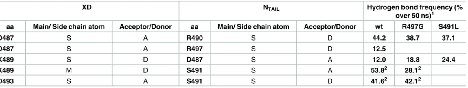
Although the association between XD and NTAILis essentially driven by hydrophobic con-tacts, the two partners also interact through hydrogen bonds that are thus expected to play a role in the binding affinity. Two intramolecular hydrogen-bond interactions are present in the crystallographic structure of the complex (Table 1). These interactions involve the side-chain
of NTAILresidue Ser491 and side-chain of Asp493 and main-chain of Lys489 from XD. These interactions are preserved in the simulations of thewt and R497G complex (Table 1andS2 Fig). Due to the absence of the polar OH group in leucine, hydrogen bonds involving the OH group of Ser491 were lost in the simulations of the S491L complex. Three additional hydrogen bonds that are not present in the X-ray structure were observed in the MD trajectories of the
wt complex (Table 1andS2 Fig). Two of them involve the side-chain of Asp487 from XD and side-chains of either Arg490 or Arg497 of NTAIL. Only the former was also observed in the simulations of both variants (Table 1andS2 Fig). The third one was formed between the side-chains of Lys489 from XD and Asp487 from NTAILand was detected in the simulation of the three complexes with the two variants exhibiting even a higher frequency (Table 1andS2
Table 1. Frequency of major intermolecular hydrogen bonds during the 50 ns MD trajectories of XD/α-MoRE complexes (wt and mutated). MD
shows the key role of NTAILS491 for stable binding to XD.
XD NTAIL Hydrogen bond frequency (%
over 50 ns)1
aa Main/ Side chain atom Acceptor/Donor aa Main/ Side chain atom Acceptor/Donor wt R497G S491L
D487 S A R490 S D 44.2 38.7 37.1 D487 S A R497 S D 12.5 K489 S D D487 S A 12.0 18.8 24.4 K489 M D S491 S A 53.82 28.12 D493 S A S491 S D 41.62 42.12 1
Transiently observed hydrogen bonds during less than 1 ns are not shown
2
Hydrogen bonds found in the X-ray structure (PDB code 1T6O) doi:10.1371/journal.ppat.1006058.t001
Fig). In addition, a water-mediated hydrogen bond could be identified between the side-chains of Tyr480 of XD and Arg497 of NTAIL, 55 and 41 percent of the time in thewt and S491L
com-plex, respectively. This interaction was not maintained with the same water molecule through-out simulation. However, when a water molecule moved away from this site it was almost immediately replaced by another water molecule. This interaction could not occur in the R497G complex and was not compensated by another interaction. The presence of this water-mediated interaction correlates with the stabilization of the aromatic ring of Tyr480. The side-chain of Tyr480 was found in almost only one conformation corresponding to aχ2 angle (CA-CB-CG-CD) of approximately -130˚ in bothwt and S491L complexes, whereas in the
R497G complex, the ring oscillates between 2 conformations (50 and -130˚) corresponding to a 180˚ rotation. Although the position of this water molecule in the crystal structure cannot be estimated with precision because the molecule is poorly defined in the electron density, the fact that a water molecule is systematically observed at this position during the simulation argues for its critical role in stabilizing the Arg497-Tyr480 interaction. That water molecules can play crucial roles in stabilizing protein-protein interactions has been widely documented [56].
To further investigate the importance of the effect of the substitutions on the binding affin-ity, additional MD simulations were carried out using the free energy perturbation (FEP) method (see details of the method in the Materials and Methods section). The calculations were based on the thermodynamic cycle shown inS3 Figwhich allowed us to estimate the impact of an amino acid substitution on the binding energy by measuring theΔΔG between thewt and mutated complexes at 300K. Replacement of Ser491 of NTAILby Leu led to an aver-age binding free energy change ranging from 3.22 to 3.91 kcal.mol-1. TheseΔΔG values corre-spond to a 200-fold to 700-fold reduction in binding affinities for the S491L variant which is compatible, although a bit more pronounced, with the KDcalculated for this variant using the empirically determined equation between luminescence and KDvalues (see above andFig 4). In a similar manner, substitution of R497 with Gly led toΔΔG values ranging from 1.29 to 1.85 kcal.mol-1. This corresponds to a 10 to 20-fold reduction of binding affinity which nicely cor-relates with the KDreduction-fold as measured by ITC.
The dissociation of the XD/NTAILcomplex cannot be observed during the time course of free MD simulations. To obtain more insights into the dissociation process, we therefore per-formed simulations using adaptive biasing force (ABF), a method that allows overcoming bar-riers of the free-energy landscape [57]. The center of geometry between the two partners was selected as ordering parameter and both proteins were allowed to diffuse reversibly along this reaction coordinate during the different stages of the simulations (no average force was exerted along the ordering parameter). The free energy profiles of thewt and mutated complexes are
shownFig 5A. The global minimum corresponds to a distance around 11.3Å, very close to the distance observed in the X-ray structure (11.03Å). Analysis of the wt complex reveals that the dissociation between the two partners proceeds from the C-terminal part of NTAIL correspond-ing to the more hydrophobic residues (Fig 5BandS2 Fig). The final step of the dissociation corresponds to the disruption of hydrogen bonds between Ser491 of NTAILand Lys489 and Asp493 of XD. The R497G complex exhibits an energy profile similar to that of thewt complex
with slightly lower energy values indicating a lower resistance against disruption. In the case of the S49IL complex, the disruption can occur from either end of theα-helix of NTAILdepending on the trajectory. This behavior can be explained by the loss of hydrogen bonding with Lys489 and Asp493 of XD. As a consequence, the energy profile is profoundly affected and this variant shows less resistance toward disruption.
Altogether, these data provide a mechanistic basis illuminating the critical role played by NTAILresidues Ser491 and Arg497 in stabilizing the NTAIL-XD complex.
Ability of N variants to support re-initiation of transcription at intergenic
regions as a function of N
TAIL/XD binding strength
In order to investigate the functional consequences of attenuating the interaction between NTAILand XD we tested the ability of each N variant to support the expression of a reporter gene from a minigenome rescued into a functional nucleocapsid by cotransfecting a plasmid coding for the minigenome under theT7 promoter together with P and L expression plasmid Fig 5. Dissociation of wt XD/α-MoRE complex computed with the adaptive bias force (ABF) molecular dynamics method. (a) Free energy profile for wt complex and the two R497G and S491L variants. (b) Snapshots
of the wt complex illustrating the dissociation through the C-terminal end of theα-MoRE (light blue arrow). XD and NTAILare represented in cartoon mode and colored orange and red, respectively. Hydrogen bonds involving S491
of NTAILare shown as green dashed lines. See alsoS2andS3Figs.
[58]. To take into account the transcription re-initiation at IGRs, we conceived and built new dual-luciferase minigenomes coding for Firefly andOplophorus gracilirostris (NanoLuc)
lucif-erase as first and second reporter gene respectively separated by each of the five IGRs of MeV genome (Fig 6A). To this end, the NanoLuc luciferase was chosen because it has a ~150-times higher specific activity compared to Firefly luciferase [59]. Like many other paramyxoviruses, MeV polymerase has the ability to edit P mRNA by adding one non-templated G when tran-scribing the specific sequence termed P editing site (3’-uguggguaauuuuuccc-5’) [12,60]. We introduced this editing site just downstream the 3’-UAC-5’ START codon of the NanoLuc gene so as to condition the creation of the NanoLuc ORF and the ensuing translation of Nano-Luc to the co-transcriptional insertion of one non-templated G by MeV polymerase. If minige-nome RNA transcripts made by the T7 RNA polymerase are basally translated in spite of the lack of both cap and polyA signals (S4A Fig), the T7 RNA polymerase does not recognize the P editing signal [61]. As a result, while the signal to noise ratio is ~24 for Firefly, it reaches ~521 for the edited NanoLuc, i.e. a 20-fold increase of the dynamic range (S4B Fig).
As a measure of the efficacy of each N variant to support the rescue of each minigenome, Firefly luciferase signals specifically driven by MeV polymerase from the first gene (as obtained after subtraction of background levels observed in the absence of a functional L, (seeS5 Fig)) were compared. They were all found to be of similar magnitude irrespective of the MeV IGR within the minigenome and of the N variant, thus indicating comparable efficiencies of the res-cuing step which relies on the random but ordinated encapsidation by the N protein of the naked RNA minigenome transcribed by the T7 polymerase ([62] see [63] for review) (S5A and S5B Fig).
We then verified that these newly built dual-luciferase minigenomes harboring individually one of the five IGR faithfully reproduce the expected re-initiation strength gradient. Indeed, when normalized to the NanoLuc/Firefly signal ratio observed with a minigenome carrying the N-P IGR, the ratios observed for the minigenomes harboring the downstream IGRs decrease with their remoteness from the genome 3’end with P-M being equivalent to N-P, M-F and F-H being significantly lower and H-L being the lowest of all (Fig 6B,wt N). These
results are in agreement with the transcription gradient observed in MeV infected cells [12,64,65,66] and with the efficacy of Sendai virus re-initiation at each IGR as determined using recombinant viruses [67]. Interestingly, this trend was absolutely conserved for every NTAILvariant upon normalization to the ratio observed with N-P IGR minigenome (Fig 6B) indicating that the observed re-initiation strength gradient is an intrinsic property of each IGR region. When NanoLuc/Firefly ratios observed for each N variant were plotted without nor-malization as a function of NTAIL/XD binding strength for each of the five MeV IGR minige-nome, the NanoLuc/Firefly signal ratio was found to decrease with decreasing binding strength, with the correlation being significant at p~0.05 or below for N-P, P-M, M-F and F-H IGR minigenomes (Fig 6C–6E), and the trend conserved for the minigenomes bearing the remotest H-L IGR (Fig 6F and 6G). Since in the natural situation MeV polymerase has to travel through every IGR, we estimated for each individual variant a mean re-initiation rate through all MeV IGRs by calculating the mean NanoLuc/Firefly ratio of the 5 IGR regions for each N variant. Remarkably this mean re-initiation rate correlates with the NTAIL/XD binding strength (Fig 6H, p = 0.021).
Ability of N variants to misrecognize IGR and generate read-through
transcripts
In few percent cases, the viral polymerase fails to recognize an intergenic region. This results in through transcripts. To investigate the possible impact of NTAIL/XD binding on
read-Fig 6. Ability of N variants to support transcription re-initiation at every MeV intergenic region (IGR) as determined using dual-luciferase 2-gene minigenomes coding for Firefly and NanoLuc luciferases. (a) Schematic structure of the minigenomes encoding the Firefly luciferase
gene at the 3’ end of the genomic sequence just downstream the leader and N gene UTR as a first gene and NanoLuc luciferase as a second reporter gene. NanoLuc luciferase is conditionally expressed by MeV polymerase mediated-edition of the transcript thanks to an editing site grafted just after the AUG codon in such a way that, without the non-templated addition of one G, the downstream coding sequence is out of frame because of the presence of an in-frame stop codon. The two genes are separated by either the N 5’UTR and the P 3’UTR separated by the natural N-P
un-through generation, a 3-gene minigenome was built as follows: the first gene code for the Fire-fly luciferase, the second gene codes for an irrelevant inactive protein (here the C-terminal half of the Gaussia luciferase (Glu2)) followed by a linker that remains in the same coding phase throughout the second downstream N-P IGR and the third gene which contains the NanoLuc luciferase coding sequence devoid of a start codon and out of frame by one missing nucleotide that can be restored by the editing signal. Consequently, among all possible viral transcripts, only the edited read-through mRNA over gene 2 and gene 3 can give rise to a NanoLuc lucifer-ase activity (Fig 7A and 7B). Therefore, with the 3-gene minigenome, the NanoLuc/Firefly ratio is dependent on two IGR-related effects: the re-initiation of the transcription at the first IGR and the failure to recognize the second. As expected, the NanoLuc/Firefly signal ratios obtained with this 3-gene minigenome were found to be of a much lower level (i.e. few per-cent) than those observed with the 2-gene minigenome shown inFig 6C. We normalized the NanoLuc/Firefly signal obtained with the 3-gene minigenome by the signal obtained with the 2-gene minigenome in order to cancel out the effect on the re-initiation at the first IGR and to focus on the generation of read-through transcripts at the second IGR. The resulting ratios are similar for all the variants, thus indicating they all roughly produce the same amount of read-though transcripts (Fig 7C). We conclude that the NTAIL/XD binding strength does not signifi-cantly impact the failure of the viral polymerase to recognize the N-P intergenic region.
Ability of N variants to support re-initiation of transcription during
polymerase scanning over an elongated un-transcribed IGR
Upon crossing an IGR, the polymerase fromMononegavirales having ceased RNA synthesis at
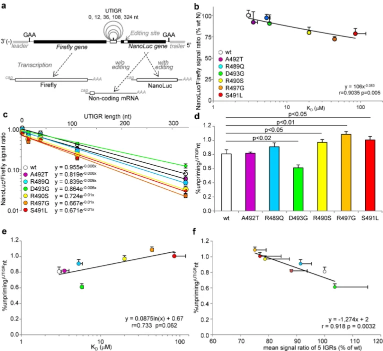
the GE is able to scan forward and backward the genome template until it recognizes the tran-scription re-initiation site GS of the next downstream gene. This search for next GS had been initially observed as measurable temporal pause in transcription [68] (see viral transcription scheme inS6 Figand [13,69] for reviews). Since the frequency of re-initiation decreases with the length of the un-transcribed IGR (UTIGR) [70,71] dual-luciferase Firefly/NanoLuc 2-gene minigenomes with elongated UTIGR based on MeV N-P IGR were also built (see schemeFig 8A) according to previous work based on the related Sendai virus that has served as the refer-ence study model forParamyxoviridae [70]. The Firefly signals specifically driven by MeV
polymerase (as obtained after subtraction of background levels observed in the presence of an inactive L protein) observed with each combination of minigenome of variable UTIGR length and N variant were of similar magnitude irrespective of the UTIGR length and of NTAIL vari-ant (S7A Fig) and did not show any correlation with the NTAIL/XD binding strength (S7B Fig). These data confirmed that the rescue of the minigenome, is neither dependent on the
sequence of the minigenome nor on the N variant. Incidentally, these experiments also allowed appreciating the reproducibility of our dual-luciferase minigenome-based experiments, as judged by comparingS5AandS7AFigs.
transcribed 3’-GAA-5’ triplet that characterizes the N-P IGR region or by P-M, M-F, F-H or H-L IGR regions, i.e. un-transcribed 3’-GAA-5’ (or 3’-GCA-5’ for H-L) triplet flanked by canonical upstream and downstream gene end and gene start sequences arbitrarily fixed to 15 nt. (b) Homogenous and comparable decrease of the efficiency of the transcription re-initiation mediated by every MeV IGR from 3’ to 5’ gene position observed with every N variant when the NanoLuc/Firefly signal ratio is normalized as a function of that observed with the first N-P IGR. MeV IGRs can be grouped into three subsets of re-initiation efficiency, high for N-P and P-M, medium for M-F and F-H and low for H-L. (c-g) Variation in the efficiency of re-initiation as a function of the 5 different MeV IGRs as determined from the NanoLuc/Firefly signal ratios (i.e. same data as in (b) but without normalization to the N-P IGR) and expressed as a function of KDof the NTAIL/XD pair. Note the progressive loss of correlation from N-P to H-L IGRs. (h) Correlation of the
mean re-initiation rate through the five MeV IGRs (estimated as mean NanoLuc/Firefly signal ratio observed over the five IGR regions) with NTAIL/XD
KD. Minigenome data are expressed as the mean +/- SD of at least 3 independent experiments, with each combination being done in triplicate. See
alsoS5 Fig.
As observed in the previous set of experiments, the NanoLuc/Firefly signal ratios obtained with the N-P minigenome (i.e. UTIGR “+0”) nicely correlate with the NTAIL/XD binding strengths (Fig 8B, compare also withFig 6Cfor data reproducibility). When N variants were tested with elongated UTIGR minigenomes, the NanoLuc/Firefly signal ratio exponentially
Fig 7. Ability of N variants to misrecognize IGR and generate read-through transcripts. Schematic structures of
2-gene (a) and 3-gene (b) N-P minigenomes. SeeFig 6Afor detailed description of 2-gene minigenome. The 3-gene minigenome comprises from its 3’-end the Firefly CDS as a first gene, a second gene encoding the C-terminal domain of Gaussia luciferase lacking a stop codon and a third gene encoding the NanoLuc CDS devoid of start codon and in frame with the upstream Gaussia CDS only after the upstream insertion of one additional G by RNA editing. Consequently the
expression of the NanoLuc luciferase (as chimeric Glu2-linker-NanoLuc protein) requires the re-initiation of the transcription at the IGR separating gene 1 and gene 2, the reading through the IGR separating gene 2 and 3 and the edition of the read-through transcript. Therefore, if the expression of NanoLuc luciferase from both 2-gene and 3-gene minigenomes relies on both the accuracy of the re-initiation of the second gene over the same N-P IGR and the editing of the transcript, the expression of the NanoLuc luciferase from the 3-gene minigenome additionally relies on the efficiency of the read-through between gene 2 and gene 3. (c) Ratios of NanoLuc signals (normalized by their upstream Firefly signal) observed with each N variant with 3-gene (numerator) and 2-gene (denominator) minigenome observed with each N variants. Data are expressed in % of the ratio observed with wt N.
declined with UTIGR elongation (Fig 8C, p<0.001 for every N variant). However the
declin-ing rate varied between N variants (compare the slopes inFig 8C). This allowed us to calculate
Fig 8. Ability of N variants to support transcription re-initiation over untranscribed IGRs (UTIGR) of variable length as determined using dual-luciferase minigenomes. (a) Principle of the dual-luciferase editing-dependent minigenome assay allowing the expression of the Firefly as the
first gene and the NanoLuc as the second gene, the translation of which relies on the addition of one non-templated G at the editing site (seeFig 6A for details). The two genes are separated by a modified N-P IGR where the polyadenylation site is ended by a canonical G and is followed by 12, 36, 108 or 324 nucleotides followed in their turn by a modified inactive polyadenylation site and the canonical GAA-5’ intergenic triplet i.e. 3’-auauuuuuuG[n]auCauuuuuuGAA-5’ as validated for SeV minigenomes [70] (b) Correlation between NanoLuc/Firefly signal ratios and NTAIL/XD KD
as observed with N-P intergenic minigenome (i.e. UTIGR n = 0). (c) NanoLuc/Firefly signal ratio observed with individual NTAILvariants as a function
of UTIGR length. The correlation is statistically significant at p<0.001 for all N variants. (d) Calculated unpriming rate per un-transcribed intergenic nucleotide from data shown in (c) and (e) their relationship with the NTAILto XD binding strength. (f) Correlation between unpriming rate per
un-transcribed intergenic nucleotide and mean re-initiation rate through the five MeV IGR regions (estimated as mean NanoLuc/Firefly signal ratio observed over the five IGR regions, as also shown inFig 6H). Minigenome data are mean values and SD from three independent experiments, each data point being made in triplicates. Statistically significant differences are as determined using the Student’s t or Spearman’ R test. See alsoS7 Fig. doi:10.1371/journal.ppat.1006058.g008
and compare the percentage of unprimingper UTIGR nt (%unpriming/UTIGRnt). The D493G variant exhibits a significantly lower %unpriming/UTIGRnt compared towt N, whereas that of
R490S, R497G and S491L variant was significantly higher (Fig 8D). Furthermore, the %unpri-ming/UTIGRnt of N variants tends to vary according to thelog of the NTAIL/XD KD, (Fig 8E, p = 0.062). Remarkably, the %unpriming/UTIGRnt and mean re-initiation rate through the five MeV IGR regions significantly correlate to each other (Fig 8F, p = 0.0032). Overall these data reveal that lowering the NTAIL/XD binding strength significantly increases the unpriming rate of MeV polymerase during transcription re-initiation and its scanning over un-transcribed genomic sequences, i.e. over each UTIGR.
Impact of N
TAILamino acid substitutions introduced into recombinant
unigene and biG-biS viruses on virus production
Since even NTAILvariants with the highest KDfor XD were able to reconstitute functional dual-luciferase minigenomes, we sought at evaluating the impact of substitutions in the viral context by expressing N variants into two types of recombinant viruses, namely unigene and biG-biS viruses. Unigene viruses possess only one copy of the N gene and thus express solely the mutated N protein. By contrast, biG-biS viruses contain a duplicated viral gene, here the N gene, one encoding thewt N protein (wt Flag-N1) and one encoding the mutated N protein
with a HA tag (HA-N2), the expression of which can be independently silenced thanks to the use of two cell lines expressing shRNA that selectively target one of the two N genes (S8 Fig)
[48]. Unigene viruses harboring NTAILvariants were all rescued. The biG-biS viruses were also all rescued in cells allowing the selective expression of thewt Flag-N1 gene copy, although the
too low virus production by the R489Q and R490S viruses prevented further analysis. Virus production by recombinant viruses at 3 d.p.i. were determined for unigene viruses in Vero cells, while that of biG-biS viruses was measured in three host cells allowing selective expres-sion of either thewt Flag-N1 gene copy, the HA-N2 gene variant, or both of the N gene copies
simultaneously (Fig 9A). Virus production was found to be very low (at least 2 log reduction with respect to thewt counterpart) in the case of unigene and biG-biS S491L viruses. Note that
the possibility that the observed differences in virus production of unigene viruses could be ascribed to a defect in N variant expression (S1andS9AFigs) or to a significant
contamina-tion by defective interfering (DI) mini-replicons was checked (S9B–S9D Fig) and ruled out.
When plotted against the NTAIL/XD binding strength as determined by glu-PCA, the virus production of unigene NTAILvariants does not significantly correlate with binding strength (Fig 9B). However, the virus titer of biG-biS viruses under the selective expression of HA-N2 variant and under the combined expression of both N copies were found to correlate with NTAIL/XD binding strength (p = 0.04 and p = 0.008, respectively) (Fig 9C and 9D), while no such a correlation was found upon selective expression of thewt Flag-N1 copy as expected
(Fig 9E). We noticed that the coexpression of Nwt with D493G variant appears deleterious for
virus production (Fig 9D). However, in a minigenome assay such a mixture of N was as effi-cient as Nwt alone (S10 Fig), thus ruling out the possibility that NTAILheterogeneity could directly impact the polymerase activity. Overall these data indicate that the NTAIL/XD binding strength may control the virus production to some extent.
Viral transcription gradient correlates with XD-binding affinity of Box2
variants
We then took advantage of unigene viruses expressing the single-site Box2 variants to deter-mine which activity of the viral polymerase could be affected by a change in the NTAIL/XD binding affinity. Vero cells were infected withwt, R489G, R490S, A492T, D493G and R497G
Fig 9. Virus production at 3 d.p.i. and in the context of unigene and biG-biS viruses and relationships with NTAIL/XD interaction strength.
(a) Infectious virus production after infection with recombinant virus coding for NTAILvariant (unigene virus) or biG-biS virus bearing two copies of
the N gene, one coding for wt Flag-N1and either wt or variant HA-N2in conditions allowing selective expression of the wt Flag-N1copy, wt or variant
HA-N2copy or both of them (data expressed in % of wt, mean±SD). Although all biG-biS viruses were successfully rescued, R489Q, R490S and
S491L could not be further studied because of a too low virus production. (b-e) Virus titers expressed in % of wt virus (for unigene viruses) or wt Flag-N1/wt HA-N2(for biG-biS viruses) were plotted (mean±SD) against NTAIL/XD binding strength as determined by glu-PCA (mean±SD). Panel b shows virus production from unigene N variants. Virus production from biG-biS viruses with (c) selective expression of variant HA-N2copy, or (d)
simultaneous expression of wt Flag-N1and variant HA-N2copies, or (e) selective expression of the wt Flag-N1copy. Same color codes as inFig 1B,
unigene viruses. Note that the S491L variant was not investigated since it could not be further amplified to reach a workable titer. RNA synthesis parameters reflecting primary transcription (i.e. mostly, if not solely, transcription, mediated by the active polymerases brought by infect-ing virions), secondary transcription and replication were determined by quantification of (+) and (-) RNA accumulation at different times post-infection as previously reported [48,65]. When RNA synthesis parameters were plotted along with NTAIL/XD KD, it appeared that both (+) RNA transcript accumulation rate and ratios between P (or F) and N transcripts could be roughly predicted from the interaction strength between the NTAILvariant and XD as mea-sured by either method (Fig 10). The correlations were statistically significant between the accumulation rate of P (+) transcripts and NTAIL/XD KD(Fig 10A) and between the F/N tran-script ratios measured at 24 h.p.i. and the KD(Fig 10B). In further support of the coherence of the results, a good correlation was found between the accumulated levels of N and P (+) RNAs during primary transcription and at 24 h.p.i. (S11A and S11B Fig), and between both N (+) and P (+) RNA transcripts and (-) genomic RNA (S11C and S11D Fig).
When the F/N mRNA ratios at 24 h.p.i. observed with unigene viruses were plotted against the calculated mean re-initiation rate of the 5 IGRs and the %unpriming/UTIGRnt a significant positive and a negative correlation were found, respectively (Fig 11). Altogether, these data
support that the NTAIL/XD binding strength controls, at least in part, the steepness of the viral transcription gradient.
Discussion
By combiningin vitro biophysical and biochemical studies, in silico analyses (i.e. MD
simula-tions) andin cellula polymerase functional investigations using recombinant viruses and
dual-luciferase editing-dependent minigenome assays, we deciphered key molecular parameters that govern the NTAIL/XD interaction. Specifically, we uncovered a correlation between inter-action strength and efficiency of transcription re-initiation at intergenic regions.
Fig 10. Relationship of transcripts accumulation rates and transcripts ratios with NTAIL/XD binding affinity. Relationship of (a) accumulation
rate of N (+) or P (+) RNA during primary transcription with NTAIL/XD binding affinity and of (b) P/N or F/N (+) RNA ratios at either 0–8 h.p.i. or at 24 h.
p.i. with NTAIL/XD binding affinity. Data are expressed as % of wt. See alsoS11 Fig.
Impact of preconfiguration of the
α
-MoRE on the interaction with XD
For most of the NTAILvariants the observed variations in binding affinities cannot be ascribed to differences in the extent ofα-helical sampling of the free form of the α-MoRE, nor to differ-ences in the ability of the latter to undergo inducedα-helical folding. However, the R489Q substitution represents an exception in this respect: indeed, it has a reduced extent ofα-helicity and a slightly increased KDtowards XD. The reducedα-helical content of this variant is in line with secondary structure predictions, as obtained using the Psipred server (http://bioinf.cs.ucl. ac.uk/psipred/) [72], that predicts a slightly lower helical propensity. Whether the experimen-tally observed reduction in affinity towards XD arises from this lower helicity or from other attributes, including charge-related ones, remains to be established. This variant also displays a reduced accumulation rate of primary transcripts. The subtle molecular mechanisms underly-ing the peculiar behavior of this variant remain however to be elucidated.
N
TAILbinding to XD involves a hydrogen bonding network including three
α
-MoRE and three XD residues
The complex hydrogen bonding revealed by MD simulations of NTAIL/XD complexes allows the drops in binding affinities experimentally observed for the S491L, R497G and R490S vari-ants to be rationalized. Interestingly, these substitutions, which have the most dramatic effects in terms of binding affinities, are also the ones that have the strongest effect on virus replica-tion, with the S491L substitution being very poorly tolerated even in biG-biS viruses. The poor ability of the low-affinity S491L variant in mediating efficient virus replication is reminiscent of the comparable deleterious effect of the F497D XD substitution [48] and of the detrimental effect of the deletion of the NTAILregion encompassing theα-MoRE [49].
The lower the N
TAIL/XD binding strength the lower the efficiency of
transcription re-initiation at intergenic regions
We provide here compelling evidence indicating that the strength of the NTAIL/XD interaction controls, at least in part, the ability of the P+L polymerase complex to re-initiate at IGRs: data obtained using our highly sensitive and reproducible dual-luciferase minigenome assay reveal a significant correlation between the NTAIL/XD binding strength and the efficiency of the
Fig 11. Relationship of F/N mRNA ratio at 24 h.p.i. from unigene viruses with the mean re-initiation rate over the five IGRs (a) and the % unpriming/UTIGRnt (b) (from data shown in Figs6,7and9).
transcription re-initiation. Since our minigenome assays rely on the edition of the second reporter gene, we cannot formally exclude that the editing may be also impacted by the NTAIL/ XD binding strength. However, the calculated %unpriming/UTIGRnt only depends on the decrease of the NanoLuc/Firefly signals ratios with the length of the UTIGR. The observed effect is therefore independent of any potential effect on the edition (i.e. if N mutations only had an effect on editing, then this effect should be the same irrespective of the IGR under study and of its length, which is not the phenotype we observed). Moreover, the correlation in the viral context between the P/N and F/N mRNA ratio and the KD, supports a role for the XD/NTAIL interaction strength in the re-initiation at IGRs.
A N protein truncated of its last 86 C-terminal amino acids, i.e. truncated of most of NTAIL including the XD binding site, had been shown to be active in transcription and replication both in a minigenome assay and when introduced into a recombinant virus [49]. We con-firmed that the N1-439 truncated protein is as good as, if not better than, thewt N in
transcrib-ing the Firefly gene from our N-P 2-gene minigenome construct (S12A Fig). However, its ability to support transcription re-initiation over the N-P junction was significantly reduced, with the extent of reduction being comparable with that observed with the low affinity R497G variant (S12B Fig, UTIGR 0 nt), thus confirming the role of NTAIL/XD interaction in tran-scription re-initiation. This low efficiency of trantran-scription re-initiation may explain the extreme growth defect of the recombinant virus bearing the truncated N until reversion to a
wt N [49].
Assuming a very slow degradation of viral mRNA [65,66,73], the transcripts accumulation rate in cells infected with unigene viruses reflects the RNA synthesis rate by the polymerase, the number of active polymerases (and their recruitment onto the nucleocapsid template), and the number of polymerases that are recruited per time unit on a given gene. For the same rea-son, the transcript ratios between the different genes are likely mostly governed by the effi-ciency with which the polymerase re-initiates the transcription at each IGR. Assuming this being a conserved feature for every N variant, we can reasonably interpret the inverse correla-tion we observed between multiple transcript ratios and KDas reflecting a direct control of the NTAIL/XD binding strength on the efficiency of the re-initiation at each IGR. A lower binding strength leads to lower levels of downstream transcripts. After completion of the polyadenyla-tion of the messenger encoded by the upstream gene, the polymerase may remain firmly in contact with its genomic RNA template embedded into the nucleocapsid only if maintained by the anchoring of its P subunit via a dynamic binding of its X domain to the TAIL domain of N subunits located at the IGR (Fig 12). Therefore, a decrease in the XD/NTAILaffinity may favour the unpriming of the polymerase. Whether unprimed polymerases can detach from the nucle-ocapsid or stay on the template and move forward to the end of the nuclenucle-ocapsid remain to be established. Hence, XD to NTAILanchoring would tightly control the re-initiation level of the RNA synthesis by the polymerase in the transcription mode, thus determining the steepness of the transcription gradient (Fig 12, see alsoS6B Fig).
The N
TAIL/XD affinity affects the processivity of the polymerase both on
the “transcription” and on the “scanning” modes
What could be the functional significance of the relationship between the accumulation rate of primary N and P transcripts and the XD/NTAILbinding strength? As speculated, the dynamics of XD/NTAILbinding and release may also affect the polymerase processivity on the nucleocap-sid [48]. The XD/NTAILinteraction may act as a brake and slows down the polymerase: the weaker is the interaction, the weaker is the brake. Also, because of the efficient recycling of the polymerases on the promoter [65], if, in the absence of transcription re-initiation, the
polymerase detaches from the RNA template, a steeper gradient would release more polymer-ases available for transcription of the first genes. With weaker NTAIL/XD interactions, the viral production by unigene viruses tends to be negatively affected although the correlation was not statistically significant likely because of the small number of available virus variants and the too high variability of the result due to the multiple intervening parameters (seeS6A Figand the complete scheme of virus replication dynamics in [65]). However with biG-biS viruses, we did observe a significant correlation between virus production and NTAIL/XD binding strength in conditions where the N variant was selectively expressed. This significance may reflect both the higher number of available virus variants and/or the higher impact of the modulation of the transcription re-initiation process in viruses possessing an additional transcription unit (i.e. where the polymerase has to go through one additional IGR). The similar correlation observed upon the co-expression of bothwt Flag-N1 and variant HA-N2 copies may indicate
similar impact on transcription re-initiation because of the tetrameric valence of the P anchor-ing on (contiguous?) heterogeneous NTAILappendages. Alternatively, it is possible that the het-erogeneity of NTAILwithin a given nucleocapsid template may have a negative impact on other mechanisms such as nucleocapsids packaging into particles since NTAILalso recruits the M protein [74], a key virion assembly factor [75]. The discrepancy we observed between virus production from biG-biS viruses and minigenome data with mixed NTAILsargues for this later hypothesis.
Using minigenomes with elongated UTIGR, we were able to measure the unpriming rate of the polymerase in the “scanning mode” and we show that a decrease in the NTAIL/XD affinity induces an increase of the unpriming rate. In this situation, without the stabilization and the
Fig 12. Model of transcription re-initiation. (1) The polymerase complex, composed of L and P proteins,
transcribes the genome. (2) After addition of the poly(A) tail and release of the mRNA, the polymerase complex may re-initiate transcription and transcribe the next gene (a) or stop transcribing (b). Whether the polymerase complex detaches from the genome template (b.i) or travels on it until reaching the 5’ end of the genome (b.ii) remains to be determined. The higher is the KD, the less efficient is the re-initiation of transcription, thus leading to a steeper
mRNA gradient.
active motion of the polymerase due to the RNA synthesis, the role of the NTAIL/XD interac-tion in maintaining the polymerase on the nucleocapsid may overcome the “brake” effect. Alternatively, as suggested by Krummet al [49], the NTAILmay need to be rearranged by P to allow an efficient RNA synthesis. In this case, a too low NTAIL/XD affinity may weaken the effi-ciency of P in rearranging NTAILand would favor the unpriming of the polymerases. The fact that the N1-439 variant, that lacks most of NTAIL, has the lowest unpriming rate on UTIGR supports this second hypothesis (0.6 vs 0.81%unpriming/UTIGRnt for N1-439 andwt N
respec-tively) (S12C Fig).
A mechanism among others to control both scanning and re-initiation by
MeV polymerase
In conclusion, the XD/NTAILinteraction may play a critical role in the polymerase processivity, in maintaining the polymerase anchored to the nucleocapsid during its scanning upon cross-ing the intergenic regions, and/or in the transcription re-initiation at each intergenic region. Since both increasing [48] or decreasing (this study) the XD/NTAILaffinity negatively affect the viral growth, the wild type XD/NTAILbinding strength seems to have been selected to mediate an optimal equilibrium between polymerase recruitment, polymerase processivity and transcription re-initiation efficiency. A corollary of this is that substitutions that strongly affect affinity towards XD are poorly tolerated. Consistent with this, the substitutions with the most dramatic impact herein investigated (i.e. R490S, S491L and R497G) do not naturally occur in any of the 1,218 non-redundant MeV sequences, while those that have a less drastic impact (i.e. R489Q, A492T and D493G) are found in circulating measles strains [47]. Interest-ingly, in the case of Ebola virus (EBOV), an additional protein, i.e. VP30, serves as an anti-ter-minator transcription factor, and mutations that either decrease or increase the binding affinity between N and VP30, decrease RNA synthesis [76] thus arguing for a similarly tightly regulated interaction. According to our work, the NTAILto XD binding strength tightly con-trols the transcription gradient. However, this does not rule out the possibility that other mechanisms may be at work in controlling the steepness of the gradient. Indeed, in the brain of three patients suffering from subacute sclerosis encephalitis (SSPE) or measles inclusion bodies encephalitis (MIBE) the transcription gradient was found to be steeper than the one measured inin vitro infected cells [77] although the amino acid sequences of NTAILand XD were found to be unvaried [78]. Furthermore, in the absence of the C protein, a steeper tran-scription gradient is also observed [79]. These two lines of evidence advocate for a multi-parametric control of the transcription gradient.
The conserved bipartite P to N interaction of Paramyxoviridae members
is also shared by other families of the Mononegavirales order
The major role of the N binding site on the C-terminus of P has been postulated to mediate L anchoring to the nucleocapsid without understanding the implication of such anchoring on the polymerase and/or on the nucleocapsid dynamics. The need for an optimized interaction between the P and N proteins might be one of the major evolution constraints to which the polymerase machinery of MeV, and possibly of paramyxoviruses in general, is subjected. Our findings raise also the question as to whether binding of the C-terminus of P to the globular moiety of N, as observed in otherMononegavirales members, needs to be similarly controlled
reflecting a similar functional role.
The bipartite nature of P to N binding (see schemeFig 1A) is remarkably conserved throughout theMononegavirales order [80]. Anα-MoRE located at N-terminus of P binds to the C-terminal globular domain of the NCOREto form the so-called N0P complex that is used

![Fig 2. Secondary structure content of NTAIL variants (a) Crystal structure of the chimera between XD (orange) and the α-MoRE of N TAIL (red) (PDB code 1T6O) [44]](https://thumb-eu.123doks.com/thumbv2/123doknet/14482411.524370/7.918.100.866.111.855/secondary-structure-content-ntail-variants-crystal-structure-chimera.webp)