CD133 positive progenitor endothelial cell lines from human cord blood
Maria Paprocka a,b, Agnieszka Krawczenkoa, Danuta Dusa, Aneta Kantorb, Aude Carreaub, Catherine Grillonb and Claudine Kiedab
aLudwik Hirszfeld Institute of Immunology and Experimental Therapy, PAN, 53114 Wrocław, Poland; bCentre de Biophysique Moléculaire, CNRS, 45071 Orléans, Cedex2, France.
Running head: human progenitor endothelial cell lines
Author contribution: M. P.: conception and design, collection of data, manuscript writing; A.K.: collection of data, manuscript writing; D. D.: conception and design, final approval of manuscript; A. K.: collection of data; A. S.: collection of data; C. G.: conception and design, collection of data, manuscript writing; C. K.: conception and design, manuscript writing, final approval of manuscript.
Correspondence to: Catherine Grillon, Centre de Biophysique Moléculaire, CNRS, 45071 Orléans, Cedex2, France; phone: 00 33 238 25 78 04 ; FAX: 00 33 238 25 54 59, e- mail : [email protected]; web site http://www.cnrs-orleans.fr.
Disclaimers: the Authors declare no potential conflicts of interest.
Acknowledgements: this work was supported by the Polish French GDRE 182 from CNRS, the Jerôme Lejeune Foundation, the Canceropôle Grand Ouest.
Authors want to thank Professor Jerzy Heimrath for the kind providing of biological samples, Michèle Mitterrand for her skilful technical help.
Abstract
Endothelial progenitor cells (EPCs) modulate postnatal vascularization and contribute to vessel regeneration in adults. Stem cells and progenitor cells were found in umbilical cord blood, bone marrow and mobilized peripheral blood cells, from where they were isolated and cultured. However, the yield of progenitor cells is usually not sufficient for clinical application and the quality of progenitor cells varies.
The aim of the study was the immortalization of early progenitor cells with high proliferative potential, capable to differentiate to EPCs and, further, toward endothelial cells. Two cell lines, namely HEPC-CB.1 and HEPC-CB.2 (human endothelial progenitor cells – cord blood) were isolated. As assessed by specific antibody labeling and flow cytometric analysis, they express a panel of stem cell markers: CD133, CD13, CD271, CD90 and also endothelial cell markers: CD202b, CD309 (VEGFR2), CD146, CD105 and CD143 but they do not present markers of finally differentiated endothelial cells: CD31, vWf, nor CD45 which is a specific hematopoietic cell marker. Using the multiplex Cytometric Bead Assay, the simultaneous production of proangiogenic cytokines IL8, angiogenin and VEGF was demonstrated in normoxia and was shown to be increased by hypoxia. Both cell lines, similarly as mature endothelial cells, underwent in vitro pre-angiogenic process, formed pseudovessel structures and present an accelerated angiogenesis in hypoxic conditions. To date, these are the first CD133 positive established cell lines from human cord blood cells.
Introduction
Endothelial progenitor cells (EPCs) may be found in many tissues as well as in the circulation (1). These cells are endowed with the ability to actively find and adhere to damaged endothelial surface and differentiate into endothelial cells to reconstitute vascular integrity (2-4). This process is especially important during the repair of damaged myocardial tissue after infarction (5). In animal models of ischemia, it has been proven that EPCs incorporated into sites of active angiogenesis (6). EPCs participate also to pathological processes like tumor growth or retinopathy. EPCs were discovered 10 years ago but their identification, characterization and actual differentiation potential are still controversial. EPCs identification is complex because they represent a very rare population both in bone marrow and circulation. The main problem for EPCs identification arises from the absence of specific markers and the lack of strict criteria to define their functions.
Putative endothelial cell progenitors i.e. angioblasts were first isolated from human peripheral blood by magnetic bead selectionon the basis of cell surface expression of CD34 antigen (7). In vitro, thesecells were demonstrated to differentiate into endothelial cells (ECs) (8). Presently, a combination of CD133 and/or CD34 markers and sometimes specific ECs markers like VEGFR2 (CD309), VE-Cadherin (CD144) and Mel-CAM (CD146) are used to separate stem and progenitor cells (9,10). In a recent review (11), Fadini and Avogaro define endothelial progenitor cells as cells expressing at least one immaturity/stem cell antigen (CD133 or CD34) and at least one endothelial antigen (VEGFR2, CD31).
EPCs were then propagated in various conditions and characterized by in vitro and/or in vivo methods which led to conflicting results. Additionally, in each individual EPCs donor, different stages of their differentiation are supposed to be present and, furthermore, EPC phenotype may differ according to the nature of the tissue they originate from (12).
populations and to discriminate the putative EPCs from hematopoietic stem cells (HSCs) (12), (13). Keeping in mind that both endothelial and hematopoietic lineages originate from a common progenitor – hemangioblast – the closer the progenitors are, the higher are their similarities.
As a marker helping to discriminate hematopoietic and endothelial lineages, the common leukocyte antigen CD45 was suggested as it is not expressed on embryonic hemangioblast and is acquired during differentiation by cells of the hematopoietic lineage only (14). It was also demonstrated that in adults, CD34+ CD45- cells only were able to differentiate towards EPCs (15).
Additional functional assays may facilitate the discrimination of both lineages. In vitro tube formation assay seems to mimic endothelial cell abilities to migrate, align, branch and form polygonal network structures. All these functions are not achieved by CD45 positive cells (15, 16).
In this work we have isolated and successfully immortalized human endothelial progenitor cells from the cord blood. Two cell lines were established and studied for their properties and potentials of providing a cell model of endothelial precursors in terms of phenotype and angiogenic properties.
Materials and Methods
Human umbilical cord blood
Cord blood was collected from four normal, full-term deliveries at the Chair of Obstetrics and Gynecology of Wrocław Medical University. The umbilical vein was punctured with a 17-gauge needle attached to a closed system collection bag containing citrate phosphate dextrose solution as an anticoagulant blood packs (MACO Pharma, Wrocław, Poland). The harvested volume was an average of 50-110 ml from a single placenta. Written informed consent was obtained from all mothers before delivery. Protocols for sampling human umbilical cord blood were approved by the Commission of Bioethics at Wrocław Medical University.
Isolation of mononuclear cells
Umbilical cord blood mononuclear cells were isolated by density-gradient centrifugation using Lymphoflot (1.077 g/ml, Biotest, Dreieich, Germany). The mononuclear cell layer was collected; cells were washed twice with 1 mM EDTA in PBS and stored frozen in liquid nitrogen until use.
Mononuclear cell culture and immortalization
Isolated mononuclear cells (1 x 10⁶ ml) were cultured on human fibronectin-coated tissue culture dishes (Biocoat; BD Biosciences, Grenoble, France) at 37oC in 95% air/5% CO2 atmosphere. Cells were cultured in EBM-2 medium (Clonetics, Lonza, Saint Beauzire, France), supplemented with 10% Fetal Bovine Serum (Hyclon, Longan, Utah, USA) and rhSCF (100 ng/ml, from Sigma Aldrich, Saint Quentin Fallavier, France ), rhSCGF (100 ng/ml from Chemicon Int, Temacula, CA), rhFlt-3 ligand (50 ng/ml) and rhVEGF (50 ng/ml) purchased from R&D systems, Lille, France.
After 3 days of culture, the medium was supplemented with cell free supernatant (50% vol/vol) of retroviral TE FLY GA hTERT cell line obtained in our laboratory (17). These cells produce retrovirus containing human telomerase reverse transcriptase gene. 8μg/ml of
polybrene (Sigma) was added to the infection supernatant to increase the transduction efficiency. Transduction procedure was repeated 3 times every 24 hr. After 3 weeks of culture, 2 clones of proliferating cells were replicated. Cells were kept for one month in culture in EBM-2 medium, supplemented with 10% FBS and growth factors. The medium was then gradually changed to OptiMEM (Invitrogen, Fischer Bioblock, Illkirch, France) supplemented with 3% of FBS. The two clones obtained were named CB.1 and HEPC-CB.2. Pictures of growing cells were taken using Axiovert S Zeiss microscope (Jena, Germany) equipped with a digital camera Fine Pix 5602 (Fuji Film).
For hypoxia treatments, cells were placed in a humidified atmosphere containing 1% of oxygen. This oxygen pressure was obtained by introducing 95% N2/5% CO2 gas mixture (Air Liquide, Paris, France) in an automated PROOX in vitro chamber (C-174; BioSpherix, Redfield, NY) under the control of a PROOX sensor-model 110 (BioSpherix).
Ability of HEPC-CB.1 and HEPC-CB.2 cell lines to grow and differentiate toward hematopoietic cells in semisolid medium
Ability of HEPC-CB.1 and HEPC-CB.2 cells to grow and differentiate toward hematopoietic cells was evaluated using MethoCult GF H4434 (StemCell Technologies Inc. Vancouver, Canada), containing the following recombinant cytokines: rh SCF (50ng/mL), rh GM-CSF (10ng/mL), rh IL-3 (10ng/mL) and rh erythropoietin (3units/mL).
103 cells of both tested cell lines were suspended in 1 mL of semisolid MethoCult medium and transferred to 24 well plate wells. After 7 days of culture, pictures of growing cell clusters were taken using Axiovert S Zeiss microscope (Jena, Germany) equipped with a digital camera Fine Pix 5602 (Fuji Film).
Immunostaining of HEPC-CB.1 and HEPC-CB.2 cell lines
Phenotype of cultured cells was analyzed with the use of non conjugated polyclonal rabbit anti-von Willebrand factor antibodies (catalog no. F3520; Sigma) and the following monoclonal mouse antibodies: PE-conjugated anti-CD133 (catalog no.130-090-853) and FITC-conjugated anti-CD271 (catalog no.130-091-917) from Miltenyi Biotec (Paris, France);
PE-conjugated anti-CD105 (catalog no. FAB10971P), anti-CD143 (catalog no. FAB929P), anti-CD146 (catalog no. FAB932P), anti-CXCR4 (catalog no. FAB173P), anti-hVEGFR2 (catalog no. FAB357P) and non-conjugated anti-CD202 (catalog no. AF313) from R&D Systems; PE-conjugated anti-CD13 (catalog no. 347837), anti-CD34 (catalog no. 345802), anti-CD38 (catalog no. 345806), anti-CD54 (catalog no. 347977), anti-CD90 (catalog no. 555596), anti CD117 (catalog no. 332785) from BD Biosciences; FITC-conjugated anti-CD45 (catalog no. 345808), anti-CD44 (catalog no. 347943) and non-conjugated anti-CD15s (catalog no. 551344) from BD Biosciences; FITC-conjugated anti-CD31 (catalog no. F8402, Sigma). All directly labeled antibodies were used at the concentration of 1-5μL of antibody for 1 x 10⁵ cells in 50μL of PBS supplemented with 1% FBS, as suggested by the manufacturer, and incubated for 30 min at 4oC. After incubation cells were carefully washed. IgG isotype matched, PE or FITC labeled immunoglobulins were used as controls.
Similarly three non-labeled antibodies were used at the concentrations of 0.1μL (anti-von Willebrand factor antibody) and 1μL (anti-CD15s and anti-CD202) for 1 x 10⁵ cells in 50μL of PBS supplemented with 1% FBS, as suggested by the manufacturer, and incubated for 30 min at 4oC. After incubation, cells were carefully washed and second anti-mouse or anti-rabbit FITC labeled antibodies were applied for subsequent 30 min at 4oC. Before FACS analysis cells were washed with PBS. Isotype-matched mouse or rabbit immunoglobulins were used as controls.
Before CD143 and von Willebrand factor labeling, cells were fixed and permeabilized with 2% paraformaldehyde and 0.2% saponin solution in PBS for 10 min to allow intracellular labeling.
On the other hand, cells were incubated with 10 μg/mL Dil-Ac-LDL (Molecular Probes, Cergy Pontoise, France) for 4h at 37°C, then detached, washed and incubated with 10 μg/mL FITC-UEA-1 lectin (Sigma) for 30 min at 4°C. A primary culture of HUVEC was used as positive control.
After checking fluorescent labeling under the microscope, cells were analyzed by flow cytometry using FACSCalibur (Becton Dickinson, CA, USA) equipped with a 488 nm laser and an filter for FITC analysis (530 BP). As single labeling were performed, no compensation setting was required. Data were recorded for 5000-10000 events using CellQuest version 3.3 software (Becton Dickinson), analyzed on ungated population (except for debris) and presented without any transformation as histograms using WINMDI 2.7 software. Komogolov-Smirnov statistics, included in CellQuest software were applied to compare histogram plots for isotypic control versus specific antibody staining. The D values coming from the comparison vary between 0 and 1. Values from 0.0 to 0.2 show a non-significant labeling, values from 0.2 to 0.3 mean a weak expression of the marker and above 0.3 a high expression.
Secretion of cytokines by HEPC-CB.1 and HEPC-CB.2 cell lines
Secretion of cytokines (IP10, IL6, IL8, VEGF, angiogenin) was evaluated by using the Cytometric Bead Assay from Becton Dickinson Biosciences. Supernatants from HEPC-CB.1 and HEPC-CB.2 cell lines were collected after culture for 16h either in normoxia (20% O2) or in hypoxia (1% O2) and kept frozen at -80°C before use. Experiments were performed
according to the manufacturer’s protocol. Flow cytometric analysis of bead labeling was performed using LSR (Becton Dickinson) and results were analyzed using FCAP array software (Becton Dickinson).
Migration assay
The migration assay with HEPC.CB1 and HEPC.CB2 cells was done using polycarbonate Nunc 10 mm Tissue Culture Inserts with 8.0 pore diameter. Tested cells were gently detached, washed and resuspended in OptiMEM medium supplemented with 1% of FCS. Chemotaxis chamber was prepared by adding 100, 10 ng/ml or no VEGF to the lower compartment, containing 1 ml of OptiMEM medium supplemented with 1% of FCS, and by adding 200 l of the same medium containing 5x10 5 cells to the upper compartment. The number of cells
capable to pass through filters and attached to the bottom of the well was evaluated under the microscope after 12 h.
Formation of pseudovessels
Matrigel™matrix (BD Biosciences) was diluted ½ in OptiMEM basal medium at 4°C, distributed in 96 well microplates in a volume of 50 l and allowed to polymerize at 37°C for 30 min. Cells (1.2 x 10⁴) of HSkMEC (human skin microvascular endothelial cells), HEPC-CB.1 and HEPC-CB.2 lines were seeded on MatrigelTM-coated microplates in a volume of 100 l and cultures were performed either in normoxic (20% O2) or hypoxic (1% O2)
conditions. The pictures were taken with the help of an inverted microscope (Axiovert 200M; Zeiss, Le Pecq, France) equipped with an Axiocam high resolution numeric camera linked to a computer driving the acquisition software Axiovision (Zeiss). The direct real-time
visualisation of the formation of pseudo-vessels was monitored during 24 h. Angiogenesis was quantified by the determination of pseudovessel number, longer than 50 micrometers, by the measurement of pseudovessel mean length using Axiovision software and by the
Results
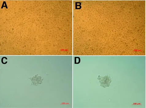
Human mononuclear cord blood cells were transduced with a retrovirus containing the gene of human telomerase reverse transcriptase. Starting from four different samples of cord blood mononuclear cells, only two clones from one sample were obtained and named HEPC-CB.1 and HEPC-CB.2. Morphology of both cell lines is presented in Figure 1A and 1B. Both cell lines were evaluated for their abilities to differentiate toward hematopoietic cells. Both appeared to be able to grow in semisolid medium but not to differentiate. After 7-9 days in culture, the cells formed compact, multicellular spheroids, as presented in Figure 1C and 1D. They displayed no sign of differentiation towards erythrocytes, granulocytes or monocytes: no “starburst” pattern characteristic for CFU-GM, capable to differentiate into granulocytes and macrophages; no large, mixt colonies, dense in the center but with loose, individual cells at the periphery and associated with dark brown colonies originated from CFU-GEMM (colony forming unit-granulocyte, erythrocyte, macrophage megakaryocyte).
Immunophenotype of HEPC-CB.1 and HEPC-CB.2 cell lines
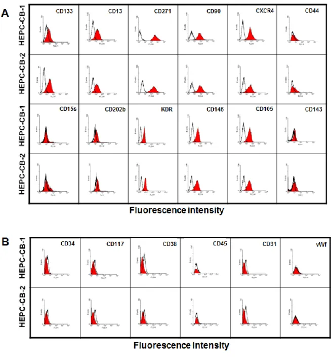
Both cell lines were first analyzed for the expression of stem cell/progenitor cell markers and for the expression of endothelial cell markers, including selected growth factor receptors and adhesion molecules (Figure 2A, Table I). Both cell lines expressed CD133 which is known to be a general marker of stem cells. They expressed also CD13 (marker of hematopoietic stem cells), CD271 (marker of non hematopoietic stem cells) and CD90 (marker of mesenchymal stem cells). Both cell lines were positive for the expression of CXCR4 and weakly positive for CD44 and CD15s, which may be expressed both on stem cells and endothelial cells. They were weakly positive for UEA-1 and Dil-Ac-LDL, markers of progenitors and differentiated endothelial cells. Additionally, they expressed endothelial cell markers: CD202b, VEGFR2, CD146, CD105 and, weakly, CD143. Markers which were not expressed by the cell lines were gathered in Figure 2B. Both CB.1 and HEPC-CB.2 lines were negative for CD34, CD117, CD38 and CD45 which suggests these cells were
not from hematopoietic origin. They were also negative for CD31 and vWf, markers of differentiated endothelial cells.
Cytokine secretion by HEPC-CB.1 and HEPC-CB.2 cell lines
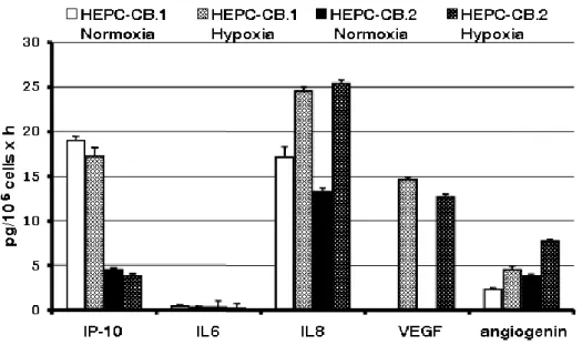
The secretion of several cytokines by HEPC-CB.1 and HEPC-CB.2 cells growing in normoxic and hypoxic conditions was evaluated. The following molecules were assessed: IP-10 as antiangiogenic factor, IL-8, VEGF and angiogenin as proangiogenic factors, and IL-6 as an irrelevant cytokine. Figure 3 shows that the alteration of partial oxygen pressure influenced the production of proangiogenic cytokines to variable extents by the two cell lines. Indeed, the production of proangiogenic cytokines IL8 and angiogenin was substantially augmented in hypoxia. Both cell lines did not produce VEGF in normoxia but the expression of this proangiogenic factor was induced by hypoxia in both cell lines.
IP10, which is an antiangiogenic cytokine, was produced at much higher levels by HEPC-CB.1 compared to HEPC-CB.2 and was not changed by hypoxia.
Migration ability of HEPC-CB.1 and HEPC-CB.2 cell lines
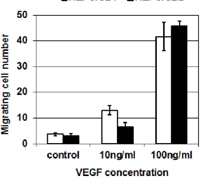
As HEPC cells do not produce VEGF in normoxia but only in hypoxia, the ability of cell lines to migrate towards VEGF as chemotactic factor agent was assessed. Figure 4 shows that, in the absence of VEGF, few cells are able to cross the filter, whatever the cell line used. This migration is increased in the presence of VEGF, by a factor 2 to 3 with 10 ng/mL, and by a factor 10 to 15 with 100 ng/mL VEGF. The whole activity of endogenous VEGF production in hypoxia was also assessed in an in vitro angiogenesis model.
Angiogenic properties of HEPC-CB.1 and HEPC-CB.2 cell lines
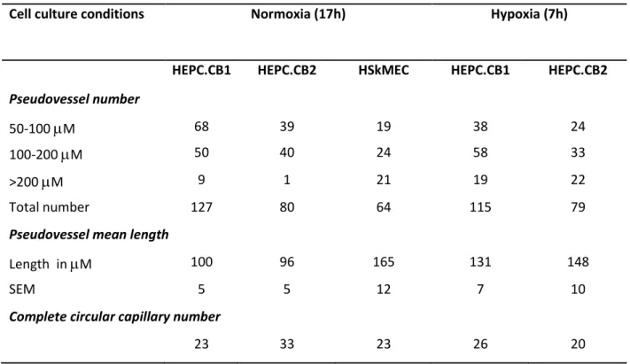
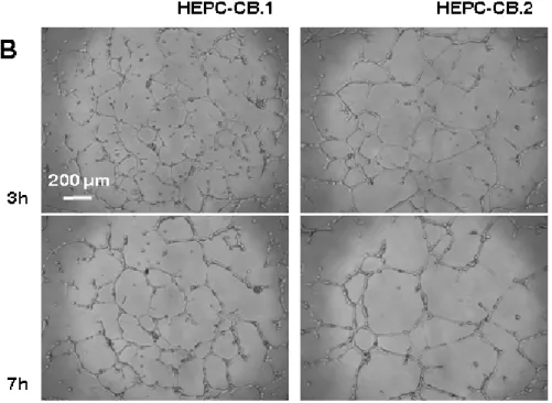
Both cell lines were assessed for their angiogenic potential on Matrigel and compared with the differentiated human skin-derived microvascular endothelial cell line, HSkMEC, which is able to form pseudovessels (18, 19). Figure 5A shows that CB.1 and HEPC-CB.2 cell lines were both able to start the angiogenic process in a comparable way, similarly to the reference HSkMEC. It means that cells are connected together to form a network. The
HEPC-CB.2 cell lines and then stopped, while it was completed by HSkMECs in 17 h. After 7 h HEPC cells stayed at the same place and continued to proliferate. Pseudovessel mean length with HEPC cell lines was lower than for HSkMEC (Table II) and the number of short vessels was higher, confirming that angiogenesis was undertaken but not achieved.
Hypoxia is the microenvironmental condition which turns on angiogenesis in vivo. It accelerated very efficiently the process of tube formation for HEPC-CB.2 cells, as it is shown in Figure 5B. This process was also accelerated for the HEPC-CB.1 line although it remained slower than in the case of HEPC-CB.2 hypoxia-dependent tube formation. Besides tube formation acceleration, hypoxia caused a more complete differentiation towards vessel-like structure that was not reached in normoxia by any of the HEPC-CB.1 and HEPC-CB.2 cell lines. Pseudovessel mean length was higher in hypoxia than in normoxia. At the same time, the number of pseudovessels > 200 m increased strongly (Table II).
Discussion
The aim of our study was the immortalization of early stem/progenitor cells with high proliferative and differentiation potentials. For this purpose human cord blood mononuclear cells were selected and transduced with the use of TE FLY GA hTERT retrovirus containing human telomerase reverse transcriptase gene. Because the transduction of resting stem and progenitor cells is usually non effective, cord blood mononuclear cells were first cultured for 3 days with a mixture of growth factors to augment their proliferation and viability. Four cord blood samples were treated in the same way but only in one case two cell clones appeared and two cell lines named HEPC-CB.1 and HEPC-CB.2 were established.
The cell lines obtained expressed CD133 and CD271, markers specific for early stem/progenitor cells. To our knowledge these two cell lines are the first CD133 positive cell lines obtained from human cord blood cells. Interestingly enough, despite the positive reaction with anti CD133 antibody these cells were negative for CD34 molecule. Other CD133+ CD34- endothelial progenitor cells have previously been isolated and characterized from blood (20) and bone marrow (21). These cells were shown to acquire the CD34 marker upon culture or differentiation in the presence of VEGF, suggesting that they are immature endothelial progenitor cells and are precursors of the CD34+ CD133+ EPC population. HEPC-CB.1 and HEPC-CB.2 cell lines expressed also CD13, activated by angiogenic signals molecule essential for capillary tube formation (22) and CD90 molecule that, besides their expression on differentiated cells, are considered as hematopoietic and mesenchymal stem cell markers, respectively.
Expression of CXCR4, CD44 and CD15s antigens was found on both stem and more differentiated cells especially of hematopoietic origin but data concerning their expression on EPCs are rather scarce. CXCR4 is a general marker of cells able to translocate and home to bone marrow and other tissues especially during inflammation. Its role was studied in HSCs differentiation and migration (23) as well as in cancer cells spreading (24). In the case of
blood than those coming from bone marrow (25). CD44 presents a variety of forms that modulate its activity in various cellular functions including lymphocyte activation, adhesion, recirculation and homing. For example, during hematopoiesis, its specific sialofucosylated glycoform, called HCELL, is found on human hematopoietic stem cells and functions as a "bone homing receptor", directing migration of human hematopoietic stem cells and mesenchymal stem cells to the bone marrow (26). Studies on hematopoietic cells differentiation demonstrated that the expression of CD15s (sLex) determinant is associated with the most primitive subset of bone marrow-derived cells. Myeloid cell maturation is accompanied by a relative loss of CD15s and a gain of CD15 expression (27).
The expression studies of main markers for stem cells and progenitor cells, as documented in this work, were completed by the markers of more differentiated cells, namely endothelial cell typical antigens. Both HEPC-CB lines were positive for the following early endothelial markers: CD202b, VEGFR2, CD146, CD105, CD143 but both were negative for markers of differentiated endothelial cells: CD31 and vWF. They were also negative for CD45 antigen, suggesting that they are not from hematopoietic lineage but rather of endothelial origin. Furthermore, endothelial nitric oxide synthase mRNA was shown to be present in both cell lines (data not shown) confirming the endothelial character of these cells.
Experiments performed on isolated endothelial progenitor cells maintained in culture have shown that EPC expressing CD133 and VEGFR2 acquire CD34 marker but also CD31 and vWF during the culture (20, 21), As CD133 is thought to be expressed on more immature cells than CD34, and, as CD31 is expressed later than VEGFR2 in endothelial differentiation, we can conclude that HEPC-CB, which are CD133+, CD34-, VEGFR2+ and CD31-, are immature endothelial progenitor cells.
The relative expression and activity of these markers along with the differentiation process of endothelial precursors might influence the release of cells in the circulation and modulate their recruitment into tissues. This may explain the variety of differentiation markers levels on circulating precursors (13).
Both HEPC-CB cell lines seem to be of endothelial origin, however, some differences between them were uncovered. CD133 and CD271 markers were not expressed equally. HEPC-CB.1 cell line displayed invariably a higher level of CD271 and a lower level of CD133 than HEPC-CB.2 line did. The differences between both cell lines in their antigens expression indicate that they may represent distinct steps of differentiation. Another feature discriminating these two cell lines was the production of IP-10, which is an antiangiogenic cytokine. It is produced at much higher level by HEPC-CB.1 compared to HEPC-CB.2 and its production is not changed by hypoxia. A feature pointing at endothelial rather than hematopoietic origin of HEPC-CB.1 and HEPC-CB.2 lines is their ability to respond to hypoxic conditions. Immunophenotype of both lines was not significantly changed after 24hr culture in hypoxia (data not shown). Moreover, functional assays performed on cells cultured in hypoxia gave different results when compared to normoxia. Production of proangiogenic cytokines IL8 and angiogenin was augmented after a 16 h culture in hypoxia and the most meaningful endothelial feature was evidenced by the hypoxia-induced production of VEGF by both cell lines, like mature endothelial cells do.
Pseudovessel formation in Matrigel is a property of endothelial cells. Both cell lines presented here are endowed in this ability although they do not finish the formation of the network like more differentiated endothelial cells do. This suggests that growth and differentiation factors are needed to augment this ability. This is indeed shown by the effect of hypoxic conditions which prompted the angiogenic process compared to normoxia. Furthermore, HEPC-CB cells were able to achieve a completed network in hypoxia as differentiated endothelial cells did in normoxia.
Because of their capacity to home into the pathological sites, EPCs are natural tools for repair of endothelia after ischemia or other injury. Phenotype and angiogenic properties of HEPC-CB.1 and HEPC-CB.2 allow us considering these cell lines as suitable in vitro models
studies of EPC differentiation and future use for the design of cell-mediated tissue repair therapies.
Literature cited
1. Dome B, Timar J, Ladanyi A, Paku S, Renyi-Vamos F, Klepetko W, Lang G, Dome P, Bogos K, Tovari J. Circulating endothelial cells, bone marrow-derived endothelial progenitor cells and proangiogenic hematopoietic cells in cancer: From biology to therapy. Crit Rev Oncol Hematol 2009;69(2):108-24.
2. Yamahara K, Itoh H. Potential use of endothelial progenitor cells for regeneration of the vasculature. Ther Adv Cardiovasc Dis 2009;3(1):17-27.
3. Suh W, Kim KL, Kim JM, Shin IS, Lee YS, Lee JY, Jang HS, Lee JS, Byun J, Choi JH and others. Transplantation of endothelial progenitor cells accelerates dermal wound healing with increased recruitment of monocytes/macrophages and neovascularization. Stem Cells 2005;23(10):1571-8.
4. Slayton WB, Li XM, Butler J, Guthrie SM, Jorgensen ML, Wingard JR, Scott EW. The role of the donor in the repair of the marrow vascular niche following
hematopoietic stem cell transplant. Stem Cells 2007;25(11):2945-55.
5. Mobius-Winkler S, Hollriegel R, Schuler G, Adams V. Endothelial progenitor cells: implications for cardiovascular disease. Cytometry A 2009;75(1):25-37.
6. Arbab AS, Pandit SD, Anderson SA, Yocum GT, Bur M, Frenkel V, Khuu HM, Read EJ, Frank JA. Magnetic resonance imaging and confocal microscopy studies of magnetically labeled endothelial progenitor cells trafficking to sites of tumor angiogenesis. Stem Cells 2006;24(3):671-8.
7. Asahara T, Murohara T, Sullivan A, Silver M, van der Zee R, Li T, Witzenbichler B, Schatteman G, Isner JM. Isolation of putative progenitor endothelial cells for
angiogenesis. Science 1997;275(5302):964-7.
8. Shi Q, Rafii S, Wu MH, Wijelath ES, Yu C, Ishida A, Fujita Y, Kothari S, Mohle R, Sauvage LR and others. Evidence for circulating bone marrow-derived endothelial cells. Blood 1998;92(2):362-7.
9. Lin Y, Weisdorf DJ, Solovey A, Hebbel RP. Origins of circulating endothelial cells and endothelial outgrowth from blood. J Clin Invest 2000;105(1):71-7.
10. Peichev M, Naiyer AJ, Pereira D, Zhu Z, Lane WJ, Williams M, Oz MC, Hicklin DJ, Witte L, Moore MA and others. Expression of VEGFR-2 and AC133 by circulating human CD34(+) cells identifies a population of functional endothelial precursors. Blood 2000;95(3):952-8.
11. Fadini GP and Avogaro A. Cell-based methods for ex vivo evaluation of human endothelial biology. Cardiovascular Research 2010;87:12–21
Fadini and Avogaro
12. Tarnok A, Ulrich H, Bocsi J. Phenotypes of stem cells from diverse origin. Cytometry A 2010;77(1):6-10.13. Timmermans F, Plum J, Yoder MC, Ingram DA,
Vandekerckhove B, Case J. Endothelial progenitor cells: identity defined? J Cell Mol Med 2009;13(1):87-102.
14. Bertolini F, Shaked Y, Mancuso P, Kerbel RS. The multifaceted circulating
endothelial cell in cancer: towards marker and target identification. Nat Rev Cancer 2006;6(11):835-45.
15. Timmermans F, Van Hauwermeiren F, De Smedt M, Raedt R, Plasschaert F, De Buyzere ML, Gillebert TC, Plum J, Vandekerckhove B. Endothelial outgrowth cells are not derived from CD133+ cells or CD45+ hematopoietic precursors. Arterioscler Thromb Vasc Biol 2007;27(7):1572-9.
16. Case J, Mead LE, Bessler WK, Prater D, White HA, Saadatzadeh MR, Bhavsar JR, Yoder MC, Haneline LS, Ingram DA. Human CD34+AC133+VEGFR-2+ cells are not endothelial progenitor cells but distinct, primitive hematopoietic progenitors. Exp Hematol 2007;35(7):1109-18.
17. Szyda A, Paprocka M, Krawczenko A, Lenart K, Heimrath J, Grabarczyk P,
Mackiewicz A, Dus D. Optimization of a retroviral vector for transduction of human CD34 positive cells. Acta Biochim Pol 2006;53(4):815-23.
18. Kieda C, Paprocka M, Krawczenko A, Zalecki P, Dupuis P, Monsigny M,
Radzikowski C, Dus D. New human microvascular endothelial cell lines with specific adhesion molecules phenotypes. Endothelium 2002;9(4):247-61.
19. Carreau A, Kieda C, Grillon C. Nitric oxide modulates the expression of endothelial cell adhesion molecules involved in angiogenesis and leukocyte recruitment. Exp Cell Res 2011;317:29-41.
20. Friedrich EB, Walenta K, Scharlau J, Nickenig G, Werner N.
CD34-/CD133+/VEGFR-2+ endothelial progenitor cell subpopulation with potent vasoregenerative capacities. Circ Res 2006;98:e20-e25.
21. Reyes M, Dudek A, Jahagirdar B, Koodie L, Marker PH, Verfaille CM. Origin of endothelial progenitors in human postnatal bone marrow. J Clin Invest
2002;109:337-46.22. Bhagwat SV, Lahdenranta J, Giordano R, Arap W, Pasqualini R, Shapiro LH. CD13/APN is activated by angiogenic signals and is essential for
capillary tube formation. Blood 2001;97(3):652-9.
23. Kahn J, Byk T, Jansson-Sjostrand L, Petit I, Shivtiel S, Nagler A, Hardan I, Deutsch V, Gazit Z, Gazit D and others. Overexpression of CXCR4 on human CD34+ progenitors increases their proliferation, migration, and NOD/SCID repopulation. Blood 2004;103(8):2942-9.
24. Dewan MZ, Ahmed S, Iwasaki Y, Ohba K, Toi M, Yamamoto N. Stromal cell-derived factor-1 and CXCR4 receptor interaction in tumor growth and metastasis of breast cancer. Biomed Pharmacother 2006;60(6):273-6.
25. Finney MR, Greco NJ, Haynesworth SE, Martin JM, Hedrick DP, Swan JZ, Winter DG, Kadereit S, Joseph ME, Fu P and others. Direct comparison of umbilical cord blood versus bone marrow-derived endothelial precursor cells in mediating
neovascularization in response to vascular ischemia. Biol Blood Marrow Transplant 2006;12(5):585-93.
26. Gunthert U. CD44: a multitude of isoforms with diverse functions. Curr Top Microbiol Immunol 1993;184:47-63.
27. Gadhoum SZ, Sackstein R. CD15 expression in human myeloid cell differentiation is regulated by sialidase activity. Nat Chem Biol 2008;4(12):751-7.
Tables
Table I: Expression of positive markers on HEPC-CB.1 and HEPC-CB.2 cell lines
Expression of the markers was evaluated using Kolmogorov-Smirnov statistics. Markers were evaluated as positive when D value was > than 0.20
Table II : Pseudovessels formation in Matrigel by HEPC-CB.1, HEPC-CB.2 and HSkMEC cell lines - Angiogenesis quantification –
Cell culture conditions Normoxia (17h) Hypoxia (7h)
HEPC.CB1 HEPC.CB2 HSkMEC HEPC.CB1 HEPC.CB2
Pseudovessel number
50-100 M 68 39 19 38 24
100-200 M 50 40 24 58 33
>200 M 9 1 21 19 22
Total number 127 80 64 115 79
Pseudovessel mean length
Length in M 100 96 165 131 148
SEM 5 5 12 7 10
Complete circular capillary number
23 33 23 26 20 HEPC-CB.1 Mean fluorescence intensity Kolmogorov-Smirnov (D) HEPC-CB.2 Mean fluorescence intensity Kolmogorov-Smirnov (D) Control isotype Antibody Control isotype Antibody CD133 2.1 27.0 0.96 2.1 51.7 0.99 CD13 3.8 18.7 0.83 3.7 14.9 0.80 CD271 1.2 291.5 1.00 3.8 160.3 0.98 CD90 5.2 66.3 0.93 4.4 82.0 0.93 CXCR4 3.8 73.5 0.97 3.7 29.3 0.86 CD44 3.6 4.4 0.22 4.4 7.7 0.35 CD15s 8.6 10.7 0.21 9.7 12.2 0.22 CD202b 9.3 15.7 0.25 9.3 19.3 0.27 KDR 2.1 7.5 0.91 2.5 9.6 0.90 CD146 3.8 114.0 0.99 3.6 89.8 0.98 CD105 3.8 31.3 0.93 4.1 23.5 0.86 CD143 9.3 11.2 0.32 8.1 12.3 0.27 Dil-Ac-LDL 3.4 5.1 0.14 3.0 4.5 0.32 UEA-1 3.4 7.2 0.70 3.0 7.8 0.75
Figures
Figure 1. Morphology of human cord blood derived HEPC-CB.1 and HEPC-CB.2 cell lines cultured as monolayers in liquid cell culture medium and as spheroids in semisolid, hematopoietic cell differentiation medium
HEPC-CB.1 (A) and HEPC-CB.2 (B) lines were grown for 24 h in OptiMEM medium supplemented with 3% of FBS. HEPC-CB.1 (C) and HEPC-CB.2 (D) lines were grown for 7 days in MethoCult GF H4434 differentiation medium. Pictures of growing cells were taken using Axiovert S Zeiss microscope equipped with digital camera Fine Pix 5602.
Figure 2. Positive (A) and negative (B) markers on HEPC-CB.1 and HEPC-CB.2 cell lines
HEPC-CB.1 and HEPC-CB.2 cell were labelled with selected antibodies as described in Methods. After washing cells were analysed by flow cytometry . Data were recorded for 5000-10000 events using CellQuest software and presented as histogram overlays. Empty histograms represent isotypic controls and coloured histograms the cell labelled with various antibodies. Values on the X axis indicate fluorescence intensity expressed in log scale and the number of events is expressed on Y linear scale.
Figure 3. Secretion of selected cytokines produced by HEPC-CB.1 and HEPC-CB.2 cell lines in normoxic and hypoxic conditions as evaluated by the multiplex BD Cytometric Bead Assay
Supernatants from HEPC-CB.1 and HEPC-CB.2 cell lines were collected after culture for 16h either in normoxia (20% O2) or in hypoxia (1% O2). Secretion of cytokines was evaluated by using the Cytometric Bead Assay. Flow cytometric analysis of bead labeling (about 300 beads were analyzed for each cytokine) was performed and results analyzed using FCAP array software. Results are expressed as pg/million cells x hour ± SEM.
Figure 4. Migration of HEPC-CB.1 and HEPC-CB.2 towards VEGF
VEGF (0, 10 or 100 nM) in OptiMEM supplemented with 1% SVF was added in the lower compartment of a chemotaxis chamber. Cells (5x10⁵) in the same medium were added to the upper compartment. After 12h, cells were counted in the lower compartment. Results are expressed as the number of cells crossing the filter (mean +/- SD from triplicates).
Figure 5. Pseudovessels formation in Matrigel by HEPC-CB.1, HEPC-CB.2 and HSkMEC cell lines
HEPC-CB.1, HEPC-CB.2 and HSkMEC cells (1.2 x 10⁴ cells) were seeded on MatrigelTM -coated microplates in a volume of 100 l and cultures were performed either in (A) normoxic (20% O2) or in (B) hypoxic (1% O2) conditions. The pictures were taken with the use of the microscope Axiovert 200M equipped with an Axiocam high resolution numeric camera linked to a computer driving the Axiovision acquisition software. The direct real-time visualisation of the formation of pseudo-vessels was monitored over 24h.