Arteries are constantly exposed to dynamic mechanical forces comprising shear stress and cyclic circumferential stretch. These stresses determine arterial structure and morphology, modulation of which leads to chronic adaptive remodeling within the arterial wall, a situation which has been linked to the progression of vascular diseases such as hypertension1 and atherosclerosis.2,3
It has been documented that areas of the vasculature exposed to low mean shear stress and cyclic reversal of flow direction also called oscillatory flow, (OSC) are at higher risk of aggres-sive inward vascular remodeling, marked by matrix reorgani-zation and cellular proliferation,4 than those exposed to a high unidirectional shear stress (HSS). Similarly, it has been suggested that reduced arterial compliance may also promote
vascular remodeling,5 in which the mechanisms also involve matrix reorganization and intimal cellular proliferation. In most previous works, these two stimuli have been simultane-ously coupled in vivo, making it very difficult to understand the individual contributions of cyclic stretch and shear stress to vascular remodeling. A recent in vivo study has addressed how altered circumferential wall tension and shear stress affect aortic remodeling in rats,6 yet how these mechanical forces are perceived and transformed into cellular signals through-out the vascular wall are still unclear. Some attempts have also been made to study the individualistic effects of these stimuli
in vitro; however, the use of different cell lines in a wide variety
of stretch systems and flow chambers has made it very difficult to draw definitive conclusions. In the present study, we found a compromise between the in vivo and in vitro works of the past by studying the autonomous effects of shear stress and cyclic stretch, and their relation to remodeling throughout the arterial wall.
Mechanisms implicated in the initiation of chronic adap-tive arterial remodeling are numerous, but recent evidence has implicated the production of reactive oxygen species (ROS) to be an important mediator of hypertensive vascular remodeling.7 All vascular cells are capable of producing ROS, 1Institute of bioengineering, Laboratory of Hemodynamics and cardiovascular
Technology (LHTc), Swiss Federal Institute of Technology Lausanne (ePFL), Lausanne, Switzerland; 2department of Neurosurgery, Geneva University Hospital, Geneva, Switzerland; 3department of Neurosciences, University medical center, University of Geneva medical School, Geneva, Switzerland. correspondence: Tyler Thacher ([email protected])
Received 25 June 2009; first decision 28 July 2009; accepted 30 August 2009; advance online publication 8 October 2009. doi:10.1038/ajh.2009.193 © 2009 American Journal of Hypertension, Ltd.
Differential Effects of Reduced Cyclic Stretch
and Perturbed Shear Stress Within the Arterial
Wall and on Smooth Muscle Function
Tyler Thacher
1, Rafaela F. da Silva
1–3and Nikos Stergiopulos
1Background
cyclic circumferential stretch and shear stress act in concert and yet are capable of independently mediating arterial smooth muscle function, modulating the production of superoxide and stimulating arterial remodeling.
Methods
Porcine carotid arteries were perfused ex vivo for 72 h. Groups combining normal (5%) and reduced (1%) stretch with high shear (6 ± 3 dyn/cm2) and oscillatory shear (0.3 ± 3 dyn/cm2) stress were
created, while maintaining a pulse pressure of 80 ± 10 mm Hg.
results
Total superoxide production, fibronectin expression, and gelatinase activation were mediated by shear stress, but expression in the endothelial region was mediated by reduced cyclic stretch. by plotting intensity vs. radius, we saw that superoxide and gelatinase activity were in part mediated by stress distributions throughout
the vascular wall, whereas fibronectin and p22-phox were much less or not at all. These findings, when coupled with our results from tissue reactive studies, suggest that the arterial remodeling process triggered in the endothelial region due to reduced stretch causes the most significant changes in arterial smooth muscle function.
conclusions
We have found that the remodeling process triggered by reduced compliance in the endothelial region of large conduit arteries has a more profound detrimental effect to smooth muscle function than that brought on by perturbed shear stress. This work provides new insight by suggesting that although mechanical stimuli such as cyclic stretch and shear stress are known to augment similar markers of vascular remodeling, the location of their expression throughout the vascular wall differs greatly and this can have dramatic effects on vascular function.
chronic increases of which have been shown to be mediated by the membrane-bound NADPH oxidase subunit p22-phox.8 Vascular remodeling is a process that requires the breakdown and reorganization of extracellular matrix (ECM), allowing enhanced cellular migration and proliferation in the intima region. Matrix metalloproteinases (MMPs) are a family of enzymes capable of degrading the ECM, of which MMP-2 has been shown to play a principal role.9 Another important mediator of remodeling is fibronectin, which is deposited by a cellular-mediated process and helps rebuild the ECM. Fibronectin has also been shown to be an important regulator of cell proliferation and migration in vascular tissue.10
The first goal of this study is to understand how shear stress and cyclic stretch individually modulate crucial components of vascular remodeling, such as superoxide production, fibronec-tin expression, MMP expression/activation, and relate this to arterial smooth muscle cell functional changes. The second goal of the study is to see how the expression of these factors is modulated regionally throughout the vascular wall.
Methods
Arterial preparation. Left internal carotid arteries of 6-month-old pigs weighing between 120 and 150 kg were taken from a local slaughter house (Bell, Cheseaux-sur-Lausanne, Switzerland). Loosely attached adventitial tissue was removed, and a 3.5 cm segment, 1 cm distal to the primary bifurcation, was obtained. Arterial segments were then mounted on to the
ex vivo arterial support system (see details below). The
longitu-dinal stretch ratio was determined to be 1.3 times the unpres-surized unstretched length. To reduce arterial compliance, a silicon cuff (Statice Sante, Besançon, France) of 6–8 mm (depending on the outer diameter) and of 0.2 ± 0.05 mm thick was placed around the arterial segment. Reduction of cyclic circumferential stretch obtained with the cuff was roughly 80%, when compared to the uncuffed segments.
Ex vivo arterial support system. The ex vivo arterial support system used in this study offers a nice compromise between
in vitro and in vivo setups, allowing the perfusion of arterial
tissue segments while allowing precise control of perfusion pressure and flow. More precise details on ex vivo arterial support system have been given previously.11 The arterial perfusions were maintained for 72 h with a medium cocktail, as previously described.5 Perfusion flow was adapted to cre-ate either a pulsatile HSS with a mean value of 6 dyn/cm2 and amplitude of 3 dyn/cm2, or an oscillatory shear stress (OSC) with a mean value of 0.3 dyn/cm2 and amplitude of 3 dyn/ cm2, both with a frequency of 1 s. Perfusion pressure was fixed at 80 mm Hg with a pulse pressure amplitude of ±10 mm Hg. Resulting strains were 4–5% for the uncuffed segment, which is in the physiologic range of pulsatile stretch for the porcine carotid, and <1% for the cuffed segment, mimicking a less compliant arterial segment.
Arterial function analysis. Arterial rings were tested before and after the perfusion to determine their capacity to achieve
smooth muscle–mediated contraction. Arterial rings were mounted in an organ bath (Emka Technology, Paris, France), equilibrated in a Krebs buffer solution at 37 °C, infused with 5% CO2 and 95% O2. During the equilibration period, rest-ing tension was repeatedly adjusted to 2 g. Arterial rrest-ings were precontracted with 80 mmol/l KCl until a constant maximum contraction was achieved. Next, tissues were rinsed with Krebs solution until baseline tension was achieved; doses from 10−8 to 10−4 mol/l of norepinephrine were introduced, and dose– response curves were obtained. Endothelial functionality was a criteria for inclusion in our study and was assessed using bradykinin (data not shown).
Immunofluorescence, DHE, and gelatinase. After the per-fusion, a 5 mm long segment of the artery was rinsed with 0.9% NaCl, frozen in OCT compound (Tissue-Tek; Sakura Finetek Europe, Zoeterwoude, the Netherlands) and stored at −80 °C for further analysis. For p22-phox and fibronectin staining, sections of 5 µm were cut, air dried and fixed in 100% acetone for 5 min at −20 °C. Sections were permeabilized with 0.1% Triton X-100 in phosphate-buffered saline (PBS) for 10 min, then incubated for 60 min with rabbit anti-p22phox (p22-phox) (1:500; Santa Cruz Biotechnology, Santa Cruz, CA) or rabbit antifibronectin (fibronectin) (1:500; Sigma-Aldrich, St Louis, MO) in 10% normal goat serum in PBS. The p22-phox sections were then incubated with Alexa Fluor 568 goat anti-rabbit IgG (Invitrogen, Carlsbad, CA) as a secondary antibody for 45 min. Sections were examined on a Leica DMI 4000 microscope (Leica, Wetzlar, Germany) at a magnification of ×10. All steps were performed at room temperature.
For gelatinase activity detection, 5 µm thick arterial cryo-sections (as described above) were incubated for 4 h at 37 °C with 25 µg/ml of DQ gelatin (EnzChek gelatinase assay kit; Molecular Probes, Carlsbad, CA) in 10% agarose (Sigma-Aldrich) in PBS. Sections were examined on a Leica DMI 4000 microscope at a magnification of ×10. All steps were per-formed in the dark.
Dihydroethidium (DHE) is a cell permeable compound that forms ethidium upon reacting with superoxide anion. For detection, 5 µm arterial cryosections (as described above) were incubated for 30 min at 37 °C with 5 µmol/l DHE (FluoProbes; Interchim, Montlucon, France) in PBS, then rinsed for 1 min in PBS. Sections were examined on a Leica DM5500 at 20×. All steps were performed in the dark.
Protein extraction and western blot analysis. Protein expres-sion was assessed using standard western blot techniques. Protein was extracted from the samples with a Brij-35 lyses buffer (50 mmol/l Tris pH 7.5, 1 mol/l NaCl, 2 mol/l urea, 0.1% Brij-35, and 1 protease inhibitor cocktail (Roche, Basel, Switzerland)). Twenty micrograms of protein was electro-phoresed, after which the proteins were transferred to a nitro-cellulose filter (Amersham, Succursale, Switzerland). Filters were incubated with mouse anti-MMP-2 (MMP-2) (1:500, Millipore, Billerica, MA) and mouse anti- glyceraldehyde-3-phosphate dehydrogenase (anti-GAPDH) (1:1,000, Chemicon
International, Temecula, CA) followed by enhanced chemi-luminescence-peroxidase-labeled mouse secondary anti-bodies (1:5,000, Amersham). MMP-2 protein expression was normalized to GAPDH.
Image analysis. Images were analyzed using MetaMorph (Meta Imaging Series 7.0; Molecular Devices, Sunnyvale, CA). Autofluorescence of elastin was subtracted from each image so as to not interfere with staining quantifications. Quantifications were performed by multiplying the percent area of the arterial region of interest expressing the stain, by the average intensity of the stain. Endothelial regions were defined by hand using a Wacom graphic tablet (Wacom, Saitama, Japan).
Statistics. The data are reported as mean values ± standard devi-ation. Two-way analysis of variance statistics were performed to assess significant differences between shear stress and cyclic stretch groups. A value of P < 0.05 was considered significant.
results
reduced stretch modulates smooth muscle contractile capacity
Smooth muscle contractile function, as assessed at the 10−4 mol/l dosage of norepinephrine, was significantly modulated for both HSS and OSC groups by reduced stretch with a 20 and 24% decrease, respectively (Figure 1, P < 0.05).
radial expression of superoxide anion is modulated by reduced cyclic stretch and oscillatory shear
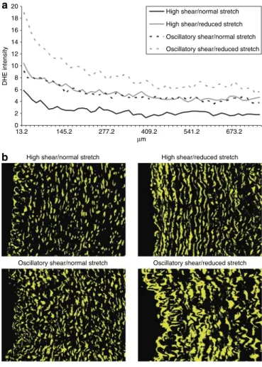
Radial vascular ROS magnitude and distribution are modu-lated almost identically by both reduced stretch and oscillatory flow, which maintain a sustained level of ROS throughout the measured region. The combination of the two stimuli are addi-tive, nearly doubling the amount of superoxide anion present in the vascular wall, which is sustained throughout the region of interest (Figure 2a,b).
radial expression of p22-phox correlates with superoxide ion production, but suggests it is not the only factor responsible for its measured increases
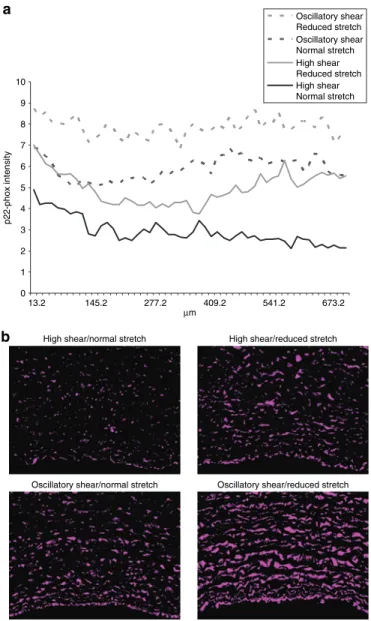
The radial magnitude of p22-phox expression is modulated by reduced stretch and oscillatory shear stress, the two effects
being additive and correlating well with the magnitude of DHE expression. In the first 130 µm, reduced stretch and oscil-latory shear stimulate nearly identical amounts of p22-phox, at which point they diverge, and oscillatory shear is shown to be the more prominent stimulus of p22-phox. The combination of both reduced stretch and oscillatory shear creates a sustained amount of vascular p22-phox (Figure 3a,b).
MMP-2 is significantly modulated by shear stress, but not stretch
In arteries exposed to OSC shear stress, MMP-2 protein expression was significantly upregulated in both normal and reduced stretch groups (21 and 18%, respectively, Figure 4a,b,
P < 0.05).
reduced stretch activates gelatinase in the endothelial region, whereas oscillatory shear increases gelatinase activity throughout the vascular wall
When focusing on the endothelial region, arteries exposed to a reduced stretch show significant increases in gelatinase
activ-0 20 40 60 80 100 Norepinephrine (mol/l) High shear/normal stretch
High shear/reduced stretch Oscillatory shear/normal stretch Oscillatory shear/reduced stretch
% Contraction
10−8 10−7 10−6 10−5 10−4
* *
Figure 1 | Norepinephrine (Ne) dose-dependent contraction capacity of
porcine carotid arterial segments after 72 h of ex vivo perfusion. data are represented as mean ± s.e.m., n = 6, *P < 0.05.
0 2 4 6 8 10 12 14 16 18 20 13.2 145.2 277.2 409.2 541.2 673.2 µm DHE intensity
Oscillatory shear/reduced stretch Oscillatory shear/normal stretch
High shear/reduced stretch High shear/normal stretch
High shear/normal stretch High shear/reduced stretch Oscillatory shear/normal stretch Oscillatory shear/reduced stretch
a
b
Figure 2 | Superoxide expression and distribution. (a) The effect of reduced
cyclic stretch and oscillatory shear stress on dihydroethidium (dHe) staining vs. arterial wall radius. curves represent the average of four images from each artery, n = 6. (b) dHe is represented in yellow; all images were taken at the same contrast and luminescence levels at ×10 magnification. Arterial lumen orientation is facing left.
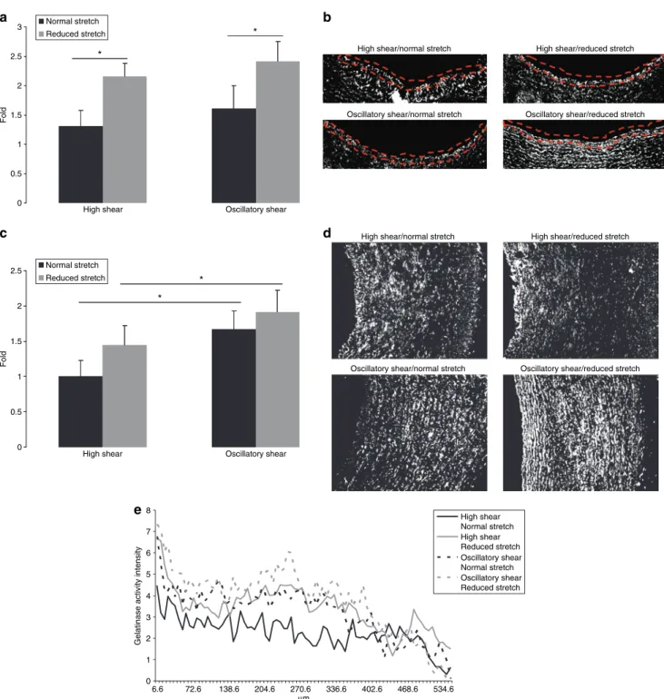
ity for both HSS and OSC groups (66 and 50%, respectively, Figure 5a, P < 0.05). Representative gelatinase images used for analysis have been included (Figure 5b).
Analysis of total vascular gelatinase activation was sig-nificantly increased by OSC shear for both the normal and reduced stretch groups (67 and 32%, respectively, Figure 5c,
P < 0.05). Representative gelatinase images used for analysis
have been included (Figure 5d).
reduced cyclic stretch and oscillatory shear stress modulate gelatinase activity differently in the arterial wall
In the first 100 µm of the arterial wall, we see that the mag-nitude of gelatinase is modulated by reduced stretch and OSC shear, and the shapes of the curves are similar. From
100 to 400 µm, gelatinase activity for the HSS 5% group steadily declines and introduction of either reduced stretch or OSC shear reduces this decline. The combination of reduced stretch and OSC shear causes the activation of gela-tinase to peak in this region. From 400 to 600 µm, we see all four groups come together and begin to behave in a similar manner (Figure 5e).
reduced stretch modulates the expression of fibronectin in the endothelial region, whereas oscillatory shear increases gelatinase activity throughout the vascular wall
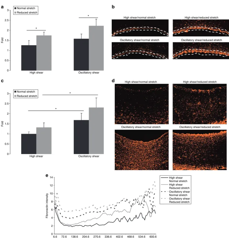
When focusing on the endothelial region, arteries exposed to reduced stretch show significant increases in fibronectin expression for both HSS and OSC groups (40 and 41%, respec-tively, Figure 6a, P < 0.05). Representative fibronectin images used for analysis have been included (Figure 6b).
Total vascular fibronectin expression was significantly increased by OSC shear for both the normal and reduced stretch groups (68 and 74%, respectively, Figure 6c, P < 0.05). Representative fibronectin images used for analysis have been included (Figure 6d).
reduced cyclic stretch and oscillatory shear stress modulate fibronectin expression differently in the arterial wall
From the endothelium penetrating 0.05 mm into the arte-rial wall, we see that the magnitude of fibronectin expression is modulated by both reduced stretch and OSC shear, but the shapes of the curves fall into two distinct groups, modulated by shear. The expression of fibronectin in the HSS groups decreases exponentially with radius, whereas the OSC groups have a more sustained expression before diminishing. As the radius increases, we see that reduced stretch, OSC shear, and the combination of the two still modulate the magnitude of 0 1 2 3 4 5 6 7 8 9 10 13.2 145.2 277.2 409.2 541.2 673.2 µm p22-phox intensity Oscillatory shear Reduced stretch Oscillatory shear Normal stretch High shear Reduced stretch High shear Normal stretch a
Oscillatory shear/reduced stretch Oscillatory shear/normal stretch
High shear/reduced stretch High shear/normal stretch
b
Figure 3 | p22-phox expression and distribution. (a) The effect of reduced
cyclic stretch and oscillatory shear stress on p22-phox expression vs. arterial wall radius. curves represent the average of four images from each artery,
n = 6. (b) p22-phox is represented in purple; all images were taken at the
same contrast and luminescence levels at ×10 magnification. Arterial lumen orientation is facing down.
0 0.2 0.4 0.6 0.8 1 1.2 1.4 1.6
High shear Oscillatory shear
Fold * * Normal stretch Normal stretch Reduced stretch Reduced stretch High shear Oscillatory shear
MMP-2 GAPDH Reduced stretch Normal stretch a b
Figure 4 | mmP-2 expression. (a) The effect of reduced cyclic stretch and
oscillatory shear stress on mmP-2 expression as evaluated by immunoblot. data are expressed as mean ± s.e.m., n = 6, *P < 0.05. (b) representative immunoblot. mmP-2, matrix metalloproteinase-2.
fibronectin expression. The shapes of the curves for the four groups follow a similar trend, steadily increasing with radius.
discussion
Previous works have shown that reduced arterial compliance12 and oscillatory shear stress13 stimulate the
production of vascular superoxide,14 which in turn con-tributed to the processes of arterial remodeling.15 However, the experimental conditions traditionally used to study shear stress and cyclic stretch have often left these two very important stimuli coupled, or exploited in such a way that is less physiologic, making it difficult to determine their 0 0.5 1 1.5 2 2.5 3
High shear Oscillatory shear
Fold * * Normal stretch Reduced stretch a c b d
High shear/normal stretch
Oscillatory shear/reduced stretch Oscillatory shear/normal stretch
High shear/reduced stretch
High shear/normal stretch
Oscillatory shear/reduced stretch Oscillatory shear/normal stretch
High shear/reduced stretch
0 0.5 1 1.5 2 2.5
High shear Oscillatory shear
Fold * * Normal stretch Reduced stretch 0 1 2 3 4 5 6 7 8 6.6 72.6 138.6 204.6 270.6 336.6 402.6 468.6 534.6 µm
Gelatinase activity intensity
High shear Normal stretch High shear Reduced stretch Oscillatory shear Normal stretch Oscillatory shear Reduced stretch e
Figure 5 | Gelatinase activity and distribution. (a) The effect of reduced cyclic stretch and oscillatory shear stress on endothelial gelatinase activity as evaluated
by image analysis. data are expressed as mean ± s.e.m., n = 6, *P < 0.05. (b) Gelatinase activity is represented in white and was evaluated at the endothelium. All images were taken at the same contrast and luminescence levels at ×10 magnification. Arterial lumen orientation is facing up. (c) The effect of reduced cyclic stretch and oscillatory shear stress on total gelatinase activity as evaluated by image analysis. data are expressed as mean ± s.e.m., n = 6, *P < 0.05. (d) Gelatinase is represented in white and was quantified over the entire region. All images were taken at the same contrast and luminescence levels at ×10 magnification. Arterial lumen orientation is facing left. (e) The effect of reduced cyclic stretch and oscillatory shear stress on gelatinase activity vs. arterial wall radius. curves represent the average of four images from each artery, n = 6.
individual contributions to vascular disease. In our study, we have performed experiments coupling high and oscil-latory shear stress with normal and reduced stretch, and studied how shear stress and cyclic stretch, two different mechanical stimuli, modulate remodeling throughout the arterial wall.
Arterial stiffness is strongly affected by endothelial cell sign-aling16 and smooth muscle contractile capacity.17 By limiting the range of arterial adaptability in response to hemodynamic stimuli, the artery becomes more susceptible to vascular dis-ease, thus provoking vascular remodeling.18 To understand the effects of shear stress and cyclic stretch on arterial smooth 0 0.5 1 1.5 2 2.5 3
High shear Oscillatory shear
Fold * * Normal stretch Reduced stretch a c b d
High shear/normal stretch
Oscillatory shear/reduced stretch Oscillatory shear/normal stretch
High shear/reduced stretch
High shear/normal stretch
Oscillatory shear/reduced stretch Oscillatory shear/normal stretch
High shear/reduced stretch
0 0.5 1 1.5 2 3 2.5
High shear Oscillatory shear
Fold * * Normal stretch Reduced stretch 0 2 4 6 8 10 12 14 6.6 72.6 138.6 204.6 270.6 336.6 402.6 468.6 534.6 600.6 µm Fibronectin intensity High shear Normal stretch High shear Reduced stretch Oscillatory shear Normal stretch Oscillatory shear Reduced stretch e
Figure 6 | Fibronectin expression and distribution. (a) The effect of reduced cyclic stretch and oscillatory shear stress on endothelial fibronectin expression
as evaluated by image analysis. data are expressed as mean ± s.e.m., n = 6, *P < 0.05. (b) Fibronectin is represented in orange and was evaluated at the endothelium. All images were taken at the same contrast and luminescence levels at ×10 magnification. Arterial lumen orientation is facing down. (c) The effect of reduced cyclic stretch and oscillatory shear stress on total fibronectin expression as evaluated by image analysis. data are expressed as mean ± s.e.m., n = 6, *P < 0.05. (d) Fibronectin is represented in orange and was quantified over the entire region. All images were taken at the same contrast and luminescence levels at ×10 magnification. Arterial lumen orientation is facing up. (e) The effect of reduced cyclic stretch and oscillatory shear stress on fibronectin expression vs. radius in the arterial wall. curves represent the average of four images from each artery, n = 6.
muscle contractile capacity, we performed vascular reactiv-ity studies on tissue segments from our four experimental groups: HSS/5% stretch, HSS/1% stretch, OSC/5% stretch, and OSC/1% stretch. We noticed introducing an oscillatory shear had little effect, whereas reducing stretch caused sig-nificant decreases in smooth muscle contractile capacity for both the HSS and OSC groups. Indicating that on a functional level, reduction of cyclic stretch has a more dramatic effect on smooth muscle contractile capacity than oscillatory flow.
Fluctuations in superoxide ion production increasing vascular levels of ROS are understood to have dramatic effects on vascular health, especially those occurring in the endothe-lial region.19 Basal levels of ROS are necessary for vascular cell physiologic function, increases of which have been shown to accelerate and contribute to the signaling required for vascular remodeling.20,21 Dramatic increases of ROS have been shown to scavenge vasoprotective molecules such as NO, thus mod-ulating vascular tone.22 Although ROS production is medi-ated at many levels, the predominant system producing ROS in vascular cells is recognized as being the NADPH oxidase cascade,23 which is mediated, in part, by the expression of p22-phox.24 Thus, we found it interesting to see how shear stress and cyclic stretch modulate ROS production and the expres-sion of p22-phox in the vascular wall. Upon staining with DHE, we found that oscillatory flow upregulated the total formation of superoxide anion as compared to reduced stretch.25 Yet when we focused on just the endothelial region, it was found that reduced stretch is a more significant modulator of super-oxide than oscillatory flow.25 Upon measuring the intensity of DHE staining vs. radius, both reduced stretch and oscillatory shear have similar effects in the expression of DHE and when combined, they have a significant additive effect. This distri-bution suggests that increased superoxide is a process that is at least partly mediated by the variation of stress throughout the vascular wall due to change in radius. When looking at the expression of p22-phox vs. arterial wall radius, we see that its expression follows a similar trend, but the shape of the curves indicates that expression is nearly independent of radius. This feature could help explain the sustained increases of superoxide production as radius increases throughout the arterial wall.
Arterial wall remodeling in response to mechanical stimuli is a process that involves increased cellular proliferation and reorganization of the ECM.26 MMP-2 is a very important ECM protein that is suspected to play a significant role in arterial matrix reorganization, a process governed by both its expression and activation. Also known to be actively involved in ECM remodeling, as well as cellular migration and pro-liferation, is fibronectin.27 Our results showed that total MMP-2 expression was significantly upregulated by oscillatory shear stress for both the normal and reduced stretch groups. Comparing this to total MMP activation, as assessed by gela-tinase activity, we observed a similar trend. Yet when we observed gelatinase activation at just the endothelial region, we saw it was significantly modulated by reduced stretch. To further investigate this regional distribution of gelatinase activity, we plotted gelatinase activity vs. arterial radius and
found a trend suggesting the distribution of gelatinase acti-vation is strongly mediated by stress variations due to radius. When the same analysis was performed for fibronectin expres-sion, we found that again, total arterial fibronectin expression was mediated by oscillatory shear stress for both normal and reduced stretch groups. Analysis of just the endothelial region showed that reduced stretch was the significant upregulator. When plotting fibronectin expression vs. radius, we observed that the endothelial region expression is strongly related to the stress distribution in this area, yet as radius increases, this rela-tion is slightly inversed.
To conclude, this study demonstrates for the first time in an arterial tissue model the individual contributions of shear stress and cyclic stretch to arterial remodeling while empha-sizing how these processes are mediated and distributed throughout the vascular wall. Our results showed that total superoxide production, fibronectin expression, and gelati-nase activation are predominantly mediated by oscillatory shear stress, whereas their expression in the endothelial region is mediated by reduced cyclic stretch, correlating well with results from total MMP-2 expression. By plotting intensity vs. radius for these markers of vascular remodeling, we were able to see that superoxide production and gelatinase activity followed trends indicating their expression was in part medi-ated by stress distributions throughout the vascular wall due to changing radius, whereas fibronectin and p22-phox were much less or not at all. Most importantly, these findings, when coupled with our results from tissue reactive studies, suggest that the arterial remodeling process triggered in the endothe-lial region due to reduced stretch causes the most significant changes in arterial smooth muscle function. Because per-turbed shear stress and reduced arterial compliance have both been implicated in the initiation and progression of vascular disease, this work provides a new perspective into how these stimuli are perceived throughout the vascular wall.
Acknowledgments: This work was supported by the Swiss National Science Foundation: grant 310000118274.
disclosure: The authors declared no conflict of interest.
1. Segers D, Helderman F, Cheng C, van Damme LC, Tempel D, Boersma E, Serruys PW, de Crom R, van der Steen AF , Holvoet P, Krams R. Gelatinolytic activity in atherosclerotic plaques is highly localized and is associated with both macrophages and smooth muscle cells in vivo. Circulation 2007; 115: 609–616.
2. Riccioni G. Statins and carotid intima-media thickness reduction: an up-to-date review. Curr Med Chem 2009; 16:1799–1805.
3. Baldassarre D, Amato M, Bondioli A, Sirtori CR, Tremoli E. Carotid artery intima-media thickness measured by ultrasonography in normal clinical practice correlates well with atherosclerosis risk factors. Stroke 2000; 31: 2426–2430.
4. Gambillara V, Montorzi G, Haziza-Pigeon C, Stergiopulos N, Silacci P. Arterial wall response to ex vivo exposure to oscillatory shear stress. J Vasc Res 2005; 42:535–544.
5. Gambillara V, Thacher T, Silacci P, Stergiopulos N. Effects of reduced cyclic stretch on vascular smooth muscle cell function of pig carotids perfused ex vivo.
Am J Hypertens 2008; 21:425–431.
6. Prado CM, Ramos SG, Alves-Filho JC, Elias J Jr, Cunha FQ, Rossi MA. Turbulent flow/low wall shear stress and stretch differentially affect aorta remodeling in rats.
J Hypertens 2006; 24:503–515.
7. Paravicini TM, Touyz RM. Redox signaling in hypertension. Cardiovasc Res 2006; 71:247–258.
8. Valko M, Leibfritz D, Moncol J, Cronin MT, Mazur M, Telser J. Free radicals and antioxidants in normal physiological functions and human disease. Int J Biochem
Cell Biol 2007; 39:44–84.
9. Abbruzzese TA, Guzman RJ, Martin RL, Yee C, Zarins CK, Dalman RL. Matrix metalloproteinase inhibition limits arterial enlargements in a rodent arteriovenous fistula model. Surgery 1998; 124:328–334; discussion 334–335. 10. Chiang HY, Korshunov VA, Serour A, Shi F, Sottile J. Fibronectin is an important
regulator of flow-induced vascular remodeling. Arterioscler Thromb Vasc Biol 2009; 29:1074–1079.
11. Montorzi G, Silacci P, Zulliger M, Stergiopulos N. Functional, mechanical and geometrical adaptation of the arterial wall of a non-axisymmetric artery in vitro.
J Hypertens 2004; 22:339–347.
12. Oliver JJ, Webb DJ. Noninvasive assessment of arterial stiffness and risk of atherosclerotic events. Arterioscler Thromb Vasc Biol 2003; 23:554–566. 13. Caro CG. Discovery of the role of wall shear in atherosclerosis. Arterioscler
Thromb Vasc Biol 2009; 29:158–161.
14. Touyz RM, Schiffrin EL. Reactive oxygen species in vascular biology: implications in hypertension. Histochem Cell Biol 2004; 122:339–352.
15. Touyz RM. Reactive oxygen species, vascular oxidative stress, and redox signaling in hypertension: what is the clinical significance? Hypertension 2004; 44:248–252.
16. Tsutsui M, Shimokawa H, Otsuji Y, Ueta Y, Sasaguri Y, Yanagihara N. Nitric oxide synthases and cardiovascular diseases: insights from genetically modified mice.
Circ J 2009; 73:986–993.
17. Sonoyama K, Greenstein A, Price A, Khavandi K, Heagerty T. Vascular remodeling: implications for small artery function and target organ damage. Ther Adv
Cardiovasc Dis 2007; 1:129–137.
18. Zieman SJ, Melenovsky V, Kass DA. Mechanisms, pathophysiology, and therapy of arterial stiffness. Arterioscler Thromb Vasc Biol 2005; 25:932–943.
19. Matlung HL, Bakker EN, Vanbavel E. Shear stress, reactive oxygen species and arterial structure and function. Antioxid Redox Signal 2009; e-pub ahead of print 2 Feb 2009.
20. Skyschally A, Schulz R, Gres P, Korth HG, Heusch G. Attenuation of ischemic preconditioning in pigs by scavenging of free oxyradicals with ascorbic acid.
Am J Physiol Heart Circ Physiol 2003; 284:H698–H703.
21. Chen W, Gabel S, Steenbergen C, Murphy E. A redox-based mechanism for cardioprotection induced by ischemic preconditioning in perfused rat heart.
Circ Res 1995; 77:424–429.
22. Sedeek M, Hébert RL, Kennedy CR, Burns KD, Touyz RM. Molecular mechanisms of hypertension: role of Nox family NADPH oxidases. Curr Opin Nephrol Hypertens 2009; 18:122–127.
23. Violi F, Basili S, Nigro C, Pignatelli P. Role of NADPH oxidase in atherosclerosis.
Future Cardiol 2009; 5:83–92.
24. Cave A. Selective targeting of NADPH oxidase for cardiovascular protection.
Curr Opin Pharmacol 2009; 9:208–213.
25. Thacher T, Silacci P, Stergiopulos N, da Silva RF. Autonomous effects of shear stress and cyclic circumferential stretch regarding endothelial dysfunction and oxidative stress: an ex-vivo arterial model. J Vasc Res, in press.
26. Lehoux S, Castier Y, Tedgui A. Molecular mechanisms of the vascular responses to haemodynamic forces. J Intern Med 2006; 259:381–392.
27. Astrof S, Hynes RO. Fibronectins in vascular morphogenesis. Angiogenesis 2009; 12:165–175.