DOI 10.1007/s10353-007-0350-4 # Springer-Verlag 2007
Breast reconstruction with flaps from the lower abdomen
J. Farhadiand G. Pierer
Department of Plastic, Reconstructive and Aesthetic Surgery, University Hospital Basel, Basel, Switzerland Received June 21, 2007; accepted July 15, 2007
# Springer-Verlag 2007
Wiederherstellung der Brust mit Lappen vom Abdomen
Zusammenfassung. Grundlagen: Die Abdominalre-gion ist eine vielseitige Quelle fu¨r mehrere Gewebe, welche als Lappen zur Wiederherstellung der weiblichen Brust geeignet sind. Diese beinhalten den gestielten und freien Transverse Rectus Abdominis Myocutaneous Lap-pen (TRAM), den Deep Inferior Epigastric Perforator Lappen (DIEAP) und den Superficial Inferior Epigastric Lappen (SIEA).
Methodik: Diese U¨ bersicht stellt Geschichte und Technik der Wiederherstellung der Brust mittels Lappen vom Abdo-men dar. Alle relevanten Studien und Erfahrungen wurden beru¨ cksichtigt.
Ergebnisse: Der Entscheidungsprozess zur Lappen-wahl ist komplex und abha¨ ngig von einer Reihe von Fakto-ren, wobei Lappen mit niedrigerer Hebemorbidita¨ t deutliche Vorteile zeigen und ein postoperativ verbessertes a¨sthe-tisches Resultat der Hebestelle die Patientenakzeptanz deutlich erho¨ hen kann.
Schlussfolgerungen: In dieser U¨ bersicht werden Anato-mie und pra¨operative Planung in dieser Spenderregion, Fragen zur intraoperativen Entscheidungsfindung sowie Resultate und Patientenzufriedenheit behandelt.
Schlu¨ sselwo¨ rter: Perforator-Lappen, DIEP, TRAM, SIEA, Hautlappennekrose, Mammarekonstruktion.
Summary. Background: The lower abdominal region offers a multitude of flaps for breast reconstruction, which include the pedicled and free transverse rectus abdominis myocutaneous flap (TRAM), the deep inferior epigastric ar-tery perforator flap (DIEAP), and the superficial inferior epi-gastric artery flap (SIEA).
Methods: This literature overview examines the history and the practice of breast reconstruction using flaps from the lower abdomen. All relevant studies and experiences have been included.
Results: The decision-making process in regard of the choice of flap to use is complex and depends on a variety of factors. A clear trend in using flaps which lower donor site morbidity and higher aesthetic outcome is seen.
Conclusions: This article serves to review surgical anat-omy, preoperative planning, intraoperative decision making in flap elevation, and reported outcomes in terms of compli-cations and patient satisfaction.
Key words: Perforator flap, DIEP, TRAM, SIEA, skin flap necrosis, breast reconstruction.
Introduction
Breast reconstruction today is regarded as one of the components in the overall treatment plan of breast cancer patients. Modern breast reconstruction techniques have evolved steadily over the last decades, in order to improve the aesthetic appearance and feel of the reconstructed breast, to improve the long term results and reduce the donor site morbidity. Today they compromise the use of implants, autologous tissues or a combination of the two. The use of autologous breast reconstruction techniques does meet the demands of an ideal breast reconstruction technique best (Table 1). Because of the variable need of individual patients, the reconstructive breast surgeon should be able to provide the full spectrum of options [1]. These needs include subjective parameters (e.g. tim-ing of surgery), oncologic parameters (e.g. radiotherapy, chemotherapy) and availability of donor tissue (Table 2). Autologous tissue can be supplied from the back, abdo-men, buttocks and the inner and outer thigh region. This review will focus on the abdomen as a source of flaps for breast reconstruction.
Up to 1979, tubed flaps from the abdomen were used as the standard in reconstruction of the breast. Then, Robbins described the vertical rectus flap as a pedicled
Correspondence: Jian Farhadi, M.D., Ph.D., Department of Plastic, Reconstructive and Aesthetic Surgery, University Hos-pital Basel, SHos-pitalstrasse 21, 4031 Basel, Switzerland. Fax:þþ41-61-265 7301
flap [2], and in the same year Holmstro¨m performed the first free abdominal flap for breast reconstruction, termed as the ‘‘free abdominoplasty flap’’ [3]. A year later, in 1982, Hartrampf changed the orientation of the skin island into a transverse one across the abdomen [4] and by so doing, a larger volume of tissue could be transferred to the breast. This pedicled TRAM flap (Transverse Rectus Abdominis Muscle) based on the superior epigastric artery became subsequently the workhorse in breast reconstruc-tion. Because of the dual blood supply of the rectus abdom-inis muscle supplying the typical TRAM skin island,
the flap can be raised as well as a free flap based on the inferior epigastric artery system. Grotting described the use of the free TRAM flap in 1989 [5] and concluded that the free TRAM flap appeared to be as safe as the conven-tional technique with the advantages of a more limited rectus muscle harvest, improved medial contour of the breast due to the lack of tunneling, and a healthier flap because of the donor vessels. These findings have been conformed in a multiple of studies [6, 7]. Perforator flaps from the abdomen, originally pioneered by Koshima in Japan in 1989 [8], have provided the next significant step towards the ideal breast reconstruction. The flap was based on a single paraumbilical perforator vessel from the deep inferior epigastric artery, consisting of only fat and skin. Allen and Treece used this flap in 1992 to perform a breast reconstruction by transferring only fat and skin from the abdomen whilst sparing the rectus abdominis muscle [9]. The deep inferior epigastric artery perforator (DIEAP) flap relies therefore on microdissection of the branches of the deep inferior epigastric system that perfo-rate the rectus abdominis to provide for a pedicle without the sacrifice of the muscle and the fascia. The decreased donor site morbidity, in terms of abdominal bulging, her-nia and reduced strength, could be shown in several stud-ies [10, 7]. In the course of the learning curve from free TRAM flaps to free DIEAP flaps, many surgeons per-formed a muscle sparing TRAM flap (MS-TRAM). It
Table 2. Parameters in decision-making of options in breast reconstruction Table 1. Comparison of autologous vs implant based
recon-struction
Autologous Implant
Patients own tissue No donor site morbidity No prosthesis associated
complications
Simple surgical procedure Versatility in creation of
breast (e.g. ptosis)
Short operating time Postoperative radiotherapy
possible
Improved wound healing in previous irradiated field Less need of opposite adjustment surgery
could be shown that there was no significant difference in flap-related complications or donor-site morbidity be-tween the free MS-TRAM or the free DIEAP flap [11]. In terms of donor-site morbidity, the superficial inferior epigastric artery (SIEA) flap allows the harvest of ab-dominal fat and skin tissue without the opening of the abdominal fascia [12]. Unfortunately, this flap has a few disadvantages: inconsistent anatomy, short pedicle, limit-ed vascular territory. But it is a good alternative for recon-struction of a small breast in the presence of good size diameter vessels [13].
The reconstructive breast surgeon today has a broad spectrum of flaps from the abdomen (pedicled TRAM, free TRAM, free MS-TRAM, DIEAP and SIEA) and other body areas at his disposal. Depending on objective, sub-jective and oncologic parameters and the surgeon’s exper-tise, the safest method of reconstruction will be chosen. Due to better vascular supply to the flap, reduced donor-site morbidity and improved medial contour of the breast, we advocate the use of free flaps over pedicled flaps. In free flaps, we aim for the use of either free DIEAP or a free MS-TRAM over a full TRAM.
Methods
This literature overview examines the history and the practice of breast reconstruction using flaps from the low-er abdomen. A review of the litlow-erature on this subject was conducted using the Pub Med search. We are quoting 55 papers from to 1979–2007. Furthermore unpublished data from our own department have been included.
Results
Indications for an abdominal flap
Breast reconstruction by an abdominal flap may be considered for any patient who is undergoing mastectomy or who has an existing defect associated with prior mas-tectomy. The use of perforator flap based reconstruction e.g. DIEAP flap provides the best option with least donor site morbidity in reconstruction of any mastectomy defect. Autologous tissue reconstruction is an important option as well in patients who present with an unsatisfactory or previously failed implant reconstruction. Replacement of implants often has to be considered with severe capsular contracture, which is more often found in patients who required radiation therapy. For those with deformities or volume loss due to prior lumpectomy, radiation or subcu-taneous mastectomy, autologous tissue reconstruction may be considered for correction. Congenital breast absence or underdevelopment (Poland syndrome) may also be cor-rected with soft tissue flap techniques [14]. Perforator flap transfer is also an appropriate consideration in any setting in which autologous tissue is preferred over implant and one wishes to avoid muscle harvesting.
Contraindication for an abdominal flap
Patients in poor general health condition, uncon-trolled diabetes, obesity and heavy smokers should be
offered an alternative to free flap breast reconstruction as they are all associated with a significant increase in the rate of major complications [15, 16]. Obesity (body mass index >30 kg=m2) is a major independent predictor of postoperative complications (partial flap loss, donor site complications, hernia=laxity), increasing the incidence of adverse events as much as 3-folds [17, 15]. Previous abdominal surgery consisting of subcostal or midline incisions are regarded as bearing a higher risk of com-plications [18, 19]. Prior abdominal operations (i.e. hys-terectomy, c-section, appendectomy) on the other hand are no contraindications for the use of an abdominal flap. The possibility of raising a perforator flap after lipo-suction has been demonstrated, but caution should be taken in doing so [20]. A prior abdominoplasty does though exclude the use of an abdominal flap as do those cases in which the abdominal skin and fat tissue is insufficient.
Fig. 1. Anatomical specimen of an abdominal flap. (A) The deep inferior epigastric artery (base) runs through the rectus abdominis muscle connecting to the superior epigastric artery (top). (B) The rectus sheath has been reflected to the left, the penetration of a perforator through this fascia and its further course into the fat and skin tissue of the flap to the right can be seen
Surgical anatomy
The skin and fat of the lower abdomen and periumbil-ical area are supplied by perforators arising from the su-perior epigastric vessels, deep inferior epigastric vessels, superficial inferior epigastric vessels, intercostal segmental vessels and terminal branches of the superficial and deep circumflex iliac vessels. The predominant blood supply to the lower abdomen is from the deep inferior epigastric vessels [21, 22]. The vessels from both epigastric systems penetrate the rectus muscles on their deep surfaces and travel as single or duplicated vessels up and down and find communication in the periumbilical region [23] (Fig. 1A). During their course, perforators are sent to the overlying fat and skin tissue (Fig. 1B). The course of these perforators through the muscle is highly variable. Pedicled TRAM flaps are raised on the superior epigastric vessel together with the entire rectus muscle. Free TRAM flaps are raised
on the deep inferior epigastric vessels together with the en-tire rectus muscle in the area of vessel perforation. Like free TRAM flap, the DIEAP flap is also based on the deep inferior epigastric vessels, but in contrast no rectus muscle or fascia must be sacrificed [9]. Instead, the perforating vessels are followed through the rectus muscle by atraumat-ically spreading it apart in the direction of the muscle fibers to their origins from the deep inferior epigastric vessels [24]. Alternatively, a ‘‘free abdominoplasty flap’’ can be raised based on the superficial inferior epigastric vessels [12]. This superficial vessel generally arises from the com-mon femoral artery or has a comcom-mon origin with the su-perficial circumflex iliac artery. In its course it pierces the scarpa fascia and proceeds in the superficial subcutaneous tissue [25, 26]. The major advantage of this SIEA flap is the fact that it can be raised with no violation of the ab-dominal wall muscle or fascia and therefore lacks the pos-sibility of postoperative hernia or abdominal bulging.
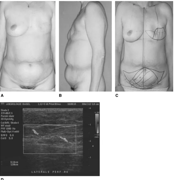
Fig. 2. (A, B) Preoperative views of a patient with a left mastectomy scar and ample abdominal tissue for breast reconstruction. (C) Typical preoperative markings on abdomen and breast. The necessary amount of skin and fat tissue is marked on the abdomen. The red crosses represent perforators found by a Doppler signal. (D) Ultrasound image: a perforator penetrates the fascia before entering the deep fat tissue
Planning
All flaps from the abdomen are planned in a similar fashion: a fusiform ellipse is outlined on the lower abdomen extending from the anterior superior iliac spine over to the umbilicus and just above the suprapubic crease inferiorly (Fig. 2A and B). The amount of tissue that can be safely taken and still allowing for a tension free closure is estimated.
Raising of a pedicled or free TRAM flap does not necessitate any further preoperative planning; only in case of previous abdominal surgery might it be wise to ensure vessel patency by a Doppler investigation. The assessment of the location of the perforators in DIEAP flap aids the operative procedure in terms of time-saving. A handheld probe can be used in order to identify the location of the perforators. Duplex Doppler scan can provide information on the position, flow, and diameter of the intramuscular perforators [27] (Fig. 2C and D). A newer development is the use of CT angiography in providing a preoperative roadmap for flap elevation [28]. This technique provides more accurate information than the duplex Doppler and
has the advantage of supplying images that can be easily studied and referred to during surgery. The most re-cent development is the use of 3-dimensional Magnetic Resonance Angiography.
Surgical technique
A two-team approach is used, simultaneously per-forming the flap harvest and either mastectomy and=or preparation of the recipient vessels. The internal mammary artery and vein have become the preferred recipient vessels. The thoracodorsal vessels have lost popularity due to sev-eral disadvantages: unusable vessels in secondary recon-struction up to 15% [29], the need of a longer pedicle, risks of nerve injury, loss of Latissimus dorsi flap as back-up. Alternatively, the circumflex scapular vessels can be dissected from a non-scarred area in order to increase pedi-cle length. Vessel hook-up in the axilla should be avoided in the case of sentinel lymph node biopsy and the prospect of an axillary node dissection after completed anastomosis [30]. The main disadvantage of the use of the internal
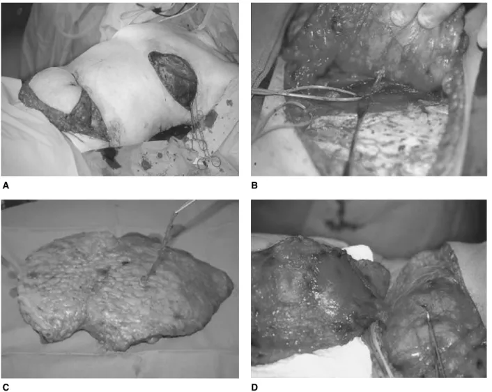
Fig. 3. (A) Situs in secondary reconstruction. The mastectomy scar has been excised and the flap incised according to the preoperative markings. (B) Once a sufficient size perforator has been encountered the fascia is opened and the perforator is dissected out between the fibres of rectus abdominis muscle. (C) A DIEAP flap on his pedicle with no rectus abdominis muscle sacrificed. (D) A TRAM flap for comparison with the full rectus muscle harvested
mammary vessels is the impairment of future cardiac by-pass surgery. Alternatively the perforators of the internal mammary artery and vein can be used if available [31].
The flap is raised by circumferential incision of skin down to fascia (Fig. 3A). During this procedure the infe-rior superficial epigastric vessels are explored routinely. If available and of significant size and quality, they are then followed down to their origin and a SIEA flap is per-formed (if the hemiabdomen provides sufficient tissue for reconstruction). If only the superficial vein is available, then it is dissected out and preserved as ‘‘lifeboat’’ in case of venous discharge problems [32]. The skin island is elevated from lateral to medial until the lateral row of perforators are encountered (Fig. 3B). If a sufficiently large perforator is found, then the flap is raised on that. Alternatively, two or three perforators in the same row can be used. If no adequate perforators are found, then the medial row is approached. The contralateral side is kept intact in order to raise a TRAM flap if no perforators can be found. Once the perforator of choice is found, the rectus sheath is opened and the vessel is dissected down through the rectus muscle to the source, the deep inferior
epigastric vessel. If two parallel perforators are to be in-cluded in the pedicle, then the cuff of muscle in between is included. Dissection continues until the pedicle is of suf-ficient length and the vessel diameter of sufsuf-ficient caliber to match the recipient vessels (Fig. 3C).
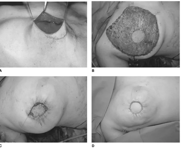
A TRAM flap is raised by incising the anterior rectus sheath and instead of tracing down single perforators, the entire rectus muscle and fascia is included in the flap (Fig. 3D). Once the mastectomy is completed (Fig. 4A) and the recipient vessels are ready, the artery and vein of the pedicle are ligated and the flap transferred to the chest (Fig. 4B). The vein and artery are anastomosed by micro-surgical techniques. The vein can either be joined by 9=0 Nylon sutures or by using an anastomotic coupling device (Microvascular Anastomotic COUPLER System). A cou-pling device has the advantage of making the venous anas-tomosis quicker and easier. The arterial anasanas-tomosis is performed with a 9=0 Nylon suture. The flap is secured on to the chest wall and trimmed and shaped according to the needs of symmetry of the contralateral breast. In pri-mary reconstruction, after skin-sparing mastectomy has been performed, the flap is deepithelized and placed in
Fig. 4. (A) Situs in a primary reconstruction. The skin sparing mastectomy has been completed. (B) The entire abdominal flap has been deepithelized with only a small skin patch left for areola reconstruction and monitoring. (C) The flap inserted and the anastomosis completed. (D) Final view at the end of the procedure
the empty skin envelope (Fig. 4C). Detailed shaping of the tissue is usually not necessary, as this is given by the skin envelope (Fig. 4D). In secondary reconstruction, the flap is folded under itself inferiorly (Fig. 5A) to provide a more natural look, projection and ptosis (Fig. 5B). The abdomi-nal fascia is closed directly if a DIEAP flap was harvested. In a TRAM flap, where the entire rectus muscle has been removed with part of the anterior fascia, an abdominal wall reinforcement by a synthetic mesh is advisable to prevent hernia formation. The wound is closed in an ab-dominoplasty fashion, where an upper abdominal flap is mobilized and the umbilicus is brought out through the flap and secured in place.
Postoperatively, the patient is observed in a surgical in-tensive care unit overnight. Perfusion of the microvascular flaps is closely monitored for three days to detect and revise vascular failures as early as possible, thus saving flaps. The patients are discharged usually after 5 to 7 days. Minor corrections and adjustment surgery of the opposite breast if necessary are carried out 8 to 12 weeks after reconstruc-tion with creareconstruc-tion of the nipple at the same time [33].
Special issues in bilateral breast reconstruction
There are a number of reasons for a patient seeking bilateral breast reconstruction: bilateral cancer, contralat-eral prophylaxis, bilatcontralat-eral ductal carcinoma in situ (DCIS), BRCA1-2 genetic diagnoses with a plea for prophylactic
mastectomy and failed implant reconstruction. As the number of bilateral reconstructions are increasing [34], this has major implications for the reconstructive team with regard to manpower. The safety of this procedure has been shown in several studies for free TRAM flaps [35, 36]. But despite these data it is apparent that the use of bilateral rectus abdominis muscle will lead to signifi-cant donor-site morbidity in the long-term [34]. Therefore the use of DIEAP or SIEA flaps in these cases would help not only to reduce the donor-site morbidity but to reduce the length of hospital stay as well [34, 37].
Complications
Microsurgical breast reconstruction has no higher com-plication rates than conventional pedicled flap breast recon-struction [15], the perfusion of the flap itself being better with less fat necrosis. The main factors contributing to these complications are patient factors as mentioned earlier.
Intraoperative problems
There can be a lack of adequate recipient vessels, especially veins or problems due to insufficient venous outflow from the flap. The internal mammary vein, partic-ularly the left, has been found to be unsuitable for micro-surgical transfer in approximately 20% of patients [38, 39]. Even though these figures are rather high in comparison to our own experience, nevertheless alternative options of recipient vessels can become necessary. The thoracodorsal vessels as an alternative can become unavailable as well due to significant scarring after previous axillary dissec-tions. In addition, the patency of these vessels, particularly that of the thoracodorsal vein, may be difficult to assess. Occlusion of the thoracodorsal vein may necessitate al-ternative venous drainage in the axilla [40]. The circum-flex scapular artery is well described as an alternative recipient vessel in the axilla [41]; however, it may not be available as well due to scarring. In these instances, and for revision of failed microvascular anastomoses to the thoracodorsal or circumflex scapular veins, the cephal-ic vein has been used [40]. This recipient vessel anasto-mosed to the superficial inferior epigastric vessel can be used as an additional venous discharge in compromised venous outflow.
Postoperative flap complications
Many studies have compared the complication rate of pedicled vs free TRAM vs DIEAP flaps. Fat necrosis is a common problem in autologous breast reconstruction. It is defined as the formation of a small firm area (or areas) of scar tissue in the periphery of a flap caused by ischemic necrosis of subcutaneous fat in the absence of necrosis of overlying flap skin [6]. Fat necrosis usually resolves spon-taneously over months, but occasionally it becomes infected and requires drainage or even resection. In addi-tion, fat necrosis can lead to concerns about possible tu-mor recurrence and may require needle biopsy or even excisional biopsy, causing patient anxiety, expense and
Fig. 5. (A) The abdominal flap is folded within itself on the abdomen, before the transfer to the breast. (B) The shaped flap is secured on the chest
inconvenience [6]. As the anterior abdominal wall is main-ly perfused by the deep inferior epigastric vessels [23, 42], a lower incidence of fat necrosis should be expected in using the free TRAM or DIEAP flap as compared to the pedicled TRAM. This could be confirmed in numerous studies, where the fat necrosis rate of pedicled TRAM was noted between 40 and 58% [6, 43], free TRAM flaps 10% [6] and free DIEAP flaps 6 to 18% [43–45]. The reason for the increased rate of fat necrosis in DIEAP flaps has not been yet fully understood, it is hypothesized that the free TRAM flap has a more robust perfusion of the flap that the DIEAP flap. Partial flap loss rates for pedicled flaps are 5 to 15% [46–49], for free TRAM 0 to 2.2% [6, 50] and for DIEAP flaps 0 to 8.7% [44, 45, 50]. Complete flap loss in pedicled TRAM flap has been found to be 0 to 4% [48, 51], 0 to 4.3% in free TRAM [48, 52] and 1 to 2.5% in DIEAP flaps [45, 50]. Problems with the vein or venous anastomosis are almost eight times more likely than problems with the artery or arterial anastomosis [45]. This stresses the importance of close postoperative monitoring by trained personnel, who are able to identify flap-related complications immediately. This again implies the necessity of a microsurgical team to be available post-operatively in order to revise and salvage the flap.
Donor-site related complications
There is a significant difference in donor-site related complications depending on the type of flap harvested and
the patients’ risks factors associated. In a 10 year retro-spective study of pedicled flap, the rate of donor-site re-lated complications was as high as 35%. The analysis indicated that the most common donor-site complication was seroma, which occurred in 31% of patients. Delayed wound healing was the next most frequent complication, occurring in 3.5% of patients. Donor-site hematoma, in-fection and hernia all occurred in less than 2% of patients [53]. In other studies, the hernia rate in pedicle flap was as high as 15.6%. The rate of hernia in free TRAM flap has been shown to be between 3 and 10% [48, 54] and in DIEAP flap from 0.7 to 5% [45, 55]. The differences in functional outcome with or without damage of the rectus abdominis muscle has been a matter of controversy. Blondeel et al. [10] were able to demonstrate the reduced donor-site morbidity in DIEAP flaps, by evaluation of trunk flexion and rotation, abdominal symmetry, and ac-tivities of daily living. A more recent study, which com-pared DIEAP flap vs MS-TRAM failed to show any difference [55]. This may be due to the muscle-sparing TRAM technique.
Conclusion
Autologous breast reconstructions with flaps from the abdomen have evolved to be the state of the art in breast reconstruction. As microsurgery has become a routine technique in reconstructive procedures, with failure rates of less than 2%, pedicled flaps are losing their popularity.
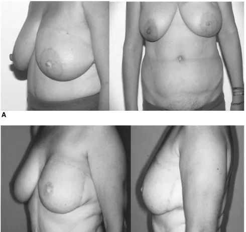
Fig. 6. (A, B) Final views of patient after delayed reconstruction, even very full breasts can be reconstructed without having to reduce the contralateral side
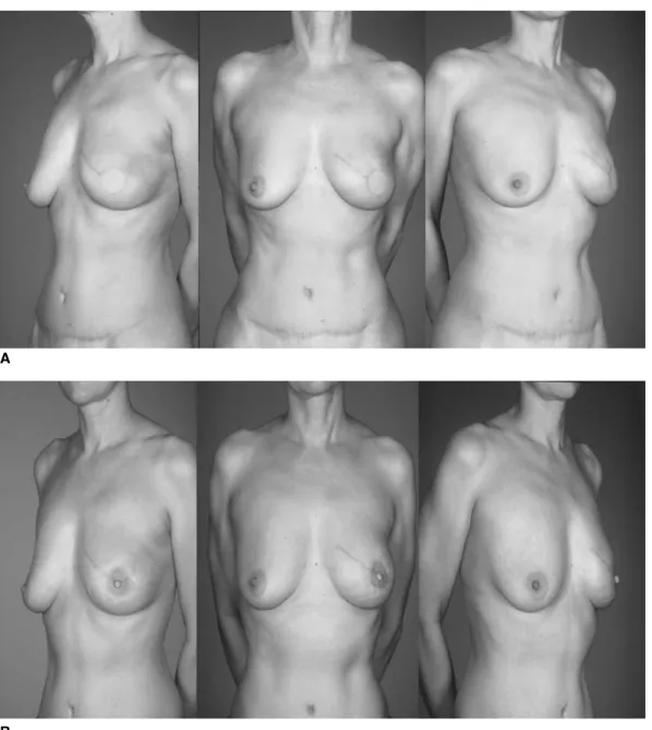
The advantages of free TRAM and DIEAP flaps over pedicled flaps have been shown in many studies. Patient satisfaction is very high in delayed (Fig. 6) and immediate reconstruction (Fig. 7). Careful patient selection is still a key to success, as most major complications are associated with patient’s comorbidity. Preoperative studies of the per-forator vessels may help to speed up the dissection period and save time intraoperatively but can as well lead to a higher success rate of DIEAP flaps by choosing the right perforator vessel. Nevertheless the final decision for har-vesting either a muscle-sparing TRAM flap or a DIEAP flap will depend on the actual caliber of the perforator and the experience of the surgeon. As it has been shown that donor-site morbidity is not increased in MS-TRAM com-pared to DIEAP flap [55], safety should be the first issue in raising these flaps. In conclusion, DIEAP, MS-TRAM or SIEA flaps have become the gold standard flaps in autologous breast reconstruction.
Products used in breast reconstruction
Handheld Doppler by Huntleigh Healthcare Products Suture Materials by Johnson&Johnson
Microsurgical Instruments and Suture Material by S&T Microvascular Anastomotic COUPLER System by Synovis
Surgical Microscope by Leica Magnifying Loupes by Zeiss
Web-sites dealing with breast reconstruction
http:==www.mdanderson.org=diseases=breastcancer= breastsurgery= http:==www.cancer.org=docroot=CRI=content=CRI_ 2_6X_Breast_Reconstruction_After_Mastectomy_5.asp http:==www.diepflap.com= http:==www.optionsforbreastreconstruction.com=
Fig. 7. (A) Primary reconstruction of left breast: result after skin sparing mastectomy and DIEAP flap and (B) NAC reconstruction after 8 weeks without any surgery to the healthy side
References
1. Bostwick J III (1999) Plastic and Reconstructive Breast Surgery. Quality Medical Publishing
2. Robbins TH (1979) Rectus abdominis myocutaneous flap for breast reconstruction. Aust N Z J Surg 49: 527–530 3. Holmstrom H (1979) The free abdominoplasty flap and
its use in breast reconstruction. An experimental study and clinical case report. Scand J Plast Reconstr Surg 13: 423–427
4. Hartrampf CR, Scheflan M, Black PW (1982) Breast recon-struction with a transverse abdominal island flap. Plast Reconstr Surg 69: 216–225
5. Grotting JC, Urist MM, Maddox WA, Vasconez LO (1989) Conventional TRAM flap versus free microsurgical TRAM flap for immediate breast reconstruction. Plast Reconstr Surg 83: 828–841
6. Kroll SS, Gherardini G, Martin JE, Reece GP, Miller MJ, Evans GR, Robb GL, Wang BG (1998) Fat necrosis in free and pedicled TRAM flaps. Plast Reconstr Surg 102: 1502–1507
7. Nahabedian MY, Dooley W, Singh N, Manson PN (2002) Contour abnormalities of the abdomen after breast recon-struction with abdominal flaps: the role of muscle preserva-tion. Plast Reconstr Surg 109: 91–101
8. Koshima I, Soeda S (1989) Inferior epigastric artery skin flaps without rectus abdominis muscle. Br J Plast Surg 42: 645–648
9. Allen RJ, Treece P (1994) Deep inferior epigastric per-forator flap for breast reconstruction. Ann Plast Surg 32: 32–38
10. Blondeel N, Vanderstraeten GG, Monstrey SJ, Van Landuyt K, Tonnard P, Lysens R, Boeckx WD, Matton G (1997) The donor site morbidity of free DIEP flaps and free TRAM flaps for breast reconstruction. Br J Plast Surg 50: 322–330 11. Bajaj AK, Chevray PM, Chang DW (2006) Comparison of
donor-site complications and functional outcomes in free muscle-sparing TRAM flap and free DIEP flap breast re-construction. Plast Reconstr Surg 117: 737–746
12. Grotting JC (1991) The free abdominoplasty flap for imme-diate breast reconstruction. Ann Plast Surg 27: 351–354 13. Wolfram D, Schoeller T, Hussl H, Wechselberger G (2006)
The superficial inferior epigastric artery (SIEA) flap: in-dications for breast reconstruction. Ann Plast Surg 57: 593–596
14. Liao HT, Cheng MH, Ulusal BG, Wei FC (2005) Deep inferior epigastric perforator flap for successful simul-taneous breast and chest wall reconstruction in a Poland anomaly patient. Ann Plast Surg 55: 422–426
15. Mehrara BJ, Santoro TD, Arcilla E, Watson JP, Shaw WW, Da Lio AL (2006) Complications after microvascular breast reconstruction: experience with 1195 flaps. Plast Reconstr Surg 118: 1100–1109
16. Chang DW, Reece GP, Wang B, Robb GL, Miller MJ, Evans GR, Langstein HN, Kroll SS (2000) Effect of smoking on complications in patients undergoing free TRAM flap breast reconstruction. Plast Reconstr Surg 105: 2374–2380 17. Chang DW, Wang B, Robb GL, Reece GP, Miller MJ,
Evans GR, Langstein HN, Kroll SS (2000) Effect of obesity on flap and donor-site complications in free transverse rectus abdominis myocutaneous flap breast reconstruction. Plast Reconstr Surg 105: 1640–1648
18. Losken A, Carlson GW, Jones GE, Culbertson JH, Schoemann M, Bostwick J III (2002) Importance of right subcostal incisions in patients undergoing TRAM flap breast reconstruction. Ann Plast Surg 49: 115–119
19. Takeishi M, Shaw WW, Ahn CY, Borud LJ (1997) TRAM flaps in patients with abdominal scars. Plast Reconstr Surg 99: 713–722
20. De Frene B, Van Landuyt K, Hamdi M, Blondeel P, Roche N, Voet D, Monstrey S (2006) Free DIEAP and SGAP flap breast reconstruction after abdominal=gluteal liposuction. J Plast Reconstr Aesthet Surg 59: 1031–1036
21. Taylor GI, Palmer JH (1987) The vascular territories (angio-somes) of the body: experimental study and clinical appli-cations. Br J Plast Surg 40: 113–141
22. Scheflan M, Dinner MI (1983) The transverse abdominal island flap: part I. Indications, contraindications, results, and complications. Ann Plast Surg 10: 24–35
23. Moon HK, Taylor GI (1988) The vascular anatomy of rectus abdominis musculocutaneous flaps based on the deep superior epigastric system. Plast Reconstr Surg 82: 815–832
24. Granzow JW, Levine JL, Chiu ES, Allen RJ (2006) Breast reconstruction with the deep inferior epigastric perforator flap: history and an update on current technique. J Plast Reconstr Aesthet Surg 59: 571–579
25. Hester TR Jr, Nahai F, Beegle PE, Bostwick J III (1984) Blood supply of the abdomen revisited, with emphasis on the superficial inferior epigastric artery. Plast Reconstr Surg 74: 657–670
26. Taylor GI, Daniel RK (1975) The anatomy of several free flap donor sites. Plast Reconstr Surg 56: 243–253 27. Blondeel PN, Beyens G, Verhaeghe R, Van Landuyt K,
Tonnard P, Monstrey SJ, Matton G (1998) Doppler flowme-try in the planning of perforator flaps. Br J Plast Surg 51: 202–209
28. Masia J, Clavero JA, Larranaga JR, Alomar X, Pons G, Serret P (2006) Multidetector-row computed tomography in the planning of abdominal perforator flaps. J Plast Reconstr Aesthet Surg 59: 594–599
29. Serletti JM, Moran SL, Orlando GS, Fox I (1999) Thora-codorsal vessels as recipient vessels for the free TRAM flap in delayed breast reconstruction. Plast Reconstr Surg 104: 1649–1655
30. Kronowitz SJ, Chang DW, Robb GL, Hunt KK, Ames FC, Ross MI, Singletary SE, Symmans WF, Kroll SS, Kuerer HM (2002) Implications of axillary sentinel lymph node biopsy in immediate autologous breast reconstruction. Plast Reconstr Surg 109: 1888–1896
31. Munhoz AM, Ishida LH, Montag E, Sturtz GP, Saito FL, Rodrigues L, Gemperli R, Ferreira MC (2004) Perforator flap breast reconstruction using internal mammary perfora-tor branches as a recipient site: an anatomical and clinical analysis. Plast Reconstr Surg 114: 62–68
32. Villafane O, Gahankari D, Webster M (1999) Superficial inferior epigastric vein (SIEV): ‘lifeboat’ for DIEP=TRAM flaps. Br J Plast Surg 52: 599
33. Farhadi J, Maksvytyte GK, Schaefer DJ, Pierer G, Scheufler O (2006) Reconstruction of the nipple-areola complex: an update. J Plast Reconstr Aesthet Surg 59: 40–53
34. Vega SJ, Bossert RP, Serletti JM (2006) Improving out-comes in bilateral breast reconstruction using autogenous tissue. Ann Plast Surg 56: 487–490
35. Kovacs L, Papadopulos NA, Ammar SA, Kloppel M, Herschbach P, Heinrich G, Baumann A, Biemer E (2004) Clinical outcome and patients’ satisfaction after simulta-neous bilateral breast reconstruction with free transverse rectus abdominis muscle (TRAM) flap. Ann Plast Surg 53: 199–204
36. Khouri RK, Ahn CY, Salzhauer MA, Scherff D, Shaw WW (1997) Simultaneous bilateral breast reconstruction with the
transverse rectus abdominus musculocutaneous free flap. Ann Surg 226: 25–34
37. Guerra AB, Metzinger SE, Bidros RS, Rizzuto RP, Gill PS, Nguyen AH, Dupin CL, Allen RJ (2004) Bilateral breast reconstruction with the deep inferior epigastric perforator (DIEP) flap: an experience with 280 flaps. Ann Plast Surg 52: 246–252
38. Feng LJ (1997) Recipient vessels in free-flap breast recon-struction: a study of the internal mammary and thoracodor-sal vessels. Plast Reconstr Surg 99: 405–416
39. Clark CP III, Rohrich RJ, Copit S, Pittman CE, Robinson J (1997) An anatomic study of the internal mammary veins: clinical implications for free-tissue-transfer breast recon-struction. Plast Reconstr Surg 99: 400–404
40. Mehrara BJ, Santoro T, Smith A, Arcilla EA, Watson JP, Shaw WW, Da Lio AL (2003) Alternative venous out-flow vessels in microvascular breast reconstruction. Plast Reconstr Surg 112: 448–455
41. Nieminen T, Asko-Seljavaara S, Suominen E, Kuokkanen H, von Smitten K (1999) Free microvascular tram flaps: report of 185 breast reconstructions. Scand J Plast Reconstr Surg Hand Surg 33: 295–300
42. Boyd JB, Taylor GI, Corlett R (1984) The vascular territo-ries of the superior epigastric and the deep inferior epigastric systems. Plast Reconstr Surg 73: 1–16
43. Garvey PB, Buchel EW, Pockaj BA, Casey WJ III, Gray RJ, Hernandez JL, Samson TD (2006) DIEP and pedicled TRAM flaps: a comparison of outcomes. Plast Reconstr Surg 117: 1711–1719
44. Kroll SS (2000) Fat necrosis in free transverse rectus abdominis myocutaneous and deep inferior epigastric per-forator flaps. Plast Reconstr Surg 106: 576–583
45. Gill PS, Hunt JP, Guerra AB, Dellacroce FJ, Sullivan SK, Boraski J, Metzinger SE, Dupin CL, Allen RJ (2004) A 10-year retrospective review of 758 DIEP flaps for breast reconstruction. Plast Reconstr Surg 113: 1153–1160
46. Hartrampf CR Jr (1988) The transverse abdominal island flap for breast reconstruction. A 7-year experience. Clin Plast Surg 15: 703–716
47. Watterson PA, Bostwick J III, Hester TR Jr, Bried JT, Taylor GI (1995) TRAM flap anatomy correlated with a 10-year clinical experience with 556 patients. Plast Reconstr Surg 95: 1185–1194
48. Moran SL, Serletti JM (2001) Outcome comparison between free and pedicled TRAM flap breast recon-struction in the obese patient. Plast Reconstr Surg 108: 1954–1960
49. Kroll SS, Netscher DT (1989) Complications of TRAM flap breast reconstruction in obese patients. Plast Reconstr Surg 84: 886–892
50. Nahabedian MY, Momen B, Galdino G, Manson PN (2002) Breast Reconstruction with the free TRAM or DIEP flap: patient selection, choice of flap, and outcome. Plast Reconstr Surg 110: 466–475
51. Trabulsy PP, Anthony JP, Mathes SJ (1994) Changing trends in postmastectomy breast reconstruction: a 13-year experi-ence. Plast Reconstr Surg 93: 1418–1427
52. Padubidri AN, Yetman R, Browne E, Lucas A, Papay F, Larive B, Zins J (2001) Complications of postmastectomy breast reconstructions in smokers, ex-smokers, and non-smokers. Plast Reconstr Surg 107: 342–349
53. Ducic I, Spear SL, Cuoco F, Hannan C (2005) Safety and risk factors for breast reconstruction with pedicled trans-verse rectus abdominis musculocutaneous flaps: a 10-year analysis. Ann Plast Surg 55: 559–564
54. Kroll SS, Baldwin B (1992) A comparison of outcomes using three different methods of breast reconstruction. Plast Reconstr Surg 90: 455–462
55. Nahabedian MY, Tsangaris T, Momen B (2005) Breast reconstruction with the DIEP flap or the muscle-sparing (MS-2) free TRAM flap: is there a difference? Plast Reconstr Surg 115: 436–444