HAL Id: inserm-00265734
https://www.hal.inserm.fr/inserm-00265734
Submitted on 20 Mar 2008HAL is a multi-disciplinary open access
archive for the deposit and dissemination of sci-entific research documents, whether they are pub-lished or not. The documents may come from teaching and research institutions in France or abroad, or from public or private research centers.
L’archive ouverte pluridisciplinaire HAL, est destinée au dépôt et à la diffusion de documents scientifiques de niveau recherche, publiés ou non, émanant des établissements d’enseignement et de recherche français ou étrangers, des laboratoires publics ou privés.
patients: ANRS CO3 Aquitaine cohort.
Marc-Arthur Loko, Laurent Castera, François Dabis, Brigitte Le Bail, Maria
Winnock, Gaëlle Coureau, Paulette Bioulac-Sage, Victor de Ledinghen, Didier
Neau
To cite this version:
Marc-Arthur Loko, Laurent Castera, François Dabis, Brigitte Le Bail, Maria Winnock, et al.. Valida-tion and comparison of simple noninvasive indexes for predicting liver fibrosis in HIV-HCV-coinfected patients: ANRS CO3 Aquitaine cohort.: Non invasive fibrosis indexes in HIV-HCV coinfected pa-tients. American Journal of Gastroenterology, Nature Publishing Group: Open Access Hybrid Model Option A, 2008, 103 (8), pp.1973-80. �10.1111/j.1572-0241.2008.01954.x�. �inserm-00265734�
AQUITAINE COHORT
Loko Marc-Arthur1, Castera Laurent2, Dabis François1,3, Le Bail Brigitte4, Winnock Maria1, Coureau Gaëlle1, Bioulac-Sage Paulette4, de Ledinghen Victor2, Neau Didier1,5, and the Groupe d'Epidémiologie Clinique du SIDA en Aquitaine (GECSA)1,3*
AFFILIATIONS
1 INSERM, U593, Bordeaux, France, and Institut de Santé Publique, d’Epidémiologie et de Développement (ISPED), Université Victor Segalen Bordeaux2, Bordeaux, France
2 Services d'Hépato-Gastroentérologie, Hôpital Haut-Lévêque, Centre Hospitalier Universitaire (CHU) Bordeaux, Pessac, France
3 CISIH, Hôpital Pellegrin, CHU Bordeaux, France
4 Laboratoire d'Anatomie Pathologique, Hôpital Pellegrin, CHU/Université Victor Segalen, Bordeaux, France
5 Fédération des Maladies Infectieuses et Tropicales, Hôpital Pellegrin, CHU Bordeaux, France
CORRESPONDENCE AND REPRINTS
Laurent Castera, M.D. Ph.D.
Service d’Hépato-Gastroenterologie, Hôpital Haut Lévêque, C.H.U. de Bordeaux, Avenue Magellan, 33604 Pessac, France
Tel: 00 33 5 57 65 64 39 Tel: 00 33 5 57 65 64 45
e-mail: laurent.castera@chu-bordeaux.fr
This is a preprint of an Article accepted for publication in the
RUNNING HEAD: non invasive fibrosis indexes in HIV-HCV coinfected patients
*Composition of the GECSA (Aquitaine Cohort):
Scientific committee: G. Chêne, F. Dabis (Chair), M. Dupon, M. Longy-Boursier, P. Morlat,
JL. Pellegrin, JM. Ragnaud and R. Salamon (Chair).
Methodological coordination: F. Dabis, G. Chêne, R. Thiébaut, C. Lewden and S.
Lawson-Ayayi.
Medical coordination: M. Dupon, P. Mercié, JF. Moreau, P. Morlat, JL. Pellegrin, JM.
Ragnaud, N. Bernard, D. Lacoste, D. Malvy and D. Neau.
Data Management and Analysis: MJ. Blaizeau, M. Decoin, S. Delveaux, C. Hannapier, S.
Labarrère, V. Lavignolle-Aurillac, B. Uwamaliya-Nziyumvira, G. Palmer, D. Touchard, M. Winnock, E. Balestre, A. Alioum, H. Jacqmin-Gadda and R. Thiébaut.
Participating physicians: Bordeaux University Hospital: P. Morlat, N. Bernard, M. Bonarek,
F. Bonnet, P. Gellie, D. Lacoste, K. Lacombe; M. Dupon, F. Dauchy, H. Dutronc, S. Lafarie; M. Longy-Boursier, P. Mercié, D. Malvy, T. Pistonne, P. Thibaut; JM. Ragnaud, D. Chambon, C. De La Taille, C. Cazorla, T. Galperine, D. Neau, A. Ochoa; JL.Pellegrin, JF. Viallard, O. Caubet, E. Lazaro C. Nouts; P.Couzigou, L. Castera; H. Fleury, ME. Lafon, B. Masquelier, I. Pellegrin; D. Breilh; JF. Moreau, P. Blanco. Dax Hospital: P. Loste, L. Caunègre. Bayonne Hospital: F. Bonnal, S. Farbos, MC Gemain. Libourne Hospital: J.Ceccaldi, S. Tchamgoué. Mont de Marsan Hospital: S. De Witte.
ABSTRACT
Background: Although an increasing number of non-invasive fibrosis markers is available in
HCV monoinfected patients, data on the performance of these tests in HIV-HCV coinfected patients are lacking.
Objective: To assess the diagnostic performance for predicting hepatic fibrosis stage of 4
simple and inexpensive non-invasive indexes (FIB-4, APRI, Forns, and platelet count) in HIV-HCV coinfected patients.
Methods: 200 consecutive HIV-HCV coinfected patients from the ANRS-CO3 Aquitaine
cohort who underwent liver biopsy were studied. Fibrosis stage was assessed according to Metavir scoring system by a single pathologist unaware of the data of the patients. Diagnostic performances were assessed by measuring areas under the receiver operating characteristic curves (AUROC) and the percentage of patients correctly identified (PCI).
Results: For predicting significant fibrosis (F≥2), APRI, Forns index, and FIB-4 had AUROCS
of 0.77, 0.75 and 0.79, with 39%, 25% and 70% of PCI, respectively. For predicting severe fibrosis (F≥3), 4 had AUROC of 0.77 with 56% of PCI. For predicting cirrhosis (F4), FIB-4, APRI and platelet count had AUROCs of 0.80, 0.79 and 0.78, with 59%, 60% and 76% of PCI, respectively. Overall, diagnostic performances of the different indexes did not differ significantly for both significant fibrosis and cirrhosis.
Conclusion: The use of these non invasive indexes could save liver biopsies in up to 56 to 76 %
of cases for the prediction of severe fibrosis-cirrhosis. However, given the high percentage of misclassified cases for significant fibrosis, such indexes do not appear currently suitable for use in clinical practice in HIV-HCV coinfected patients.
INTRODUCTION
Chronic hepatitis C virus (HCV) infection is common in patients with human immunodeficiency virus (HIV) (1, 2). Among the 40 million HIV-infected persons worldwide, an estimated 4-5 million are also chronically infected with HCV (1). HIV infection notably modifies the natural history of HCV infection with an accelerated progression of HCV-related liver disease towards cirrhosis (3-7). In addition, response to antiviral therapy in HIV-HCV coinfected is poorer than in HCV monoinfected patients with higher discontinuation rates (8-10). Therefore, assessment of liver fibrosis is of critical importance in HIV-HCV coinfected patients not only for prognosis but also for antiviral therapy indications.
Until recently, liver biopsy was the only way to evaluate fibrosis (11). However, liver biopsy is an invasive and painful procedure with rare but potentially life-threatening complications (12, 13). Thus many patients are reluctant to undergo liver biopsies and HIV-HCV coinfected patients may be discouraged to start anti-HCV treatment for this reason. The accuracy of liver biopsy to assess fibrosis has also been questioned, in relation to sampling errors and intra- and inter-observer variability that may lead to over- or under-staging (14-16). These findings thus emphasize the need for accurate non-invasive methods to measure the degree of liver fibrosis. Several markers and models have been proposed over the past few years for the prediction of fibrosis in HCV monoinfected patients, including the aspartate aminotransferase (AST) to platelet ratio index (17), the Forns index (18) and platelet count (19). However, very little information regarding the performance and utility of these tests in HIV-HCV coinfected patients is available. Finally, FIB-4 a new index based on age, alanine aminotransferase (ALT), AST and platelets has been specifically designed for predicting severe liver fibrosis in HIV-HCV co-infected patients (20). Although this index has been very recently studied in HCV monoco-infected patients (21), it has not been validated independently in HIV-HCV coinfected patients.
The aim of our study was to evaluate the diagnostic performance of APRI, Forns index, FIB-4, and platelet count, for predicting liver fibrosis in HIV-HCV coinfected patients.
PATIENTS AND METHODS
Patients
This retrospective study included patients with HIV-HCV coinfection who were referred for liver biopsy prior to HCV antiviral therapy at the University Hospital of Bordeaux between January 1999 and January 2005. Inclusion criteria were: age over 18, positive serum antibodies to HCV by means of a second- or third-generation HCV enzyme-linked immunosorbent assay (Ortho Diagnostic, Raritan NJ, USA) and detectable serum HCV RNA (AmplicorTM HCV, Roche Molecular Systems, Pleasanton, California, USA). Exclusion criteria were: coinfection with hepatitis B, other known causes of liver disease, alcohol intake of more than 50g/day. A total of 200 patients of the Aquitaine Cohort met those criteria.
The ANRS CO 3 Aquitaine Cohort, a prospective hospital-based cohort of HIV-1-infected patients under routine clinical management, was initiated in 1987 in the Bordeaux University Hospital and four other public hospitals in the Aquitaine region, Southwestern France, by the Groupe d’Epidémiologie Clinique du SIDA en Aquitaine (GECSA) (22). All adult patients with HIV-1 infection confirmed by Western-Blot testing, and who have given an informed consent are enrolled in the cohort, whatever their clinical stage, gender or HIV transmission group. Additionally, information from at least one follow-up visit after the baseline assessment, or known date of death has to be available. At each hospital contact, a standardized questionnaire including epidemiological, clinical, biological and therapeutic data is filled in by clinicians and entered into the database. The schedule of follow-up visits is based on clinical practice, and an active search of patients lost to follow-up is performed annually.
Histological assessment
The liver biopsy specimen was fixed in 10% formalin, paraffin embedded, and serially sectioned. Sections were stained with hematoxylin-eosin-safran, Masson’s trichrome, picrosirius red, and Gordon and Sweet’s stains. Biopsy samples were read by a single experienced pathologist (BLB) who was unaware of the clinical data of the patients, using the METAVIR scoring system for fibrosis (23) : F0 = no fibrosis; F1= portal fibrosis without septa ; F2 = portal fibrosis with rare septa ; F3 = numerous septa without cirrhosis ; F4= cirrhosis.
Non invasive indexes for prediction of fibrosis
AST, ALT, gamma-GT, cholesterol and platelet count, routinely determined for all patients, were available on the day of liver biopsy for 55% of patients, and within 1 month of the time of liver biopsy for 45%. APRI, Forns index and FIB-4 were calculated using the formula originally described (17, 18, 20). The cut-offs used were those proposed in the original studies. For platelet count, a cut-off of 150 109/L (19) was used for predicting cirrhosis.
Statistical analysis
Continuous variables were described by their mean and standard deviation (SD) or median and interquartile range (IQR) and categorical variables by percentages. To evaluate the diagnostic performance of the different indexes, sensitivity (Se), specificity (Spe), positive predictive value (PPV) and negative predictive value (NPV) were calculated, using cut-offs previously described for each index (17, 18, 20). The overall diagnostic performance of scores was evaluated by area under the receiver operating characteristic curves (AUROC). The percentage of patients correctly identified (PCI) was also estimated for each index. AUROCs were compared
according to the procedure proposed by Hanley and McNeil (24). Statistical analyses were performed using Stata Statistical software, version 9.2 (Stata corporation, College station, TX).
RESULTS
Study population
The main characteristics of the 200 HIV-HCV coinfected patients included are presented in Table 1. The mean age was 39.8±6.3 years and 67% of patients were male. Their median CD4 cell counts was 500 (78-1644) cell/µl. Most patients (87%) were taking highly active antiretroviral therapy (HAART) at the time of non invasive evaluation of liver fibrosis. The mean platelet count did not differ between patients receiving HAART (n=174) or not (n=26): 191±68 vs. 171±55 109/L, respectively (p=NS). Significant fibrosis (F≥2) was present in 157 patients (78.5%) and cirrhosis in 40 (20%). The mean liver biopsy length was 15.7±7.5 mm. Biopsy length was greater than 10 mm in 154 patients (81%), and greater than 15 mm in 89 (46.8%) (Table 2).
Diagnostic value of the indexes
AUROCs were calculated to assess the overall diagnostic performance of each index for prediction of significant (F≥2), severe fibrosis (F≥3) and cirrhosis (F4) (Table 2).
AUROCs of Forns index, APRI and FIB4 for discriminating F0F1 versus F2F3F4 were 0.75, 0.77 and 0.79, respectively. AUROC of FIB-4 for discriminating F0F1F2 versus F3F4, was 0.77. AUROCs of FIB4, APRI and platelet count for discriminating F0F1F2F3 versus F4, were 0.80, 0.79 and 0.78, respectively.
Comparison of AUROC values of the different indexes did not show any statistically significant difference for both significant fibrosis and cirrhosis. Also, no difference was found according to liver biopsy length or between patients receiving HAART or not.
Indexes aimed at predicting significant fibrosis
For a FIB4 index ≤0.6, 9 of 43 patients (20.9%) without significant fibrosis at liver biopsy were correctly identified (Table 3). In addition, the presence of significant fibrosis could not be excluded as 3 out of 12 of patients with a FIB4 index ≤0.6 had significant fibrosis at liver biopsy (NPV=75%). For a FIB4 ≥1, 131 of 157 patients (83.4%) with significant fibrosis at liver biopsy were correctly identified. In addition, 131 of 151 patients with FIB4 ≥1 had significant fibrosis at liver biopsy (PPV=86.7%).
For an APRI ≤0.5, 21 of 43 patients (48.8%) without significant fibrosis at liver biopsy were correctly identified (Table 3). In addition, the presence of significant fibrosis could not be excluded with certainty, as 19 of 40 patients with APRI ≤0.5 had significant fibrosis at liver biopsy (NPV=52.5%). For an APRI ≥1.5, only 57 of 157 patients (36.3%) with significant fibrosis at liver biopsy were correctly identified. In addition, 57 of 59 patients with APRI ≥1.5 had significant fibrosis at liver biopsy (PPV=96.6%).
For a Forns index <4.2, only 34.6% of patients without significant fibrosis at liver biopsy were correctly identified (Table 3). The presence of significant fibrosis could not be excluded as 69% of patients with a Forns index <4.2 had significant fibrosis at liver biopsy (NPV=31%). For a Forns index >6.9, only 23% of patients with significant fibrosis at liver biopsy were correctly identified. In addition, all the patients with a Forns index >6.9 had significant fibrosis at liver biopsy (PPV=100%).
Overall, using FIB4, APRI and Forns index, liver biopsy could have been avoided in 70%, 39% and 25% of patients, respectively.
For a FIB-4 ≤1.45, 90 of 129 patients (69.8%) without severe fibrosis at liver biopsy were correctly identified (Table 4). But, the presence of severe fibrosis could not be totally excluded, as 19 of 109 patients (17.4%) with FIB-4 ≤1.45 had severe fibrosis (NPV=82.6%). For a FIB-4 ≥3.25, only 22 of 71 patients (31%) with severe fibrosis at liver biopsy were correctly identified. In addition, 22 of 31 patients with a FIB-4≥3.25 had severe fibrosis at liver biopsy (PPV=71%). Using FIB-4, liver biopsy could have been avoided in 56% of patients.
Indexes aimed at predicting cirrhosis
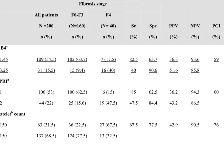
For a FIB4 ≤1.45, 102 of 160 patients (63.7%) without cirrhosis at liver biopsy were correctly identified (Table 5). In addition, the presence of cirrhosis could not be excluded totally, as 7 of 109 patients with an FIB4 ≤1.45 had cirrhosis at liver biopsy (NPV=93.6%). For a FIB4 ≥3.25, 16 of 40 patients (40%) with cirrhosis at liver biopsy were correctly identified. In addition, 16 of 31 patients with an FIB4 ≥3.25 had cirrhosis at liver biopsy (PPV=51.6%).
For an APRI ≤1, 100 of 160 patients (62.5%) without cirrhosis at liver biopsy were correctly identified (Table 5). In addition, the presence of cirrhosis could not be excluded totally, as 6 of 106 patients with an APRI ≤1 had cirrhosis at liver biopsy (NPV=94.3%). For an APRI >2, 19 of 40 patients (47.5%) with cirrhosis at liver biopsy were correctly identified. In addition, 19 of 44 patients with an APRI >2 had cirrhosis at liver biopsy (PPV=43.2%).
Platelet count <150.109g/l, identified correctly 27 of 40 patients (67.5%) with cirrhosis at liver biopsy (Table 5). The absence of cirrhosis could not be excluded totally as 13 of 137 (9.5%) patients with platelets count ≥150.109g/l had cirrhosis at liver biopsy (NPV=90.5%).
Overall, using FIB4, APRI and platelet count, liver biopsy could have been avoided in 59%, 60% and 76% of patients, respectively.
DISCUSSION
Although non invasive markers of liver fibrosis are being increasingly used in clinical practice in HCV monoinfected patients (25, 26), data on the performance of these tests in HIV-HCV coinfected patients are lacking. The Fibrotest (27) was one of the few tests evaluated in HIV-HCV coinfection. In a retrospective study in 130 patients (28), the AUROC for detection of significant fibrosis (Metavir F≥2) was 0.85. For a Fibrotest value <0.2, significant fibrosis could be excluded with 93% certainty (NPV 93%) whereas for a value >0.6, the presence of significant fibrosis could be predicted with 86% certainty (PPV 86%). Overall, liver biopsy could have been avoided in approximately 55% of patients. Recently, an index based on hyaluronic acid, albumin and AST (SHASTA) has been proposed in HIV-HCV coinfected patients (29). For a value <0.3, fibrosis could be excluded with 94% certainty (NPV 94%) whereas for a value >0.8 the presence of significant fibrosis could be predicted with 100% certainty. Overall, liver biopsy could have been avoided in approximately one-third of patients. It must be stressed, however, that Fibrotest and SHASTA are based on laboratory parameters not routinely performed and use a complex formula that limit their clinical applicability.
In the present study, we assessed the diagnostic performance of several simple and inexpensive non invasive indexes based on routinely available laboratory tests (APRI, Forns index, FIB-4, and platelet count), for the prediction of liver fibrosis in HIV-HCV coinfected patients. Liver biopsy was used as the reference for the diagnosis of fibrosis. We assessed the performance of the different indexes by measuring areas under the ROC curve. Overall, areas under the ROC curves did not differ significantly between the indexes (Forns, APRI, FIB-4 or platelet count) for both significant fibrosis and cirrhosis. Also no difference was found according to liver biopsy length. Finally, the diagnostic performance of these scores was lower in HIV-HCV coinfected patients than that found in the original studies performed in HCV monoinfected patients. For instance, the diagnostic accuracy of APRI and Forns index for significant fibrosis (with AUROCs of 0.77 and 0.75, respectively) was lower than that found in the original studies (17, 18) performed in HCV monoinfected patients (in which the AUROCs were 0.88 and 0.81,
respectively) but similar to the latest independent reports (19, 30, 31). APRI predicted the presence of significant fibrosis with 96.6% certainty (only 3.4% of patients with a score ≥1.5 did not have significant fibrosis), and overall 39% of patients were correctly classified. Similarly, Forns index predicted the presence of significant fibrosis with 100% certainty. However, no more than 25% of patients could be correctly classified. Finally, liver biopsy could have been avoided in 39% and 25% of our patients using APRI and Forns index, respectively as compared with around 50% in the original studies. These findings are in line with those of Macias et al. (32) in 263 HIV-HCV coinfected patients in whom liver biopsy could have been avoided in only one-third of patients using APRI and Forns index for prediction of significant fibrosis.
One possible explanation for such a discrepancy between our findings and those from original studies could be the difference for the prevalence of significant fibrosis among studies: higher in our HIV-HCV coinfected population (78.5%) than in the original studies, ranging from 26 to 50% (17, 18). Also, performance of APRI and Forns index could be affected in HIV-HCV coinfected patients by factors such as HAART-associated hepatotoxicity and HIV-induced thrombocytopenia (33-35). However, in the present study, diagnostic performance (as measured by AUROCs) of Forns index and APRI as well as mean platelet count did not differ significantly between patients receiving HAART or not. On the contrary, FIB4 when used for the exclusion or the prediction of significant fibrosis (at cut-offs of 0.6 and 1, respectively) performed better than APRI and Forns index. Surprisingly, these performances (AUROC 0.79 and 70% of PCI) are better than those published in the original study (AUROC 0.71 and 52% of PCI) (20). We have no clear explanation for this discrepancy, except, as stated before, differences in the prevalence of significant fibrosis between the 2 studies. It should be stressed, however, that specificity was very poor (20.9% and 53.5% for cut-offs ≤0.6 and ≥1, respectively) making FIB-4 as well as APRI and Forns index currently not suitable for confident use in clinical practice in HIV-HCV coinfected patients, especially for making treatment decision.
When APRI was used for the prediction of cirrhosis, its diagnostic performance was better than that observed for significant fibrosis. For instance an APRI ≤1 could exclude cirrhosis with 94% certainty (NPV=94.3%). Similarly, FIB4≤1.45 and platelet count when ≥150 x109/l could exclude cirrhosis with 94% (NPV=93.6%) and 90% certainty (NPV=90.5%). Although this may be important for reassuring patients, it is of little clinical use as these patients still need a liver biopsy for treatment decision. By contrast, APRI, FIB4 and platelet count did not confidently predict the presence of cirrhosis. For an APRI>2, a FIB4≥3.25 and a platelet <150 x109/l, the positive predictive values for cirrhosis were low (43.2%, 51.6% and 42.9%, respectively), which indicated a need of liver biopsy to stage for half of the patients. Overall, 60%, 59% and 76% of patients could be correctly classified with APRI, FIB4 and platelet count. These results are in agreement with those obtained in HIV-HCV coinfection (32) as well as in HCV monoinfection (19, 30).
With regards to the prediction of severe fibrosis (F3-F4), the diagnostic performance of FIB-4 with an AUROC of 0.77 was close to that reported in the original study (20). For instance, a FIB-4 ≤ 1.45 could exclude severe fibrosis with 82% certainty (NPV=82.6%). Conversely, a FIB-4 ≥3.25 could predict the presence of severe fibrosis with 71% certainty (PPV=71.0%). However, the percentage of patients in whom liver biopsy could have been avoided (56%) was lower than in the original study (71%) (20). Also on a clinical standpoint, 19 patients with severe fibrosis on liver biopsy and a FIB-4 ≤ 1.45 would have been falsely reassured with the risk of inappropriate management. Such a misclassification rate remains to high for confident use of FIB-4 in clinical practice.
One way to increase diagnostic accuracy of non invasive markers in HIV-HCV coinfected patients might be to use sequential algorithms combining several markers as recently suggested in HCV monoinfected patients (36). Further studies are needed to validate these algorithms in HIV-HCV coinfected patients.
In conclusion, the overall diagnostic performance of these indexes was lower in HIV-HCV coinfected patients than originally reported in HIV-HCV monoinfected patients. The use of
FIB-4, APRI or platelet count could avoid liver biopsy for the diagnosis of significant fibrosis, severe fibrosis and cirrhosis in up to 56 to 76 % of cases in HIV-HCV coinfected patients. However, given the high percentage of misclassified patients, these indexes do not currently appear to be suitable for routine clinical use in HIV-HCV coinfected patients. Further external validations in larger HIV-HCV coinfected populations are still needed to optimize the use of these non-invasive methods in such patients.
What is current knowledge
- Non invasive markers of liver fibrosis are gaining popularity in HCV monoinfected patients. - Data on the performance of these markers in HIV-HCV coinfected patients are still lacking.
What is new here
- The diagnostic performance of these markers is lower in HIV-HCV coinfected patients than that originally reported in HCV monoinfected patients.
- The use of these markers could save liver biopsies in up to 56 to 76 % of cases for the prediction of severe fibrosis-cirrhosis.
- However, given the high percentage of misclassified cases for significant fibrosis, such markers do not appear currently suitable for use in clinical practice in HIV-HCV coinfected patients.
1. Alter MJ. Epidemiology of viral hepatitis and HIV co-infection. J Hepatol 2006;44 (Suppl 1)::S6-S9.
2. Sherman KE, Rouster SD, Chung RT, et al. Hepatitis C Virus prevalence among patients infected with Human Immunodeficiency Virus: a cross-sectional analysis of the US adult AIDS Clinical Trials Group. Clin Infect Dis 2002;34:831-837.
3. Graham CS, Baden LR, Yu E, et al. Influence of human immunodeficiency virus infection on the course of hepatitis C virus infection: a meta-analysis. Clin Infect Dis 2001;33:562-569.
4. Martinez-Sierra C, Arizcorreta A, Diaz F, et al. Progression of chronic hepatitis C to liver fibrosis and cirrhosis in patients coinfected with hepatitis C virus and human immunodeficiency virus. Clin Infect Dis 2003;36:491-498.
5. Martin-Carbonero L, Benhamou Y, Puoti M, et al. Incidence and predictors of severe liver fibrosis in human immunodeficiency virus-infected patients with chronic hepatitis C: a European collaborative study. Clin Infect Dis 2004;38:128 - 133.
6. Mohsen AH, Easterbrook PJ, Taylor C, et al. Impact of human immunodeficiency virus (HIV) infection on the progression of liver fibrosis in hepatitis C virus infected patients. Gut 2003;52:1035-1040.
7. Salmon-Ceron D, Lewden C, Morlat P, et al. Liver disease as a major cause of death among HIV infected patients: role of hepatitis C and B viruses and alcohol. J Hepatol 2005;42:799-805. 8. Chung RT, Andersen J, Volberding P, et al. Peginterferon Alfa-2a plus ribavirin versus
interferon alfa-2a plus ribavirin for chronic hepatitis C in HIV-coinfected persons. N Engl J Med 2004;351:451-459.
9. Carrat F, Bani-Sadr F, Pol S, et al. Pegylated interferon alfa-2b vs standard interferon alfa-2b, plus ribavirin, for chronic hepatitis C in HIV-infected patients: a randomized controlled trial. JAMA 2004;292:2839-2848.
10. Torriani FJ, Rodriguez-Torres M, Rockstroh JK, et al. Peginterferon Alfa-2a plus ribavirin for chronic hepatitis C virus infection in HIV-infected patients. N Engl J Med 2004;351:438-450. 11. Bravo AA, Sheth SG, S C. Liver biopsy. N Engl J Med 2001;344:495-500.
Hepatology 1999;30:1529-1530.
13. Cadranel JF, Rufat P, F D. Practices of liver biopsy in France: results of a prospective nationwide survey. For the Group of Epidemiology of the French Association for the Study of the Liver (AFEF). Hepatology 2000; 32:477-481.
14. Bedossa P, Dargère D, Paradis V. Sampling variability of liver fibrosis in chronic hepatitis C. Hepatology 2003;38:1449-1457.
15. Colloredo G, Guido M, Sonzogni A, et al. Impact of liver biopsy size on histological evaluation of chronic viral hepatitis: the smaller the sample, the milder the disease. Journal of Hepatology 2003;39:239-244.
16. Regev A, Berho M, Jeffers LJ, et al. Sampling error and intraobserver variation in liver biopsy in patients with chronic HCV infection. Am J Gastroenterol 2002;97:2614-2618.
17. Wai CT, Greenson JK, Fontana RJ, et al. A simple noninvasive index can predict both significant fibrosis and cirrhosis in patients with chronic hepatitis C. Hepatology 2003;38:518-526.
18. Forns X, Ampurdanes S, Llovet JM, et al. Identification of chronic hepatitis C patients without hepatic fibrosis by a simple predictive model. Hepatology 2002;36:986-992.
19. Lackner C, Struber G, Liegl B, et al. Comparison and validation of simple noninvasive tests for prediction of fibrosis in chronic hepatitis C. Hepatology 2005;41:1376-1382.
20. Sterling RK, Lissen E, Clumeck N, et al. Development of a simple noninvasive index to predict significant fibrosis in patients with HIV/HCV coinfection. Hepatology 2006;43:1317-1325. 21. Vallet-Pichard A, Mallet V, Nalpas B, et al. FIB-4: an inexpensive and accurate marker of
fibrosis in HCV infection. comparison with liver biopsy and fibrotest. Hepatology 2007;46:32-6. 22. Binquet C, Chene G, Jacqmin-Gadda H, et al. Modeling changes in CD4-positive T-lymphocyte
counts after the start of highly active antiretroviral therapy and the relation with risk of opportunistic infections: the Aquitaine Cohort, 1996-1997. Am J Epidemiol 2001;153:386-393. 23. Intraobserver and interobserver variations in liver biopsy interpretation in patients with chronic
hepatitis C. The French METAVIR Cooperative Study Group. Hepatology 1994;20:15-20. 24. Hanley JA, McNeil BJ. A method of comparing the areas under receiver operating characteristic
2004;99:1160-1174.
26. Castera L, Denis J, Babany G, et al. Evolving practices of non-invasive markers of liver fibrosis in patients with chronic hepatitis C in France: Time for new guidelines? J Hepatol 2007;46:528-529.
27. Imbert-Bismut F, Ratziu V, Pieroni L, et al. Biochemical markers of liver fibrosis in patients with hepatitis C virus infection: a prospective study. Lancet 2001;357:1069-1075.
28. Myers RP, Benhamou Y, Imbert-Bismut F, et al. Serum biochemical markers accurately predict liver fibrosis in HIV and hepatitis C virus co-infected patients. AIDS 2003;17:721-725.
29. Kelleher TB, Mehtaa SH, Bhaskarb R, et al. Prediction of hepatic fibrosis in HIV/HCV co-infected patients using serum fibrosis markers: The SHASTA index. Journal of Hepatology 2005;43:78-84.
30. Leroy V, Hilleret MN, Sturm N, et al. Prospective comparison of six non-invasive scores for the diagnosis of liver fibrosis in chronic hepatitis C. J Hepatol 2007;46:775-782.
31. Castera L, Vergniol J, Foucher J, et al. Prospective comparison of transient elastography, Fibrotest, APRI, and liver biopsy for the assessment of fibrosis in chronic hepatitis C. Gastroenterology 2005;128:343-350.
32. Macias J, Giron-Gonzalez JA, Gonzalez-Serrano M, et al. Prediction of liver fibrosis in human immunodeficiency virus/hepatitis C virus coinfected patients by simple non-invasive indexes. Gut 2006;55:310-312.
33. Cole JL, Marzec UM, Gunthel CJ, et al. Ineffective platelet production in thrombocytopenic human immunodeficiency virus-infected patients. Blood 1998;91:3239-3246.
34. Kontorinis N, D. D. Hepatotoxicity of antiretroviral therapy. AIDS Rev 2003;5:36-43.
35. Bonacini M. Liver injury during highly active antiretroviral therapy: the effect of hepatitis C coinfection. Clin Infect Dis 2004;38 Suppl 2:S104-108.
36. Sebastiani G, Vario A, Guido M, et al. Stepwise combination algorithms of non-invasive markers to diagnose significant fibrosis in chronic hepatitis C. J Hepatol 2006;44:686-693.
coinfected patients of the ANRS CO 3 Aquitaine cohort
N=200
Age (years) 39.8 (6.3)
Male gender n (%) 133 (67.0)
Body mass index (kg/m²) 22.1 (3.2)
Source of HIV infection n (%)
Intravenous drug use 136 (68.0)
Others 64 (32.0) AST/ULN 2.2 (1.6) ALT/ULN 2.3 (1.9) Gamma-GT a (IU/l) 154 (175.8) Cholesterol a (mg/dl) 171.8 (50.3) Platelet count (x109/l) 188 (67.2)
Liver biopsies length (mm) 15.7 (7.5)
CD4 cell count b (/µl) n (%)
≤200 11 (6.1)
200-350 35 (19.6)
>350 133 (74.3)
HIV plasma RNA c n (%)
Undetectable (<50copies/ml) 84 (49)
Antiretroviral therapy n (%) 174 (87)
PI 79 (39.5)
NRTI 170 (85)
NNRTI 62 (31)
Fibrosis stage (METAVIR score) n (%)
F0-F1 43 (21.5)
F2 86 (43)
Data missing for: a 48 , b 21, c 30
AST: aspartate aminotransferase, ALT : Alanine aminotransferease, ULN: upper limit of normal,
PI: protease inhibitor, NRTI: nucleoside reverse transcriptase inhibitor, NNRTI: non nucleoside reverse transcriptase inhibitor
invasive indexes for predicting significant fibrosis (F≥2), severe fibrosis (F≥3) or cirrhosis (F=4), in HIV-HCV coinfected patients, according to liver biopsy length or higly active antiretroviral therapy (HAART).
Significant fibrosis Severe fibrosis Cirrhosis
N FORNS APRI FIB4 FIB4 FIB4 APRI Platelet
count Biopsy length (mm) All 200 0.75 a (0.66-0.84) 0.77 (0.70-0.85) 0.79 (0.72-0.86) 0.77 (0.70-0.84) 0.80 (0.73-0.87) 0.79 (0.72-0.86) 0.78 (0.69-0.87) ≥ 10 154 0.69 b (0.55-0.82) 0.77 (0.68-0.86) 0.80 (0.72-0.88) 0.75 (0.66-0.83) 0.78 (0.70-0.86) 0.76 (0.67-0.85) 0.76 (0.66-0.87) ≥ 15 89 0.68 c (0.51-0.84) 0.75 (0.61-0.88) 0.74 (0.61-0.87) 0.72 (0.61-0.84) 0.77 (0.67-0.88) 0.78 (0.67-0.88) 0.74 (0.60-0.87) HAART 0.75 d Yes 174 0.77 0.79 0.76 0.81 0.78 0.80 (0.65-0.85) (0.69-0.85) (0.72-0.87) (0.69-0.84) (0.73-0.88) (0.70-0.87) (0.71-0.90) 0.82 e (0.62-1.0) 0.81 (0.61-1.0) 0.84 (0.67-1.0) 0.84 (0.69-1.0) 0.76 (0.54-0.98) 0.85 (0.69-1.0) 0.62 No 26 (0.34-0.89) a in 152 patients; b
Se: sensitivity; Spe: specificity; PPV: positive predictive value; NPV: negative predictive value; PCI : patients correctly identified; AUROC: area under the receiver operating characteristic curve.
< cut-off means absence of significant fibrosis ; >cut-off means presence of significant fibrosis
Fibrosis stage All patients N =200 n (%) F0-F1 (N=43) n (%) F2-F4 (N= 157) n (%) Se (%) Spe (%) PPV (%) NPV (%) PCI (%) FIB4 ≤ 0.6 12 (6) 9 (20.9) 3 (1.9) 98.1 20.9 81.9 75.0 70 ≥ 1 151 (75.5) 20 (46.5) 131 (83.4) 83.4 53.5 86.7 46.9 APRI ≤ 0.5 40 (20) 21 (48.8) 19 (12.1) 87.9 48.8 87.9 52.5 39 ≥ 1.5 59 (29.5) 2 (4.7) 57 (36.3) 36.1 95.4 96.6 29.1 Fibrosis stage FORNS N =152 n (%) F0-F1 (N=26) n (%) F2-F4 (N= 126) n (%) < 4.2 29 (19.1) 9 (35) 20 (15.9) 84.1 34.6 86.2 31 25 > 6.9 29 (19.1) 0 (0) 29 (23) 23 100 100 21
Se: sensitivity; Spe: specificity; PPV: positive predictive value; NPV: negative predictive value; PCI : patients correctly identified; AUROC: area under the receiver operating characteristic curve.
< cut-off means absence of severe fibrosis ; >cut-off means presence of severe fibrosis
Fibrosis stage FIB-4 All patients N =200 n (%) F0-F2 (N=129) n (%) F3-F4 (N= 71) n (%) Se (%) Spe (%) PPV (%) NPV (%) PCI (%) ≤ 1.45 109 (54.5) 90 (69.8) 19 (26.8) 73.2 69.8 57.1 82.6 56 ≥ 3.25 31 (15.5) 9 (7) 22 (31) 31 93 71 71
Table 5. Diagnostic performance of the indexes aimed at predicting cirrhosis (F4) in the HIV-HCV coinfected patients.
Se: sensitivity; Spe: specificity; PPV: positive predictive value; NPV: negative predictive value; PCI : patients correctly identified; AUROC: area under the receiver operating characteristic curve.
a : < cut-off means absence of cirrhosis and; >cut-off means presence of cirrhosis b : < cut-off means presence of cirrhosis and > cut-off means absence of cirrhosis
Fibrosis stage All patients N =200 n (%) F0-F3 (N=160) n (%) F4 (N= 40) n (%) Se (%) Spe (%) PPV (%) NPV (%) PCI (%) FIB4a ≤ 1.45 109 (54.5) 102 (63.7) 7 (17.5) 82.5 63.7 36.3 93.6 59 ≥ 3.25 31 (15.5) 15 (9.4) 16 (40) 40 90.6 51.6 85.8 APRIa ≤ 1 106 (53) 100 (62.5) 6 (15) 85 62.5 36.2 94.3 60 > 2 44 (22) 25 (15.6) 19 (47.5) 47.5 84.4 43.2 86.5 Plateletb count < 150 63 (31.5) 36 (22.5) 27 (67.5) 67.5 77.5 42.9 90.5 76 ≥ 150 137 (68.5) 124 (77.5) 13 (32.5)