ORIGINAL ARTICLE
In situ RHAMM protein expression in acute myeloid
leukemia blasts suggests poor overall survival
Alexandar Tzankov&Ulrich Strasser&Stephan Dirnhofer&Thomas Menter&
Caroline Arber&Martine Jotterand&Alicia Rovo&Andre Tichelli&
Reinhard Stauder&Ursula Günthert
Received: 17 September 2010 / Accepted: 10 January 2011 / Published online: 28 January 2011 # Springer-Verlag 2011
Abstract Treatment options for patients with high-risk acute myeloid leukemia (AML) include high-dose chemotherapy regimens in combination with allogeneic hematopoietic stem cell transplantation, which takes advantage of the donor T-cell-mediated graft-versus-leukemia effect. Together with beneficial responses observed in assays targeted at leukemia-associated antigens (LAA), this encouraged research on cancer vaccines and adoptive cellular therapies in AML. The receptor for hyaluronic acid-mediated motility (RHAMM, CD168) was identified as one of the most promising LAA in AML. Thus far, little is known about in situ expression in leukemic bone marrow blasts or the prognostic role of RHAMM and its interaction partners in AML. We immunohistochemically analyzed the expression
and prognostic significance of RHAMM on trephine bone marrow biopsies from 71 AML cases that had been evaluated
for cytogenetics and presence of FLT3-internal tandem
duplications and NPM1 mutations. Fifty-five patients
(77%) were treated with curative intent, while 16 (23%) received the most appropriate supportive care. Twenty of 71 (28%) AML cases were considered RHAMM+. Receiver operating characteristic curves showed significant discrimi-natory power considering overall survival (OS) in AML patients treated curatively for RHAMM (p=0.015). Multi-variable analysis revealed that expression of RHAMM in >5% of leukemic blasts identifies a subgroup of curatively treated cases with adverse OS independent of failures to achieve complete remission. RHAMM not only represents a promising LAA with specific T-cell responses in AML but, if assessed in situ on blasts, also a probable prognostic factor. Keywords AML . RHAMM . CD44 . Prognosis
Introduction
Acute myeloid leukemias (AML) are neoplastic hemato-poietic stem cell disorders characterized by maturation arrest, uncontrolled proliferation, and resistance to
apopto-sis [1,2]. Decision making for treatment of AML patients is
risk adapted to the World Health Organization (WHO)
defined “clinico-pathologic-genetic” entities [3, 4] and
further depends on the patient’s health status and age. Treatment options range from best supportive care to intensive multiagent chemotherapy alone or high-dose regimens in combination with, primarily, allogeneic
hema-topoietic stem cell transplantation (allo-HSCT) [5]. In the
latter, the graft-versus-leukemia effect suggests that leukemia-specific T lymphocytes play a major role in attacking and Alexandar Tzankov and Ulrich Strasser equal contribution.
A. Tzankov (*)
:
U. Strasser:
S. Dirnhofer:
T. Menter:
U. GünthertInstitute of Pathology, University Hospital Basel, Schönbeinstr. 40,
4031 Basel, Switzerland e-mail: [email protected] U. Strasser
Institute of Pathology, Medical University of Innsbruck, Innsbruck, Austria
C. Arber
:
A. Rovo:
A. Tichelli Hematology, University Hospital Basel, Basel, SwitzerlandM. Jotterand
Service de génétique médicale, CHUV, Lausanne, Switzerland
R. Stauder
Department of Internal Medicine V (Hematology and Oncology), Medical University of Innsbruck,
Innsbruck, Austria
eliminating leukemic cells, thus supporting long-term remission. Along with several observed immunological and clinical responses using assays targeted at leukemia-associated antigens, these observations encouraged research on cancer vaccines and adoptive cellular therapies in AML
[6–8]. The receptor for hyaluronic acid-mediated motility
(RHAMM, CD168) was identified as one of the most
promising leukemia-associated antigens in AML [7,9–13].
RHAMM has several known functions that vary with respect to its different interaction partners. Interaction with CD44 in the presence of hyaluronic acid plays a role in cellular motility, wound healing, and modification of the Rat
sarcoma (Ras)-signaling cascade [14–17]. Through
interac-tions with signaling cascades related to extracellular signal-regulated protein kinase 1/2 (ERK1/2), breast cancer 1 (BRCA1), and BRCA1-associated RING domain 1 (BARD1), RHAMM, alone or in complex with CD44, osteopontin (OPN), and integrins, can promote cancer cell invasion and is essential for tumorigenicity, most likely by
oncogenic inhibition of apoptosis [13–15,18–23].
Further-more, intracellular RHAMM is expressed in a cell cycle-dependent manner and helps maintaining genomic stability,
thus acting as a tumor suppressor [21, 24]. Finally, as a
surface molecule on tumor cells, RHAMM has been shown to play an important role in humoral anti-tumor responses
[11, 25]. RHAMM has been identified as an important
prognostic factor of significance in carcinomas and
lympho-mas, and also in leukemias [10,25–33]. A study of Greiner
et al. suggested a probable but statistically not significant favorable prognostic impact of elevated RHAMM mRNA
expression in AML [7].
Along with RHAMM, CD44, a transmembrane glyco-protein, is also a receptor for hyaluronic acid. CD44 can interact with various molecules, like OPN, matrix metal-loproteinases, CD95, and also with extracellular RHAMM to promote invasion, angiogenesis, inflammation, and
apoptotic resistance [19,33,34]. Like RHAMM, CD44 is
involved in signaling pathways including cellular sarcoma
(c-Src) family kinases and Ras-like family GTPases [34].
CD44 arouse attention in AML research when a monoclo-nal antibody against this molecule eradicated AML stem
cells [35].
Despite the potentially promising observations on RHAMM in AML, little is known about its in situ expression on bone marrow blasts or its prognostic significance. In addition, there are no data on the expression of its interaction partners, OPN, CD44 and signal transducers, and activators of transcription (STAT), or about its influence on the rate of spontaneous apoptosis in AML. Therefore, we performed immunohistochemical analyses on bone marrow trephine biopsies from a cohort of 71 AML cases to determine the expression and prognostic importance of RHAMM and interacting molecules.
Patients, materials, and methods Patients
A retrospective cohort of 71 patients with AML was analyzed. All newly diagnosed patients with AML between 2005 and 2007 at the University Hospital Basel, who had been fully evaluated for cytogenetics and molecular markers, including the presence of internal tandem
dupli-cations of FMS-like tyrosine kinase 3 (FLT3-ITD) and, in
cases with normal cytogenetics, for Nucleophosmin 1
(NPM1) mutations, were included in this study if paraffin blocks of the bone marrow trephine biopsies were ade-quately preserved for immunohistochemical analyses. All
cases were reclassified according to WHO criteria [4].
Clinical and follow-up data were obtained by reviewing the charts. Retrieval of tissue and clinical data were performed according to data safety laws.
Of the 71 patients included in this study, 33 were male (mean age 58, median age 57, range 26–95) and 38 female (mean age 56, median 59, range 20–89). Therapy
was risk adapted to the “clinico-pathologic-genetic” AML
entity and to the patient’s health status according to the protocols of the Dutch-Belgian Cooperative Trial Group
for Hematology and Oncology (HOVON, http://www.
hovon.nl/trials/trials-by-type/aml.html) at the time of diag-nosis. The distribution of cases according to the WHO
classification is presented in Table 1. Responses to
treatment were defined as published by the International Working Group for Diagnosis, Standardization of Response Criteria, Treatment Outcomes, and Reporting Standards for
Therapeutic Trials in AML [36].
Cytogenetic analysis was performed at the Centre Hospitalier Universitaire Vaudois in Lausanne (Unité cytogénétique du cancer). Karyotypes were described according to the International System for Human
Cytoge-netic Nomenclature [37]. Cases were grouped according to
cytogenetics into three risk groups as suggested by
Grimwade [38]. FLT3-ITD and NMP1 mutation analysis
was performed as described [39].
Immunohistochemical staining
Paraffin blocks of the 71 bone marrow trephine biopsies were processed in a standardized manner for immunohis-tochemistry. Eight cases with reactive bone marrow changes (non-hemolytic anemia and thrombocytopenia)
served as controls. Table 2 lists the primary antibodies
used as well as the incubation and pre-treatment conditions. Visualization of bound secondary antibodies was done by avidin–biotin–peroxidase techniques using either diamino-benzidine (DAB) or aminoethylcarbazole (AEC) as chrom-ogens. Since the staining intensity varied between cases
[40, 41], only the relative proportion (percentage) of positively staining tumor cells was quantified. To study reproducibility of stainings, a second observer reassessed 20% of cases.
Statistics
Statistical analyses were performed using SPSS version 15.0 (SPSS, Chicago, IL). Incomplete data were not excluded from the tests. The degree of agreement between the observers for the quantitative immunohistochemical values was evaluated by intra/interclass correlation coefficients using the Cronbach’s alpha analysis. The Spearman’s test was used to analyze relationships between the markers. Only correlation coefficients >0.300 or <−0.300 were further considered. The Fisher’s exact test was used to analyze differences in distribution of case numbers between groups. Mann–Whitney or Kruskal–Wallis tests were applied as appropriate to assess differences of means between groups. The prognostic performance of the variables and determination of optimal cut-off values was established by receiver operating charac-teristic (ROC curves plotting sensitivity versus 1-specificity with special consideration of the respective area under the ROC (AUROC)). The optimal cut-off point was calculated
using Youden’s index (Y), denoting Y = sensitivity +
specificity− 1, since this method can be applied to find the
optimal cut-off value with the highest sensitivity and specificity when there is no particular requirement on
sensitivity and/or specificity [42]. OS was analyzed by the
Kaplan–Meier method and compared by the log-rank test in
univariable modus and by the Cox’ regression analysis in
multivariable modus. Only factors that were determined to be of at least trend prognostic significance in the univariable
modus (p<0.1) were entered into the multivariable analysis.
Statistical significance was defined asp<0.05 and corrected
for multiple testing, when necessary. Two-sided tests were used throughout.
Results
Response to therapy and clinical outcomes
The average follow-up time was 16.7 months (median 10; range 0−62). Within this time, 32 patients died. Most of the patients (n=55) were treated with curative intent
(chemo-therapy alone: n=25; intensive chemotherapy followed by
autologous HSCT: n=4; intensive chemotherapy followed
by allo-HSCT: n=26). Sixteen patients did not qualify for
intensive treatment. Five were given hydroxyurea and the others received best supportive care. The mean OS of patients treated without intention to cure was 4.4 months and the median 1.3 months, while the OS of patients treated
T able 1 Immunophenotypic characteristics of the 75 AML cases assessed on formalin-fixed, paraf fin-embedded trephine bone marrow biopsies according to WH O subtype AML, classified according to WHO N Mean age M/F ratio Therapy Outcome (dead) F ACR panCD44 (%) RHAMM (%) OPN (%) Active caspase-3 (%) CT SCT Other Mean ± S D Cases > cut-of f Mean ± S D Cases > cut-of f Mean ± S D Cases > cut-off Mean ± S D Cases > cut-off AML with recurrent genetic aberrations 13 54 6:7 9 2 2 4 4 9 + 2 2 2 5 + 9 3 21 + 2 3 5 0.5 + 0.4 2 AML with myelodysplasia-related changes 17 59 7:10 3 9 5 7 5 0 0 2 + 3 2 2 8 + 23 8 0.9 + 0.6 2 AML, therapy related 7 6 8 3:4 2 1 4 4 4 2 9 + 37 3 8 + 1 0 2 25 + 3 2 2 0.4 + 0.1 1 AML, not otherwise specified 34 54 1:1 1 1 18 5 1 4 5 10 + 1 9 1 1 8 + 1 2 1 3 3 4 + 27 20 0.5 + 0.4 10 Intermediate risk group a 26 53 1 1:15 9 1 5 2 10 2 6 + 9 9 6 + 9 9 3 9 + 30 18 0.5 + 0.3 7 Poor risk group a 8 6 1 3:1 2 3 3 4 3 1 5 + 15 2 1 5 + 16 4 1 5 + 19 2 0.8 + 0.6 3 p value n.s. n.s. 0.003 n.a. n.s. 0.014 0.031 n.s n.s n.s. n.s. 0.019 n.s. Cut-off scores were determined by ROC curves: panCD44 >5%, RHAMM >5%, osteopontin (OPN) >25%, active caspase-3 >0.5% FACR failure to achieve complete remission, CT chemotherapy ,SCT stem cell transplantation a Th e risk groups amo ng AML , not othe rwise specified were def ined according to Grimwa de et al. [ 38 ], i.e., the inter mediate grou p encomp assed A M L w it h normal kar yotype and othe r noncom plex abnorm alities while the poor risk group included AML with com plex karyoty pes
with intention to cure was 45.3 months (mean) and the median was not reached. Of the 55 patients treated with curative intent, 29 achieved a complete remission after induction, 12 a partial remission, while 14 had a resistant disease. Responses to induction therapy did not correlate with leukemia subtype, neither subtypes according to WHO
nor with Grimwade’s prognostic groups [4, 38], patients’
age, regimen composition nor any of the studied phenotypic and genetic markers. Failure to achieve complete remission was a prognostic factor for OS (p=0.039), while patients’
age, gender, leukemia subtype were not, yet the Kaplan–
Meier survival curves indicated poorer outcomes for elderly patients and patients with therapy-related AML as well as such with complex karyotypes. These observations were particularly due to the fact that patients treated with curative intent were treated in different ways according to their “clinico-pathologic-genetic” risk and patients being at risk were treated in a more intensive way, which probably
evened outcomes (Table1).
One third of AML express cytoplasmic/membranous RHAMM in approximately 20% of blasts
Assessment of RHAMM and active caspase-3 expression was highly reproducible (average interobserver correlation
coefficient α=0.92 and 0.89, respectively), and that of
panCD44 and OPN was good (α=0.82 and 0.79,
respec-tively). Except for 10 AML cases that were not assessable for active caspase-3 quantification, all cases were evaluable by immunohistochemical analysis.
In reactive bone marrows, panCD44 was expressed in
erythropoietic precursors, as expected [43], and the same was
found with OPN. RHAMM was detectable in single myelo-/ monopoietic precursors and mature mononuclear cells, as
formerly shown [29,44]. Active caspase-3 stained two to 16
apoptotic bodies per 0.283 mm2, especially in areas rich in
erythropoiesis. Qualitative impression of the various
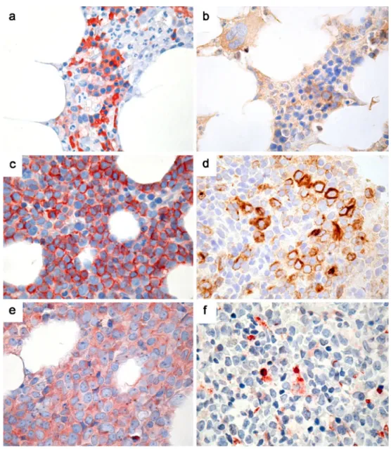
anti-body staining patterns can be extracted from Fig.1a and b.
Compared to controls, AML cases showed primarily qualitative staining differences, expressing the respective molecules in different cell populations. PanCD44 was strongly expressed in a mean of 38% of AML blasts in the 16 positive cases (cut-off score >5%), RHAMM in a mean of 19% of AML blasts in the 20 positive cases (cut-off score >5%) and OPN in 52% of AML blasts in the 35 positive cases
(cut-off score >25%). The sub-cellular localization of stain-able RHAMM was predominantly cytoplasmic. In AML cases with blasts negative for the respective marker, there were few erythropoietic or mononuclear cells remaining, occasionally expressing panCD44, OPN, or/and RHAMM, which served as internal positive control. Active caspase-3
stained 0 to 29 apoptotic bodies per 0.283 mm2, which were
morphologically attributable to leukemic blasts. Quantitative
immunohistochemical data are shown in Table1. Qualitative
impression of the various antibodies’ staining patterns can be
extracted from Fig.1c–f.
Expression of RHAMM in AML correlates with panCD44, pSTAT3, and pSTAT5, and with the spontaneous apoptotic activity
Expression of the analyzed markers was correlated with each other and to known clinical and pathological param-eters previously determined on the study collective, such as WHO leukemia subtype, patient age and gender, lack of complete remission, expression of pSTAT1, pSTAT3, and
pSTAT5, and presence ofFLT3-ITD and NPM1 mutations
[39]. RHAMM correlated with expression of panCD44
(ρ=0.560), of pSTAT3 and pSTAT5 (ρ=0.521 and 0.347,
respectively) and with spontaneous apoptosis as determined by the expression of active caspase-3 (ρ=0.321). The correlations of RHAMM and panCD44 as well as pSTAT3 and pSTAT5 expression were also observable in AML with recurrent genetic aberrations and AML, not otherwise specified; in addition in AML with normal karyotype and
noncomplex abnormalities according to Grimwade [38],
presence of FLT3-ITD correlated with failures to achieve
c o m p l e t e r e m i s s i o n (ρ = 0.504). In AML with
myelodysplasia-related changes RHAMM correlated with pSTAT1 expression in blasts (ρ=0.565), while this correla-tion was weaker (ρ=0.288) in the AML collective as a whole. There were no correlations between the studied
markers andNPM1 mutation status.
RHAMM is a WHO subtype-, age-, treatment type-, and response to treatment-independent negative prognostic factor in AML
Patients treated with curative intent (n=55) and those receiving best supportive care (n=16) were analyzed
Antibody Clone Source Dilution Retrieval Dye
Active caspase3 AF835 R&D 1:2,000 MW, EDTA pH 8, 100°C, 15′ AEC OPN OP3N Novocastra 1:50 MW, citrate pH 6, 100°C, 30′ AEC panCD44 IM7.8.1 ATCC 1:100 MW, citrate pH 6, 100°C, 30′ AEC RHAMM 2D6 Novocastra 1:25 Bond max, EDTA pH 8, 100°C, 20′ DAB Table 2 Applied antibodies and
staining conditions
AEC aminoethylcarbazole, DAB diaminobenzidine,EDTA ethylenediaminetetraacetate, MW microwave oven
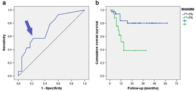
separately to exclude treatment effect bias. Of the markers analyzed, ROC curve-based analysis showed significant discriminatory power for survival of curatively treated AML
patients only for RHAMM (AUROC = 0.721, p=0.015;
Fig.2a). In a further step, this was linked to survival by the
Kaplan–Meier method and compared by the log-rank test using the cut-off score suggested by ROC, i.e., >5% blasts; the expression of RHAMM showed a significant negative
prognostic effect (Fig. 2b) that was particularly evident in
patients who failed to achieve complete remission after
induction treatment (Fig.3a and b). Importantly, expression
of RHAMM in patients who did or did not achieve complete remission was similar, and the lack of complete remission did not correlate to any of the above-mentioned clinical or pathological parameters. Analyzed with respect to the WHO entities and the Grimwade’s cytogenetic prognostic
groups [38], the negative prognostic effect of RHAMM was
apparent in the Kaplan–Meier curves in AML with recurrent
genetic aberrations, in AML, not otherwise specified, in the favorable risk group AML and in AML with normal karyotypes and other noncomplex abnormalities (intermediate risk group) according to Grimwade. Except for AML with normal karyotypes and other noncomplex abnormalities (intermediate risk group), where statistical significance was reached (p=0.029) patient numbers were too low in any of the groups to draw statistical conclusions. Considering the type of curative treatment regimens, this effect remained significant only for patients treated with multiagent
chemo-therapy (p=0.031), and not with high-dose regimens with
HSCT (p=0.119). In a multivariable model, expression of RHAMM turned out to be a negative prognostic factor independent of failure to achieve complete remission in curatively treated AML (p=0.002, relative risk 5.38, 95% confidence interval 1.75–16.49 for RHAMM; p=0.017,
relative risk 4.23, 95% confidence interval 1.29–13.78 for
failure to achieve complete remission). Fig. 1 Expression of the
studied markers. a Expression of panCD44 on erythroid precursors in normal bone marrow. b Expression of RHAMM in a single
mononuclear cell in the normal bone marrow. c PanCD44-positive AML. d AML case with 20% RHAMM-positive blasts. e Osteopontin-positive AML. f AML case with increased (>1%) spontaneous apoptotic activity as assessed by the expression of active caspase-3; all original magnifications ×400
Discussion
Our study is the first histopathology-based qualitative and quantitative examination of the expression of RHAMM and its partners in AML patients by immunohistochemistry on trephine bone marrow biopsies. Expression of RHAMM in >5% of leukemic blasts identified a subgroup of AML patients (treated curatively) with poor prognoses indepen-dent of AML WHO subtype or age. This effect was
particularly pronounced in patients who failed to achieve complete remission after induction therapy. Thus, our results considerably extend previous data on RHAMM in AML, showing that RHAMM not only represents a promising leukemia-associated antigen, similar to the preferentially expressed antigen in melanoma and the carbonic anhydrase IX (G250), with specific T-cell responses that suggest the possibility of tumor vaccination
[11,13], but also a prognostic tumor-related factor with a
Fig. 2 Prognostic significance of RHAMM in AML. a ROC curve of the prognostic performance of RHAMM in AML cases treated with curative intent. The area under the ROC of 0.721 (95% confidence interval 0.568–0.873) indicates that consideration of RHAMM is 22% more accurate in predicting survival than a random guess (p=0.015). The curve point (arrow) at sensitivity of 0.57 (57%) and specificity of 0.775 (77.5%; 1-specificity = 0.225) is at
greatest distance from the reference line. The relevant cut-off score of RHAMM at that point is >5% positive blasts. b Kaplan–Meier overall survival curves in AML patients treated with curative intent with respect to the expression of RHAMM; six out of the 37 RHAMM-negative AML patients died, mean survival 52 months, median not reached, compared to eight out of 17 RHAMM-positive patients, mean survival 21 months, median 15 months,p=0.006
Fig. 3 Prognostic significance of RHAMM in AML with respect to treatment responses. Kaplan–Meier overall survival curves in AML patients achieving (a) and failing to achieve complete remission (b) with respect to the expression of RHAMM; p=0.391 for (a);
among the patients who failed to achieve complete remission four out of 20 RHAMM-negative patients died, mean survival 47 months, median not reached, compared to all six RHAMM-positive patients, mean survival 10 months, median 9 months,p=0.001 for (b)
particular prognostic significance for patients failing to achieve complete remission after induction treatment. The latter might be of special interest since there are no established prognostic factors for AML in patients who failed to achieve complete remission.
RHAMM expression has been shown to be of prognostic importance in hematological neoplasia like B-cell chronic lymphocytic leukemia, plasma cell myeloma, and diffuse
large B-cell lymphoma [10, 27–29]. The biological
back-ground of the observed prognostic effects of RHAMM is not known. Because it is, on the one hand, a surface receptor molecule, and on the other, a functional intracel-lular protein, RHAMM plays different roles that vary with respect to its numerous interaction partners. Together with CD44, RHAMM promotes cellular motility, wound healing,
and the Ras-signaling cascade [15–17]. Through
interac-tions with signaling related to ERK1/2, BRCA1 and BARD1, RHAMM, alone or in complexes with CD44, OPN, and integrins, RHAMM can be tumorigenic, most
likely by inhibition of apoptosis [13–15, 18–23]. In
addition, intracellular RHAMM helps maintaining genomic
stability [21,24]. Finally, as a surface molecule on tumor
cells, RHAMM has been shown to play an important role in
humoral anti-tumor responses [11,25].
A former study of Greiner et al. suggest a possible yet not statistically significant (p=0.284) favorable prognostic
impact of RHAMM in AML [7]. These authors studied
gene expression levels of tumor-associated antigens from peripheral blood and bone marrow samples utilizing mRNA quantification of extracts from frozen mononuclear cells. The probable positive prognostic effect of RHAMM was attributed to anti-leukemic activity of the immune system represented by killing residual tumor cells expressing such tumor-associated antigens. The discrepancy in the observed prognostic impact of RHAMM between our study and that of Greiner et al. might be due to the different methodolo-gies employed to determine RHAMM expression, i.e., mRNA quantification and in situ analysis of blast counts with surface/cytoplasmic protein expression. This could reflect different RHAMM functions as an oncogene (surface, cytoplasmic protein), a tumor-suppressor gene (nuclear protein) or leukemia-associated antigen (surface and extracellular protein). Lastly, considering the contro-versies on the prognostic impact of expression levels of
another leukemia-associated antigen,WT1, in the study of
Greiner et al. and previous studies [7, 45, 46], and the
minor, if any, statistical significance of the gene expression of all studied leukemia-associated antigens in the former study, the in situ approach chosen in our study might more ostensibly mirror the oncogenic function of RHAMM in AML. In our AML collective, RHAMM was predominantly cytoplasmically expressed in a proportion of blasts, and closely correlated with expression of CD44 and pSTAT3
and pSTAT5 as well as with spontaneous leukemia apoptosis, but did not correlate with OPN. This indicates that RHAMM is probably not constitutively expressed by all blasts, and that its observed effects might be related to the interference of OPN-independent RHAMM with AML cell signaling or locomotion, and rather not with its anti-apoptotic functions.
In summary, beyond being a promising leukemia-associated antigen in AML, consideration of RHAMM surface/cytoplasmic expression in blasts may allow identi-fication of a subgroup of curatively treated patients with poor prognosis independent of AML WHO subtype, especially in patients who failed to achieve complete remission after induction therapy, which might merit further evaluation.
Acknowledgment The study was supported by the Oncosuisse grant OCS-01792-10-2005.
References
1. Estey E, Dohner H (2006) Acute myeloid leukaemia. Lancet 368:1894–1907
2. Löwenberg B (2008) Diagnosis and prognosis in acute myeloid leukemia—the art of distinction. N Engl J Med 358:1960–1962 3. Santamaria CM, Chillon MC, Garcia-Sanz R, Perez C, Caballero
MD, Ramos F et al (2009) Molecular stratification model for prognosis in cytogenetically normal acute myeloid leukemia. Blood 114:148–152
4. Swerdlow S, Campo E, Harris N, Jaffe E, Pileri S, Stein H, Thiele J, Vardiman J (2008) WHO classification of tumours of haematopoietic and lymphoid tissues. IARC, Lyon
5. Wahlin A, Billstrom R, Bjor O, Ahlgren T, Hedenus M, Hoglund M et al (2009) Results of risk-adapted therapy in acute myeloid leukaemia. A long-term population-based follow-up study. Eur J Haematol 83:99–107
6. Greiner J, Bullinger L, Guinn BA, Dohner H, Schmitt M (2008) Leukemia-associated antigens are critical for the proliferation of acute myeloid leukemia cells. Clin Cancer Res 14:7161–7166 7. Greiner J, Schmitt M, Li L, Giannopoulos K, Bosch K, Schmitt A
et al (2006) Expression of tumor-associated antigens in acute myeloid leukemia: implications for specific immunotherapeutic approaches. Blood 108:4109–4117
8. Li L, Reinhardt P, Schmitt A, Barth TF, Greiner J, Ringhoffer M et al (2005) Dendritic cells generated from acute myeloid leukemia (AML) blasts maintain the expression of immunogenic leukemia associated antigens. Cancer Immunol Immunother 54:685–693 9. Giannopoulos K, Li L, Bojarska-Junak A, Rolinski J, Dmoszynska A,
Hus I et al (2006) Expression of RHAMM/CD168 and other tumor-associated antigens in patients with B-cell chronic lymphocytic leukemia. Int J Oncol 29:95–103
10. Giannopoulos K, Mertens D, Buhler A, Barth TF, Idler I, Moller P et al (2009) The candidate immunotherapeutical target, the receptor for hyaluronic acid-mediated motility, is associated with proliferation and shows prognostic value in B-cell chronic lymphocytic leukemia. Leukemia 23:519–527
11. Greiner J, Ringhoffer M, Taniguchi M, Hauser T, Schmitt A, Dohner H, Schmitt M (2003) Characterization of several leukemia-associated antigens inducing humoral immune responses in acute and chronic myeloid leukemia. Int J Cancer 106:224–231
12. Greiner J, Ringhoffer M, Taniguchi M, Li L, Schmitt A, Shiku H, Dohner H, Schmitt M (2004) mRNA expression of leukemia-associated antigens in patients with acute myeloid leukemia for the development of specific immunotherapies. Int J Cancer 108:704–711
13. Greiner J, Ringhoffer M, Taniguchi M, Schmitt A, Kirchner D, Krahn G et al (2002) Receptor for hyaluronan acid-mediated motility (RHAMM) is a new immunogenic leukemia-associated antigen in acute and chronic myeloid leukemia. Exp Hematol 30:1029–1035
14. Sherman L, Sleeman J, Herrlich P, Ponta H (1994) Hyaluronate receptors: key players in growth, differentiation, migration and tumor progression. Curr Opin Cell Biol 6:726–733
15. Tölg C, Hamilton SR, Nakrieko KA, Kooshesh F, Walton P, McCarthy JB, Bissell MJ, Turley EA (2006) Rhamm-/- fibroblasts are defective in CD44-mediated ERK1, 2 motogenic signaling, leading to defective skin wound repair. J Cell Biol 175:1017–1028 16. Turley EA, Belch AJ, Poppema S, Pilarski LM (1993) Expression and function of a receptor for hyaluronan-mediated motility on normal and malignant B lymphocytes. Blood 81:446–453 17. Wang C, Thor AD, Moore DH 2nd, Zhao Y, Kerschmann R, Stern
R, Watson PH, Turley EA (1998) The overexpression of RHAMM, a hyaluronan-binding protein that regulates ras signaling, correlates with overexpression of mitogen-activated protein kinase and is a significant parameter in breast cancer progression. Clin Cancer Res 4:567–576
18. Hamilton SR, Fard SF, Paiwand FF, Tölg C, Veiseh M, Wang C et al (2007) The hyaluronan receptors CD44 and Rhamm (CD168) form complexes with ERK1, 2 that sustain high basal motility in breast cancer cells. J Biol Chem 282:16667–16680
19. Maxwell CA, McCarthy J, Turley E (2008) Cell-surface and mitotic-spindle RHAMM: moonlighting or dual oncogenic functions? J Cell Sci 121:925–932
20. Bourguignon LY, Gunja-Smith Z, Iida N, Zhu HB, Young LJ, Muller WJ, Cardiff RD (1998) CD44v(3, 8–10) is involved in cytoskeleton-mediated tumor cell migration and matrix metal-loproteinase (MMP-9) association in metastatic breast cancer cells. J Cell Physiol 176:206–215
21. Godar S, Weinberg RA (2008) Filling the mosaic of p53 actions: p53 represses RHAMM expression. Cell Cycle 7:3479
22. Khan SA, Cook AC, Kappil M, Günthert U, Chambers AF, Tuck AB, Denhardt DT (2005) Enhanced cell surface CD44 variant (v6, v9) expression by osteopontin in breast cancer epithelial cells facilitates tumor cell migration: novel transcriptional, post-translational regulation. Clin Exp Metastasis 22:663–673 23. Tzircotis G, Thorne RF, Isacke CM (2005) Chemotaxis towards
hyaluronan is dependent on CD44 expression and modulated by cell type variation in CD44-hyaluronan binding. J Cell Sci 118:5119–5128
24. Sohr S, Engeland K (2008) RHAMM is differentially expressed in the cell cycle and downregulated by the tumor suppressor p53. Cell Cycle 7:3448–3460
25. Schmitt A, Barth TF, Beyer E, Borchert F, Rojewski M, Chen J et al (2009) The tumor antigens RHAMM and G250/CAIX are expressed in head and neck squamous cell carcinomas and elicit specific CD8+ T cell responses. Int J Oncol 34:629–639 26. Gust KM, Hofer MD, Perner SR, Kim R, Chinnaiyan AM,
Varambally S et al (2009) RHAMM (CD168) is overexpressed at the protein level and may constitute an immunogenic antigen in advanced prostate cancer disease. Neoplasia 11:956–963 27. Maxwell CA, Rasmussen E, Zhan F, Keats JJ, Adamia S, Strachan
E et al (2004) RHAMM expression and isoform balance predict aggressive disease and poor survival in multiple myeloma. Blood 104:1151–1158
28. Nagel S, Hirschmann P, Dirnhofer S, Günthert U, Tzankov A (2010) Coexpression of CD44 variant isoforms and receptor for
hyaluronic acid-mediated motility (RHAMM, CD168) is an International Prognostic Index and C-MYC gene status-independent predictor of poor outcome in diffuse large B-cell lymphomas. Exp Hematol 38:38–45
29. Pilarski LM, Masellis-Smith A, Belch AR, Yang B, Savani RC, Turley EA (1994) RHAMM, a receptor for hyaluronan-mediated motility, on normal human lymphocytes, thymocytes and malignant B cells: a mediator in B cell malignancy? Leuk Lymphoma 14:363–374 30. Rein DT, Roehrig K, Schondorf T, Lazar A, Fleisch M, Niederacher D, Bender HG, Dall P (2003) Expression of the hyaluronan receptor RHAMM in endometrial carcinomas suggests a role in tumour progression and metastasis. J Cancer Res Clin Oncol 129:161–164
31. Shigeishi H, Fujimoto S, Hiraoka M, Ono S, Taki M, Ohta K, Higashikawa K, Kamata N (2009) Overexpression of the receptor for hyaluronan-mediated motility, correlates with expression of microtubule-associated protein in human oral squamous cell carcinomas. Int J Oncol 34:1565–1571
32. Zlobec I, Terracciano L, Tornillo L, Günthert U, Vuong T, Jass JR, Lugli A (2008) Role of RHAMM within the hierarchy of well-established prognostic factors in colorectal cancer. Gut 57:1413–1419 33. Mielgo A, van Driel M, Bloem A, Landmann L, Günthert U (2006) A novel antiapoptotic mechanism based on interference of Fas signaling by CD44 variant isoforms. Cell Death Differ 13:465–477
34. Ponta H, Sherman L, Herrlich PA (2003) CD44: from adhesion molecules to signalling regulators. Nat Rev Mol Cell Biol 4:33–45 35. Jin L, Hope KJ, Zhai Q, Smadja-Joffe F, Dick JE (2006) Targeting of CD44 eradicates human acute myeloid leukemic stem cells. Nat Med 12:1167–1174
36. Cheson BD, Bennett JM, Kopecky KJ, Buchner T, Willman CL, Estey EH et al (2003) Revised recommendations of the interna-tional working group for diagnosis, standardization of response criteria, treatment outcomes, and reporting standards for therapeutic trials in acute myeloid leukemia. J Clin Oncol 21:4642–4649
37. Schreck RR, Distèche CM (2001) Chromosome banding techniques. In: Haines JL, Korf BR, Morton CC, Seidman EG, Seidman JG, Smith DR, Sharer JD (eds) Current protocols in human genetics. Wiley, New York, pp 4.2.1–4.2.36
38. Grimwade D, Walker H, Harrison G, Oliver F, Chatters S, Harrison CJ et al (2001) The predictive value of hierarchical cytogenetic classification in older adults with acute myeloid leukemia (AML): analysis of 1065 patients entered into the United Kingdom Medical Research Council AML11 trial. Blood 98:1312–1320
39. Obermann EC, Arber C, Jotterand M, Tichelli A, Hirschmann P, Tzankov A (2010) Expression of pSTAT5 predicts FLT3 internal tandem duplications in acute myeloid leukemia. Ann Hematol 89:663–669
40. Atkins D, Reiffen KA, Tegtmeier CL, Winther H, Bonato MS, Storkel S (2004) Immunohistochemical detection of EGFR in paraffin-embedded tumor tissues: variation in staining intensity due to choice of fixative and storage time of tissue sections. J Histochem Cytochem 52:893–901
41. Zlobec I, Terracciano L, Jass JR, Lugli A (2007) Value of staining intensity in the interpretation of immunohistochemistry for tumor markers in colorectal cancer. Virchows Arch 451:763–769 42. Tzankov A, Zlobec I, Went P, Robl H, Hoeller S, Dirnhofer S
(2010) Prognostic immunophenotypic biomarker studies in diffuse large B cell lymphoma with special emphasis on rational determination of cut-off scores. Leuk Lymphoma 51:199–212 43. Chen K, Liu J, Heck S, Chasis JA, An X, Mohandas N (2009)
Resolving the distinct stages in erythroid differentiation based on dynamic changes in membrane protein expression during erythropoiesis. Proc Natl Acad Sci U S A 106:17413–17418
44. Pilarski LM, Pruski E, Wizniak J, Paine D, Seeberger K, Mant MJ, Brown CB, Belch AR (1999) Potential role for hyaluronan and the hyaluronan receptor RHAMM in mobilization and trafficking of hematopoietic progenitor cells. Blood 93:2918–2927 45. Bergmann L, Miething C, Maurer U, Brieger J, Karakas T, Weidmann E, Hoelzer D (1997) High levels of Wilms’ tumor gene
(wt1) mRNA in acute myeloid leukemias are associated with a worse long-term outcome. Blood 90:1217–1225
46. Yanada M, Terakura S, Yokozawa T, Yamamoto K, Kiyoi H, Emi N et al (2004) Multiplex real-time RT-PCR for prospective evaluation of WT1 and fusion gene transcripts in newly diagnosed de novo acute myeloid leukemia. Leuk Lymphoma 45:1803–1808