HAL Id: hal-01134234
https://hal-univ-rennes1.archives-ouvertes.fr/hal-01134234
Submitted on 29 Oct 2015
HAL is a multi-disciplinary open access
archive for the deposit and dissemination of sci-entific research documents, whether they are pub-lished or not. The documents may come from teaching and research institutions in France or abroad, or from public or private research centers.
L’archive ouverte pluridisciplinaire HAL, est destinée au dépôt et à la diffusion de documents scientifiques de niveau recherche, publiés ou non, émanant des établissements d’enseignement et de recherche français ou étrangers, des laboratoires publics ou privés.
Evidence for a vasomotor cyclo-oxygenase dependent
mechanism of sensitization at the cutaneous level.
Guillaume Mahé, Pierre Abraham, Anne Humeau-Heurtier, Lydie Gascoin,
Georges Lefthériotis, Sylvain Durand
To cite this version:
Guillaume Mahé, Pierre Abraham, Anne Humeau-Heurtier, Lydie Gascoin, Georges Lefthériotis, et al.. Evidence for a vasomotor cyclo-oxygenase dependent mechanism of sensitization at the cutaneous level.. British Journal of Clinical Pharmacology, Wiley, 2015, 80 (2), pp.185-192. �10.1111/bcp.12623�. �hal-01134234�
Title: Evidence for a vasomotor cyclo-oxygenase dependent mechanism of sensitization at the
cutaneous level.
Running title: Prostaglandin and cutaneous vasodilatation
Key words: blood flow, iontophoresis, skin, prostaglandins, laser Doppler flowmetry
Authors: G Mahé 2,3,5,6,7, P Abraham2-3, A Humeau-Heurtier4, L Gascoin3, G Lefthériotis2-3,
and S Durand1.
Affiliations:
1: LUNAM Université, Université du Maine, <EA 4334>, Motricité, Interactions,
Performance, Avenue Olivier Messiaen, 72085 LE MANS CEDEX 9, France ;
- , Angers, France;
3: Laboratory of Vascular Investigations, University Hospital of Angers, France;
4: Université d'Angers, LARIS - Laboratoire Angevin de Recherche en Ingénierie des
Systèmes, 62 avenue Notre-Dame du Lac, 49000 Angers, France.
5 H œ -vaisseaux, F-35033 Rennes, France.
6 : INSERM, Clinical Investigation Center CIC 1414, F-34043 Rennes, France.
7 : Université de Rennes 1, F-34043 Rennes, France.
This article has been accepted for publication and undergone full peer review but has not been through the copyediting, typesetting, pagination and proofreading process, which may lead to differences between this version and the Version of Record. Please cite this article as doi: 10.1111/bcp.12623
Corresponding author and principal investigator: Sylvain Durand. LUNAM Université.
Université du Maine, <EA 4334>, Motricité, Interactions, Performance, Département
STAPS, Avenue Olivier Messiaen, 72085 LE MANS CEDEX 9, France Email:
sylvain.durand@univ-lemans.fr. Tel: +33243832794.
Word count: 3215
Number of figures: 3
SUMMARY:
AIMS: Current-induced vasodilation (CIV) is an axon-reflex response observed during
monopolar current application such as iontophoresis. Cyclo-oxygenase derivates (COD)
participate in CIV and act as sensitizing agents at the anodal level. Mechanisms involved
during cathodal current application (CCA) are partially unknown. In a randomized double
blinded crossover trial, we tested in sixteen healthy subjects (i) the influence of the
inter-stimulation interval (I-I) by comparing CIV following all-at-once 10 s CCA against 2x5 s CCA with intervals ranging 15 s-16 min, (ii) the participation of COD in CIV using 1g aspirin or placebo intake.
METHODS: Measurements were repeated 2 h and 14 days after treatment. Laser Doppler
flowmetry assessed cutaneous blood flow, reported in multiple of baseline.
RESULTS: Before treatment, peak vasodilation 10 min after the last current application
(CVCstim2) increased compared to baseline whatever the I-I. Increase in CVCstim2 from
baseline was greater for the 4 min (9.4 (5.3, 10.9) times; median (1rst percentile, 3rd
percentile)) and higher I-Is compared to all-at-once delivery (3.0 (2.1, 4.3) times, p<0.05).
The response was similar after placebo but aspirin abolished this vasodilation (increase by 1.2
(1.1, 1.3) times for all-at-once delivery and by 1.5 (1.3, 1.7) ± 0.3 times for 4 min interval, 2
h after aspirin intake)that was recovered after 14 days.
CONCLUSIONS: This confirms the participation of COD in CIV with CCA and their
sensitizing action. This model can represent an attractive way to study the axon-reflex and
What is already known about this subject:
-Monopolar current applications (segmented or not) at the cutaneous level can elicit
vasodilatation.
-Cyclo-oxygenase derivates (propably prostaglandins) participate in this phenomenon and,
during segmented current applications, can have a sensitizing effect on the nervous afferent
stimulated.
-This effect is well known at the anodal level but must be better described for cathodal
current.
What this study adds:
-Participation of cyclo-oxygenase derivates is confirmed for cathodal segmented current
application.
-A minimal interval must be respected between two current applications to observe the
sensitizing effect of cyclo-oxygenase derivates.
-This model is usefull to study the sensitizing function of cyclo-oxygenase derivates in
INTRODUCTION:
Monopolar current application as those performed during iontophoresis can elicit a
current-induced vasodilation (CIV) at the skin microcirculation level [1, 2]. This phenomenon is
considered an adverse effect of the technique since CIV can possibly make confused the
interpretation of the effect of the drug applied through iontophoresis [2]. This response is
assumed to result from an axon reflex because it is abolished under local anesthesia or after
chronic capsaicin desensitization [3]. Among the different mechanisms involved in CIV,
Gohin et al. (2011) showed that prostacyclin (PGI2), a potent vasodilator produced by the
vascular endothelium participates in CIV in healthy rat skin [4]. Furthermore, it has been
revealed that for the same amount of electrical charge, segmented current application leads to
a greater CIV than all-at-once delivery [5]. Despite the fact that this phenomenon has been
observed both at the anode and at the cathode, it has been poorly described at this latter level.
However, some observations confirmed that this mechanism of sensitization is
aspirin-sensitive suggesting the involvement of cyclo-oxygenase derivates (COD) (probably
prostaglandins) [5, 6].
It has been previously reported that the recovery of CIV after aspirin intake and following
segmented cathodal or anodal current delivery was long lasting, since sensitization is still
abolished or dramatically reduced two days after aspirin intake [5, 6]. Such results suggest
that COD are a major factor in the development of CIV promoting vasodilation either directly
or through a mechanism of sensitization. We consider that this mechanism likely represents
an interesting physiological model of the role of COD as neural sensitizing agent and
deserves that we focus on it.
It has been previously observed, for anodal segmented current application and an
Yet, whereas Durand et al. (2002) observed an influence of the length of the interstimulation
interval on the amplitude of sensitization at the anode, Tartas et al. (2005) did not report any
change in CIV amplitude whatever the interstimulation interval (10, 20 or 40 min) at the
cathode [6]. The reason for this discrepancy remains unclear but could rely on a long lasting
effect of the mediators of sensitization.
Consequently, we screened whether a minimal interval (inferior to 10 min) during cathodal
segmented current application was required to lead to amplification of CIV compared to
all-at-once delivery. We crossed these measurements with aspirin or placebo intake to confirm
the involvement of COD in CIV in the responses observed before and at 2 hours and 14 days
after a single oral dose of 1g aspirin or placebo.
METHODS:
Population
Sixteen healthy subjects participated in the study. The local ethics committee
approved the study. All volunteers provided written, informed consent prior to participation.
This study was carried out in accordance with the Declaration of Helsinki. It is registered to
the American National Institutes of Health database under reference No: NCT 01572961.
None of the investigators involved in the clinical and microvascular assessments were aware f bj ’ y y. T y w in Figure 1. Before each microvascular assessment, subjects were asked to refrain from
consuming caffeine in the four hours preceding the test. After baseline microvascular
assessment (i.e. standard response), subjects were randomly assigned to either one
orange juice alone. Two hours and 14 days after the allocation, microvascular assessments
were repeated and a global wash out period of 14 days was respected before the protocol was
repeated [7].
Subjects’ preparation
Participants were placed supine in a quiet room with the ambient temperature set at
23±1°C, and left at rest for 15 min before each experiment for temperature and
cardiovascular adaptations [8].
Assessment of skin microcirculation:
For microvascular recordings, we studied successively skin blood flow on the volar
aspect of both forearms using laser Doppler flowmetry (LDF). On each forearm, different
probes were used. “A ” were specifically designed to allow for simultaneous
blood flow recording and current application, and are refer “ ”
(probe 481-1, Perimed, Sweden) (Figure 2 . T “ ” b
of ∼1 cm2 allowing for the positioning of the specially designed disposable sponge for
iontophoresis. At the center of the sponge, skin LDF was measured through a multifiber laser
probe (780 nm, 1 mW maximal emission, and bandwidth for Doppler shift 20–20,000 Hz). A
third electrode (PF408, Perimed, Jarfalla, Sweden) was used as a reference to confirm the
absence of response to the current application at an adjacent unstimulated site, and to account
for eventual systemic variations. The electrodes were placed at different points on the
forearm, each measured area being at a minimal distance of 4 cm from each of the others.
P f P 5 P w . T “ ” w suppliers (Periiont micropharmacology System, PF 382 Perimed, Jarfalla, Sweden). For those
electrodes, a disposable adhesive electrode (Kendall) was positioned 5 cm away from the
laser probes, forming the anode and closing the current system [9] (Figure 2).
The changes of skin blood flow in response to cathodal current application of 0.1 mA,
through deionized water, were recorded on each forearm and expressed in perfusion unit (PU)
or multiple of baseline. The current application was delivered on each active probe either
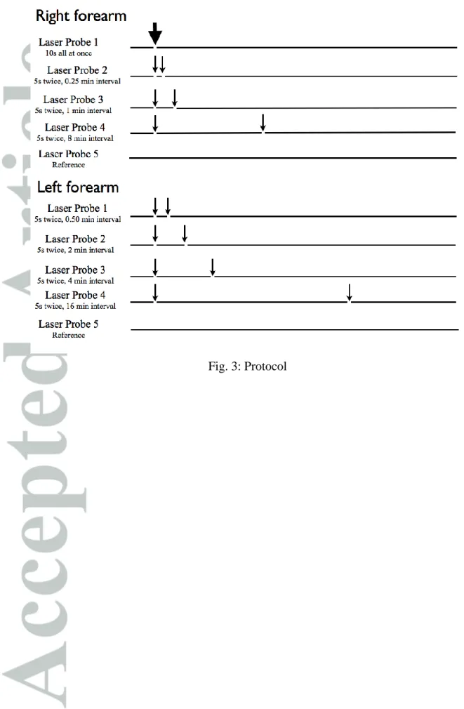
all-at-once (10 s) or in two consecutive 5-s applications with different intersimulation intervals.
Four different intervals were tested: 15 s (1/4 min), 30 s (1/2 min), 1 min, 2 min, 4 min, 8 min
and 16 min. The procedure is described on Figure 3.
Cardiovascular and temperature recordings
Blood pressure was continuously recorded from the left middle finger (Finapres,
Ohmeda, Madison, WI, USA) with mean arterial pressure (MAP) obtained from the
continuous blood pressure signal. The MAP signal was recorded at a sampling rate of 18 Hz
using an analog to digital converter (MP100, Biopac sys, Goleta, USA) and analyzed offline
using the Acknowledge® software V3.5.4 (Biopac sys, Goleta, USA).
Local skin temperature was measured using a surface thermocouple probe positioned
5 cm from any of the other electrodes [8]. The thermocouple was connected to an electronic
thermometer (BAT-12, Physitemp Instruments, Inc., Clifton, NJ, USA).
Data and statistical analyses
Results are presented as mean SD, and for CVC, mediane (1rst
percentile, 3rd
Results for raw data were expressed in CVC or in multiple of baseline CVC values.
CVC was calculated as the ratio of cutaneous blood flow to mean arterial pressure (mmHg)
and expressed in PU/mmHg [10].
The values recorded before placebo or aspirin intake have been considered as the
standard response.
For further analysis, the following points were determined for each subject and
measurement:
- mean reference CVC (average of the CVC values during the first min of the
reference period, CVCref),
- CVCstim2 corresponding to the CVC values 10 min after the end of the second
current application for the segmented current applications or 10 min after the
10s current application during all-at-once delivery.
Parametric or non parametric tests were performed according to the distribution of
values using SPSS (IBM SPSS statistics V15.0, Chicago, USA). Differences in CVCref and
CVCstim2 were analyzed via the test of Wilcoxon using. Increases in CVCstim2 from baseline in
function of the delivery mode and the interstimulation interval were compared via repeated
measures ANOVA. When a significant difference was found, Tukey post hoc analysis was
performed. For each statistical analysis, a two- p ≤ . 5 w
RESULTS:
Subjects were 22 ± 2 years old (10 males, 6 females). Mean weight and height were
68.1 ± 9.4 kg and 172.8 ± 9.5 cm, respectively. At the beginning of the protocol, their blood
characteristics were the following: glycemia: 4.6 ± 0.5 mmol/L, LDL cholesterol: 2.7 ± 0.6
mmol/L, HDL cholesterol: 1.5 ± 0.3 mmol/L, creatinemia: 73.3 ± 9.2 µmol/L and number of
platelets: 247.6 ± 49.8 (giga/L).
There was no significant change in cutaneous temperature during the measurement,
whatever the day considered. Mean skin temperature ranged between 33.8 ± 1.5 to 34.6 ± 1.1
°C.
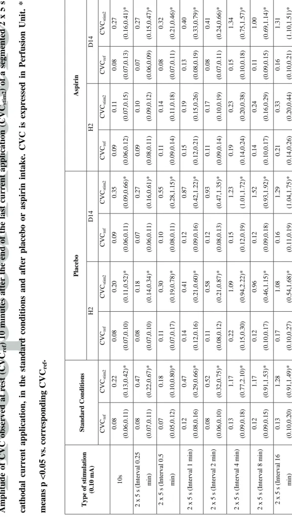
Table 1 presents the CVC values (CVCref and CVCstim2) recorded in standard
conditions, i.e. before any kind of treatment, and the CVC values observed after placebo or
aspirin intake depending on the delivery mode of current application (10 s or 2 x 5 s with
various intervals).
In standard condition, i.e. before treatment, CVC increased significantly from 0.08 (0.06,
0.11) to 0.22 (0.13, 0.42) PU/mmHg following an all-at-once 10 s continuous current
application. When the current application was segmented (i.e. 2x5 s with different intervals),
CVC also increased significantly from baseline whatever the interval considered. However,
CVCstim2 was significantly greater when the total current charge was segmented compared to
all-at-once delivery when the interval between the two 5 s current application was at least of
4 min. For example, CVC increased by 3.1 (2.5, 5.8) times for a 2x5 s current application
with an interval of 1 min (non significant (NS) vs. all-at-once delivery). On the contrary, it
increased by 9.4 (5.3, 10.9) times with an interval of 4 min (p<0.05 vs. all-at-once delivery).
We did not observe any change in CVCstim2 amplitude when the interstimulation interval
Placebo treatment did not affect significantly the responses observed in standard conditions
and the values recorded 14 days after placebo intake were not different from the standard
response (Table 1).
Intake of 1g aspirin resulted in the disappearance of CIV following the all-at-once 10 s
current application (Table 1). Two hours after intake, CVCref was 0.09 (0.06, 0.012)
PU/mmHg and CVCstim2 was 0.11 (0.07, 0.15) PU/mmHg (NS vs. CVCref). Aspirin also
abolished the CIV induced by segmented current application whatever the interval considered
and there was no difference in CVCstim2 between the continuous 10 s current application and
the segmented current application. For example, CVC increased by 1.2 (1.1, 1.3) times for all
at once 10 s current application and by 1.5 (1.3, 1.7) times for a segmented current
application with a 4 min interval.
Fourteen days after aspirin intake, values recorded were not different from those recorded
before the treatment. Again, after current application (all at once or segmented), CVC
increased significantly from baseline (Table 1). In addition, increase in CVCstim2 was
significantly greater when the total current charge was segmented compared to all-at-once
delivery when the interstimulation interval between was 4 min and more. CVC increased by
3.9 (3.4, 9.7) times for a 2 x 5 s current application with an interval of 2 min (NS vs. all at
once); but it increased by 8.0 (5.9, 10.9) (p<0.05 vs. all at once) with an interval of 4 min.
We did not observe any change in CVC all along the different microvascular assessments at
DISCUSSION:
This study confirms that segmented application of cathodal low intensity (0.10 mA)
current leads to a significant and greater vasodilation in healthy human skin compared to the
one observed when the current total charge is applied all-at-once. This suggests that the first
current application can activate a sensitization mechanism that is abolished by aspirin. The
magnitude of this sensitization mechanism relies on the length of the interstimulation interval
and a minimum of 4 minutes is necessary to observe a significant sensitization mechanism.
Due to its sensitivity to aspirin, this mechanism likely relies on the release of prostaglandins.
CIV is probably based on an axon reflex [2, 11] but the mechanisms participating in
CIV with all-at-once or segmented cathodal current application are not perfectly known [3].
CIV in response to anodal or cathodal current application (whatever the delivery mode) is
abolished by aspirin suggesting the involvement of COD (prostaglandins?) in the vasodilator
mechanism [5, 6]. Gohin et al. confirmed the participation of PGI2 in the CIV in response to
a single prolonged (240 s) current application in rat skin [4]. PGE2 and PGI2 are powerful
vasodilators on their own but prostaglandins are also able to sensitize the response of afferent
unmyelinated fibres in animal [12, 13].
We previously hypothesized the participation of COD in the neural sensitization to
anodal current application [5]. This would explain the amplified CIV observed for an amount
of electrical charge delivered segmented compared to all-at-once [5]. This hypothesis was
comforted latter at the cathodal level by the observation of Tartas et al. [6]. Durand et al. in
supplementary material brought initially the evidence that this mechanism of sensitization
was long lasting since still occurring 90 min after the first current application but yet its
intensity was declining with time [5]. Using segmented current application (two sequences of
anodal current, 0.1mA, 1 min with interstimulation interval of 5, 10, 20, 40, 60 or 90 min),
level and progressively diminished with time. We hypothesized that the initial anodal current
application had lead to the release of prostaglandins acting as sensitizing agent on the neural
fibres involved in CIV leading to a large CIV when current application is repeated and that
the progressive catabolism of prostaglandins would explain the blunting of this sensitizing
mechanism [5]. At the cathodal level, the CIV is usually more important than the CIV at the
anodal level for the same intensity and duration of current application [3]. Consequently,
Tartas et al. used shorter segmented current application (two sequences of cathodal current,
0.10 mA, 10 s with interstimulation interval of 10, 20 or 40 min) to investigate the presence
or not of the mechanism of sensitization at the cathodal level [6]. This mechanism was
observed again with an amplitude in CIV, for a given total electric charge, superior when the
current application was segmented compared to all-at-once. However, the authors did not
report any difference in CIV amplitude whatever the interval considered. Considering the fact
that cathodal current applications elicit more CIV than anodal current, this observation could
be explained by an important and long lasting release of prostaglandins during the first
current application leading to an important and long lasting mechanism of sensitization.
However, some questions remain concerning this mechanism.
In the aim to better characterize this mechanism of sensitization at the cathodal level,
we used half of the amount of electrical charge used by Tartas et al. (i.e. in our study two
sequences of cathodal current, 0.10 mA, 5 s) and proposed shorter interstimulation interval
(from 2 min to 16 min) [6]. The hypothesis here was that a minimal interval was necessary to
have the mediators of sensitization released. We observed that after the second current
application, a significant amplification of CIV, compared to CIV measured after a single
all-at-once 10 s cathodal current application, appears for an intersimulation interval of 4 min.
This amplification was abolished 2 hours after aspirin intake whatever the interstimulation
current application. Indeed, we previously reported a disappearance of CIV following 2 x
1min current applications with a 10 min interstimulation interval 2 hours after aspirin intake
[5]. COD like prostaglandins are known to act as sensitizing agent and to be aspirin sensitive.
It is likely that the same kind of COD participate in the mechanism of sensitization at the
anode and at the cathode. Consequently we hypothesized that a single 5 s cathodal current
application is sufficient to induce a significant release of COD acting as sensitizing agents
when a cathodal current application is repeated. A minimal delay between two monopolar
current applications has to be respected to observe the sensitizing effect of COD leading to
amplification of CIV (4 min in our experimental conditions). The exact nature of these COD
remains to be determined.
In conclusion, several tests exist to investigate cutaneous vasomotor function. For
example, post occlusive reactive hyperemia stimulates both the endothelial and the
non-endothelial pathway [14, 15]. Sodium nitroprusside and acetylcholine iontophoresis
investigate the non-endothelial and the endothelial pathways, respectively [9, 10, 16, 17].
Gohin et al. proposed to use CIV to investigate the vascular function of prostacyclin in rats
[4]. We confirm the involvement of COD in CIV and the existence of a sensitizing
mechanism leading to amplification of CIV when cathodal current application is repeated.
The same observation has been previously made for segmented anodal current application.
This suggests that the study of CIV in response to segmented anodal or cathodal current
application, when the appropriate charge is applied and a minimal interstimulation interval
respected, can be an attractive way to study the neural sensitizing function of
Limits
This study has been conducted in young and healthy subjects. We cannot exclude a
change in the amplitude and mechanisms in other populations such as elderly people, diabetic
subjects or patients suffering from cardiovascular disease. Furthermore, to guarantee the
same cardiovascular state for determination of CVC, all measurements (i.e. the tests of the
different interstimulation intervals) have been performed during the same experimental
session on the two arms. It could be consequently hypothesized that the different current
applications performed could interfere one over another on a same arm and that the responses
could differ between the two arms. However, sites of current applications on the forearms
were randomized and the current was locally applied. There is no report of a difference in
CIV in function of the arm (left or right), there was no change in CVC at the reference probe
and we clearly observed an influence of the interstimulation interval on the apparition of
CIV. All these elements lead us to the conclusion that there was no interference between the
LIST OF SYMBOLS AND ABBREVIATIONS
CIV: Current Induced Vasodilation
CVC: Cutaneous Vascular Conductance
COD: Cyclo-Oxygenase Derivates
PGE2: Prostaglandin E2
PGI2: Prostaglandin I2
NS: Non Significant
AUTHORS CONTRIBUTIONS
Conceived and designed the experiments: SD, GM, PA. Performed the experiments: LG, GM
Analyzed the data: SD, GM. Wrote the paper: SD, GM. Statistical analysis: SD, GM. Final
approval of the article: SD, GM, AH, LG, GL, PA.
FUNDING
This study was supported by the university hospital of Angers (Microtec study) and was
STATEMENT OF CONFLICTS OF INTEREST
"All authors have completed the Unified Competing Interest form at
http://www.icmje.org/coi_disclosure.pdf (available on request from the corresponding author)
and declare: no support from any organisation for the submitted work; no financial
relationships with any organisations that might have an interest in the submitted work in the
previous 3 years; no other relationships or activities that could appear to have influenced the
REFERENCES
1. Berliner MN. Skin microcirculation during tapwater iontophoresis in humans: cathode stimulates more than anode. Microvasc Res 1997; 54: 74-80.
2. Grossmann M, Jamieson MJ, Kellogg DL, Jr., Kosiba WA, Pergola PE, Crandall CG, Shepherd AM. The effect of iontophoresis on the cutaneous vasculature: evidence for
current-induced hyperemia. Microvasc Res 1995; 50: 444-52.
3. Durand S, Fromy B, Bouye P, Saumet JL, Abraham P. Current-induced vasodilation during water iontophoresis (5 min, 0.10 mA) is delayed from current onset and involves aspirin sensitive mechanisms. J Vasc Res 2002; 39: 59-71.
4. Gohin S, Sigaudo-Roussel D, Conjard-Duplany A, Dubourg L, Saumet JL, Fromy B. What can current stimulation tell us about the vascular function of endogenous prostacyclin in healthy rat skin in vivo? J Invest Dermatol 2011; 131: 237-44.
5. Durand S, Fromy B, Bouye P, Saumet JL, Abraham P. Vasodilatation in response to repeated anodal current application in the human skin relies on aspirin-sensitive mechanisms. J Physiol 2002; 540: 261-9.
6. Tartas M, Bouye P, Koitka A, Jaquinandi V, Tan L, Saumet JL, Abraham P. Cathodal current-induced vasodilation to single application and the amplified response to repeated application in humans rely on aspirin-sensitive mechanisms. J Appl Physiol 2005; 99: 1538-44.
7. Durand S, Fromy B, Koitka A, Tartas M, Saumet JL, Abraham P. Oral single high-dose aspirin results in a long-lived inhibition of anodal current-induced vasodilatation. Br J Pharmacol 2002; 137: 384-90.
8. Abraham P, Bourgeau M, Camo M, Humeau-Heurtier A, Durand S, Rousseau P, Mahe G. Effect of skin temperature on skin endothelial function assessment. Microvasc Res 2013; 88: 56-60.
9. Puissant C, Abraham P, Durand S, Humeau-Heurtier A, Faure S, Leftheriotis G, Mahe G. Assessment of endothelial function by acetylcholine iontophoresis: Impact of
inter-electrode distance and electrical cutaneous resistance. Microvasc Res 2014; 93: 114-8. 10. Mahe G, Humeau-Heurtier A, Durand S, Leftheriotis G, Abraham P. Assessment of skin microvascular function and dysfunction with laser speckle contrast imaging. Circulation Cardiovascular imaging 2012; 5: 155-63.
11. Ferrell WR, Ramsay JE, Brooks N, Lockhart JC, Dickson S, McNeece GM, Greer IA, Sattar N. Elimination of electrically induced iontophoretic artefacts: implications for non-invasive assessment of peripheral microvascular function. J Vasc Res 2002; 39: 447-55. 12. Lopshire JC, Nicol GD. Activation and recovery of the PGE2-mediated sensitization of the capsaicin response in rat sensory neurons. Journal of neurophysiology 1997; 78: 3154-64.
13. Minami T, Okuda-Ashitaka E, Hori Y, Sakuma S, Sugimoto T, Sakimura K, Mishina M, Ito S. Involvement of primary afferent C-fibres in touch-evoked pain (allodynia) induced by prostaglandin E2. The European journal of neuroscience 1999; 11: 1849-56.
14. Cracowski JL, Gaillard-Bigot F, Cracowski C, Sors C, Roustit M, Millet C.
Involvement of cytochrome epoxygenase metabolites in cutaneous postocclusive hyperemia in humans. J Appl Physiol (1985) 2013; 114: 245-51.
15. Lorenzo S, Minson CT. Human cutaneous reactive hyperaemia: role of BKCa channels and sensory nerves. J Physiol 2007; 585: 295-303.
16. Puissant C, Abraham P, Durand S, Humeau-Heurtier A, Faure S, Leftheriotis G, Rousseau P, Mahe G. Reproducibility of non-invasive assessment of skin endothelial
function using laser Doppler flowmetry and laser speckle contrast imaging. PloS one 2013; 8: e61320.
17. Puissant C, Abraham P, Durand S, Humeau-Heurtier A, Faure S, Rousseau P, Mahe G. [Endothelial function: role, assessment and limits]. J Mal Vasc 2014; 39: 47-56.
T ABL E 1 A m p li tud e of CV C ob se rve d at r est (C VC ref ) 10 m in u te s af te r the en d of the last cu rr en t ap p li cat ion ( CVC st im 2 ) of a se gme n te d 2 x 5 s cat h od al cu rr en t ap p li cat ion , in the stand ar d c on d ition s an d a fte r p lace b o or asp irin in take. CV C is exp re ss ed in Pe rf u sion Uni t. * m ean s p <0.05 vs. cor re sp on d in g CVC re f . Ty p e o f sti m u la tio n (0 .1 0 m A) S ta n d a r d C o n d iti o n s Pl a ce b o As p iri n H2 D 1 4 H2 D 1 4 CV Cre f CV Cst im 2 CV Cre f CV Cst im 2 CV Cre f CV Cst im 2 CV Cre f CV Cst im 2 CV Cre f CV Cst im 2 10 s 0 .0 8 (0 .0 6 ,0 .1 1 ) 0 .2 2 (0 .1 3 ,0 .4 2 )* 0 .0 8 (0 .0 7 ,0 .1 0 ) 0 .2 0 (0 .1 1 ,0 .5 2 )* 0 .0 9 (0 .0 6 ,0 .1 1 ) 0 .3 5 (0 .0 9 ,0 .6 6 )* 0 .0 9 (0 .0 6 ,0 .1 2 ) 0 .1 1 (0 .0 7 ,0 .1 5 ) 0 .0 8 (0 .0 7 ,0 .1 3 ) 0 .2 7 (0 .1 6 ,0 .4 1 )* 2 x 5 s (I n terv al 0 .2 5 m in ) 0 .0 8 (0 .0 7 ,0 .1 1 ) 0 .4 7 (0 .2 2 ,0 .6 7 )* 0 .0 8 (0 .0 7 ,0 .1 0 ) 0 .1 8 (0 .1 4 ,0 .3 4 )* 0 .0 7 (0 .0 6 ,0 .1 1 ) 0 .2 7 (0 .1 6 ,0 .6 1 )* 0 .0 9 (0 .0 8 ,0 .1 1 ) 0 .1 0 (0 .0 9 ,0 .1 2 ) 0 .0 7 (0 .0 6 ,0 .0 9 ) 0 .2 7 (0 .1 5 ,0 .4 7 )* 2 x 5 s (I n terv al 0 .5 m in ) 0 .0 7 (0 .0 5 ,0 .1 2 ) 0 .1 8 (0 .1 0 ,0 .8 0 )* 0 .1 1 (0 .0 7 ,0 .1 7 ) 0 .3 0 (0 .1 9 ,0 .7 8 )* 0 .1 0 (0 .0 8 ,0 .1 1 ) 0 .5 5 (0 .2 8 ,1 .1 5 )* 0 .1 1 (0 .0 9 ,0 .1 4 ) 0 .1 4 (0 .1 1 ,0 .1 8 ) 0 .0 8 (0 .0 7 ,0 .1 1 ) 0 .3 2 (0 .2 1 ,0 .4 6 )* 2 x 5 s (I n terv al 1 m in ) 0 .1 2 (0 .0 8 ,0 .1 6 ) 0 .4 7 (0 .2 9 ,0 .6 6 )* 0 .1 4 (0 .1 2 ,0 .1 6 ) 0 .4 1 (0 .2 1 ,0 .6 0 )* 0 .1 2 (0 .0 9 ,0 .1 6 ) 0 .8 7 (0 .4 2 ,1 .2 2 )* 0 .1 5 (0 .1 2 ,0 .2 1 ) 0 .1 9 (0 .1 5 ,0 .2 6 ) 0 .1 3 (0 .0 8 ,0 .1 9 ) 0 .4 0 (0 .3 3 ,0 .7 9 )* 2 x 5 s (I n terv al 2 m in ) 0 .0 8 (0 .0 6 ,0 .1 0 ) 0 .5 2 (0 .3 2 ,0 .7 5 )* 0 .1 1 (0 .0 8 ,0 .1 2 ) 0 .5 8 (0 .2 1 ,0 .8 7 )* 0 .1 2 (0 .0 8 ,0 .1 3 ) 0 .9 3 (0 .4 7 ,1 .3 5 )* 0 .1 1 (0 .0 9 ,0 .1 4 ) 0 .1 7 (0 .1 0 ,0 .1 9 ) 0 .0 8 (0 .0 7 ,0 .1 1 ) 0 .4 1 (0 .2 4 ,0 .6 6 )* 2 x 5 s (I n terv al 4 m in ) 0 .1 3 (0 .0 9 ,0 .1 8 ) 1 .1 7 (0 .7 7 ,2 .1 0 )* 0 .2 2 (0 .1 5 ,0 .3 0 ) 1 .0 9 (0 .9 4 ,2 .2 2 )* 0 .1 5 (0 .1 2 ,0 .1 9 ) 1 .2 3 (1 .0 1 ,1 .7 2 )* 0 .1 9 (0 .1 4 ,0 .2 4 ) 0 .2 3 (0 .2 0 ,0 .3 8 ) 0 .1 5 (0 .1 0 ,0 .1 8 ) 1 .3 4 (0 .7 5 ,1 .5 7 )* 2 x 5 s (I n terv al 8 m in ) 0 .1 2 (0 .0 9 ,0 .1 5 ) 1 .1 7 (0 .9 1 ,1 .5 3 )* 0 .1 2 (0 .1 0 ,0 .1 7 ) 0 .9 6 (0 .4 6 ,1 .1 5 )* 0 .1 2 (0 .0 9 ,0 .1 8 ) 1 .5 2 (0 .9 3 ,1 .9 2 )* 0 .1 4 (0 .1 0 ,0 .1 7 ) 0 .2 4 (0 .1 6 ,0 .2 9 ) 0 .1 1 (0 .0 9 ,0 .1 5 ) 1 .0 0 (0 .6 9 ,1 .1 4 )* 2 x 5 s (I n terv al 1 6 m in ) 0 .1 3 (0 .1 0 ,0 .2 0 ) 1 .2 8 (0 .9 1 ,1 .4 9 )* 0 .1 7 (0 .1 0 ,0 .2 7 ) 1 .0 8 (0 .5 4 ,1 .6 8 )* 0 .1 6 (0 .1 1 ,0 .1 9 ) 1 .2 9 (1 .0 4 ,1 .7 5 )* 0 .2 1 (0 .1 4 ,0 .2 6 ) 0 .3 3 (0 .2 0 ,0 .4 4 ) 0 .1 6 (0 .1 0 ,0 .2 1 ) 1 .3 1 (1 .1 0 ,1 .5 1 )*
Fig. 1: Design of the study. D0 means day of treatment. H2 means time of measurement 2