HAL Id: hal-01824325
https://hal.umontpellier.fr/hal-01824325
Submitted on 13 Apr 2020
HAL is a multi-disciplinary open access
archive for the deposit and dissemination of
sci-entific research documents, whether they are
pub-lished or not. The documents may come from
teaching and research institutions in France or
abroad, or from public or private research centers.
L’archive ouverte pluridisciplinaire HAL, est
destinée au dépôt et à la diffusion de documents
scientifiques de niveau recherche, publiés ou non,
émanant des établissements d’enseignement et de
recherche français ou étrangers, des laboratoires
publics ou privés.
natriuretic peptide in cardiac remodelling and
pro-arrhythmogenicity
J Thireau, Sarah Karam, J. Fauconnier, Stéphanie Roberge, Cécile Cassan,
Olivier Cazorla, Franck Aimond, Alain Lacampagne, Dominique Babuty,
Sylvain Richard
To cite this version:
J Thireau, Sarah Karam, J. Fauconnier, Stéphanie Roberge, Cécile Cassan, et al.. Functional evidence
for an active role of B-type natriuretic peptide in cardiac remodelling and pro-arrhythmogenicity.
Car-diovascular Research, Oxford University Press (OUP), 2012, 95 (1), pp.59 - 68. �10.1093/cvr/cvs167�.
�hal-01824325�
... ... .... ... ... ... .... ... ... ... .... ... ... ... ... .... ... ... ... .... ... ... ... .... ... ... ... .... ... ... ... .... ... ... ... ... .... ... ..
... ... .... ... ... ... .... ... ... ... .... ... ... ... ... .... ... ... ... .... ... ... ... .... ... ... ... .... ... ... ... .... ... ... ... ... .... ... ..
Functional evidence for an active role of B-type
natriuretic peptide in cardiac remodelling
and pro-arrhythmogenicity
Je´roˆme Thireau
1, Sarah Karam
1, Je´re´my Fauconnier
1, Ste´phanie Roberge
1,
Ce´cile Cassan
1, Olivier Cazorla
1, Franck Aimond
1, Alain Lacampagne
1,
Dominique Babuty
2, and Sylvain Richard
1*
1INSERM U1046, Physiologie and Me´decine Expe´rimentale du Cœur et des Muscles, Universite´ Montpellier-1, Universite´ Montpellier-2, CHU Arnaud de Villeneuve, 371, Rue du doyen
G. Giraud, Montpellier 34295, France; and2Cardiologie B, Hoˆpital Trousseau, Tours, France
Received 14 February 2012; revised 9 May 2012; accepted 16 May 2012; online publish-ahead-of-print 22 May 2012 Time for primary review: 23 days
Aims During heart failure (HF), the left ventricle (LV) releases B-type natriuretic peptide (BNP), possibly contributing to adverse cardiovascular events including ventricular arrhythmias (VAs) and LV remodelling. We investigated the cardiac effects of chronic BNP elevation in healthy mice and compared the results with a model of HF after myocar-dial infarction (PMI mice).
Methods and results
Healthy mice were exposed to circulating BNP levels (BNP-Sham) similar to those measured in PMI mice. Telemetric surface electrocardiograms showed that in contrast with fibrotic PMI mice, electrical conduction was not affected in BNP-Sham mice. VAs were observed in both BNP-Sham and PMI but not in Sham mice. Analysis of heart rate vari-ability indicated that chronic BNP infusion increased cardiac sympathetic tone. At the cellular level, BNP reduced Ca2+transients and impaired Ca2+reuptake in the sarcoplasmic reticulum, in line with blunted SR Ca2+ATPase
2a and S100A1 expression. BNP increased Ca2+spark frequency, reflecting Ca2+leak through ryanodine receptors,
elevated diastolic Ca2+, and promoted spontaneous Ca2+waves. Similar effects were observed in PMI mice. Most of
these effects were reduced in BNP-Sham and PMI mice by the selective b1-adrenergic blocker metoprolol.
Conclusion Elevated BNP levels, by inducing sympathetic overdrive and altering Ca2+handling, promote adverse cardiac
remod-elling and VAs, which could account in part for the progression of HF after MI. The early use of b-blockers to prevent the deleterious effects of chronic BNP exposure may be beneficial in HF.
-Keywords Natriuretic peptide † Heart failure † Arrhythmia † Calcium † Remodelling
1. Introduction
B-type natriuretic peptide (BNP) is synthesized in cardiac myocytes and released in excess into the blood circulation following left ven-tricular (LV) wall stretching.1During the onset of ventricular
remod-elling after myocardial infarction (PMI), BNP is chronically elevated and is a strong indicator of heart failure (HF) severity.1Despite
fa-vourable haemodynamic effects, high blood BNP is associated with the risk of ventricular arrhythmias (VAs) and sudden cardiac death (SCD).2,3 Consequently, the effectiveness of therapies aimed at
in-creasing BNP during HF is still questionable.4 While short-term
BNP infusion is of haemodynamic benefit, with natriuretic, diuretic,
and vasorelaxant effects, chronic BNP treatment increases the risk of mortality.3,5,6 Sympathetic activation related to a reflex response
triggered by these haemodynamic effects could be implicated in this process.7,8Although the signalling pathways mediating the effects of
BNP are poorly understood, a therapeutic strategy that could prevent the deleterious effects of BNP without affecting its benefits remains of interest.
Altered Ca2+cycling, characterized by increased sarcoplasmic
re-ticulum (SR) Ca2+leak, decreased SR Ca2+uptake leading to diastolic
Ca2+ elevation, and decreased systolic Ca2+ levels, is a common
feature in chronic HF.9–11 Such altered Ca2+-handling and chronic
sympathetic overdrive are established components of chronic HF
*Corresponding author. Tel: +33 4 67 41 52 40; fax: +33 4 67 41 52 42, Email: sylvain.richard@inserm.fr
Published on behalf of the European Society of Cardiology. All rights reserved.&The Author 2012. For permissions please email: journals.permissions@oup.com.
Cardiovascular Research (2012) 95, 59–68 doi:10.1093/cvr/cvs167 Downloaded from ht tps: //academic. oup. com/ cardiovascres/ art icle-abst ract /95/ 1/ 59/ 331506 by guest on 13 A pril 2020
triggering VA.12BNP decreases the expression of SR Ca2+ATPase 2a
(SERCA2a)13,14 and induces pro-adrenergic effects.15 We thus
hypothesized that BNP could influence two key players in the trigger-ing of VA and, more globally, in cardiac remodelltrigger-ing durtrigger-ing HF.
Our present study attempts to determine the effect of chronic BNP treatment on LV function both in vivo in healthy mice and at the cel-lular level. Here, we show, by heart rate variability (HRV) analysis and the use of the b-blocker metoprolol, that increased sympathetic tone associated with Ca2+-handling alterations contributes to
BNP-mediated LV remodelling and VA.
2. Methods
2.1 Animals and BNP
All procedures conformed to European Parliament Directive 2010/63/EU and the 22 September 2010 Council on the protection of animals and were approved by the local institutional animal research committee (Languedoc Roussillon, No. CE-LR-0714).
For cardiomyocyte experiments, the heart was explanted after euthan-asia by cervical dislocation. Seven-week-old male C57BL/6 mice (Janvier, Le Genest-Saint-Isle, France) were randomly assigned to the following groups: (i) Sham; (ii) Sham treated with BNP (BNP-Sham); (iii) mice sub-jected to HF by left coronary artery ligation (PMI); (iv) BNP-Sham mice treated with the b1-adrenergic blocker metoprolol (BB-BNP-Sham); and (v) PMI mice treated with metoprolol (BB-PMI). For PMI, a left thora-cotomy was performed under anaesthesia and cardiac monitoring (2% iso-flurane/O2, Aerranew, Baxter, France). The artery was ligated 1–2 mm
beyond its point of emergence from the top of the left atrium, using an 8-0 suture. A subcutaneous injection of 0.01 mL buprenorphine solution (0.3 mg/mL) was administered for post-operative analgesia. Sham mice were subjected to the same surgical procedure but without coronary artery ligation. Two mice died during surgery, i.e. before their inclusion in the study, and were replaced. Metoprolol (Sigma-Aldrich, 100 mg/kg/ day) was administered in the drinking water. The dose was determined based on the literature. In the range of 60–350 mg/kg/day, metoprolol has beneficial effects on cardiac and cellular function.16–20 Here, the
dose of 100 mg/kg/day reduced the heart rate and improved the HRV parameters in PMI mice without affecting the water intake. Mouse BNP(1–45) (Ref 14-5-30A, American Peptide, Sunnyvale, CA, USA) was administered to Shams at a rate of 0.03 mg/kg/min for 14 days, using a micro-osmotic pump (Alzet 1002, Charles River, France), to achieve plasma BNP levels similar to the steady-state level observed in PMI mice. Circulating BNP levels were measured in duplicate using a commer-cial kit (Phoenix Pharmaceuticals, Belmont, CA, USA). The timeline of experiments is shown in the Supplementary material online, Figure S1.
2.2 Histology
Haematoxylin–eosin and Sirius red staining were performed as described.21Results indicate the area of Sirius red-stained tissue as a
per-centage of the total area of myocardial tissue.
2.3 In vivo analysis
Cardiac function was assessed by echocardiography (Vivid7Pro, GE Medical Systems, USA). LV mass, LV shortening fraction (SF), end-diastolic LV dimension (LVEDd), and end-systolic LV dimension (LVEDs) were measured.10 Electrocardiograms (ECGs) were recorded by telemetry
(DSI, St Paul, MN, USA, and EMKA Technologies, France).10HRV, the
PR, QRS, and QTc intervals, and arrhythmias were analysed using 12 h nocturnal ECGs (ECG-auto, EMKA Technologies).21
2.4 Ca
21handling
LV myocytes were enzymatically dissociated, loaded with a fluorescent Ca2+indicator Fluo-4 AM (5 mmol/L, Molecular Probes, Paris, France),
and field-stimulated at 1.0 Hz to assess intracellular Ca2+ transients
and cell shortening.10 Ca2+ sparks were recorded in quiescent cells
(1.5 ms/line, LSM510 Zeiss confocal microscope; ×63 water-immersion objective; NA: 1.2) at 258C.10Cell volume was estimated using Z-stack
(x–y projection, front view) image acquisition. Data were analysed using ImageJ and ‘SparkMaster’. Cellular arrhythmias and diastolic Ca2+ levels
were measured with ratiometric Indo-1 AM (10 mM, Invitrogen, France; IonOptix System, Hilton, USA).10 Ca2+ fluorescence was measured
during a 30 s pacing period (1.0 Hz), followed by a 30 s rest period. Dia-stolic Ca2+levels and the number of cells developing ectopic Ca2+
tran-sients during the rest period were quantified.
2.5 Ca
21-handling proteins
LV proteins were separated using 2–20% SDSi–PAGE, blotted onto PVDF membranes (Protean, Germany) and incubated overnight at 48C with primary antibodies: RyR-2 (Covalab, France), Phospho Ser2809-RyR-2 (A010-30, Badrilla, UK), SERCA2a (A010-20, Badrilla),
phospholamban (PLB; A010-14, Badrilla), PhosphoSer16-PLB (A010-12, Badrilla), NCX1 (R3F1, Swant, Switzerland), and S100A1 (SP5355P, Acris Antibodies GmbH, Germany). Protein levels were expressed rela-tive to calsequestrin (PA1-913, ABR, USA). Immunodetection was per-formed using the ECL Plus system (Amersham, UK).
2.6 Statistical analysis
All data are reported as means + standard deviation. Statistical analyses were performed using GraphPad Prism and Origin Softwares. One-way ANOVA for multiple comparisons was used, followed by a parametric t-test with Bonferroni’s correction for all parameters. A P-value of 0.05 or less indicated a statistically significant difference.
3. Results
3.1 Plasma BNP levels in BNP-Sham
and PMI mice
Sham mice had levels of circulating BNP lower than the detection limit (,0.34 ng/mL). In PMI mice, BNP increased to 5.4 + 1.2 ng/mL (n ¼ 8) 14 days after MI and remained stable over the next 2 weeks. At week 4, BNP-Sham mice presented BNP levels (6.8 + 1.4 ng/mL, n ¼ 8) similar to those measured in PMI animals (5.1 + 0.9 ng/mL, n ¼ 7).
3.2 Morphofunctional and
electrocardiographic effects of BNP
Chronic BNP treatment increased the heart weight–body weight ratio (HWR) of BNP-Sham and PMI mice (Table1). Unlike modifica-tions in PMI mice, echocardiography revealed an unchanged LVEDd and SF in BNP-Sham mice (Table 1). ECG showed an unchanged heart rate in BNP-Sham mice, whereas it was increased in PMI mice as indicated by the decreased RR interval (Figure1A). The PR interval, corresponding to the conduction time from the sinus node through the atrioventricular (AV) node to the ventricle, was unaltered in both BNP-Sham and PMI mice (Figure1A), whereas the QRS duration, representing the time to ventricular depolarization and early repolar-ization, was lengthened in PMI mice (Figure1C). In addition, multiple spikes within the complex (fragmented QRS),22probably caused by
myocardial scarring, were observed in PMI but not in BNP-Sham mice. The QTc interval was increased in both BNP-Sham and PMI
Downloaded from ht tps: //academic. oup. com/ cardiovascres/ art icle-abst ract /95/ 1/ 59/ 331506 by guest on 13 A pril 2020
Figure 1 ECG analysis. Parameters estimated from 12 h nocturnal ECGs: heart rate (RR interval) (A), PR interval (B), QRS duration (C), and QTc interval (D). Heart rate variability analysed in the time-domain (SDNN) (E) and frequency-domain with low-frequency (LF; F) and high-frequency bands (HF; G) and LF/HF ratio (H ). *P , 0.05, **P , 0.01, BNP-Sham/PMI vs. Sham animals;£P , 0.05, ££P , 0.01 for metoprolol-treated vs.
untreated animals, n ¼ 12 for each group.
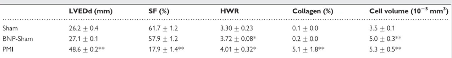
... ... ... ... ... ... ... ... ... ... ... ... Table 1 Echocardiography, histology, and cellular hypertrophy
LVEDd (mm) SF (%) HWR Collagen (%) Cell volume (1025mm3)
Sham 26.2 + 0.4 61.7 + 1.2 3.30 + 0.23 0.1 + 0.0 3.5 + 0.1 BNP-Sham 27.1 + 0.1 57.9 + 1.2 3.72 + 0.08* 0.2 + 0.0 5.0 + 0.3** PMI 48.6 + 0.2** 17.9 + 1.4** 4.01 + 0.32* 5.1 + 1.8** 5.3 + 0.5**
End-diastolic diameter of left ventricular cavity (LVEDd), shortening fraction (SF), and heart weight–body weight ratio (HWR) (10 mice/group). Collagen content, expressed as a percentage of the total area of myocardial tissue analysed (7 mice/group) and cell volume (n ¼ 30 cells, 3 mice/group) were estimated *P , 0.05, **P , 0.01, BNP-Sham/PMI vs. Sham animals.
BNP induces adrenergic-dependent Ca2+-cycling alterations 61
Downloaded from ht tps: //academic. oup. com/ cardiovascres/ art icle-abst ract /95/ 1/ 59/ 331506 by guest on 13 A pril 2020
mice (Figure1D). ECG analyses showed that chronic BNP treatment did not alter electrical conduction. In accordance with this finding, BNP had no effect on fibrosis as quantified by collagen content, whereas a large increase was observed in PMI mice (Table1).
3.3 Effect of BNP on cardiac
sympathovagal regulation
We examined the regulation of the cardiac rhythm by the autonomic nervous system (ANS) by examining the HRV.10,21,23,24 The HRV,
assessed by the standard deviation of the mean of all normal sinus intervals (SDNN), was comparably decreased in BNP-Sham and PMI mice (Figure 1E), suggesting an elevation of sympathetic tone.23,24
Frequency-domain analysis showed that oscillations in ‘low’-frequency bands (LF), reflecting parasympathetic and sympathetic components, were increased in BNP-Sham mice (Figure 1F), whereas ‘high’-frequency oscillations (HF), reflecting exclusively vagal activity, were unchanged (Figure1G). The increase in the LF bands in BNP-Sham mice is consistent with increased sympathetic activity without a de-tectable modification of the mean heart rate.25 The typical profile
of HRV parameters measured in PMI mice, with a blunted LF band and LF/HF ratio (Figure1F–H), confirmed the loss of rhythmicity of ANS activity on the heart and the severity of cardiac pathology in PMI animals.24,26,27 This blunting could result from the saturating
influence of persistent high sympathetic tone on the sinus node24
(as attested by the decreased RR interval), or from an impairment of b-adrenergic receptor signalling following chronic sympathetic activation.26,27
3.4 BNP promotes rhythm disorders
Mice, like humans, display spontaneous arrhythmias (Figure 2A–C). Sham, BNP-Sham, and PMI mice exhibited comparable incidences of sinus arrests (Figure 2D) and AV blocks (Figure 2E). In contrast, more VAs were observed in BNP-Sham and PMI than in Sham mice (Figure2F). Ventricular tachycardia (VT, defined as more than five con-secutive ectopic beats; Figure 2G) was observed in BNP-Sham (2/8 mice) and PMI (3/12) but not Sham mice (0/12).
3.5 Preventive effect of the b-blocker
metoprolol
The influence of the BNP-induced neurohormonal imbalance on the ANS was further investigated using the b1-adrenergic blocker meto-prolol. Metoprolol reduced the heart rate, as shown by the increased RR (Figure 1A) and PR intervals (Figure 1B) in BB-BNP-Sham and BB-PMI mice. It had no effect on QRS lengthening either in BB-BNP-Sham, or in BB-PMI mice, as would be expected from the
Figure 2 Arrhythmic events. Sinus arrests (A), atrioventricular blocks (B), and PVC (C) were counted and averaged (D–F). Typical spontaneous non-sustained VT recorded in a BNP-Sham mouse (G). *P , 0.05, BNP-Sham/PMI vs. Sham animals; **P , 0.01;£P , 0.05 for metoprolol-treated
vs. untreated animals. Downloaded from ht tps: //academic. oup. com/ cardiovascres/ art icle-abst ract /95/ 1/ 59/ 331506 by guest on 13 A pril 2020
structural alterations (fibrosis) in PMI mice (Figure 1B). In addition, myocardial collagen deposits were unchanged in BB-BNP-Sham when compared with BNP-Sham mice (0.2 + 0.0, n ¼ 7 vs. 0.2 + 0.0%, n ¼ 4) and in BB-PMI when compared with PMI mice (4.4 + 1.9, n ¼ 7 vs. 5.2 + 1.8%, n ¼ 4), consistent with a previous study.28
Metoprolol had a weak effect on morphofunctional parameters such as LVEDd (26.6 + 0.4 and 39 + 0.6 mm) and SF (60.1 + 4.6 and 22 + 2.2%, respectively, in BB-BNP-Sham and BB-PMI mice, n ¼ 5 each). However, metoprolol decreased the QTc in both BB-BNP-Sham and BB-PMI mice (Figure1D).
Metoprolol also abolished the decrease in the SDNN in both BB-BNP-Sham and BB-PMI mice (Figure 1E). In the frequency domain, metoprolol decreased the LF in both BB-BNP-Sham and BB-PMI mice (Figure 1F), without modifying the HF (Figure 1G). Overall, metoprolol prevented the sympathovagal imbalance observed both after BNP treatment and in PMI mice (Figure 1H). Metoprolol also reduced premature ventricular contractions (PVC) both in BB-PMI and BB-BNP-Sham mice (Figure 2F). No VT was observed with metoprolol, but two of eight BB-BNP-Sham and one of eight BB-PMI mice presented a high incidence of sinus arrests and AV blocks (first degree), in line with the slowing of the AV con-duction time by metoprolol.29
3.6 BNP promotes alterations in Ca
21handling
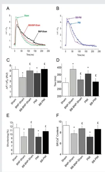
The detection of VAs in the absence of conduction disorders or fibro-sis led us to investigate the cellular mechanisms underlying the pro-arrhythmogenic effect of BNP in single LV cardiomyocytes. First, we found that both BNP and MI increased cell size. BNP increased the cell volume (Table1) and cell length when compared with Shams, consistent with cell hypertrophy, which was prevented by metoprolol (data not shown). We next measured intracellular Ca2+ transients using Fluo-4 AM (Figure 3A–F) and Indo-1 AM
(Figure 4A–C). In BNP-Sham and PMI mice, the amplitude of Ca2+
transients was smaller (Figure. 3A–C) and decay kinetics were slower (Figure3D) than in Sham mice. Cell shortening was similarly decreased in BNP-Sham and PMI when compared with Sham mice (Figure 3E). Cardiomyocyte shortening depends on the amount of Ca2+ released from the SR by ryanodine receptors (RyRs) during
systole. We assessed SR Ca2+content by measuring the Ca2+
transi-ent induced by rapid caffeine application, which instantly opens all RyRs. SR Ca2+ content was lower in Sham-BNP and PMI than in
Sham mice (Figure 3F). Metoprolol prevented the effects of BNP and MI on Ca2+ transient amplitude, Ca2+reuptake, and SR Ca2+
-store depletion, resulting in improved cell shortening in BB-BNP-Sham and BB-PMI mice (Figure3E).
We next examined the arrhythmogenic propensity of cardiomyo-cytes. In BNP-Sham and PMI mice, diastolic Ca2+ was higher and
spontaneous Ca2+waves more frequent than in Shams (Figure 4A–
C). Metoprolol prevented the diastolic Ca2+ increase similarly in
BB-BNP-Sham and BB-PMI mice and decreased Ca2+ wave
occur-rence (Figure 4C). Since elevated diastolic Ca2+ and ectopic Ca2+
waves are suggestive of abnormal spontaneous RyR opening, we investigated RyR activity by directly visualizing Ca2+sparks.9,10
Repre-sentative linescan images are shown in Figure4D. Both BNP-Sham and PMI cells exhibited more Ca2+ sparks, with lower amplitudes, than
Sham cells, a change prevented by metoprolol (Figure4E and F).
3.7 BNP decreases SERCA2a and S100A1
expression
Although SR Ca2+leak reduces SR Ca2+content and cell shortening,
it does not explain the slowing of Ca2+ transient decay kinetics,
known to result mainly from Ca2+ reuptake into the SR via
SERCA2a, and Ca2+ extrusion by the Na+/Ca2+ exchanger
(NCX1). SERCA2a activity is inhibited by PLB, and PLB phosphoryl-ation (P-PLB) relieves this inhibition.12SERCA2a activity is also
modu-lated by the small Ca2+-binding protein S100A1.30Here, we found
reduced SERCA2a and S100A1 protein expression in the LV of BNP-Sham mice (Figure 5A and C), with no modification of PLB or the P-PLB/PLB ratio (Figure 5D–F). RyR protein content was decreased (Figure 5G), whereas the Pser2809RyR/RyR ratio remained
Figure 3 Ca2+transients and cell shortening. Ca2+transients in
single LV cardiomyocytes field-stimulated at 1.0 Hz visualized (Fluo-4 AM), expressed as DF/F0 in Sham, BNP-Sham, and
BB-BNP-Sham mice (A) and in PMI and BB-PMI mice (B); averaged Ca2+ transient amplitude (C); averaged decay time constant, Tau,
of Ca2+ transients (D); cellular shortening (E); and averaged SR
Ca2+ content, estimated from the caffeine-induced peak in Ca2+
transients (F). *P , 0.05, BNP-Sham/PMI vs. Sham animals;
£P , 0.05 for metoprolol-treated vs. untreated animals. Sham,
n ¼ 60 cells, 6 mice; BNP-Sham, BB-BNP-Sham, PMI, and BB-PMI, n ¼ 40 cells; 4 mice.
BNP induces adrenergic-dependent Ca2+-cycling alterations 63
Downloaded from ht tps: //academic. oup. com/ cardiovascres/ art icle-abst ract /95/ 1/ 59/ 331506 by guest on 13 A pril 2020
constant (Figure 5H and I). NCX1 protein content was increased (Figure 5B). In PMI mice, the results were similar overall, notably regarding SERCA2a, NCX1, and S100A1. However, the P-PLB/PLB ratio was decreased (Figure 5D–F), and the Pser2809RyR/RyR ratio
was increased (Figure 5G–I). Metoprolol had substantial beneficial effects on both BB-BNP-Sham and BB-PMI mice by preventing the re-duction in SERCA2a and S100A1 and the increase in NCX1, and by increasing the P-PLB/PLB ratio (Figure 5A–F). Metoprolol also decreased the Pser2809RyR/RyR ratio in BB-PMI mice (Figure5G–I).
4. Discussion
In the present study, we show that in healthy mice, circulating BNP, at levels consistent with those measured during HF following MI, severely alters cellular and molecular functions in cardiomyocytes. Chronically elevated BNP sets the stage for alterations in cellular contraction and Ca2+ handling and promotes the occurrence of
spontaneous cellular Ca2+ waves and VAs in vivo. Most of these
changes are prevented by the b-blocker metoprolol, suggesting a Figure 4 Diastolic Ca2+levels, spontaneous Ca2+waves, and Ca2+sparks. Ca2+transients evoked by field stimulation (1.0 Hz) and spontaneous
Ca2+waves in cardiomyocytes loaded with Indo-1 AM (A); averaged diastolic Ca2+levels (B); percentage of cells exhibiting spontaneous Ca2+waves
(C); representative DF/F linescan images of Ca2+sparks, recorded in intact Fluo-4 AM-loaded cardiomyocytes (D); averaged Ca2+spark frequency (E);
averaged Ca2+spark amplitude (F). *P , 0.05, **P , 0.01, ***P , 0.001, BNP-Sham/PMI vs. Sham animals;£P , 0.05,££P , 0.01, and£££P , 0.001 for
metoprolol-treated vs. untreated animals. Sham, n ¼ 60 cells, 6 mice; BNP-Sham, BB-BNP-Sham, PMI, and BB-PMI, n ¼ 40 cells; 4 mice.
Downloaded from ht tps: //academic. oup. com/ cardiovascres/ art icle-abst ract /95/ 1/ 59/ 331506 by guest on 13 A pril 2020
role for the sympathetic nervous system in the cardiac effects of cir-culating BNP.
4.1 High blood BNP levels promote Ca
21waves and VA in healthy mice
A striking finding was that chronic exposure of the heart to high BNP levels promoted PVC, consistent with the correlation between high BNP levels and VA and SCD in HF patients.2 VA occurred in the
absence of fibrosis, in line with the normal QRS duration, and was associated with mechanisms intrinsic to LV cardiomyocytes and con-sistent with the participation of spontaneous diastolic Ca2+ waves.
The accumulation of aberrant RyR openings in diastole, observed functionally by the higher incidence of Ca2+sparks, provided the
sub-strate for VA/VT by generating Ca2+ waves.9,10 Spontaneous Ca2+
waves are known to induce a transient inward depolarizing current (Iti) via NCX activation.31 The increase in NCX1 protein was also
likely to favour cellular arrhythmias.11
Figure 5 Ca2+-handling proteins. Normalized protein content and ratios obtained by western blotting for SERCA2a (A), NCX1 (B), S100A1 (C),
PLB (D), P-PLB (E), P-PLB/PLB (F) RyR2 (G), P-RyR (H ), and P-RyR/RyR (I). *P , 0.05, **P , 0.01, BNP-Sham/PMI vs. Sham animals;£P , 0.05 and ££P , 0.01 for metoprolol-treated vs. untreated animals; n ¼ 8 animals per group.
BNP induces adrenergic-dependent Ca2+-cycling alterations 65
Downloaded from ht tps: //academic. oup. com/ cardiovascres/ art icle-abst ract /95/ 1/ 59/ 331506 by guest on 13 A pril 2020
4.2 High blood BNP blunts SERCA2a
and S100A1
Chronic exposure to BNP increased the frequency of Ca2+sparks
and reduced SR Ca2+content, normally regulated by the activity of
SERCA2a and its regulatory proteins PLB and S100A1.12,30In a key
finding, BNP blunted the expression of both SERCA2a and S100A1, explaining both the impairment of SR Ca2+uptake and RyR-mediated
SR Ca2+leak. S100A1 is a Ca2+-dependent molecular inotrope that
regulates cardiac SR Ca2+-handling and myofibrillar Ca2+
responsive-ness.32,33S100A1 colocalizes and interacts with both the SERCA2a/
PLB complex and RyR, thereby playing a key role in the coordinated enhancement of RyR2-mediated Ca2+ release during systole and
SR-Ca2+ uptake during diastole.34,35 S100A1 enhances SR Ca2+
load without changing the PLB/SERCA2a ratio or PLB phosphoryl-ation.36S100A1 also inhibits spontaneous RyR activity and decreases
SR Ca2+leakage.34,35
The lack of change of Pser2809RyR/RyR and P-PLB/PLB ratios in
BNP-Sham mice and the fact that neither PKA nor CaMKII is involved in the regulation of RyR and PLB by S100A136together suggest that
phosphorylation is not a key player in the chronic effects of BNP. Our data are also consistent with the functional consequences of an S100A1 deficit.36Abnormal RyR openings in diastole occurred
in-dependently of increased PKA-dependent RyR phosphorylation, known to favour RyR opening.37An increase in spark frequency in
the absence of increased PKA-dependent RyR phosphorylation is already known.10Therefore, the elevation in diastolic Ca2+ due to
impaired SR Ca2+uptake may contribute to SR Ca2+leak, notably
in relation with the biphasic Ca2+-dependent effect of S100A1 on
RyR activity, which depends on cytosolic Ca2+ levels.30,36 Other
mechanisms, such as S-nitrosylation, cannot be excluded.10
4.3 The role of ANS and the b1-adrenergic
pathway
A recent report shows the beneficial effects of local BNP on SERCA2a following intramyocardial gene delivery.38This apparent
in-consistency with our data, together with other reports,13–15may in
fact reflect different aspects of BNP action: localized vs. systemic effects, and/or acute vs. chronic effects. A dose-dependent effect of circulating BNP has been described.7,39Our study points to the
crit-ical importance of systemic effects of chroncrit-ically elevated BNP on cardiac function. Indeed, BNP induces a neurohormonal imbalance and affects the myocardium through sympathetic activation. Decreased HRV is a strong adverse prognostic marker for heart dis-tress and cardiac mortality40in patients with decompensated HF7or
sympathetic overdrive.24 A decrease in SDNN is an established
marker of sympathetic activation in HF, where its reduction parallels disease severity.24The increase in the LF and the LF/HF ratio, a
sym-pathovagal index, in healthy mice treated with BNP further confirms enhanced sympathetic tone.23,24 This is consistent with findings
showing that high-dose BNP increases sympathetic activity in decom-pensated HF7or in patients with essential hypertension.8
Overactiva-tion of the sympathetic system is in part responsible for the cellular alterations observed in our model. These effects could result from a reflex response7,8 or an enhancement of the adrenergic
pathway,15,41,42which is known to promote ventricular hypertrophy
and Ca2+-cycling alterations.43
The use of metoprolol, which counteracts sympathetic overdrive, provides strong evidence for an active role of the b-adrenergic
system in the adverse cardiac remodelling induced by chronic BNP. Indeed, metoprolol prevented several functional alterations, including Ca2+mishandling and the triggering of cellular Ca2+waves and VA in
healthy BNP-treated animals. This is in line with results showing that high BNP sensitizes the b1-adrenergic response via NPR-B,42the
pre-dominant natriuretic peptide receptor in the failing heart.41 At the
protein level, metoprolol prevented SERCA2a and S100A1 blunting, thus maintaining normal SR Ca2+re-uptake, correcting RyR-mediated
Ca2+leakage, and retaining low diastolic Ca2+ levels.17,44 In short,
metoprolol contributed to preserving SR Ca2+ content and Ca2+
transient amplitude, and consequently, cardiomyocyte contraction.45
4.4 Role of endogenous BNP during
the progression of HF?
There were certain phenotypic differences between BNP-Sham and PMI mice: unlike PMI, BNP had no noticeable effect on myocardial function, heart rate, electrical conduction (QRS), or fibrosis in Shams. At the cardiomyocyte level, however, BNP-Sham mice exhib-ited established features of HF (reproduced in PMI mice) regarding Ca2+handling. In addition, both BNP treatment and MI blunted the
expression of S100A1, which contributes to Ca2+-handling alterations
and depressed contraction.36,46 S100A1 is decreased in HF,30
promotes SR Ca2+ leak by increasing the probability of RyR2
opening,47and hampers Ca2+reuptake due to reduced SERCA2a
ac-tivity.30Both altered SERCA2a expression and BNP production are
considered early indicators of HF and are inversely correlated in human cardiac hypertrophy and HF.14,48,49 Our finding regarding
blunted S100A1 and SERCA2a expression in BNP-Sham mice further strengthens the concept that BNP contributes to adverse cardiac remodelling early in the progression of HF.13,14 This could
explain the ineffectiveness of nesiritide in treating HF, despite its bene-ficial haemodynamic effects.15Both HRV analysis and the prevention
of the deleterious effects of BNP by metoprolol suggest that BNP acts in part through adrenergic overdrive. We observed similar effects of metoprolol in BB-PMI mice, confirming previous studies de-scribing an increase in Ca2+reuptake through increased SERCA2a
ex-pression, increased phosphorylation of PLB,50 and decreased RyR2
phosphorylation. Our study also highlights the functional conse-quences of the previously unsuspected but recently described cardiac pro-adrenergic property of BNP,15 which could potentially
lead to a cellular HF-like profile with Ca2+-cycling defects or
aggravate existing HF. 4.4.1 Clinical implications
Our work shows that elevated blood BNP is not only a biomarker for guided therapy in HF but contributes per se to adverse cardiac remod-elling and the triggering of VA, making the rapid lowering of BNP levels in HF patients a highly desirable end, in harmony with early in-hospital treatment aimed at decreasing BNP levels to improve sur-vival.51–53 Our study brings into question the treatment of HF
patients with synthetic natriuretic peptide-like drugs, whose efficacy is uncertain54,55and whose link to increased mortality has been
sus-pected.5,6Indeed, the ventricular remodelling induced by chronically
elevated natriuretic peptides may limit their haemodynamic benefits. Overall, our work supports the general concept that the very early normalization of SERCA2a56 or/and S100A1 expression30 is critical
in limiting adverse cardiac remodelling in HF. Last, but not least, b-blocker therapy should be considered as soon as BNP levels rise, even in patients without cardiac symptoms.
Downloaded from ht tps: //academic. oup. com/ cardiovascres/ art icle-abst ract /95/ 1/ 59/ 331506 by guest on 13 A pril 2020
Supplementary material
Supplementary material is available at Cardiovascular Research online.
Acknowledgments
We thank Dr S. Rasika of Gap Junction for English editing. Conflict of interest: none declared.
Funding
This work was supported by funding from the Fondation de France (Project PepNaRhythm, no. 2068001722) and the Institut National de la Sante´ et de la Recherche Me´dicale (INSERM). SR, JF, OC and AL hold CNRS positions.
References
1. Morita E, Yasue H, Yoshimura M, Ogawa H, Jougasaki M, Matsumura T et al. Increased plasma levels of brain natriuretic peptide in patients with acute myocardial infarction. Circulation 1993;88:82–91.
2. Simon T, Becker R, Voss F, Bikou O, Hauck M, Licka M et al. Elevated B-type natri-uretic peptide levels in patients with nonischemic cardiomyopathy predict occurrence of arrhythmic events. Clin Res Cardiol 2008;97:306–309.
3. Wang TJ, Larson MG, Levy D, Benjamin EJ, Leip EP, Omland T et al. Plasma natriuretic peptide levels and the risk of cardiovascular events and death. N Engl J Med 2004;350: 655–663.
4. Cleland JG, Coletta AP, Buga L, Antony R, Pellicori P, Freemantle N et al. Clinical trials update from the American Heart Association meeting 2010: EMPHASIS-HF, RAFT, TIM-HF, Tele-HF, ASCEND-HF, ROCKET-AF, and PROTECT. Eur J Heart Fail 2011; 13:460–465.
5. Sackner-Bernstein JD, Kowalski M, Fox M, Aaronson K. Short-term risk of death after treatment with nesiritide for decompensated heart failure: a pooled analysis of rando-mized controlled trials. JAMA 2005;293:1900–1905.
6. Topol EJ. Nesiritide - not verified. N Engl J Med 2005;353:113–116.
7. Aronson D, Burger AJ. Effect of nesiritide (human b-type natriuretic peptide) and dobutamine on heart rate variability in decompensated heart failure. Am Heart J 2004;148:e16.
8. La Villa G, Bisi G, Lazzeri C, Fronzaroli C, Stefani L, Barletta G et al. Cardiovascular effects of brain natriuretic peptide in essential hypertension. Hypertension 1995;25: 1053–1057.
9. Cheng H, Lederer WJ. Calcium sparks. Physiol Rev 2008;88:1491–1545.
10. Fauconnier J, Thireau J, Reiken S, Cassan C, Richard S, Matecki S et al. Leaky RyR2 trigger ventricular arrhythmias in Duchenne muscular dystrophy. Proc Natl Acad Sci USA 2010;107:1559–1564.
11. Sipido KR, Volders PG, Vos MA, Verdonck F. Altered Na/Ca exchange activity in cardiac hypertrophy and heart failure: a new target for therapy? Cardiovasc Res 2002;53:782–805.
12. Ikeda Y, Hoshijima M, Chien KR. Toward biologically targeted therapy of calcium cycling defects in heart failure. Physiology (Bethesda) 2008;23:6–16.
13. Toischer K, Kogler H, Tenderich G, Grebe C, Seidler T, Van PN et al. Elevated after-load, neuroendocrine stimulation, and human heart failure increase BNP levels and inhibit preload-dependent SERCA upregulation. Circ Heart Fail 2008;1:265–271. 14. Kogler H, Schott P, Toischer K, Milting H, Van PN, Kohlhaas M et al. Relevance of
brain natriuretic peptide in preload-dependent regulation of cardiac sarcoplasmic re-ticulum Ca2+ATPase expression. Circulation 2006;113:2724–2732.
15. Chan NY, Seyedi N, Takano K, Levi R. An unsuspected property of natriuretic pep-tides: promotion of calcium-dependent catecholamine release via protein kinase g-mediated phosphodiesterase type 3 inhibition. Circulation 2012;125:298–307. 16. Maczewski M, Mackiewicz U. Effect of metoprolol and ivabradine on left ventricular
remodelling and Ca2+handling in the post-infarction rat heart. Cardiovasc Res 2008;
79:42–51.
17. Sun YL, Hu SJ, Wang LH, Hu Y, Zhou JY. Effect of beta-blockers on cardiac function and calcium handling protein in postinfarction heart failure rats. Chest 2005;128: 1812–1821.
18. Ahmet I, Morrell C, Lakatta EG, Talan MI. Therapeutic efficacy of a combination of a beta1-adrenoreceptor (AR) blocker and beta2-AR agonist in a rat model of postmyo-cardial infarction dilated heart failure exceeds that of a beta1-AR blocker plus angiotensin-converting enzyme inhibitor. J Pharmacol Exp Ther 2009;331:178–185. 19. Xydas S, Kherani AR, Chang JS, Klotz S, Hay I, Mutrie CJ et al. beta(2)-Adrenergic
stimulation attenuates left ventricular remodeling, decreases apoptosis, and improves calcium homeostasis in a rodent model of ischemic cardiomyopathy. J Pharmacol Exp Ther 2006;317:553–561.
20. Harding VB, Jones LR, Lefkowitz RJ, Koch WJ, Rockman HA. Cardiac beta ARK1 in-hibition prolongs survival and augments beta blocker therapy in a mouse model of severe heart failure. Proc Natl Acad Sci USA 2001;98:5809–5814.
21. Thireau J, Aimond F, Poisson D, Zhang B, Bruneval P, Eder V et al. New insights into sexual dimorphism during progression of heart failure and rhythm disorders. Endocrin-ology 2010;151:1837–1845.
22. Das MK, Maskoun W, Shen C, Michael MA, Suradi H, Desai M et al. Fragmented QRS on twelve-lead electrocardiogram predicts arrhythmic events in patients with ische-mic and nonischeische-mic cardiomyopathy. Heart Rhythm 2010;7:74–80.
23. Thireau J, Zhang BL, Poisson D, Babuty D. Heart rate variability in mice: a theoretical and practical guide. Exp Physiol 2008;93:83–94.
24. Task Force of the European Society of Cardiology the North American Society of Pacing Electrophysiology. Heart rate variability: standards of measurement, physio-logical interpretation and clinical use. Circulation 1996;93:1043–1065.
25. Thireau J, Poisson D, Zhang BL, Gillet L, Le Pecheur M, Andres C et al. Increased heart rate variability in mice overexpressing the Cu/Zn superoxide dismutase. Free Radic Biol Med 2008;45:396–403.
26. Cooley RL, Montano N, Cogliati C, van de Borne P, Richenbacher W, Oren R et al. Evidence for a central origin of the low-frequency oscillation in RR-interval variability. Circulation 1998;98:556–561.
27. van de Borne P, Montano N, Pagani M, Oren R, Somers VK. Absence of low-frequency variability of sympathetic nerve activity in severe heart failure. Circulation 1997;95:1449–1454.
28. Wei S, Chow LT, Sanderson JE. Effect of carvedilol in comparison with metoprolol on myocardial collagen postinfarction. J Am Coll Cardiol 2000;36:276–281.
29. Metoprolol in acute myocardial infarction. Other clinical findings and tolerability. The MIAMI Trial Research Group. Am J Cardiol 1985;56:39G–46G.
30. Most P, Pleger ST, Volkers M, Heidt B, Boerries M, Weichenhan D et al. Cardiac adenoviral S100A1 gene delivery rescues failing myocardium. J Clin Invest 2004;114: 1550–1563.
31. Shannon TR, Lew WY. Diastolic release of calcium from the sarcoplasmic reticulum: a potential target for treating triggered arrhythmias and heart failure. J Am Coll Cardiol 2009;53:2006–2008.
32. Most P, Bernotat J, Ehlermann P, Pleger ST, Reppel M, Borries M et al. S100A1: a regu-lator of myocardial contractility. Proc Natl Acad Sci USA 2001;98:13889–13894. 33. Remppis A, Most P, Loffler E, Ehlermann P, Bernotat J, Pleger S et al. The small
EF-hand Ca2+binding protein S100A1 increases contractility and Ca2+cycling in
rat cardiac myocytes. Basic Res Cardiol 2002;97(Suppl. 1):I56–I62.
34. Kettlewell S, Most P, Currie S, Koch WJ, Smith GL. S100A1 increases the gain of excitation-contraction coupling in isolated rabbit ventricular cardiomyocytes. J Mol Cell Cardiol 2005;39:900–910.
35. Most P, Remppis A, Pleger ST, Katus HA, Koch WJ. S100A1: a novel inotropic regu-lator of cardiac performance. Transition from molecular physiology to pathophysio-logical relevance. Am J Physiol Regul Integr Comp Physiol 2007;293:R568–R577. 36. Rohde D, Ritterhoff J, Voelkers M, Katus HA, Parker TG, Most P. S100A1: a
multi-faceted therapeutic target in cardiovascular disease. J Cardiovasc Transl Res 2010; 3:525–537.
37. Reiken S, Gaburjakova M, Guatimosim S, Gomez AM, D’Armiento J, Burkhoff D et al. Protein kinase A phosphorylation of the cardiac calcium release channel (ryanodine receptor) in normal and failing hearts. Role of phosphatases and response to isoproterenol. J Biol Chem 2003;278:444–453.
38. Moilanen AM, Rysa J, Mustonen E, Serpi R, Aro J, Tokola H et al. Intramyocardial BNP gene delivery improves cardiac function through distinct context-dependent mechanisms. Circ Heart Fail 2011;4:483–495.
39. Brunner-La Rocca HP, Kaye DM, Woods RL, Hastings J, Esler MD. Effects of intraven-ous brain natriuretic peptide on regional sympathetic activity in patients with chronic heart failure as compared with healthy control subjects. J Am Coll Cardiol 2001;37: 1221–1227.
40. Fauchier L, Babuty D, Cosnay P, Autret ML, Fauchier JP. Heart rate variability in idio-pathic dilated cardiomyopathy: characteristics and prognostic value. J Am Coll Cardiol 1997;30:1009–1014.
41. Dickey DM, Flora DR, Bryan PM, Xu X, Chen Y, Potter LR. Differential regulation of membrane guanylyl cyclases in congestive heart failure: natriuretic peptide receptor (NPR)-B, Not NPR-A, is the predominant natriuretic peptide receptor in the failing heart. Endocrinology 2007;148:3518–3522.
42. Qvigstad E, Moltzau LR, Aronsen JM, Nguyen CH, Hougen K, Sjaastad I et al. Natri-uretic peptides increase beta1-adrenoceptor signalling in failing hearts through phosphodiesterase 3 inhibition. Cardiovasc Res 2010;85:763–772.
43. Engelhardt S, Hein L, Dyachenkow V, Kranias EG, Isenberg G, Lohse MJ. Altered calcium handling is critically involved in the cardiotoxic effects of chronic beta-adrenergic stimulation. Circulation 2004;109:1154–1160.
44. Kubo H, Margulies KB, Piacentino V 3rd, Gaughan JP, Houser SR. Patients with end-stage congestive heart failure treated with beta-adrenergic receptor antagonists have improved ventricular myocyte calcium regulatory protein abundance. Circulation 2001; 104:1012–1018.
45. Reiken S, Wehrens XH, Vest JA, Barbone A, Klotz S, Mancini D et al. Beta-blockers restore calcium release channel function and improve cardiac muscle performance in human heart failure. Circulation 2003;107:2459–2466.
BNP induces adrenergic-dependent Ca2+-cycling alterations 67
Downloaded from ht tps: //academic. oup. com/ cardiovascres/ art icle-abst ract /95/ 1/ 59/ 331506 by guest on 13 A pril 2020
46. Pleger ST, Most P, Boucher M, Soltys S, Chuprun JK, Pleger W et al. Stable myocardial-specific AAV6-S100A1 gene therapy results in chronic functional heart failure rescue. Circulation 2007;115:2506–2515.
47. Volkers M, Loughrey CM, Macquaide N, Remppis A, DeGeorge BR Jr, Wegner FV et al. S100A1 decreases calcium spark frequency and alters their spatial characteristics in permeabilized adult ventricular cardiomyocytes. Cell Calcium 2007;41:135–143. 48. de Boer RA, Henning RH, Suurmeijer AJ, Pinto YM, Olthof E, Kirkels JH et al. Early
expression of natriuretic peptides and SERCA in mild heart failure: association with severity of the disease. Int J Cardiol 2001;78:5–12.
49. Takahashi T, Allen PD, Izumo S. Expression of A-, B-, and C-type natriuretic peptide genes in failing and developing human ventricles. Correlation with expression of the Ca(2+)-ATPase gene. Circ Res 1992;71:9–17.
50. MacLennan DH, Kranias EG. Phospholamban: a crucial regulator of cardiac contract-ility. Nat Rev Mol Cell Biol 2003;4:566–577.
51. Anand IS, Fisher LD, Chiang YT, Latini R, Masson S, Maggioni AP et al. Changes in brain natriuretic peptide and norepinephrine over time and mortality and morbidity in the Valsartan Heart Failure Trial (Val-HeFT). Circulation 2003;107:1278–1283. 52. Cohen-Solal A, Logeart D, Huang B, Cai D, Nieminen MS, Mebazaa A. Lowered
B-type natriuretic peptide in response to levosimendan or dobutamine treatment is
associated with improved survival in patients with severe acutely decompensated heart failure. J Am Coll Cardiol 2009;53:2343–2348.
53. Fujimura M, Akaike M, Iwase T, Yoshida S, Sumitomo Y, Yagi S et al. Decrease in plasma brain natriuretic peptide level in the early phase after the start of carvedilol therapy is a novel predictor of long-term outcome in patients with chronic heart failure. Acta Cardiol 2009;64:589–595.
54. Hunt SA, Abraham WT, Chin MH, Feldman AM, Francis GS, Ganiats TG et al. 2009 focused update incorporated into the ACC/AHA 2005 Guidelines for the Diagnosis and Management of Heart Failure in Adults: a report of the American College of Cardiology Foundation/American Heart Association Task Force on Practice Guide-lines: developed in collaboration with the International Society for Heart and Lung Transplantation. Circulation 2009;119:e391–e479.
55. O’Connor CM, Starling RC, Hernandez AF, Armstrong PW, Dickstein K, Hasselblad V et al. Effect of nesiritide in patients with acute decompensated heart failure. N Engl J Med 2011;365:32–43.
56. Byrne MJ, Power JM, Preovolos A, Mariani JA, Hajjar RJ, Kaye DM. Recirculating cardiac delivery of AAV2/1SERCA2a improves myocardial function in an experimental model of heart failure in large animals. Gene Ther 2008;15:1550–1557.
Downloaded from ht tps: //academic. oup. com/ cardiovascres/ art icle-abst ract /95/ 1/ 59/ 331506 by guest on 13 A pril 2020