Regulation of
-Synuclein Membrane
Binding and Its Implications
Robert H.C. Chen
1, Sabine Wislet-Gendebien
1,2,
Howard T.J. Mount
1and Anurag Tandon
11
Tanz Centre for Research in Neurodegenerative Diseases, University of Toronto,
2GIGA Neurosciences, University of Liège,
1Canada
2Belgium
1. Introduction
1.1 Parkinson’s disease and Lewy bodies
Parkinson’s disease (PD) is the most prevalent neurodegenerative disease that affects motor control, although dementia, depression, and other psychiatric symptoms are occasionally present (reviewed in Jankovic, 2008). The motor symptoms include resting tremors, bradykinesia/akinesia, rigidity, and postural instability, and are associated with the death of dopaminergic neurons in the substantia nigra pars compacta (SNpc) (Lloyd and Hornykiewicz, 1970; reviewed in Saper, 1999) and noradrenergic neurons in the locus coeruleus (Mann and Yates, 1983; Gaspar et al., 1991). Degeneration of these neurons disrupts basal ganglia circuitry of the brain and interferes with the initiation of voluntary movement (Levy et al., 1997).
Another pathological characteristic of the disease is the presence of the intracellular protein aggregates known as Lewy bodies and Lewy neurites (Hughes et al., 1992), described initially by Frederick Lewy in 1912, that were associated with dying dopaminergic neurons in the SNpc by Tretiakoff (reviewed in Holdorff, 2006). Lewy bodies are also present in other brain regions outside the SNpc, and may appeat first in the glossopharyngeal, vagal, and olfactory centres (Braak et al., 2003). The disruption of these regions is now linked to a preclinical phase of PD that includes perturbations of smell, gastrointestinal motility, and sleep patterns prior to the development of motor impairments coincident with neurodegeneration and inclusions in the midbrain. These intraneuronal aggregates are composed of lipids and various proteins including -synuclein, ubiquitin, and neurofilaments (Spillantini et al., 1998; reviewed in Cookson, 2005). Whether Lewy bodies directly cause cytotoxicity or are formed as compensatory activation of survival pathways remains under debate. Nevertheless, the presence of Lewy bodies is an essential component of post-mortem diagnoses of PD (Christine and Aminoff, 2004; reviewed in Jankovic, 2008).
1.2 α-Synuclein in Parkinson’s disease
-Synuclein is important to understanding the etiology of PD both because it is the main pathological component of Lewy bodies and because mutations and changes in its expression are linked to familial PD. Three missense mutations causing single amino acid
substitutions in -synuclein are linked to autosomal dominant forms of familial PD: A53T, A30P, and E46K (Polymeropoulos et al., 1997; Kruger et al., 1998; Zarranz et al., 2004). Interestingly, in many mammalian species, including mice and new world primates, threonine is the normal residue at position 53, which suggest some kind of corrective mechanism elsewhere in the coding sequence to reduce the toxicity of the A53T mutation (Hamilton, 2004). Elevated expression of normal -synuclein is also pathogenic as gene triplication or duplication is linked to early- or late-onset PD, respectively (Singleton et al., 2003; Chartier-Harlin et al., 2004).
Despite the efforts of many research laboratories since its initial discovery as an abundant presynaptic protein in cholinergic nerve terminals innervating the Torpedo electric organic (Maroteax et al, 1988), -synuclein’s function still remains somewhat of a mystery. Mice deficient in -synuclein are unremarkable, but exhibit increased release of dopamine under paired-pulse stimuli and reduced striatal tissue content of dopamine (Abeliovich et al., 2000). Over-expression of -synuclein in yeast leads to aggregation of vesicles, disruption in vesicular transport, and death (Gitler et al., 2008; Soper et al., 2008). Similarly, moderate -synuclein over-expression in mammalian neurons reduces transmitter release and the number of synaptic vesicles arrayed at presynaptic active zones (Nemani et al., 2010). These reports clearly implicate -synuclein in synaptic vesicle mobilization or fusion, but offer little information about its pathogenic role.
In vitro studies revealed that A30P and A53T mutant -synuclein form oligomers at a faster rate than wild-type -synuclein (Conway et al., 2000). Post-translational modifications may also affect -synuclein aggregation, as phosphorylation of serine at position 129 is highly upregulated in Lewy bodies (Fujiwara et al., 2002). Phosphomimic residue changes, such as serine 129 to aspartate, which simulates the charge distribution of phosphorylation, cause neuronal loss in fruit flies and substitution of serine 129 with alanine, which prevents phosphorylation, rescues this cell loss (Chen and Feany, 2005). However, experimental evidence for the toxicity of serine 129 phosphorylation is somewhat equivocal in rodents, because expression of -synuclein with alanine 129 is as toxic as aspartate 129 (Gorbatyuk et al, 2008; McFarland et al., 2009).
Although -synuclein is decreased in the cerebrospinal fluid of PD patients (Mollenhauer et al, 2011), there is an increase in -synuclein oligomers suggesting that -synuclein aggregation and its leakage from neurons is accelerated in PD (Tokuda et al., 2010). However, the mechanistic contribution of -synuclein to neuronal death in PD still remains unclear. Low nanomolar concentrations of -synuclein are neuroprotective against the oxidative insults of hydrogen peroxide and 6-hydroxydopamine, whereas higher micromolar concentrations are toxic (Batelli et al., 2008). This suggests that -synuclein may be neuroprotective and toxic at low and high levels, respectively, and is in accord with the pathogenic effects of SNCA duplication or triplication. -Synuclein has also been shown to cause oxidative stress through direct damage to mitochondria. For example, mitochondrial import of -synuclein mediated by a cryptic targeting signal can disrupt complex 1 activity (Devi et al., 2008), and -synuclein over-expression in nematodes and mammalian cells induced fragmentation of mitochondria (Kamp et al., 2010; Nakamura et al., 2011). Co-expression of PINK1, parkin, and DJ-1 protect against mitochondrial fragmentation, consistent with the notion that these three proteins function in a common mitochondrial pathway leading to PD pathology. The convergence of -synuclein to a mitochondrial role when expressed at sufficient levels could therefore be viewed as a pathogenic pathway that diverges from its normal physiological role in vesicle trafficking.
1.3 -Synuclein outside of disease
A role in synaptic function is implicated from the observation that -synuclein is highly concentrated in presynaptic terminals (Jakes et al., 1994; George et al., 1995) and that -synuclein disperses reversibly from these presynaptic terminals in response to brief neural activity (Fortin et al., 2005). The dispersion is attenuated by tetanus toxin, which inactivates vesicle fusion but not the preceding ion fluxes, suggesting that -synuclein solubility is directly linked to exocytosis. While these results do not suggest a presynaptic function for -synuclein, other studies with -synuclein knockout mice reveal increased nigrostriatal dopamine release induced by pairedpulse stimuli (Abeliovich et al., 2000), implicating -synuclein as a negative regulator of a readily-releasable vesicle pool of dopamine. In contrast, another line of -synuclein null mice showed normal response to single or paired stimuli, but deficits in response to trains of stimuli which rely on reserve vesicle recruitment and a reduction in the number distal vesicles (Cabin et al., 2002). To explore whether -synuclein compensates for loss of --synuclein, removal of both - and --synuclein decreased total brain dopamine by 20% as well as complexins and 14-3-3 proteins (Chandra et al., 2004). Complexins are a regulatory component of the SNARE (soluble N-ethylmaleimide-sensitive attachment protein receptor) complexes involved in synaptic vesicle fusion (Hu et al., 2002). However, the other previously described aberrations such as synaptic vesicle reduction, electrophysiological changes, and dopamine re-uptake were not observed in these -synuclein knockout mice. Despite the differences, -synuclein knockout mice display subtle alterations in vesicle storage and dynamics.
The converse experimental paradigm using over-expression of -synuclein also suggests that it inhibits synaptic transmission in primary neurons and intact ex vivo brain (Nemani et al., 2010). Morphological and electrophysiological analyses indicate that the reduction was due to decreased readily-releasable synaptic vesicles, but not the overall number of synaptic vesicles, consistent with the notion that -synuclein is a negative regulator of the synaptic vesicle mobilization.
Recent studies using biochemical approaches have reported that -synuclein disrupts arachidonic acid-mediated stabilization of soluble N-ethylmaleimide sensitive fusion protein receptor (SNARE) complex (comprised of syntaxin-1, SNAP25, and synaptobrevin-2) that is essential for synaptic vesicle fusion (Darios et al., 2010). Although this study did not co-immunoprecipitate -synuclein with members of the SNARE complex, thus suggesting an indirect interaction, others have reported -synuclein binding with synaptobrevin-2 and proposed that this binding stabilizes the SNARE complex (Burre et al., 2010). Interestingly, loss of assembled SNARE complexes in mice deficient for cysteine-string protein alpha (CSP ) can be rescued by -synuclein over-expression and worsened by its knockdown. The loss of all three synucleins ( -, -, and -) causes premature death due to neurological symptoms, suggesting that - and/or -synuclein perform redundant functions and compensate for -synuclein deficiency in the single knockout animals. Thus,
-synuclein exerts a regulatory effect on SNARE complex formation, but its mechanism remains poorly defined.
Some studies have suggested that -synuclein may regulate vesicle trafficking more broadly that just in synaptic transmission. Expression of -synuclein in yeast and mammalian cells causes delays in ER-to-Golgi vesicle trafficking, an effect that was more striking with the A53T mutant (Cooper et al., 2006; Thayanidhi et al., 2010). These changes in ER-to-Golgi trafficking by -synuclein may be caused by immobilizing vesicles in the cytoplasm (Soper et al., 2008). The mechanism appears to involve rab proteins, small GTPases that regulate
vesicle trafficking, as the impairment is rescued by overexpressing certain rab proteins (Gitler et al., 2008). Furthermore, as yeast do not express -synuclein endogenously, these studies suggest that -synuclein can interact with both constitutive and Ca2+-dependent
vesicle trafficking machineries that is conserved between yeast and higher organisms.
2.
-Synuclein and membrane-binding
2.1 The structure of -synuclein and how it contributes to membrane-binding
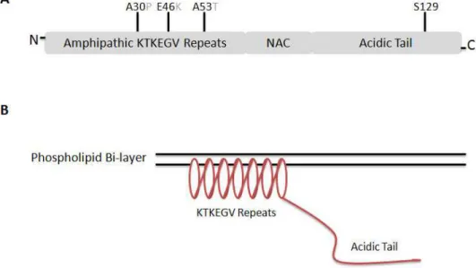
The structure of -synuclein can be divided into three domains: an amino-terminal domain with seven imperfect KTKEGV repeats, a central domain essential for aggregation called the non-amyloid component (NAC), and a hydrophilic carboxyl-terminal tail (Clayton and George, 1998) (Fig. 1.1A). The amino-terminal repeats, which are similar to sequences found in apolipoproteins, form amphipathic helices with polar and non-polar residues aligned in opposing direction and allow -synuclein to immerse partially into lipid membranes (Fig. 1.1B). The capacity of -synuclein to bind freely to membranes has been demonstrated (Jo et al., 2000; Conway et al., 2000). The helical structure of -synuclein upon binding to sodium dodecyl sulfate (SDS)-soluble micelles has been established using nuclear magnetic
Fig. 1. The primary structure of human -synuclein. (A) The structure itself can be divided into three unequal regions: the imperfect KTKEGV repeats believed to form a helical structure on phospholipid membranes, the non-A component (NAC) seen in Alzheimer disease-associated plaques, and a non-membrane-associated acidic c-terminal tail. (B) Upon binding to membranes, -synuclein assumes a helical conformation due to the amphipathic nature of the KTKEGV repeats. All three amino acid substitution sites associated with familial forms of PD (A30P, E46K, and A53T) can be found in the amphipathic repeat region. The serine 129 site commonly found to be phosphorylated in Lewy bodies can be found in the acidic tail. Note that the residue in position 53 is naturally a threonine instead of an alanine in many mammalian species, including rodents.
resonance and electron spin resonance studies (Jao et al., 2004; Ulmer et al., 2005; Borbat et al., 2006). The helical conformation is energetically favourable when compared to the unstructured conformation of cytosolic -synuclein.
2.2 Implications of -synuclein membrane-binding
Binding to and dissociation from the synaptic membrane may be linked to regulating the still-undefined function of -synuclein. Using fluorescent imaging to visualize GFP-tagged -synuclein, depolarization reversibly induced the redistribution of -synuclein from synaptic boutons to the perisynaptic region (Fortin et al., 2005), consistent with the dissociation of synuclein from synaptic vesicles prior to exocytosis. This link between synuclein, its membrane binding, and exocytosis is consistent with other reports that -synuclein regulates SNARE complex assembly (Darios et al., 2010; Burre et al, 2010).
The membrane-binding properties of -synuclein may also have implications in PD pathogenesis. The addition of lipid vesicles causes a greater proportion of -synuclein to assume an -helical conformation and decreases the fibrillization of -synuclein (Zhu et al., 2003); Jo et al., 2004), suggesting that increased membrane binding may be protective against Lewy body pathology. However, other studies adding phospholipids to -synuclein induced fibril formation (Narayanan and Scarlata, 2001; Cole et al., 2002). Mutant A30P -synuclein exhibits a lower propensity to bind to phospholipid membranes as determined in vitro (Jo et al., 2002), but forms oligomers faster than wild-type or A53T -synuclein (Conway et al., 2000). Nevertheless, the A53T mutant -synuclein forms fibrils (the likely precursor to Lewy bodies) at a higher rate. Furthermore, a rotenone model of PD revealed that lipids co-stained with -synuclein aggregates (Lee et al., 2002), suggesting that lipids may enable Lewy body formation. Taken together, the evidence is conflicting on whether the membrane-bound or cytosolic -synuclein is the precursor for aggregated -synuclein and Lewy body pathology, although it remains possible that oligomerization and fibrillization could occur in distinct cellular compartments (Auluck et al., 2010).
2.3 Regulation of -synuclein membrane-binding
While there remains some uncertainty as to whether the membranebound or cytosolic -synuclein is more pathologically relevant, it is also important to understand the mechanisms regulating the exchange between these two pools. We have previously suggested that brain cytosolic factors are critical in facilitating -synuclein dissociation and association from synaptic membranes (Wislet-Gendebien et al., 2006; 2008). The remainder of this chapter will be devoted to describing some of these cytosolic factors that were characterized using our cell-free assays.
3. Key techniques for examining the regulation of membrane-binding
3.1 Fractionation into membrane-bound and cytosolic proteins
Much of our current understanding of -synuclein conformation and membrane interactions are based on studies with recombinant -synuclein purified from bacteria and bound to artificial membranes. This approach has yielded a wealth of information on the biophysical characteristics of normal and mutant -synuclein and their affinity for specific lipids. However, recombinant -synuclein and artificial membranes provide no basis for the understanding -synuclein behaviour in vivo, in particular its ability to exchange between membrane and cytosol within neurons, where the compartmentalization of -synuclein is modulated by intracellular components and neuronal activity.
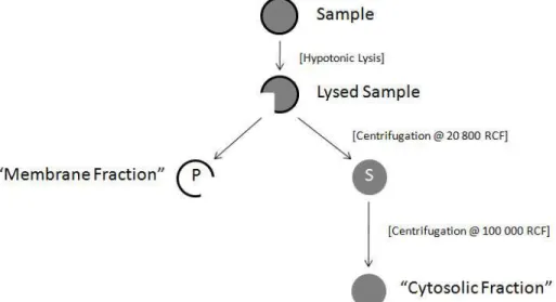
To understand endogenous intracellular regulation of -synuclein dynamics, reconstitution of its membrane binding and dissociation can be studied using semi-intact or cell-free assays. For example, the use of hypotonic lysis to disrupt neuronal plasma membrane releases freely-diffusible cytoplasmic components and permits measurement of subtle shifts in -synuclein membrane-binding (Fig. 2). Experiments of this type are best done with endogenously expressed -synuclein, although where exogenous expression is required, consideration should be given to the extent of -synuclein overexpression so as not to saturate the membrane compartment and cause artifactually low membrane-bound to cytosolic ratio of -synuclein, as excess -synuclein will accumulate in cytosol. Measurement of -synuclein dynamics in intact cells may be limited by an inability to assess whether changes in membrane-bound and cytosolic -synuclein are caused by perturbations in -synuclein dissociation or binding kinetics. This can be overcome by using cell-free assays (such as those described below) that monitor the unidirectional movement of -synuclein from a ”donor” fraction expressing --synuclein to an “acceptor” fraction derived from an -synuclein-deficient mouse or cells.
Fig. 2. A schematic overview of fractionation. Synaptosomes or cells were hypotonically lysed in either distilled water or a hypotonic buffer. The lysates were centrifuged and the supernatant (S) and pellet (P) fractions were processed separately. S fractions were centrifuged again to remove residual synaptic vesicles and supernatants were kept as cytosolic fractions. The P fractions were resuspended in buffer containing 1% CHAPS or 1% Triton X-100. Lysates were centrifuged and the supernatants were kept as membrane fraction.
3.2 -Synuclein dissociation and binding assays
While analyzing the ratio of membrane-bound to cytosolic -synuclein can yield useful data in an in-vivo or ex-vivo setting, it does not provide information on whether effects are taking place in the membrane-dissociation or membrane-binding step of -synuclein dynamics. This question can be answered by employing cell-free assays we have previously published
(Wislet-Gendebien et al., 2006). To measure -synuclein dissociation, synaptosomal membranes from either non-transgenic or -synuclein transgenic mice are incubated with brain cytosol isolated from synuclein deficient mice. By measuring the amount of -synuclein dissociated from the membrane and into the cytosol under different conditions, it is possible to determine whether specific cellular factors affect -synuclein membrane-dissociation.
A converse protocol was developed to assess -synuclein binding to synaptic membrane (Wislet-Gendebien et al., 2008). In this assay, -synuclein can be prepared from 2 different origins: either 1) purified E.coli-expressed -synuclein mixed with cytosol from -synuclein deficient mouse brains, or 2) cytosol prepared from transgenic mice overexpressing the human form of wild-type or mutant -synuclein. Either of the two sources of -synuclein is then combined with synaptic membrane prepared from -synuclein deficient mouse synaptosomes. Membranebound synuclein can then be analyzed as a measure of -synuclein membrane-binding in response to various controlled factors such as pharmacological agents or lipids. This assay can provide information on whether a certain condition or factor is affecting -synuclein membrane-binding.
The key benefit of these dissociation and binding assays is the ability to probe the intracellular milieu with specific reagents, such as antibodies, recombinant mutants, or peptide domains, which are membrane impermeant. However, because these assays depends on mixing separately-derived membrane and cytosolic fractions, one of which must be -synuclein-deficient, there is an inherent limitation to assess the dissociation or binding of other synaptic proteins due to their presence in both the membrane and the cytosol of both assays. However, these assays can be adapted to analyze the dissociation and binding of other synaptic proteins provided that transgenic animals deficient in the protein of interest are available.
4. Factors involved in regulating
-synuclein membrane-binding
4.1 Brain cytosol
The -synuclein dissociation assay revealed that stably membrane-bound -synuclein can be recruited into the cytosol in the presence of brain cytosolic proteins (Wislet-Gendebien et al., 2006). Pre-digestion with trypsin or preheating at 95 °C of the cytosol eliminated its ability to induce -synuclein dissociation, directly implicating a role for specific cytosolic proteins in controlling -synuclein solubility. Moreover, the permissive factors required to mediate -synuclein dissociation from the membrane appeared to be enriched in brain cytosol, as a 6-fold greater concentration of liver cytosol was required to achieve equivalent
-synuclein dissociation using brain cytosol. The proteins triggering -synuclein dissociation were in limited quantity in cytosol and were not regenerated under our assay conditions. A single exposure to synaptosomal membranes was sufficient to deplete the capacity of the cytosol to extract membrane -synuclein so that subsequent incubations with fresh membranes yielded no additional dissociated -synuclein. In contrast, presynaptic membranes retained ample extractable -synuclein, which could be dissociated with subsequent applications of fresh cytosol. We also showed that the cytosolic activity that mediated -synuclein dissociation clearly distinguished between wild-type -synuclein and PD-associated mutants. The cytosol-dependent off-rate for both A30P and A53T -synuclein mutant was double that of the wild-type, but had no effect on cytosol-independent dissociation.
4.2 Cytosolic lipids
Using our -synuclein binding assay, we observed that cytosol-mediated -synuclein membrane-binding was heat stable and protease insensitive. Further characterization revealed that ATP and lipids are two of the main cytosolic components that modulate -synuclein binding to synaptic membranes (Wislet-Gendebien et al., 2008). We proposed that endogenous cytosolic lipids transferred to membranes prior to -synuclein recruitment or bound directly to cytosolic -synuclein may aid -synuclein folding at the lipid-cytoplasm interface so that it is more amenable to binding directly to synaptic membranes. To provide further insight into this novel protein-lipid-protein interaction, we profiled glycerophosphocholines bound to proteins in -synuclein-deficient cytosol by nanoflow LC-ESI-MS and precursor ion scan. Our analysis identified 24 species that can potentially affect
-synuclein membrane interactions, including platelet activating factor, which was able to reconstitute the activity of delipidated cytosol.
4.3 Rab3a
Interestingly, our binding experiments also revealed that the association of -synuclein to synaptic membranes could be stabilized by brief formaldehyde-induced cross-linking, which generates very short intermolecular covalent bonds. This suggested that -synuclein binding is partly dependent on one or more synaptic vesicle proteins for recruitment. There are several candidate vesicular proteins that have been proposed to interact with -synuclein, including cysteine string protein, rab3a, and synaptobrevin-2 (Chandra et al., 2005; Gitler et al., 2008; Burre et al., 2010). As a potential test to identify the -synuclein receptive component on synaptic vesicles, when we screened the ability of antibodies against synaptic vesicle proteins to inhibit -synuclein membrane binding, rab3a antibodies effectively reduced -synuclein membrane binding. Moreover, exposure of membranes to rab3a antibody prior to incubation with synuclein was sufficient, whereas treatment of -synuclein-containing cytosol with the antibody has no effect, suggesting that -synuclein binding is facilitated by vesicle bound rab3a (Chen and Tandon, unpublished).
Similarities in localization and membrane-binding have previously suggested an interaction between rab3a and -synuclein, at least under pathological conditions. Immunoprecipitation studies revealed -synuclein interaction with rabphilin in brains from individuals with diffuse Lewy body disease and multiple system atrophy (Dalfo et al., 2004a; 2005). In addition, A30P -synuclein in transgenic mouse brain was found to co-elute with rab3a, rab5, and rab8, suggesting that -synuclein may interact with a broad range of rab proteins (Dalfo et al., 2004b). From a functional perspective, -synuclein appears to antagonize rab3a function. For example, over-expression of -synuclein in yeast interfered with ER-to-Golgi vesicle trafficking that was corrected by the simultaneous over-expression of the yeast homologue of rab1 (Cooper et al., 2006; Soper et al., 2008). Similarly, elevated toxicity in nematodes and rat primary neurons engineered to express wild-type or A53T -synuclein was rescued by the addition of rab8 or rab3a (Gitler et al., 2008). In all these studies, -synuclein toxicity was corrected by over-expressing rab proteins, suggesting that the over-expression of -synuclein disrupted rab-mediated vesicle targeting and docking.
Rab3a is a small GTPase that regulates synaptic vesicle targeting, docking, and fusion, and like other members of the rab family, it’s cycling between vesicles and cytosol is regulated by the phosphorylation state of its guanine nucleotide (GTP/GDP). Dissociation of rab3a from synaptic vesicles is coupled to the calcium-influx that initiates exocytosis (Fischer von
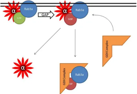
Mollard et al., 1991). The molecular machinery that mediates rab3a cycling has been extensively characterized. After GTP cleavage, rab3a is retrieved off vesicles by guanine-nucleotide dissociation inhibitor (GDI) complex that includes Hsp90, such that the Hsp90 inhibitors radicicol and geldanamycin prevent rab3a dissociation from synaptic membranes (Sakisaka et al., 2002). Using our -synuclein dissociation assay, we found that the Hsp90 inhibitors also caused an accumulation of -synuclein on synaptic membranes, providing support for argument that -synuclein membrane association is closely linked to that of rab3a and is regulated by the GDI/Hsp90 chaperone complex (Chen and Tandon, unpublished). In accordance with this, expression of a constitutively GTP-bound, dominant-negative rab3a mutant also induced an increase in membrane-bound -synuclein suggesting that GTP/GDP conversion by rab3a is a precondition for the liberation of vesicular synuclein. Together, our results suggest that rab3a and its recycling machinery regulate -synuclein membrane-binding and dissociation (Fig. 3).
Fig. 3. A model whereby rab3a regulates dissociation of -synuclein from synaptic
membranes. GTP-bound rab3a binds to -synuclein on the membrane. After GTP-hydrolysis mediated by a GTPase activating protein (GAP), rab3a is chaperoned off of the membrane by the GDP-dissociation inhibitor complex (GDI complex) and -synuclein also dissociates from the membrane. Whether -synuclein dissociation is transiently mediated by the GDI complex or is because of the loss of membrane-bound rab3a is yet to be determined.
Whether these results have any implications on the etiology of synucleinopathies is unclear and there are no rab3a mutations linked to PD. However, the interaction with rab3a reinforces -synuclein’s close relationship with the machinery controlling synaptic vesicle trafficking. Further research is needed on how -synuclein fits into the broader picture of the exocytic pathway and how subtle changes to -synuclein disposition may disturb vesicle dynamics and lead to pathology.
5. Conclusion
There is now ample evidence that -synuclein is involved in the regulation of synaptic vesicle trafficking and, more recently, in mitochondrial fission/fusion machinery. Whether its role in maintaining these different organelles occurs concurrently in healthy neurons is not known, though it may be instructive to consider the levels of expression required for apparent modulation of vesicle mobilization or mitochondrial dynamics. The effects of -synuclein on mitochondrial morphology have been noted in only two studies thus far, both requiring transient overexpression and not in models of normal or stable -synuclein expression (Kamp et al., 2010; Nakamura et al., 2011). If this observation holds in future analyses, it may represent a segregation of -synuclein activity based on its expression level, whereby lower levels of -synuclein impart direct effects on synaptic vesicle behaviour and high -synuclein concentration disrupts normal mitochondrial division and turnover. This dual activity may also explain the biphasic nature of -synuclein toxicity, being beneficial at lower expression levels and increasing in toxicity at higher concentration, particularly in combination with oxidative stress. Moreover, a gainoffunction pathogenic link between -synuclein and mitochondria fits well into a biological cascade that is opposed by expression of PINK1, parkin, or DJ-1, which are now recognized as controlling mitochondrial fission/fusion and mitophagy.
It is clear that direct interaction of -synuclein with lipid membranes is necessary to propagate its biological function. Regardless of the organelle interaction, it is reasonable to assume that the binding -synuclein to various organelle membranes is likely to involve a common mechanism whereby the amino-terminal half of -synuclein folds into an amphipathic -helix for partial immersion into the lipid bilayer. However, additional selective molecular requirements for stable -synuclein binding and retrieval may vary between different organelles, such as vesicles or mitochondria, each regulated by organelle-specific factors present on the membrane or in the cytosol. One potential means to characterize -synuclein compartmentalization on discrete organelles is by further refining the cell-free binding and dissociation assays, as described above using hypotonically permeabilized mouse brain synaptosomes, to specific organelles that are first isolated by differential density and centrifugation protocols. Alternatively, detection of organelle-specific interactors could be done by using inhibitory antibodies to screen for candidates known to exist only on defined organelles, as we have done to identify rab3a as an -synuclein binding partner on synaptic vesicles. Finally, these cell-free assays also offer an approach to validate the functional effects of -synuclein interactions identified by various genetic or biochemical methodologies by characterizing their consequences on -synuclein membrane distribution.
6. Acknowledgements
This work was supported by operating grants to AT from the Canadian Institutes for Health Research and from the Parkinson Society of Canada.
7. References
Abeliovich, A., Schmitz, Y., Farinas, I., Choi-Lundberg, D., Ho, W. H., Castillo, P. E., Shinsky, N., Verdugo, J. M., Armanini, M., Ryan, A., Hynes, M., Phillips, H., Sulzer,
D., and Rosenthal, A. (2000). Mice lacking alpha-synuclein display functional deficits in the nigrostriatal dopamine system. Neuron 25(1), 239-52.
Auluck, P. K., Caraveo, G., and Lindquist, S. (2010). alpha-Synuclein: membrane interactions and toxicity in Parkinson's disease. Annu Rev Cell Dev Biol 26, 211-33.
Batelli, S., Albani, D., Rametta, R., Polito, L., Prato, F., Pesaresi, M., Negro, A., and Forloni, G. (2008). DJ-1 modulates alpha-synuclein aggregation state in a cellular model of oxidative stress: relevance for Parkinson's disease and involvement of HSP70. PLoS
ONE 3(4), e1884.
Borbat, P., Ramlall, T. F., Freed, J. H., and Eliezer, D. (2006). Inter-helix distances in lysophospholipid micelle-bound alpha-synuclein from pulsed ESR measurements. J
Am Chem Soc 128(31), 10004-5.
Braak, H., Del Tredici, K., Rub, U., de Vos, R. A., Jansen Steur, E. N., and Braak, E. (2003). Staging of brain pathology related to sporadic Parkinson's disease. Neurobiol Aging 24(2), 197-211.
Burre, J., Sharma, M., Tsetsenis, T., Buchman, V., Etherton, M., and Sudhof, T. C. (2010). {alpha}-Synuclein Promotes SNARE-Complex Assembly in Vivo and in Vitro.
Science.
Cabin, D. E., Shimazu, K., Murphy, D., Cole, N. B., Gottschalk, W., McIlwain, K. L., Orrison, B., Chen, A., Ellis, C. E., Paylor, R., Lu, B., and Nussbaum, R. L. (2002). Synaptic vesicle depletion correlates with attenuated synaptic responses to prolonged repetitive stimulation in mice lacking alpha-synuclein. J Neurosci 22(20), 8797-807.
Chandra, S., Chen, X., Rizo, J., Jahn, R., and Sudhof, T. C. (2003). A broken alpha -helix in folded alpha -Synuclein. J Biol Chem 278(17), 15313-8.
Chandra, S., Fornai, F., Kwon, H. B., Yazdani, U., Atasoy, D., Liu, X., Hammer, R. E., Battaglia, G., German, D. C., Castillo, P. E., and Sudhof, T. C. (2004). Double-knockout mice for alpha- and beta-synucleins: effect on synaptic functions. Proc
Natl Acad Sci U S A 101(41), 14966-71.
Chandra S, Gallardo G, Fernandez-Chacon R, Schluter OM, Sudhof TC (2005) Alpha-synuclein cooperates with CSP alpha in preventing neurodegeneration. Cell, 123:383-396.
Chartier-Harlin, M. C., Kachergus, J., Roumier, C., Mouroux, V., Douay, X., Lincoln, S., Levecque, C., Larvor, L., Andrieux, J., Hulihan, M., Waucquier, N., Defebvre, L., Amouyel, P., Farrer, M., and Destee, A. (2004). Alpha-synuclein locus duplication as a cause of familial Parkinson's disease. Lancet 364(9440), 1167-9.
Chen, C. Y., and Balch, W. E. (2006). The Hsp90 chaperone complex regulates GDI-dependent Rab recycling. Mol Biol Cell 17(8), 3494-507.
Chen, C. Y., Sakisaka, T., and Balch, W. E. (2005). Use of Hsp90 inhibitors to disrupt GDI-dependent Rab recycling. Methods Enzymol 403, 339-47.
Chen, L., and Feany, M. B. (2005). Alpha-synuclein phosphorylation controls neurotoxicity and inclusion formation in a Drosophila model of Parkinson disease. Nat Neurosci 8(5), 657-63.
Christine, C. W., and Aminoff, M. J. (2004). Clinical differentiation of parkinsonian syndromes: prognostic and therapeutic relevance. Am J Med 117(6), 412-9.
Clayton, D. F., and George, J. M. (1998). The synucleins: a family of proteins involved in synaptic function, plasticity, neurodegeneration and disease. Trends Neurosci 21(6), 249-54.
Cole, N. B., Murphy, D. D., Grider, T., Rueter, S., Brasaemle, D., and Nussbaum, R. L. (2002). Lipid droplet binding and oligomerization properties of the Parkinson's disease protein alpha-synuclein. J Biol Chem 277(8), 6344-52.
Conway, K. A., Lee, S. J., Rochet, J. C., Ding, T. T., Williamson, R. E., and Lansbury, P. T., Jr. (2000). Acceleration of oligomerization, not fibrillization, is a shared property of both alpha-synuclein mutations linked to early-onset Parkinson's disease: implications for pathogenesis and therapy. Proc Natl Acad Sci U S A 97(2), 571-6.
Cookson, M. R. (2005). The biochemistry of Parkinson's disease. Annu Rev Biochem 74, 29-52.
Cooper, A. A., Gitler, A. D., Cashikar, A., Haynes, C. M., Hill, K. J., Bhullar, B., Liu, K., Xu, K., Strathearn, K. E., Liu, F., Cao, S., Caldwell, K. A., Caldwell, G. A., Marsischky, G., Kolodner, R. D., Labaer, J., Rochet, J. C., Bonini, N. M., and Lindquist, S. (2006). Alpha-synuclein blocks ER-Golgi traffic and Rab1 rescues neuron loss in Parkinson's models. Science 313(5785), 324-8.
Dalfo, E., Barrachina, M., Rosa, J. L., Ambrosio, S., and Ferrer, I. (2004a). Abnormal alpha-synuclein interactions with rab3a and rabphilin in diffuse Lewy body disease.
Neurobiol Dis 16(1), 92-7.
Dalfo, E., and Ferrer, I. (2005). Alpha-synuclein binding to rab3a in multiple system atrophy.
Neurosci Lett 380(1-2), 170-5.
Dalfo, E., Gomez-Isla, T., Rosa, J. L., Nieto Bodelon, M., Cuadrado Tejedor, M., Barrachina, M., Ambrosio, S., and Ferrer, I. (2004b). Abnormal alpha-synuclein interactions with Rab proteins in alpha-synuclein A30P transgenic mice. J Neuropathol Exp
Neurol 63(4), 302-13.
Darios, F., Ruiperez, V., Lopez, I., Villanueva, J., Gutierrez, L. M., and Davletov, B. (2010). Alpha-synuclein sequesters arachidonic acid to modulate SNARE-mediated exocytosis. EMBO Rep 11(7), 528-33.
Davidson, W. S., Jonas, A., Clayton, D. F., and George, J. M. (1998). Stabilization of alpha-synuclein secondary structure upon binding to synthetic membranes. J Biol Chem 273(16), 9443-9.
Devi, L., Raghavendran, V., Prabhu, B. M., Avadhani, N. G., and Anandatheerthavarada, H. K. (2008). Mitochondrial import and accumulation of alpha-synuclein impair complex I in human dopaminergic neuronal cultures and Parkinson disease brain. J
Biol Chem 283(14), 9089-100.
Fischer von Mollard, G., Sudhof, T. C., and Jahn, R. (1991). A small GTP-binding protein dissociates from synaptic vesicles during exocytosis. Nature 349(6304), 79-81. Fortin, D. L., Nemani, V. M., Voglmaier, S. M., Anthony, M. D., Ryan, T. A., and Edwards, R.
H. (2005). Neural activity controls the synaptic accumulation of alpha-synuclein. J
Fujiwara, H., Hasegawa, M., Dohmae, N., Kawashima, A., Masliah, E., Goldberg, M. S., Shen, J., Takio, K., and Iwatsubo, T. (2002). alpha-Synuclein is phosphorylated in synucleinopathy lesions. Nat Cell Biol 4(2), 160-4.
George, J. M., Jin, H., Woods, W. S., and Clayton, D. F. (1995). Characterization of a novel protein regulated during the critical period for song learning in the zebra finch.
Neuron 15(2), 361-72.
Georgieva, E. R., Ramlall, T. F., Borbat, P. P., Freed, J. H., and Eliezer, D. (2010). The lipid-binding domain of wild type and mutant alpha-synuclein: compactness and interconversion between the broken and extended helix forms. J Biol Chem 285(36), 28261-74.
Geppert, M., Bolshakov, V. Y., Siegelbaum, S. A., Takei, K., De Camilli, P., Hammer, R. E., and Sudhof, T. C. (1994). The role of Rab3A in neurotransmitter release. Nature 369(6480), 493-7.
Geppert, M., Goda, Y., Stevens, C. F., and Sudhof, T. C. (1997). The small GTP-binding protein Rab3A regulates a late step in synaptic vesicle fusion. Nature 387(6635), 810-4.
Gitler, A. D., Bevis, B. J., Shorter, J., Strathearn, K. E., Hamamichi, S., Su, L. J., Caldwell, K. A., Caldwell, G. A., Rochet, J. C., McCaffery, J. M., Barlowe, C., and Lindquist, S. (2008). The Parkinson's disease protein alpha-synuclein disrupts cellular Rab homeostasis. Proc Natl Acad Sci U S A 105(1), 145-50.
Gorbatyuk, O. S., Li, S., Sullivan, L. F., Chen, W., Kondrikova, G., Manfredsson, F. P., Mandel, R. J., and Muzyczka, N. (2008). The phosphorylation state of Ser-129 in human alpha-synuclein determines neurodegeneration in a rat model of Parkinson disease. Proc Natl Acad Sci U S A 105(2), 763-8.
Hamilton, B. A. (2004). alpha-Synuclein A53T substitution associated with Parkinson disease also marks the divergence of Old World and New World primates. Genomics 83(4), 739-42.
Hashimoto, M., Hsu, L. J., Rockenstein, E., Takenouchi, T., Mallory, M., and Masliah, E. (2002). alpha-Synuclein protects against oxidative stress via inactivation of the c-Jun N-terminal kinase stress-signaling pathway in neuronal cells. J Biol Chem 277(13), 11465-72.
Holdorff, B. (2006). Fritz Heinrich Lewy (1885-1950). J Neurol 253(5), 677-8.
Hu, K., Carroll, J., Rickman, C., and Davletov, B. (2002). Action of complexin on SNARE complex. J Biol Chem 277(44), 41652-6.
Jakes, R., Spillantini, M. G., and Goedert, M. (1994). Identification of two distinct synucleins from human brain. FEBS Lett 345(1), 27-32.
Jao, C. C., Der-Sarkissian, A., Chen, J., and Langen, R. (2004). Structure of membrane-bound alpha-synuclein studied by site-directed spin labeling. Proc Natl Acad Sci U S A 101(22), 8331-6.
Jankovic, J. (2008). Parkinson's disease: clinical features and diagnosis. J Neurol Neurosurg
Psychiatry 79(4), 368-76.
Jo, E., McLaurin, J., Yip, C. M., St George-Hyslop, P., and Fraser, P. E. (2000). alpha-Synuclein membrane interactions and lipid specificity. J Biol Chem 275(44), 34328-34.
Jo, E., Fuller, N., Rand, R. P., St George-Hyslop, P., and Fraser, P. E. (2002). Defective membrane interactions of familial Parkinson's disease mutant A30P alpha-synuclein. J Mol Biol 315(4), 799-807.
Jo, E., Darabie, A. A., Han, K., Tandon, A., Fraser, P. E., and McLaurin, J. (2004). alpha-Synuclein-synaptosomal membrane interactions: implications for fibrillogenesis.
Eur J Biochem 271(15), 3180-9.
Johannes, L., Doussau, F., Clabecq, A., Henry, J. P., Darchen, F., and Poulain, B. (1996). Evidence for a functional link between Rab3 and the SNARE complex. J Cell Sci 109 ( Pt 12), 2875-84.
Johannes, L., Lledo, P. M., Roa, M., Vincent, J. D., Henry, J. P., and Darchen, F. (1994). The GTPase Rab3a negatively controls calcium-dependent exocytosis in neuroendocrine cells. Embo J 13(9), 2029-37.
Kamp, F., Exner, N., Lutz, A. K., Wender, N., Hegermann, J., Brunner, B., Nuscher, B., Bartels, T., Giese, A., Beyer, K., Eimer, S., Winklhofer, K. F., and Haass, C. (2010). Inhibition of mitochondrial fusion by alpha-synuclein is rescued by PINK1, Parkin and DJ-1. Embo J 29(20), 3571-89.
Kanda, S., Bishop, J. F., Eglitis, M. A., Yang, Y., and Mouradian, M. M. (2000). Enhanced vulnerability to oxidative stress by alpha-synuclein mutations and C-terminal truncation. Neuroscience 97(2), 279-84.
Kruger, R., Kuhn, W., Muller, T., Woitalla, D., Graeber, M., Kosel, S., Przuntek, H., Epplen, J. T., Schols, L., and Riess, O. (1998). Ala30Pro mutation in the gene encoding alpha-synuclein in Parkinson's disease. Nat Genet 18(2), 106-8.
Lee, H. J., Choi, C., and Lee, S. J. (2002). Membrane-bound alpha-synuclein has a high aggregation propensity and the ability to seed the aggregation of the cytosolic form. J Biol Chem 277(1), 671-8.
Leenders, A. G., Lopes da Silva, F. H., Ghijsen, W. E., and Verhage, M. (2001). Rab3a is involved in transport of synaptic vesicles to the active zone in mouse brain nerve terminals. Mol Biol Cell 12(10), 3095-102.
Maroteaux L, Campanelli JT, Scheller RH (1988). Synuclein: a neuron-specific protein localized to the nucleus and presynaptic nerve terminal. J Neurosci 8, 2804-2815. McFarland, N. R., Fan, Z., Xu, K., Schwarzschild, M. A., Feany, M. B., Hyman, B. T., and
McLean, P. J. (2009). Alpha-synuclein S129 phosphorylation mutants do not alter nigrostriatal toxicity in a rat model of Parkinson disease. J Neuropathol Exp Neurol 68(5), 515-24.
Mollenhauer, B., Locascio, J. J., Schulz-Schaeffer, W., Sixel-Doring, F., Trenkwalder, C., and Schlossmacher, M. G. (2011). alpha-Synuclein and tau concentrations in cerebrospinal fluid of patients presenting with parkinsonism: a cohort study. Lancet
Neurol 10(3), 230-40.
Nakamura, K., Nemani, V. M., Azarbal, F., Skibinski, G., Levy, J. M., Egami, K., Munishkina, L., Zhang, J., Gardner, B., Wakabayashi, J., Sesaki, H., Cheng, Y., Finkbeiner, S., Nussbaum, R. L., Masliah, E., and Edwards, R. H. (2011). Direct membrane association drives mitochondrial fission by the Parkinson Disease-associated protein {alpha}-synuclein. J Biol Chem (Epub ahead of print).
Narayanan, V., and Scarlata, S. (2001). Membrane binding and self-association of alpha-synucleins. Biochemistry 40(33), 9927-34.
Nemani, V. M., Lu, W., Berge, V., Nakamura, K., Onoa, B., Lee, M. K., Chaudhry, F. A., Nicoll, R. A., and Edwards, R. H. (2010). Increased expression of alpha-synuclein reduces neurotransmitter release by inhibiting synaptic vesicle reclustering after endocytosis. Neuron 65(1), 66-79.
Polymeropoulos, M. H., Lavedan, C., Leroy, E., Ide, S. E., Dehejia, A., Dutra, A., Pike, B., Root, H., Rubenstein, J., Boyer, R., Stenroos, E. S., Chandrasekharappa, S., Athanassiadou, A., Papapetropoulos, T., Johnson, W. G., Lazzarini, A. M., Duvoisin, R. C., Di Iorio, G., Golbe, L. I., and Nussbaum, R. L. (1997). Mutation in the alpha-synuclein gene identified in families with Parkinson's disease. Science 276(5321), 2045-7.
Sakisaka, T., Meerlo, T., Matteson, J., Plutner, H., and Balch, W. E. (2002). Rab-alphaGDI activity is regulated by a Hsp90 chaperone complex. Embo J 21(22), 6125-35.
Saper, C. B. (1999). 'Like a thief in the night': the selectivity of degeneration in Parkinson's disease. Brain 122 ( Pt 8), 1401-2.
Singleton, A. B., Farrer, M., Johnson, J., Singleton, A., Hague, S., Kachergus, J., Hulihan, M., Peuralinna, T., Dutra, A., Nussbaum, R., Lincoln, S., Crawley, A., Hanson, M., Maraganore, D., Adler, C., Cookson, M. R., Muenter, M., Baptista, M., Miller, D., Blancato, J., Hardy, J., and Gwinn-Hardy, K. (2003). alpha-Synuclein locus triplication causes Parkinson's disease. Science 302(5646), 841.
Soper, J. H., Roy, S., Stieber, A., Lee, E., Wilson, R. B., Trojanowski, J. Q., Burd, C. G., and Lee, V. M. (2008). Alpha-synuclein-induced aggregation of cytoplasmic vesicles in Saccharomyces cerevisiae. Mol Biol Cell 19(3), 1093-103.
Spillantini, M. G., Crowther, R. A., Jakes, R., Hasegawa, M., and Goedert, M. (1998). alpha-Synuclein in filamentous inclusions of Lewy bodies from Parkinson's disease and dementia with lewy bodies. Proc Natl Acad Sci U S A 95(11), 6469-73.
Thayanidhi, N., Helm, J. R., Nycz, D. C., Bentley, M., Liang, Y., and Hay, J. C. (2010). Alpha-synuclein delays endoplasmic reticulum (ER)-to-Golgi transport in mammalian cells by antagonizing ER/Golgi SNAREs. Mol Biol Cell 21(11), 1850-63.
Ulmer, T. S., Bax, A., Cole, N. B., and Nussbaum, R. L. (2005). Structure and dynamics of micelle-bound human alpha-synuclein. J Biol Chem 280(10), 9595-603.
Wislet-Gendebien, S., D'Souza, C., Kawarai, T., St George-Hyslop, P., Westaway, D., Fraser, P., and Tandon, A. (2006). Cytosolic proteins regulate alpha-synuclein dissociation from presynaptic membranes. J Biol Chem 281(43), 32148-55.
Wislet-Gendebien, S., Visanji, N. P., Whitehead, S. N., Marsilio, D., Hou, W., Figeys, D., Fraser, P. E., Bennett, S. A., and Tandon, A. (2008). Differential regulation of wild-type and mutant alpha-synuclein binding to synaptic membranes by cytosolic factors. BMC Neurosci 9, 92.
Zarranz, J. J., Alegre, J., Gomez-Esteban, J. C., Lezcano, E., Ros, R., Ampuero, I., Vidal, L., Hoenicka, J., Rodriguez, O., Atares, B., Llorens, V., Gomez Tortosa, E., del Ser, T., Munoz, D. G., and de Yebenes, J. G. (2004). The new mutation, E46K, of alpha-synuclein causes Parkinson and Lewy body dementia. Ann Neurol 55(2), 164-73.
Zhu, M., and Fink, A. L. (2003). Lipid binding inhibits alpha-synuclein fibril formation. J Biol
Edited by Dr. Juliana Dushanova
ISBN 978-953-307-876-2 Hard cover, 582 pages
Publisher InTech
Published online 08, February, 2012 Published in print edition February, 2012
InTech Europe
University Campus STeP Ri Slavka Krautzeka 83/A 51000 Rijeka, Croatia Phone: +385 (51) 770 447 Fax: +385 (51) 686 166 www.intechopen.com
InTech China
Unit 405, Office Block, Hotel Equatorial Shanghai No.65, Yan An Road (West), Shanghai, 200040, China Phone: +86-21-62489820
Fax: +86-21-62489821
Parkinson's disease (PD) results primarily from the death of dopaminergic neurons in the substantia nigra. Current PD medications treat symptoms; none halt or retard dopaminergic neuron degeneration. The main obstacle to developing neuroprotective therapies is a limited understanding of the key molecular mechanisms that provoke neurodegeneration. The discovery of PD genes has led to the hypothesis that misfolding of proteins and dysfunction of the ubiquitin-proteasome pathway are pivotal to PD pathogenesis. Previously implicated culprits in PD neurodegeneration, mitochondrial dysfunction, and oxidative stress may also act in part by causing the accumulation of misfolded proteins, in addition to producing other deleterious events in dopaminergic neurons. Neurotoxin-based models have been important in elucidating the molecular cascade of cell death in dopaminergic neurons. PD models based on the manipulation of PD genes should prove valuable in elucidating important aspects of the disease, such as selective vulnerability of substantia nigra dopaminergic neurons to the degenerative process.
How to reference
In order to correctly reference this scholarly work, feel free to copy and paste the following:
Robert H.C. Chen, Sabine Wislet-Gendebien, Howard T.J. Mount and Anurag Tandon (2012). Regulation of a-Synuclein Membrane Binding and Its Implications, Mechanisms in Parkinson's Disease - Models and
Treatments, Dr. Juliana Dushanova (Ed.), ISBN: 978-953-307-876-2, InTech, Available from:
http://www.intechopen.com/books/mechanisms-in-parkinson-s-disease-models-and-treatments/regulation-of-synuclein-membrane-binding-and-its-implications