European Journal of Chemistry
View Journal Online View Article Online
Crystal structure and Hirshfeld surface analysis of
N-(2-(N-methylsulfamoyl)phenyl)formamide: Degradation product of
2-methyl-2H-1,2,4-benzothiadiazine 1,1-dioxide
Koffi Sénam Etsè
1,*, Guillermo Zaragoza
2and Bernard Pirotte
11 Laboratory of Medicinal Chemistry, Center for Interdisciplinary Research on Medicines (CIRM), University of Liège, Quartier Hôpital B36 Av. Hippocrate 15
B-4000 Liège, Belgium
ks.etse@uliege.be (K.S.E.), b.pirotte@uliege.be (B.P.)
2 Unidade de Difracción de Raios X, RIAIDT, Universidade de Santiago de Compostela, Campus VIDA, 15782 Santiago de Compostela, Spain
g.zaragoza@usc.es (G.Z.)
* Corresponding author at: Laboratory of Medicinal Chemistry, Center for Interdisciplinary Research on Medicines (CIRM), University of Liège, Quartier Hôpital B36 Av. Hippocrate 15 B-4000 Liège, Belgium.
Tel: +32.04.3664368 Fax: +32.04.3664361 e-mail: ks.etse@uliege.be (K.S. Etse).
10.5155/eurjchem.10.3.189-194.1903 Received: 03 June 2019
Received in revised form: 04 July 2019 Accepted: 05 July 2019
Published online: 30 September 2019 Printed: 30 September 2019
The hydrolysis of 2-methyl-2H-1,2,4-benzothiadiazine 1,1-dioxide (2) during crystallization under humidity (85 %) conditions, lead to N-(2-(N-methylsulfamoyl)phenyl)formamide as second step hydrolysis product, identified in the proposed degradation mechanism. Crystal of N-(2-(N-methylsulfamoyl)phenyl)formamide C8H10N2O3S (4), was obtained and
characterized. The molecular structure determination was carried out with MoKα X-ray and data measured at 100 K. The compound 4 crystallizes in triclinic P�1 space group with unit cell parameters a = 4.8465(4) Å, b = 8.1942(9) Å, c = 11.8686(13) Å, α = 77.080(4)°, β = 82.069(4)°, γ = 80.648(4)°, V = 450.76 (8) Å3 and Z = 2. The crystal structure is stabilized by
intramolecular N-H···O and intermolecular C-H···O and N-H···O hydrogen bonds that extended as infinite 1D chain along [100]. Stabilization is also ensured by oxygen-π stacking interaction between the aromatic ring and oxygen of the sulfonamide group. The analysis of intermolecular interactions through the mapping of dnorm and shape-index revel that the
most significant contributions to the Hirshfeld surface 40.6 and 33.9% are from H···H and O···H contacts, respectively.
Hydrolysis Formamide Crystal structure Hirshfeld surface Benzothiadiazine
Hydrogen bonding Cite this: Eur. J. Chem. 2019, 10(3), 189-194 Journal website: www.eurjchem.com
1. Introduction
Thiazide derivatives in general and benzothiadiazine 1,1-dioxides particularly are described as possessing numerous pharmacological activities [1-8]. Hydrochlorothiazide (HCTZ or 6-chloro-3,4-dihydro-2H-1,2,4-benzothiadiazine-7-sulfon-amide 1,1-dioxide) is a well-known benzothiadiazine used as diuretic drug for the treatment of hypertension [9]. Despite the importance of these heterocycles as promising drugs and the report of various molecular structures obtained by X-ray diffraction analysis [10-12], studies have shown that benzo-thiadiazine 1,1-dioxides hydrolyze slowly in aqueous media. In the case of HCTZ, studies reveal that hydrolysis led to degra-dation product identified as 4-amino-6-chlorobenzene-1,3-disulfonamide and formaldehyde [13-16]. Although various by-products are later identified during thiazide hydrolysis [17,18], isolation and full characterization of all degradation products were not reported at all. Qun Xu have proposed a plausible hydrolysis pathway of benzothiadiazine under
thermal and humidity conditions showing various inter-mediates [19]. In general, 2-aminobenzenesulfonamide derivatives are recognized as a main isolated and charac-terized product of benzothiadiazine 1,1-dioxides decompo-sition.
Our group is continuously interested in the synthesis, characterization and biological evaluation of benzoisothiazoles [20] and benzothiadiazine 1,1-dioxides [21,22]. In view to develop a new family of these interesting compounds, 2-methyl-2H-1,2,4-benzothiadiazine 1,1-dioxide was prepared as precursor. The crystallization of this latter under humidity conditions leads to mixture of by-products and monocrystal that was analyzed. In this paper, we are reporting the structural characterization by single-crystal X-ray diffraction analysis of N-(2-(N-methylsulfamoyl)phenyl)formamide (4) as hydrolysis uncommon by-product of 2-methyl-2H-1,2,4-benzothiadiazine 1,1-dioxide. Furthermore, the intermolecular interactions were examined using Hirshfeld surface analysis.
ABSTRACT RESEARCH ARTICLE
KEYWORDS
European Journal of Chemistry
ISSN 2153-2249 (Print) / ISSN 2153-2257 (Online) – Copyright © 2019 The Authors – Atlanta Publishing House LLC – Printed in the USA.
European Journal of Chemistry - This document downloaded from http://www.eurjchem.com by guest on 30 September 2019 13:27. For personal use only.
NH2 SO2 HN SO2 N N HC(OEt)3 150 °C NH SO2 HN H O SO2 N HN OH NH SO2 HN OH HO H2O H2O H OH O 1 2 3 4 5
Figure 1. Formation and proposal hydrolysis pathway of 2-methyl-2H-1,2,4-benzothiadiazine 1,1-dioxide 2 to yield N-(2-(N-methylsulfamoyl)phenyl)
formamide (4). 2. Experimental
2.1. Crystal growth
Crystal of compound 4 was obtained by dissolving appropriate amounts (30 mg) of compound 2 in a mixture of acetone and dichloromethane. Solvent was added to ensure completely solubilization of the starting compound and the solution was transferred to 1 cm diameter tube without cap and keep in cold room at 6 °C. After solvent evaporation, monocrystals suitable for X-ray diffraction analysis were isolated and analysed.
2.2. X-ray crystallography
For the crystal structure determination, the data were collected by applying the omega and phi scans method on a Bruker D8 Venture PHOTON III-14 diffractometer using INCOATEC multilayer mirror monochromated with MoKα radiation (λ = 0.71073 Å) from a microfocus sealed tube source at 100 K with detector resolution of 7.3910 pixels/mm. Computing data and reduction were made with the APEX3 [23]. The structure was solved using SHELXT [24] and finally refined by full-matrix least-squares based on F2 by SHELXL [25]. An empirical absorption correction was applied using the SADABS program. Software used to molecular graphics: ORTEP for Windows [26]. Software used to prepare material for publication: WinGX publication routines [26] and Mercury [27].
2.3. Refinement
All non-hydrogen atoms were refined anisotropically and the hydrogen atom positions were included in the model on the basis of Fourier difference electron density maps. All CH and aromatic CH hydrogen (C-H = 0.95 Å) atoms were refined using a riding model with Uiso(H) = 1.2 Ueq(C). The methyl hydrogen (C-H = 0.98 Å) atoms were refined as a rigid group with torsional freedom [Uiso(H) = 1.5 Ueq(C)] and the hydrogens H1N and H2N atom as a free atom.
3. Results and discussion
3.1. Description of crystal structure of compound 4
The crystal of the title compound was obtained from slow degradation of 2-methyl-2H-1,2,4-benzothiadiazine 1,1-dioxide (2) during crystallization. The synthesis of compound
2 was accomplished using the procedure; we have already reported in [4,28]. Indeed, the 2-amino-N-methylbenzene sulfonamide was heated in triethyl orthoformiate to reflux for 30 min. After cooling on an ice bath, the benzothiadiazine (2) was collected by filtration, washed with diethyl ether and dried. An appropriate amount (30 mg) of this product was dissolved in a mixture of acetone and dichloromethane. The amount of solvent used was sufficient to completely solubilize the compound and the solution was transferred to 1 cm diameter tube without cap and kept at 6 °C until the solvent evaporation was completed and formation of crystals. Monocrystal suitable for X-ray diffraction analysis were obtained and analyzed. Although studies report mainly 2-aminobenzenesulfonamides as the most stable and recurrent degradation product of benzothiadiazines, our present results have shown that the molecular structure correspond to N-(2-(N-methylsulfamoyl)phenyl)formamide (4) (Figure 1). Working without a cap allows solvent evaporation but also leads to solvent wetness and therefore favors the hydrolysis of the starting benzothiadiazine. The synthesis and the proposed pathway for the degradation of 2-methyl-2H-1,2,4-benzo-thiadiazine 1,1-dioxide during crystallization under humidity conditions are shown in Figure 1. Based on the described benzothiadiazines hydrolysis studies, we propose here a mechanism for the formation of product 4 [19,29].
The compound 4 crystallizes in triclinic P�1 space group. All relevant crystal data are reported in Table 1. The molecular structure of compound 4 is shown in Figure 2. The S-O (~1.43 Å) and C-S (1.77 Å) bonds length are typical for that observed in sulfonamides and benzothiadiazines [30]. For the amide function, the carbonyl bond C1-O (1.2291(17) Å) influence electronically the N2-C1 bond length that becomes shorter than that observed for N1-C8 in the sulfonamide with a value of 1.3471(17) and 1.4163(17) Å, respectively (Table 2) [31,32]. The dihedral angles between the aromatic ring and the plane formed by the atoms C7-C2-N2 (1.45(7)°) and the one formed by C2-C7-S1 (5.50(6)°) revealed slightly deviation of aniline (N2-C2) and sulfone (C7-S1) bonds from the aromatic ring (C2---C7) plane. One intramolecular short hydrogen interactions N2-H2N···O1 (Figure 2) was observed resulting from the proximity of H2N and O1 atoms 2.21 (2) Å
(Table 3) with N2-H2N and S1-O1 bonds orientation in the
same direction.
The cell unit presents two molecules around the inversion center, so that the aromatic rings are confronted in parallel with a dihedral angle of 0.00(7)° (Figure 3).
European Journal of Chemistry - This document downloaded from http://www.eurjchem.com by guest on 30 September 2019 13:27. For personal use only.
Table 1. Crystal data of compound 4.
Parameters Compound 4
Chemical formula C8H10N2O3S
Mw (g) 214.24
Crystal system Triclinic
Space group P-1 Temperature (K) 100 a (Å) 4.8465(4) b (Å) 8.1942(9) c (Å) 11.8686(13) α (°) 77.080(4) β (°) 82.069(4) γ (°) 80.648(4) V (Å3) 450.76(8) Z 2
Radiation type Mo-Kα radiation (λ = 0.71073 Å)
μ (mm−1) 0.34
Crystal size (mm) 0.33 × 0.09 × 0.05
Tmin, Tmax 0.911, 0.953
(sin θ/λ)max (Å−1) 0.667
Measured/independent/observed [I>2σ(I)] reflections 21965/2245/2053
Rint 0.040
R[F2 > 2σ(F2)], wR(F2), S 0.029, 0.073, 1.06
Δρmax/Δρmin (e.Å−3) 0.35, -0.45
Table 2. Selected geometric parameters (Å, °) for compound 4.
Bonds Length (Å) Bonds Angle (°)
C2-N2 1.4163(17) C8-N1-S1 121.40(10) C7-S1 1.7721(13) C1-N2-C2 125.73(12) C8-N1 1.4675(18) O2-S1-O1 120.04(6) C1-O3 1.2291(17) O2-S1-N1 107.58(6) C1-N2 1.3471(17) O1-S1-N1 105.57(6) N1-S1 1.6044(12) N1-S1-C7 109.22(6) O1-S1 1.4399(10) C8-N1-S1-C7 77.02(12) O2-S1 1.4339(10) O3-C1-N2-C2 3.0(2) C1-H1 0.95 C2-C7-S1-N1 72.31(12) C2-C7 1.4018(18) N2-C2-C7-S1 6.70(17) N1-H1N 0.84(2) C8-N1-S1-C7 77.02(12) N2-H2N 0.85(2) O3-C1-N2-C2 3.0(2)
Table 3. Hydrogen-bond geometry (Å, °)
D-H···A D-H H···A D···A D-H···A
N2-H2N···O1 0.85(2) 2.21(2) 2.7967(15) 127.0(17)
C1-H1···O1 i 0.95 2.4 3.2860(17) 155
N1-H1N···O3 ii 0.84(2) 2.07(2) 2.8988(16) 168(2)
N2-H2N···O3 iii 0.85(2) 2.57(2) 3.2832(16) 143.2(17)
Symmetry codes: (i) −x+2, −y, −z+1; (ii) −x+1, −y, −z+1; (iii) x+1, y, z.
Figure 2. Molecular structure and intramolecular hydrogen bond of compound 4 with thermal ellipsoids drawn at the 50% probability level.
Analysis of intermolecular interactions of the compound 4 shows that the crystal packing is stabilized by intermolecular N-H···O and C-H···O hydrogen bonds that extended as infinite 1D chain along [100] (Figure 3 and Table 3).
3.2. Hirshfeld surface analysis
The intermolecular interactions were evaluated using Hirshfeld surface analysis. Molecular Hirshfeld surfaces of compound 4 were calculated using a standard (high) surface
resolution and with the three-dimensional dnorm surfaces mapped over a fixed colour scale -0.5418 (red) to 1.0214 Å (blue) with the program CrystalExplorer 17 [33].
The Hirshfeld surface of the title compound mapped with shape-index (-1.0 to 1.0 A�) was also investigated (Figure 4). On the shape-index surface of compound 4, convex blue regions represent hydrogen-donor groups and concave red regions represent hydrogen-acceptor groups [34]. The oxygen atom act as an acceptor while all the hydrogen implicated in hydrogen bonds act as donor group.
European Journal of Chemistry - This document downloaded from http://www.eurjchem.com by guest on 30 September 2019 13:27. For personal use only.
Table 4. Intramolecular S-O1→Cg (C2→C7 ring) interactions in compounds 4.
Interactions X···Cg X-Perp Gamma Y-X···Cg Y···Cg
O1 → Cg1 [ 1+x, y, z] 3.0284(12) -2.920 15.38 109.98(5) 3.7716(8)
Figure 3. Infinite 1D chain along [100] by intermolecular hydrogen bonds observed in compound 4. Non-bonded hydrogen atoms are omitted for clarity.
Symmetry codes: (i) −x+2, −y, −z+1; (ii) −x+1, −y, −z+1; (iii) x+1, y, z.
Figure 4. (a) Hirshfeld surfaces of compound 4 mapped with shape-index (DG: Donor Group, AG: Acceptor Group). (b) O···C /C···O contacts O1···Cg (1+x, y, z).
Figure 5. View of the three-dimensional Hirshfeld surface of the title compound plotted over dnorm in the range -0.5418 (red) to 1.0214 Å (blue), highlighting
N-H···O and C-N-H···O hydrogen bonds by dashed yellow lines.
This analysis revelated the existence of an oxygen-π stacking interaction (C···O/O···C) between aromatic ring and O1 oxygen of sulfonamide group (Figure 4b). This interaction shows a distance between O1 and aromatic ring centroid (O1-Cg) of 3.0284(12) Å, an perpendicular distance of O1 respect to the aromatic ring plane of -2.920 Å, and an angle S-O-Cg of 109.98(5) ° (Table 4).
The analysis of intermolecular interactions through the mapping of dnorm is accomplished by considering the contact distances di and de from the Hirshfeld surface to the nearest atom inside and outside, respectively [35]. In compound 4, the surface mapped over dnorm highlights four well defined red spots showing distances shorter than the sum of the Van der Waals radii (Figure 5).
European Journal of Chemistry - This document downloaded from http://www.eurjchem.com by guest on 30 September 2019 13:27. For personal use only.
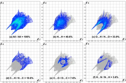
Figure 6. The Hirshfeld surface representations with the function dnorm plotted onto the surface for (a) all interactions, and (b) H···H, (c) O···H/H···O, (d)
C···H/C···H, (e) C···O/O···C and (f) N···H/N···H interactions.
These dominant interactions correspond to inter-molecular N-H···O and C-H···O hydrogen bonds and stacking interactions between the surface and the neighboring environment. The mapping also shows white spots with distances equal to the sum of the Van der Waals radii and blue regions with distances longer than the sum of the van der Waals radii [34,36]. The surfaces are transparent to allow visualization of the molecule.
We also analyzed the two-dimensional fingerprint (FP) of compound 4 plotted in Figure 6 highlighting particular close contacts of atom pairs and the contributions from different contacts. All interactions are presented in Figure 6a. The most significant contributions to the Hirshfeld surface (40.6 and 33.9%) are from H···H and O···H contacts (Figure 6b and 6c). These two interactions add to 74.5% of the intermolecular contacts of the Hirshfeld surface area. Others contributions correspond to C···H/C···H (10.6%), C···O/O···C (7.6%), N···H/N···H (3.6%), C···C (2.9%) and the remaining less-important interactions are inferior to 1%.
4. Conclusion
The hydrolysis of benzothiadiazine derivatives lead generally to isolated 2-aminobenzenesulfonamide, although substituted sulfamoylphenylformamide structures are described to be form during the first steps. In this study, we have determined the molecular structure of N-(2-(N-methyl sulfamoyl)phenyl)formamide obtained from slow degradation of 2-methyl-2H-1,2,4-benzothiadiazine 1,1-dioxide during crystallization using single crystal X-ray diffraction analysis. The molecular structure of compound 4 is stabilized by intra and intermolecular hydrogen bonds forming 1D chain along [100]. The analysis of intermolecular interactions through the mapping of Hirshfeld surface and two-dimensional fingerprint are presented and highlighted close contact of hydrogen bonds as well as acceptor and donor groups.
Acknowledgements
The authors thank the Université de Liège for a research grant.
Supporting information
CCDC-1915966 contains the supplementary crystallo-graphic data for this paper. These data can be obtained free of charge via https://www.ccdc.cam.ac.uk/structures/, or by e-mailing data_request@ccdc.cam.ac.uk, or by contacting The Cambridge Crystallographic Data Centre, 12 Union Road, Cambridge CB2 1EZ, UK; fax: +44(0)1223-336033.
Disclosure statement
Conflict of interests: The authors declare that they have no conflict of interest.
Author contributions: All authors contributed equally to this work.
Ethical approval: All ethical guidelines have been adhered. Sample availability: Samples of the compounds are available from the author.
ORCID Koffi Sénam Etsè
http://orcid.org/0000-0001-8495-4327 Guillermo Zaragoza http://orcid.org/0000-0002-2550-6628 Bernard Pirotte http://orcid.org/0000-0001-8251-8257 References
[1]. Martinez, A.; Gil, C.; Abasolo, M. I.; Castro, A.; Bruno, A. M.; Perez, C.; Prieto, C.; Otero, J. J. Med. Chem. 2000, 43, 3218-3225.
[2]. Norholm, A. B.; Francotte, P.; Olsen, L.; Krintel, C.; Frydenvang, K.; Goffin, E.; Challal, S.; Danober, L.; Botez-Pop, I.; Lestage, P.; Pirotte, B.; Kastrup, J. S. J. Med. Chem. 2013, 56, 8736-8745.
[3]. Di Bella, M.; Monzani, A.; Andrisano, M. G.; Fabio, U.; Quaglio, G. P. Farmaco. Sci. 1979, 34, 189-198.
[4]. Dintilhac, G.; Arslan, D.; Dilly, S.; Danober, L.; Botez, I.; Lestage, P.; Pirotte, B.; De Tullio, P. Med. Chem. Comm. 2011, 2, 509-523. [5]. Larsen, A. P.; Fievre, S.; Frydenvang, K.; Francotte, P.; Pirotte, B.;
Kastrup, J. S.; Mulle, C. Mol. Pharmacol. 2017, 91, 576-585. [6]. Varano, F.; Catarzi, D.; Colotta, V.; Squarcialupi, L.; Matucci, R. Arch.
Pharm. (Weinheim) 2014, 347, 777-785.
[7]. Bock, L.; Miller, G. H.; Schaper, K. J.; Seydel, J. K. J. Med. Chem. 1974, 17, 23-28. European Journal of Chemistry - This document downloaded from http://www.eurjchem.com by guest on 30 September 2019 13:27. For personal use only.
[8]. Pirotte, B.; Lebrun, P.; De Tullio, P.; Somers, F.; Delarge, J.; Hansen, J. B.; Nielsen, F. E.; Hansen, H. C.; Mogensen, J. P.; Moller-Tagmose, T. US Patent 6,242,443 B1, June 5, 2001.
[9]. Sica, D. A.; Carter, B.; Cushman, W.; Hamm, L. J. Clin. Hypertens. 2011, 13, 639-643.
[10]. Dupont, L.; De Tullio, P.; Boverie, S.; Pirotte, B. Acta Cryst. E 2002, 57, o602-o603.
[11]. Muhammad, N. A.; Tariq, M.; Ather, F. K.; Muhammad, Z. U. R.; Abdullah, M. A.; Islam, U. K.; Riffat, U. N.; Khurshid, A.; Azam, M.; Muhammad, T. S. Chinese J. Struct. Chem. 2015, 34, 15-25.
[12]. Dupont, L.; De Tullio, P.; Tinant, B.; Pirotte, B. Acta Cryst. E 2001, 57, o1050-o1051.
[13]. Baele, G.; Delbeke, F. T.; Pozo, O. J.; Van Eenoo, P.; Deventer, K. J. Pharm. Biomed. Anal. 2008, 49, 519-524.
[14]. Brigante, M.; DellaGreca, M.; Previtera, L.; Rubino, M.; Temussi, F. Environ. Chem. Lett. 2005, 2, 195-198.
[15]. Mollica, J. A.; Rehm, C. R.; Smith, J. B.; Govan, H. K. J. Pharm. Sci. 1971, 41, 1380-1384.
[16]. Yamana, T.; Mizukami, Y.; Tsuji, A.; Ichimura, F. Yakugaku Zasshi
1969, 89, 740-744.
[17]. Gumieniczek, A.; Galeza, J.; Mroczek, T.; Wojtanowski, K.; Lipska, K.; Pietras, R. Chromatographia 2018, 81, 1147-1162.
[18]. Tyrrell, R. J.; Bibart, R. T.; Reed, R. A.; McCafferty, J. F.; Harmon, P. A.; Yin, W.; Fang, X.; Mayr, S. J. Pharm. Sci. 2003, 90, 1800-1809. [19]. Xu, Q. J. Pharm. Anal. 2019, 9, 77-82.
[20]. Etsè, K. S.; Dassonneville, B.; Zaragoza, G.; Demonceau, A. Tetrahedron Lett. 2017, 58, 789-793.
[21]. Larsen, A. P.; Francotte, P.; Frydenvang, K.; Tapken, D.; Goffin, E.; Fraikin, P.; Caignard, D. H.; Lestage, P.; Danober, L.; Pirotte, B.; Kastrup, J. S. ACS Chem. Neurosci. 2016, 7, 378-390.
[22]. Drapier, T.; Geubelle, P.; Bouckaert, C.; Nielsen, L.; Laulumaa, S.; Goffin, E.; Dilly, S.; Francotte, P.; Hanson, J.; Pochet, L.; Kastrup, J. S.; Pirotte, B. J. Med. Chem. 2018, 61, 5279-5291.
[23]. Bruker, APEX II, Bruker AXS Inc., Madison, WI, USA, 2004. [24]. Sheldrick, G. M. Acta Cryst. A 2015, 71, 3-8.
[25]. Sheldrick, G. M. Acta Cryst. C 2015, 71, 3-8. [26]. Farrugia, L. J. J. Appl. Cryst. 2012, 45, 849-854.
[27]. Macrae, C. F.; Bruno, I. J.; Chisholm, J. A.; Edgington, P. R.; McCabe, P.; Pidcock, E.; Rodriguez-Monge, L.; Taylor, R.; Van de Streek, J.; Wood, P. A. J. Appl. Cryst. 2008, 41, 466-470.
[28]. Goffin, E.; Drapier, T.; Larsen, A. P.; Geubelle, P.; Ptak, C. P.; Laulumaa, S.; Rovinskaja, K.; Gilissen, J.; De Tullio, P. ; Olsen, L.; Frydenvang, K.; Pirotte, B.; Hanson, J.; Oswald, R. E.; Kastrup, J. S.; Francotte, P. J. Med. Chem. 2018, 61, 251-264.
[29]. Mahajan, A. A.; Thaker, A. K.; Mohanraj, K. J. Braz. Chem. Soc. 2012, 23, 445-452.
[30]. Dupont, L.; Pirotte, B.; De Tullio, P.; Masereel, B.; Delarge, J. Acta Cryst. C 1995, 51, 1385-1388.
[31]. Tombul, M.; Guven, K.; Svoboda, I. Acta Cryst. E 2008, 64, m246-m247.
[32]. Bozkurt, S.; Gumus, I.; Arslan H. J. Organomet. Chem. 2019, 884, 66-76.
[33]. Turner, M. A.; McKinnon, M. J.; Wolff, J. J.; Grimwood, S. K.; Spackman, D. J.; Jayatilaka, P. R.; Spackman, D.; CrystalExplorer17, University of Western Australia, 2017.
[34]. Spackman, M. A.; Jayatilaka, D. CrystEngComm. 2009, 11, 19-32. [35]. Ouedraogo, M.; Abou, A.; Djande, A.; Ouari, O.; Zoueu, J. T. Acta Cryst.
E 2018, 74, 530-534.
[36]. Samba, M.; Minnih, M. S.; Hökelek, T.; Kaur, M.; Jasinski, J. P.; Sebbarf, N. K.; Essassi, E. M. Acta Cryst. E 2019, 75, 228-232.
Copyright © 2019 by Authors. This work is published and licensed by Atlanta Publishing House LLC, Atlanta, GA, USA. The full terms of this license are available at http://www.eurjchem.com/index.php/eurjchem/pages/view/terms and incorporate the Creative Commons Attribution-Non Commercial (CC BY NC) (International, v4.0) License (http://creativecommons.org/licenses/by-nc/4.0). By accessing the work, you hereby accept the Terms. This is an open access article distributed under the terms and conditions of the CC BY NC License, which permits unrestricted non-commercial use, distribution, and reproduction in any medium, provided the original work is properly cited without any further permission from Atlanta Publishing House LLC (European Journal of Chemistry). No use, distribution or reproduction is permitted which does not comply with these terms. Permissions for commercial use of this work beyond the scope of the License (http://www.eurjchem.com/index.php/eurjchem/pages/view/terms) are administered by Atlanta Publishing House LLC (European Journal of Chemistry).
European Journal of Chemistry - This document downloaded from http://www.eurjchem.com by guest on 30 September 2019 13:27. For personal use only.


![Figure 3. Infinite 1D chain along [100] by intermolecular hydrogen bonds observed in compound 4](https://thumb-eu.123doks.com/thumbv2/123doknet/6538392.175938/4.892.225.677.519.700/figure-infinite-chain-intermolecular-hydrogen-bonds-observed-compound.webp)