Molecular structure, multipolar charge density
study and nonlinear optical properties of
4-methyl-N-[(5-nitrothiophen-2-ylmethylidene)]
aniline
N. BOUKABCHAa, A. FEDDAGa, R. RAHMANIa, A. CHOUAIHa*, F. HAMZAOUIb
a Laboratory of Technology and Solid Properties, Faculty of Sciences and Technology, University of Mostaganem, 27000
Mostaganem, Algeria
b
LPFM Académie de Montpellier - France
In this work we present a comparative structural study of the 4-methyl-N-[(5-nitrothiophen-2-ylmethylidene)] aniline molecule using X-ray diffraction and theoretical ab initio methods. The title compound is considered as a good candidate for non-linear optical applications. The experimental results were obtained from a high-resolution X-ray diffraction study using the multipolar charge density model of Hansen and Coppens (1978) in which the charge density around the atoms are described with their non-spherical parts. Where the theoretical investigations were performed using the Hartree–Fock (HF) Density Functional Theory (DFT) methods at B3LYP level of theory using 6-31G(d,p) basis set. The knowledge of the analytical function of the electron charge density distribution have led easily to the determination of the net atomic charge s, molecular dipole moment and the electrostatic potential around the studied compound. The optimized geometry of the title compound was found to be coherent with the structure determined by X-ray diffraction. The calculated HOMO and LUMO energies show also the charge transfer within the molecule. The obtained molecular electrostatic potential from the two methods confirm the nature of the electron charge transfer and locate the electropositive and the electronegative part s. The theoretical predicted non-linear optical properties are much greater than ones of urea .
(Received September 16, 2016; accepted April 5, 2018)
Keywords: Charge density, Electrostatic potential, ab initio, Nonlinear optical properties
1. Introduction
The nonlinear optical (NLO) behavior of organic materials interests several researchers , especially due to their high efficiency [1,2]. It has been shown that the knowledge of the three-dimensional structure of these materials is crucial for understanding the related phenomena observed in telecommunications and optoelectronics applications [3]. In this context, compounds derived from thiophene have recently received special attention due to their interesting NLO properties [4-9]. In addition, thiophene derivatives functionalized with the nitro group are versatile building blocks for the synthesis of donor–acceptor substituted π-conjugated systems for several optical applications [10].
In continuation of our research on the development of organic NLO compounds [11-17], this work presents the structural analysis of 4-methyl-N-[(5-nitrothiophen -2-ylmethylidene)] aniline molecule carried out from a high resolution X-ray refinements using the MOPRO package [18]. The obtained results are compared to theoretical ab
initio calculations done on an optimized molecule. This
study provides also the calculation of polarizability and first hyperpolarizability tensor describing the NLO properties using the Gaussian 03 program [19]. These theoretical investigations were performed using conventional ab initio methods based on self-consistent
field molecular orbital Hartree-Fock (HF) theory and density functional theory (DFT) with the 6-31G (d, p) basis set.
2. Structural investigation 2.1. X-Ray Structure
The crystal structure of 4-methyl-N-[(5-nitrothiophen -2-ylmethylidene)] aniline compound, with chemical formula C12H10N2O2S, has already been described in details by M. Cai and al. [20] and structures of similar compounds have been already studied by many researchers [21-25]. In this last article the refinement was based on spherical atomic charge distribution and led a reliability refinement parameter of 4%. The crystallographic details are summarized in Table 1. In order to improve the accuracy of the results we have used the same X-ray diffraction data and carried out a new refinement based on a non-spherical atomic charge distribution as described by Coppens and Hansen [26]. This new investigation led to a localization of the hydrogen atoms and the distribution of the electron charge
density around the intermolecular hydrogen bonding C–H…O.
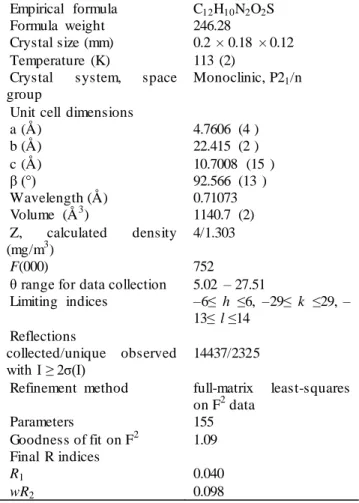
Table 1. Crystal data and spherical refinement details Empirical formula C12H10N2O2S Formula weight 246.28
Crystal size (mm) 0.2 × 0.18 × 0.12 Temperature (K) 113 (2)
Crystal system, space group
Monoclinic, P21/n Unit cell dimensions
a (Å) 4.7606 (4 ) b (Å) 22.415 (2 ) c (Å) 10.7008 (15 ) β (°) 92.566 (13 ) Wavelength (Å) 0.71073 Volume (Å3) 1140.7 (2) Z, calculated density (mg/m3) 4/1.303 F(000) 752
θ range for data collection 5.02 – 27.51
Limiting indices –6≤ h ≤6, –29≤ k ≤29, – 13≤ l ≤14 Reflections collected/unique observed with I ≥ 2σ(I) 14437/2325
Refinement method full-matrix least-squares on F2 data Parameters 155 Goodness of fit on F2 1.09 Final R indices R1 0.040 wR2 0.098 2.2. Geometry optimization
In this work, full geometry optimization has been performed using the GAUSSIAN03 package [19] and the Gauss-View molecular visualization program [27], at the Becke3-parameter hybrid exchange functions and Lee-Young–Parr correlation functional (B3LYP) level [28, 29] and HF theory [30], using the 6-31G(d,p) basis set by the Berny method [31,32]. The optimized structure of C12H10N2O2S is illustrated in Fig. 1 and the corresponding main geometrical parameters (bonds lengths, bond angles and torsion angles) are listed in Tables 2, 3 and 4.
Fig. 1. Optimized structure of the molecule C12H10N2O2S.
2.3. Electron charge density analysis
2.3.1. Multipolar refinement
The Hansen-Coppens multipole formalism, as implemented in the Mopro least-squares program for multipole refinement, was used for the structure factor fitting. This last model describes the crystal electron density as a superposition of non-spherical pseudo-atoms modeled on a multipole expansion given by the following equation: 𝜌𝑎𝑡𝑜𝑚(𝑟⃗) = 𝜌𝑐(𝑟⃗) + 𝑃𝑉𝐾′3𝜌𝑣(𝑘′𝑟⃗) + ∑ ∑+1 𝐾" 𝑚=−1 𝑙 𝑚𝑎𝑥 𝑙 𝑅𝑙(𝐾"𝑟⃗)𝑃𝑙𝑚𝑌𝑙𝑚( 𝑟⃗ 𝑟) (1) Here 𝜌𝑐 and 𝜌v are spherically averaged Hartree-Fock core and valence densities, with 𝜌v being normalized to one electron. The Slater-type radial functions are obtained by:
𝑅𝑙(𝑟⃗) = 𝑁𝑙. 𝑟𝑙 𝑛𝑗
exp (−𝑘′ξ 𝑙𝑟⃗)
These functions modulate the spherical harmonic angular functions 𝑌𝑙𝑚± and 𝑁l is a normalization factor. The values for parameters n =𝑛1 and 𝜉 were chosen according to rules provided by Coppens (1997) [33].
The sum over in equation (1) includes ±𝑙, so that for each one, 2𝑙+1 functions are included. The non-spherical charge density was described at the octapole level (𝑙=3) for the atoms C, N and O, the hexadecapole for the S atom (𝑙=4) and at the dipole levels (𝑙 =1or 2) for hydrogen atoms not involved and involved in strong H-bonds, respectively. Charge densities of all hydrogen were considered to have cylindrical symmetry along the corresponding hydrogen-heavy atom bond [34].
During the refinements, the adjustable variables were the valence-shell contraction expansion parameters k ′, k ″, the population parameter Pv, and the multipolar parameters
Plm for each atom. In order to reduce the number of these
variables, chemical constraints were imposed: atoms with similar environment were assumed to have the same deformation. Also the local symmetry of each atom has been considered.
3. Results and discussion 3.1. Structural properties
From the results, it can be seen that there is a good agreement between the calculated and the experimental values. The largest deviation between X-ray data and theoretical calculations at the HF/6-31G** level is the N2– O1 distance, around 0.02 Å, and the (C10 C11 C12) angle, which is larger than 1.48°. The B3LYP/6-31G** results deviate in the range from 0.001 to 0.014 Å for bond lengths, and from 0.02° to 1.45° (C7 C1 C6) for bond angles. It can be seen also that there is intramolecular hydrogen bonding between hydrogen atom linked to C8 with oxygen atom O2 in the nitro groups . Some possible
hydrogen bonds are presented in Table 5. Fig. 2 shows the C8H8...O2 hydrogen bond. These intermolecular interactions stabilize the crystal structure of the title compound.
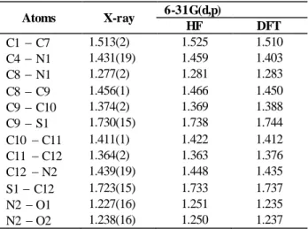
Table 2. Bond lengths of C12H10N2O2S molecule
Atoms X-ray 6-31G(d,p) HF DFT C1 C7 1.513(2) 1.525 1.510 C4 N1 1.431(19) 1.459 1.403 C8 N1 1.277(2) 1.281 1.283 C8 C9 1.456(1) 1.466 1.450 C9 C10 1.374(2) 1.369 1.388 C9 S1 1.730(15) 1.738 1.744 C10 C11 1.411(1) 1.422 1.412 C11 C12 1.364(2) 1.363 1.376 C12 N2 1.439(19) 1.448 1.435 S1 C12 1.723(15) 1.733 1.737 N2 O1 1.227(16) 1.251 1.235 N2 O2 1.238(16) 1.250 1.237
Table 3. Bond angles of C12H10N2O2S molecule
Atoms X-ray 6-31G(d,p) HF DFT N1 C4 C3 117.06(14) 116.64 116.87 C4 N1 C8 118.86(13) 124.76 118.96 C6 C4 N1 125.10(13) 124.27 125.10 C7 C1 C6 121.84(15) 120.69 120.39 C7 C1 C2 120.39(15) 121.18 120.39 N1 C8 C9 122.04(14) 121.08 122.04 C8 C9 C10 125.35(14) 126.88 125.35 C8 C9 S1 122.69(11) 120.66 122.70 C9 C10 C11 113.49(14) 113.77 113.47 C9 S1 C12 89.17(7) 87.75 89.77 C10 C9 S1 111.94(12) 112.44 111.94 C10 C11 C12 110.57(14) 112.05 110.57 C11 C12 N2 125.62(14) 124.92 125.42 S1 C12 N2 120.10(11) 121.10 120.11 S1 C12 C11 114.25(12) 113.97 114.25 C12 N2 O1 118.08(13) 117.93 118.09 C12 N2 O2 117.65(13) 117.96 117.55 O1 N2 O2 124.27(13) 124.10 124.37
Table 4. Torsion angles of C12H10N2O2S molecule
Atoms X-ray 6-31G(d,p) HF DFT C2 C3 C4 N1 -179.45(13) -179.49 -179.01 C3 C4 N1 C8 167.13(14) 155.72 145.86 C4 N1 C8 C9 179.73(13) 179.00 179.67 C5 C4 N1 C8 -14.30(2) -25.67 -36.78 C6 C5 C4 N1 -179.80(14) -179.87 -179.41 C7 C1 C2 C3 -178.92(14) -179.41 -179.46 C7 C1 C6 C5 179.55(14) -179.94 -179.86 N1 C8 C9 C10 -175.90(15) -179.60 -179.46 N1 C8 C9 S1 5.10(2) 0.36 0.49 C8 C9 C10 C11 -179.16(14) -179.91 -179.97 C8 C9 S1 C12 179.15(13) 179.99 179.97 C9 C10 C11 C12 0.09(19) 0.02 0.03 C9 S1 C12 C11 0.01(12) 0.05 0.03 C9 S1 C12 N2 -178.17(12) -179.91 -179.97 C10 C9 S1 C12 0.04(12) 0.06 0.03 C10 C1 C12 S1 -0.06(17) -0.03 -0.03 C10 C11 C12 N2 178.01(13) 179.93 197.79 C11 C12 N2 O1 2.70(2) 0.17 0.06 C11 C12 N2 O2 -179.72(14) -179.83 -179.97 C11 C12 S1 C9 0.01(12) 0.03 0.03 S1 C12 N2 O1 -179.35(11) -179.86 -179.97 S1 C12 N2 O2 1.25(18) 0.13 0.05
Table 5. Hydrogen bonds in C12H10N2O2S crystal by X-ray diffraction D – H…A D – H (Å) D – A (Å) H – A (Å) D – H….A (°) C11–H11…O15 0.930 2.813 2.658 89.80 C5–H5…O2 (1) 0.930 3.417 2.866 119.13 C6–H6…O2 (1) 0.930 3.438 2.900 118.21 C8–H8…O2 (2) 0.930 3.403 2.628 141.24 C10–H10…O2 (2) 0.930 3.533 2.853 130.96 C10–H10…N1(3) 0.930 3.367 2.759 123.84 C3–H3…O1 (4) 0.930 3.584 2.960 125.75 C7–H7A…O1 (5) 0.960 3.386 2.953 108.73 C7–H7A…O2 (6) 0.960 3.305 2.938 104.03 Equivalent positions ( 1) x+1/2, –y+1/2, z–1/2 (2) x+1/2, –y+1/2, z–1/2 ( 3) x–1/2, –y+1/2, z–1/2 ( 4) x+1/2, –y+1/2, z+1/2 (5) –x+1/2, y+1/2, –z+1/2 (6) –x+1/2, y+1/2, –z+1/2
Fig. 2. C8–H8…O2 Hydrogen bond
3.2. Electron density maps
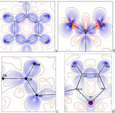
The experimental density deformation maps are shown in Figure 3, from which we can observe the
absence of the density on the atomic sites and the appearance of all the bond density peaks. The effectiveness of the formalism used for the treatment of data and their quality as proposed by Blessing [35] are confirmed by the obtained maps. This visualization is obtained using the calculated multipolar phases with the observed structure factors Fobs (h):
𝛿𝜌𝑒𝑥𝑝(𝑟) =1
𝑉∑[|𝐹𝑜𝑏𝑠(ℎ)|𝑒 −𝑖𝜃𝑚𝑢𝑙
ℎ
− |𝐹𝑠𝑝ℎ(ℎ)|𝑒−𝑖𝜃𝑠𝑝ℎ] 𝑒−2𝜋𝑖ℎ𝑟 where Fsph(h) is computed with atomic positions and thermal parameters. The experimental density map from high-order refinement is ρ exp = ρo− ρsph, where ρo is the observed electron density and 𝜌𝑠𝑝ℎ is the calculated electron density using the atomic parameters obtained from the high-order refinement.
a b
c d
Fig. 3. Electron density maps of C12H10N2O2S molecule in different planes, (a) Benzene ring, (b) Nitro group, (c) C8 = N1 double bond, (d) Thiophene cycle.
We have explored four planes to visualize the electron density distribution; the plane of the aromatic cycle containing the C1, C2, C3, C4, C5 and C6 atoms; the plane containing the electro-acceptor group (Nitro group), the plane formed by the double bond (C8 = N1) and the plane of the thiophene cycle containing the C9, C10, C11, C12 and S1 atoms. Fig. 3 (a, b, c, and d) gives the different maps cited above; all contour intervals are 0.05 e.Å−3.
The almost centered electron density distribution is observed in the middle of the chemical bonds. We also notice that the peaks (Fig. 3 (b)) of the electron density in the connection N2–O1 and N2–O2 are centered towards
the nitrogen N2 backing the electro donor character of the Nitro group.
These maps allowed us to illustrate the distribution of electronic charges of atoms and chemical bonds along the molecule. We noticed that the lone pairs of oxygen atoms are well resolved. The electron density at sulfur atom is deformed; this deformation can be explained by the effect of a thermal agitation.
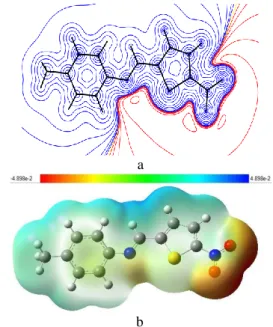
The residual map using a non-spherical atom model is shown in Fig. 4, from which we can notice the absence of the density on both the atomic sites and all the bonds. This map confirms the high quality of the data.
Fig. 4. Residual density map of the C12H10N2O2S molecule, contour 0.05 Å-3
3.3. Atomic Charges
The valence population coefficients 𝑃𝑉𝑖 were used to estimate the partial charges on the different atoms according to the following equation:
𝑞𝑖= 𝑛𝑖− 𝑃𝑉𝑖
where
n
i is the total number of electrons of atoms i. The experimental and theoretical atomic charges are presented in Table 6 and their distribution is shown in Figure 5. The experimental atomic charges derived from the X-ray experiment are generally of the same sign compared to the theoretical ones except some atoms. The expected values are in concordance to the chemical knowledge: H atoms are positively charged, where as O, S, and N atoms are negatively charged.Table 6. Atomic charges of C12H10N2O2S molecule Atoms qx-ray qab initio
S1 0.115 0.487 O1 0.102 0.367 N1 0.123 0.407 O2 0.102 0.377 C9 0.144 0.207 C5 0.049 0.242 H5 0.085 0.240 N2 0.125 0.459 C11 0.049 0.224 H11 0.085 0.282 C8 0.135 0.053 H8 0.146 0.202 C4 0.018 0.103 C6 0.049 0.231 H6 0.085 0.242 C2 0.049 0.233 H2 0.085 0.243 C10 0.049 0.256 H10 0.085 0.261 C12 0.078 0.104 C3 0.049 0.204 H3 0.085 0.257 C7 0.007 0.709 H7A 0.044 0.245 H7B 0.044 0.254 H7C 0.044 0.250 C1 0.036 0.015
Fig. 5. Atomic charges of the title molecule
3.4. HOMO and LUMO energies
The highest occupied molecular orbitals (HOMOs) and lowest-lying unoccupied molecular orbitals (LUMO) are very useful for physicists and chemists because the energy difference between these orbitals (energy gap) represents the minimum energy required to promote an electron, and is therefore often the most-frequent and important energy transfer mechanism within a system. The orbitals also provide important electron density information which can help determine which part of the
molecule is most actively participating in an energy transfer event. The HOMO-LUMO energy gap of the molecule was calculated using B3LYP/ 6-31G(d,p) level as presented in Table 7. The energy gap between HOMO and LUMO indicates molecular chemical stability. In addition, a lower HOMO-LUMO energy gap (3.22 eV) calculated at B3LYP/6-31G(d,p) explains the fact that eventual charge transfer interaction is taking place within the molecule. The HOMO-LUMO plots are given in Fig. 6. -0.5 -0.4 -0.3 -0.2 -0.1 0 0.1 0.2 0.3 0.4 S1 O1 N1 O2 C9 C5 H5 N2 C11 H11 C8 H8 C4 C6 H6 C2 H2 C10 H10 C12 C3 H3 C7 H 7 a H 7 b H 7 c C1 X-ray Ab initio
Table 7. HOMO-LUMO energies of the molecule Energies HF/6-31G(d,p) DFT/6-31G(d,p ) EHOMO 0.30855 0.22761 ELUMO 0.02852 0.10917 EGap (a.u.) 0.33707 0.11844 EGap (eV) 9.17 3.22
Fig. 6. The HOMO and LUMO plot of the title molecule at B3LYP/6-31G(d,p)
3.5. Molecular electrostatic potential
The electrostatic potential V(𝑟⃗), at a given point 𝑟⃗(x, y, z) in the space around a molecule (in atomic units) is defined in terms of the interaction energy between the electrical charge generated from the molecule electrons and nuclei and positive test charge (a proton) located at 𝑟⃗ and can be expressed as:
𝑉(𝑟⃗) = ∑ 𝑍𝐴 |𝑅⃗⃗𝐴− 𝑟⃗| 𝐴 − ∫𝜌(𝑟 ′ ⃗⃗⃗⃗)𝑑𝑟⃗′ |𝑟⃗′ − 𝑟⃗|
where 𝑍𝐴 is the charge on nucleus A, located at 𝑅⃗⃗𝐴 and 𝜌 (𝑟⃗⃗⃗) is the electronic density function for the molecule. ′
The first and second terms represent the contributions to the potential due to nuclei and electrons, respectively. V(𝑟⃗) is the resultant electrical potential at each point 𝑟⃗, which is the net electrostatic effect produced at the point 𝑟⃗ by both the electrons and nuclei of the molecule. The molecular electrostatic potential (MEP) serves as a useful quantity to explain hydrogen bonding, reactivity and structure– activity relationship of the molecules [36].
In order to predict the molecular reactive sites, the MEP for the title molecule was calculated using 6-31G (d,p) basis set and shown together with the experimental one in Fig. 7. The different values of the electrostatic potential at the surface are represented by different colors, where blue indicates the highest electrostatic potential energy and red indicates the lowest electrostatic potential energy. As it can be seen from the MEP map of the title compound, the region around oxygen atoms linked with carbon through double bond represents the most negative potential region (red). The most negative V(𝑟⃗) value is
associated also with NO2 group. The hydrogen atoms attached to nitrogen atoms posses the maximum positive charge. The MEP surface provides necessary details about the reactive sites.
a
b
Fig. 7. Molecular electrostatic potential maps (a) experimental (b) calculated at B3LYP/6-31G(d,p) level
3.6. Nonlinear optical (NLO) properties
For organic NLO materials, theoretical and experimental studies are performed in order to understand the microscopic origin of nonlinear behavior [37, 38].
In this context, this study is extended to the determination of the electric dipole moment µ, the isotropic polarizability α and the first hyperpolarizability 𝛽𝑡𝑜𝑡 of the title compound.
The dipole moment (𝜇), isotropic polarizability (𝛼), first-order hyperpolarizability (𝛽𝑡𝑜𝑡) tensor, can be obtained using the following equations:
𝜇0= (𝜇2𝑥+ 𝜇2𝑦+ 𝜇2𝑧)1/2 𝛼 =1
3(𝛼𝑥𝑥+ 𝛼𝑦𝑦+ 𝛼𝑧𝑧) 𝛽𝑡𝑜𝑡= (𝛽𝑥2+ 𝛽𝑦2+ 𝛽𝑧2)1 2⁄
The whole equation for computing the magnitude of the first hyeprpolarizability (β) from Gaussian 03 output is given below: 𝛽𝑡𝑜𝑡 = [(𝛽𝑥𝑥𝑥 + 𝛽𝑥𝑦𝑦+ 𝛽𝑥𝑧𝑧) 2 + (𝛽𝑦𝑦𝑦+ 𝛽𝑦𝑧𝑧+ 𝛽𝑦𝑥𝑥) 2 + (𝛽𝑧𝑧𝑧+ 𝛽𝑧𝑥𝑥+ 𝛽𝑧𝑦𝑦) 2 ]1 2 ⁄
The first hyperpolarizability is a third rank tensor that can be depicted by a 3 × 3 × 3 matrices. The 27 components of the 3D matrix can be reduced to 10 components because of the Kleinman symmetry [39]. The Gaussian 03 output provides 10 components of this matrix
as βxxx, βxxy, βxyy, βyyy, βxxz, βxyz, βyyz, βxzz, βyzz, βzzz
respectively.
The first hyperpolarizability tensors provided by Gaussian 03 are given in atomic units (a.u.), the computed
values were converted into electrostatic units. (α: 1 a.u. = 0.1482×10⁻²⁴ esu ; β: 1 a.u. = 8.6393×10⁻³³
esu).
The dipole moment (0), mean polarizability (α) and
first hyperpolarizability (β) are calculated at 6-31G(d,p) basis set. Table 8 and 9 give the HF and B3LYP results of the electronic dipole moment µi (i = x, y, z), polarizability
αij and the first hyperpolarizability βijk for
4-methyl-N-[(5-nitrothiophen -2-ylmethylidene)] aniline compound. Theoretical calculation plays a significant role in understanding the structure-property relationship which is able to help in designing novel NLO materials. It is well established that the higher values of dipole moment, molecular polarizability, and hyperpolarizability are very important for active NLO properties. The highest value of
dipole moment obtained from multipolar refinement is equal to 7.10 D. The highest value of dipole moment is observed for component µx. In this direction, this value is
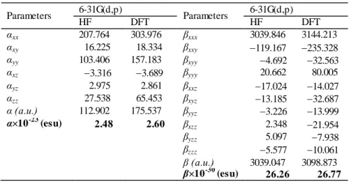
equal to 5.5421. Figure 8 shows the orientation of the molecular dipole moment of the title molecule. The calculated polarizability (α), is about to 2.5×10⁻²³ esu obtained with 6-31G(d,p) basis set. As it can be seen in Table 9, the calculated polarizability αij have non zero
values and was dominated by the diagonal components. The first hyperpolarizability value 𝛽𝑡𝑜𝑡 of the title molecule obtained with B3LYP/6-31G(d,p) is 26.77 × 10⁻³⁰ esu. The calculated results show that the title molecule might have microscopic nonlinear (NLO) behavior with non-zero values.
The calculated first order hyperpolarizability of the title molecule is found to be 137 times greater than the
βtotal value of urea (0.1947 10–30 esu), therefore, predicting that our molecule is a powerful candidate for NLO material.
Table 8. The molecular dipole moment of the C12H10N2O2S obtained by X-ray, HF and DFT/B3LYP methods. Methods Models 𝜇𝑥 (e.Å) 𝜇𝑦 (e.Å) 𝜇𝑧 (e.Å) ‖𝜇⃗‖ (D) X-Ray Multipole refinement 3.8105 4.5369 3.4954 7.10 Ab initio B3LYP/6-31G(d,p) 5.5421 3.7709 0.6531 6.74 HF/6-31G(d,p) 5.3130 4.3369 1.0518 6.94
From the cited above results, the molecule having the
greatest 𝛽𝑡𝑜𝑡 value, corresponds to the low HOMO–LUMO energy gap. These results show that
HOMO–LUMO gap have a substantial influence on the first hyperpolarizability. The high β value and low HOMO–LUMO energy gap show that the title molecule is highly NLO active material and it might be efficient for optoelectronic applications.
Consequently, we can finally infer from the above discussion that the introduction of electron correlat ion in the method applied for the analysis of the hyperpolarizability, such as DFT method, will probably predict more reasonable values as opposed to those converged upon use of the HF method.
Fig. 8. Orientation of the molecular dipole moment of C12H10N2O2S molecule
𝜇𝑀
⃗⃗⃗⃗⃗⃗ : Experimental molecular dipole moment, 𝜇⃗⃗⃗⃗⃗ : Calculated 𝐴 molecular dipole moment
Table 9. Polarizability (α) and hyperpolarizability (β) values of the C12H10N2O2S molecule obtained by HF and DFT/B3LYP using 6-31G(d,p) basis set.
Parameters 6-31G(d,p) Parameters 6-31G(d,p) HF DFT HF DFT αxx 207.764 303.976 βxxx 3039.846 3144.213 αxy 16.225 18.334 βxxy 119.167 235.328 αyy 103.406 157.183 βxyy 4.692 32.563 αxz 3.316 3.689 βyyy 20.662 80.005 αyz 2.975 2.861 βxxz 17.024 14.027 αzz 27.538 65.453 βxyz 13.185 32.687 α (a.u.) 112.902 175.537 βyyz 3.226 13.999 α×10-23 (esu) 2.48 2.60 βxzz 2.348 21.954 βyzz 5.097 7.938 βzzz 5.577 10.061 β (a.u.) 3039.047 3098.873 β×10-30 (esu) 26.26 26.77
4. Conclusion
In the present work, we have performed theoretical calculation on 4-methyl-N-[(5-nitrothiophen-2-ylmethylidene)] aniline molecule which leads to an optimized structure with bond lengths and bond angles closer to the X-ray experiment results. On the other hand, high resolution X-ray experiment crystallographic data were used to investigate the electron charge density of the title compound. We have also shown that electron density can yield to electronic experimental proprieties such as atomic charges, dipole moment and electrostatic potential. Using theoretical structural results, we have calculated atomic charges, HOMO–LUMO energy, molecular electrostatic potential, and nonlinear optical properties for the organic molecule 4-methyl-N-[(5-nitrothiophen-2-ylmethylidene)] aniline. Molecular properties have been calculated by using ab initio HF and DFT (B3LYP) methods with 6-31G(d,p) basis set. The molecular electrostatic potential map shows that the negative potential sites are on the electronegative atoms as well as the positive potential sites are around the hydro gen atoms. These sites give information about the possible regions for inter- and intramolecular hydrogen bonding. The calculated dipole moment mean value of the studied compound is 7.10 D obtained using multipolar refinement which is in agreement with the value (about 7.0 D) calculated by theoretical methods. The mean polarizability (α) and the total first static hyperpolarizability (βtotal) of the
molecule are found to be 2.5×10⁻²³ esu and 26.77 × 10⁻³⁰ esu, respectively. The computed βtotal value is nearly 137
times more than βtotal of urea, inferring that our molecule
to be a potential candidate for nonlinear optical applications. HOMO-LUMO energy gap explains the eventual charge transfer interactions taking place within the molecule. Furthermore, the first-order hyperpolarizability and total dipole moment property of the molecule show that the title molecule is an attractive target for future studies of nonlinear optical properties.
References
[1] J. Zyss, I. Ledoux, M. Bertault, E .Toupet, Chemical Physics 150, 125 (1991).
[2] I. Ledoux, J. Zyss, Molecular Nonlinear Optics: Fundamentals and Applications , (I.C. Khoo, F. Simoni, C. Umeton, Ed.) pp. 1, 1997.
[3] D.S. Chemla, J. Zyss, Nonlinear Optical Properties of Organic Molecules and Crystals, Academic Press, New York 1987.
[4] H. Qin, S. Wenger, M. Xu, F. Gao, X. Jing, P. Wang, S. M. Zakeeruddin, M. Grätzel, J. Am. Chem. Soc. 130, 9202 (2008).
[5] H. Ünver, A. Karakas, A. Elmali, J. Mol. Struct. 702, 49 (2004).
[6] H. Tanak, A. A. Ağar, O. Büyükgüngör, Spectrochim. Acta Part A 87, 15 (2012).
[7] H. Tanak, A. A. Ağar, O. Büyükgüngör, Spectrochimica Acta Part A 118, 672 (2014). [8] H. Tanak, A.A. Ağar, O. Büyükgüngör, J. Mol. Struct. 1048, 41 (2013).
[9] R. M. F. Batista, S.P.G. Costa, M. Belsley, M. M. M. Raposo, Dyes Pigm. 80, 329 (2009). [10] L. Xiao-Hong, Z. Xian-Zhou, Spectrochim. Acta Part A 105, 280 (2013).
[11] Y. Megrouss, N. Benhalima, R. Bahoussi, N. Boukabcha, A. Chouaih, F. Hamzaoui, Chin. Phys. B 24(10), 106103 (2015).
[12] F. Hamzaoui, F. Baert, J. Zyss, J. Mater. Chem. 6, 1123 (1996).
[13] F. Hamzaoui, A. Zanoun, G. Vergoten, J. Mol. Struct. 697(1–3), 17 (2004).
[14] A. Chouaih, F. Hamzaoui, G. Vergoten, J. Mol. Struct. 738(1–3), 33 (2005).
[15] N. Benhalima, K. Toubal, A. Chouaih, G. Chita, S. Maggi, A. Djafri, F. Hamzaoui, J. Chem. Crystallogr. 41(11), 1729 (2011).
[16] M. Drissi, A. Chouaih, Y. Megrouss, F. Hamzaoui, Journal of Crystallography 2013, ID 326457 (2013). [17] N. Boubegra, A. Chouaih, M. Drissi, F. Hamzaoui, Chin. Phys. B 23(1), 016103 (2014).
[18] C. Jelsch, B. Guillot, A. Lagoutte, C. Lecomte, J. Appl. Crystallogr. 38, 1 (2005).
[19] M. J. Frisch, G. W. Trucks, H. B. Schlegel, G. E. Scuseria, M. A. Robb, J. R. Cheeseman, J. A. Montgomery, T. Vreven, K. N. Kudin, J. C. Burant, J. M. Millam, S. S. Iyengar, J. Tomasi, V. Barone, B. Mennucci, M. Cossi, G. Scalmani, N. Rega, G. A. Petersson, H. Nakatsuji, M. Hada, M. Ehara, K. Toyota, R. Fukuda, J. Hasegawa, M. Ishida, T. Nakajima, Y. Honda, O. Kitao, H. Nakai, M. Klene, X. Li, J. E. Knox,
H. P. Hratchian, J. B. Cross, V. Bakken, C. Adamo, J. Jaramillo, R. Gomperts, R. E. Stratmann, O. Yazyev, A. J. Austin, R. Cammi, C. Pomelli, J. W. Ochterski, P. Y. Ayala, K. Morokuma, G. A. Voth, P. Salvador, J. J. Dannenberg, V. G. Zakrzewski, S. Dapprich, A. D. Daniels, M. C. Strain, O. Farkas, D. K. Malick, A. D. Rabuck, K. Raghavachari, J. B. Foresman, J. V. Ortiz, Q. Cui, A. G. Baboul, S. Clifford, J. Cioslowski,
B. B. Stefanov, G. Liu, A. Liashenko, P. Piskorz, I. Komaromi, R. L. Martin, D. J. Fox, T. Keith, M. A. Al-Laham, C.Y. Peng, A. Nanayakkara, M. Challacombe, P.M. W. Gill, B. Johnson, W. Chen, M.W. Wong, C. Gonzalez, J.A. Pople, Gaussian 03, Revision C.02, Gaussian, Inc., Wallingford CT, USA, 2004.
[20] M. Cai, X. Wang, T. Sun, Acta Cryst. E 67, o2218 (2011).
[21] T. Akbal, E. Agar, S. Gümüs, A. Erdönmez, Acta Cryst. E 68, o3026 (2012).
[22] T. Akbal, E. Agar, S. Gümüs, A. Erdönmez, Acta Cryst. E 68, o2673 (2012).
[23] N. Kan Kaynar, S. Gümüs, E. Agar, O. Büyükgüngör, M. Yavuz, Acta Cryst. E 68, o393 (2012).
[24] Ü. Ceylan, S. Gümüs, E. Agar, M.S. Soylu, Acta Cryst. E 68, o2116 (2012).
[25] Ü. Ceylan, H. Tanak, S. Gümüs, E. Agar, Acta Cryst. E 67, o2004 (2011).
[26] N.K. Hansen, P. Coppens, Acta Cryst. A 34, 909 (1978).
[27] A. E. Frisch, A. B. Nielsen, A. J. Holder, Gaussview. Gaussian Inc., Pittsburg, PA 2003.
[28] A. D. Becke, J. Chem. Phys. 107, 8554 (1997). [29] G. Rauhut, P. Pulay, J. Phys. Chem. 99, 3093 (1995). [30] H.D. Cohen, C.C. Roothaan, J. Chem. Phys. 43, S34 (1965).
[31] R. Fletcher, M.J.D. Powell, Comput. J. 6, 163 (1963). [32] R.F. Bader, Atoms in molecules. A quantum theory. Clarendon Press, Oxford 1990.
[33] P. Coppens, X-Ray Charge Densities and Chemical Bonding, Oxford, New York 1997.
[34] M.A. Spackman, “Charge densities from X-ray diffraction data,” Annual Reports on the Progress of Chemistry C.94, 177 (1998).
[35] R.H. Blessing, J. Appl. Cryst. 22, 396 (1989). [36] S. Chidangil, M. K. Shukla, P. C. Mishra, J. Mol. Model. 4, 250 (1998).
[37] P. Kerkoc, M. Zgonik, K. Sutter, C. Bosshard, P. Gunter, J. Opt. Soc. Am. B 7, 313 (1990) [38] D. Sajan, H.J. Ravindra, M. Neeraj, I. Hubert Joe, Vibrational Spectroscopy 54, 72 (2010).
[39] D. A. Kleinman, Phys. Rev. 126, 1977 (1962).
_____________________________