HAL Id: hal-00307890
https://hal.archives-ouvertes.fr/hal-00307890
Submitted on 29 Jul 2008
HAL is a multi-disciplinary open access
archive for the deposit and dissemination of sci-entific research documents, whether they are pub-lished or not. The documents may come from teaching and research institutions in France or abroad, or from public or private research centers.
L’archive ouverte pluridisciplinaire HAL, est destinée au dépôt et à la diffusion de documents scientifiques de niveau recherche, publiés ou non, émanant des établissements d’enseignement et de recherche français ou étrangers, des laboratoires publics ou privés.
The use of IRES-based bicistronic vectors allows the
stable expression of recombinant G-protein coupled
receptors such as NPY5 and histamine 4.
Camille Allera-Moreau, Pascale Chomarat, Valérie Audinot, Francis Cogé,
Mélanie Gillard, Yvan Martineau, Jean Boutin, Anne-Catherine Prats
To cite this version:
Camille Allera-Moreau, Pascale Chomarat, Valérie Audinot, Francis Cogé, Mélanie Gillard, et al.. The use of IRES-based bicistronic vectors allows the stable expression of recombinant G-protein coupled receptors such as NPY5 and histamine 4.. Biochimie, Elsevier, 2006, 88 (6), pp.737-46. �10.1016/j.biochi.2006.05.019�. �hal-00307890�
The use of IRES-Based bicistronic vectors allows the stable expression of recombinant G-protein coupled receptors such as NPY5 and histamine 4.
Camille Allera-Moreau1, Pascale Chomarat2, Valérie Audinot2, Francis Cogé2, Mélanie
Gillard1, Yvan Martineau1, Jean A. Boutin2*, and Anne-Catherine Prats1*
1Institut National de la Santé Et de la Recherche Médicale (INSERM), U589, "Hormones,
facteurs de croissance et physiopathologie vasculaire", Institut Louis Bugnard, IFR31, CHU Rangueil, Bât L3, Avenue Jean Poulhès, BP 84225, 31432 Toulouse Cedex 4, France
2 Institut de Recherche Servier (IdRS), Division de Pharmacologie Moléculaire et Cellulaire,
125 chemin de ronde, 78290 Croissy-sur-Seine, France
Runing title : IRES-based vector for receptor expression
* Corresponding author: Jean A. Boutin, Tel .: (33) 155 72 27 48 ; Fax : (33) 155 72 28 10; email: jean.boutin@fr.netgrs.com
ABSTRACT.
Stable expression of G protein coupled receptors in cell lines is a crucial tool for the characterization of the molecular pharmacology of receptors and the screening for new antagonists. However, in some instances, many difficulties have been encountered to obtain stable cell lines expressing functional receptors. Here, we addressed the question of vector optimization to establish cell lines expressing the human neuropeptide Y receptor 5 (NPY5-R) or histamine receptor 4 (HH4R). We have compared bicistronic vectors containing viral or cellular Internal Ribosome Entry Sites (IRES), co-expressing the receptor and the neomycine resistance gene from a single mRNA, to a bigenic vector containing two distinct promoters upstream each different genes. This study is the first one to validate the use of three cellular IRESs for long term transgene expression. Our results demonstrate for both NPY5-R and HH4R that the bicistronic vectors with EMCV, VEGF, FGF1A or FGF2 IRES provide clones expressing functional receptors with yields between 25% and 100%. In contrast, the bigenic vector provided no functional clones, related to a low expression of NPY5R mRNA. The cell lines expressing active receptor were stable after more than 50 passages. These data indicate that IRES-based bicistronic vectors are particularly appropriate to establish cell clones expressing active G-coupled protein receptors with a high yield. In the case of NPY5, it was a new way to produce such a stable cell line. Furthermore, the characteristics - presented herein - of this receptor pharmacological property are perfectly in line with those reported in the literature.
Key words: bicistronic vectors, IRES, gene transfer, NPY5 receptor, Histamine 4 receptor,
INTRODUCTION
Histamine and Neuropeptide Y receptors belong to the large family of seven transmembrane domains, G-protein coupled receptors. Three major functions for histamine, inflammatory wheal, gastric acid secretion in the gut and neurotransmitter release in the central nervous system, have been related to the H1, H2 and H3 receptor subtypes, respectively [1]. More recently, a fourth histamine receptor, H4 (HH4R), has been discovered, which, in view of its distribution might be involved in immune response. As regards to the neuropeptide Y (NPY), several receptors have been described [2]. NPY is a 36-amino acid peptide which belongs to the pancreatic polypeptide (PP) family [3]. NPY, expressed in the adrenal gland, exerts various auto- and paracrine regulatory functions. NPY is co-released with catecholamines under a variety of stimuli and stimulates catecholamines secretion while it inhibits aldosterone secretion. NPY may also be involved in the regulation of blood pressure and in the pathophysiology of pheochromocytomas. Among the NPY receptors, the receptor Y5 (NPY5-R) is claimed to be involved in feeding behavior in mammals.
The pharmacological interest of both HH4R and NPY5-R incited us to develop tools to establish their molecular pharmacology. However, difficulties have been encountered in obtaining stable cell lines expressing the human NPY5-R [4] as well as the human HH4R. Such a lack of receptor expression in cell lines could result from inadequate intracellular targeting and/or counter-selection of cells expressing high levels of functional receptors. Indeed, based on a long record of 7-transmembrane domain G-coupled receptor pharmacology studies in our lab, we established standard protocols, according to which most of our previous targets were obtained in stable expressing cell lines mainly CHO-derived, with some HEK ones. Our protocol included the adding of a M2 flag at the C-terminus of the receptor and the co-expression with a protein conferring neomycin resistance to the cells. This
process was applied over the last years to bio-aminergic receptors, such as adrenoceptors 1 and 2 [5, 6], melatoninergic receptors [7,8], MCH receptors [9,10], serotonin receptors [11], histaminergic receptors [12], a small bunch of enzymes [13,14] and even some non-reported orphan receptors. In this strategy, we used a stable transfected Gα16-CHIO cell lines, which is useful in ‘forcing’ the secondary signals downstream the receptors towards the calcium release pathway. All along these years, we only found these two receptors (H4 and NPY5) to be resistant to this otherwise successful recipe. To be complete, we should point out that NPY5 has been reported expressed in a stable cell line, in the non-classical human endometrial cancer cell line HEC-1B [15].
In order to overcome several attempts with our classical approaches that turned out all to be unsuccessful, we decided to focus on the basis of gene expression regulation. Expression cassettes may be improved by acting not only on the transcription control, but also on the translational control of the genes of interest. For a few years, research on translational control of gene expression has led, in particular, to the identification and characterization of RNA elements present in viral and cellular mRNAs, called IRESs (Internal Ribosome Entry Sites); they can be considered as translational enhancers [16,17,18,19]. These elements have been identified in mRNAs bearing a long untranslated region (5’ UTR) expected to prevent the translation initiation by classical cap-dependent mechanisms [20]. The first IRES’s were discovered in two picornaviruses, poliovirus and encephalomyocarditis virus (EMCV) [16,21]. The cellular mRNAs bearing IRES mostly encode for proteins involved in the growth control or stress response (for review, see [22]). IRESs confer to these mRNAs the capability to be translated by a cap-independent mechanism when cap-dependent translation is blocked, which occurs during the M phase of cell cycle, and when cells are subjected to various stresses (heat shock, hypoxia, ischemia…).
expression cassettes in which a single transcription unit can code for several genes [23,24]. Such a feature is not provided by the cap-dependent mechanism, according to which a given mRNA codes for a single protein. The multicistronic vector concept is possible only because of the introduction of IRESs between the genes borne by the multigenic expression cassette. The use of multicistronic vectors provides several advantages by comparison with vectors comprising several promoters. Particularly, one expects to get rid of the promoter silencing phenomenon that often appears when several promoters are present, resulting in the loss of expression of counter-selected genes of interest.
In the cases of NPY5-R and HH4R, stable cell lines expressing functional forms of these receptors have not been obtained in our laboratory using classical vectors that proved to function on a wide variety of such receptors (vide supra). In the present study, we addressed this difficulty by constructing a set of bicistronic vectors expressing from a single promoter the receptor and the neomycine resistance gene under the control of different IRESs. Stable CHO clones expressing active NPY5-R and HH4R were obtained with a high yield using the bicistronic vectors with both viral and cellular IRESs, whereas no positive clones - as previously - were obtained when the receptor and resistance genes were expressed from monocistronic cassettes under the control of two distinct promoters.
MATERIALS AND METHODS.
Reagents - Peptides NPY were obtained from Neosystem (Illkirch, France), clobenpropit from Tocris (Avonmouth, UK). Histamine as well as forskolin, were obtained from SIGMA (St Louis, Mo). The human genes NPY5R and HRH4, flagged with the M2 sequence at the 3' terminal end, were from IdRS.
Vector construction - Plasmids used in this study and cloning strategies are available upon request. The oligonucleotides used to create the polylinker (SpeI – T3 PRIMER – HindIII – NotI – SpeI – BamHI – NcoI – EcoRV – M13R PRIMER – BglII) had the following sequences: 5’ctagtATTAACCCTCACTAAAGGGAaagcttgcggccgcactagtggatccccatgggatatc-GGTCATAGCTGTTTCCTGa3’ (sense) ; 5’gatctCAGGAAACAGCTATGACCgatatcccatgg-ggatccactagtgcggccgcaagcttTCCCTTTAGTGAGGGTTAATa3’ (antisense).
Cell culture and transfection - Gα16-CHO cells derived from CHO cells were cultured in Ham-F12 medium (Invitrogen, France) supplemented with 10% fetal calf serum and 200 µg/ml of hygromycin (Invitrogen, France) [10]. NPY5-R and HH4R were expressed in Gα16-CHO cells. Selection of Y5-Gα16-Gα16-CHO or H4-Gα16-Gα16-CHO cell clones was performed in complete Ham-F12 medium supplemented with 1,2 mg/ml geneticin and 100 g/ml hygromycin. Clones were further characterized by immunofluorescence using anti M2 flag antibody. All cells were tested for the absence of mycoplasma contamination. Gα16-CHO cells were transfected with 2 µg total DNA for 65 000 cells per 3,5 cm diameter petri dishes, using FugeneTM 6 (Roche Diagnostic, France) according to the manufacturer's instruction.
expression was performed by real-time RT PCR. Total RNA was purified using the SV RNA total isolation System (Promega, France) according to the manufacturer’s instruction. An additional DNAse I treatment (DNA free kit, Ambion, France) was carried out. 1-3 µg of total RNA was used for reverse transcription assay with the Reverse Transcription Core Kit (Eurogentec, Belgium). The reaction product (cDNA) was treated with RNAse H (Invitrogen, France). Specific primers and probes were designed using TaqMan® Primer & Probe Design of Primer Express® v1.5 software (Perkin Elmer/Applied Biosystem, USA). Primers and probes were purchased from Eurogentec (Belgium). Quantification of ribosomal 18S RNA was used as an internal control (Pre-developed Taqman® Assay Reagent, 18S rRNA - Applied Biosystem, USA). Primers and probes efficiencies were determined in preliminary experiments. The reaction mix contained 50 ng of cDNA, primers and the fluorophore Sybrgreen® (Applied Biosystem, USA). The reaction assay was performed on Gene Amp 5700 sequence detection system (Applied Biosystems, USA).
cAMPRadioactive Immuno-Assay - 1,5.106 Y5- or H4-Gα16-CHO cells were cultivated for 48h in 10 cm diameter Petri dishes. Cells were harvested and counted using a coulter counter. 70 000 cells were used to quantify the production of cAMP in the presence of forskolin, with or without neuropeptide Y (NPY) or histamine for Y5-Gα16-CHO cell clones or H4-Gα16-CHO clones, respectively. The cells were used after 6 to 9 passages in culture. Cells were washed two times with PBS at 37 °C, then harvested with PBS, EDTA 0,5 M at 37°C during 30 minutes. Cells were counted in Isoton, then diluted in HamF12, Hepes 1M, IBMX 1 M at 37 °C to obtain a suspension of 70 000 cells per 160 µl. 160 µl of these cells was added to samples containing either forskolin to stimulate directly adenylate cyclase (AC) or forskolin and NPY or histamin, expected to inhibit AC by stimulation of the corresponding G protein coupled receptor (GPCR). The reaction was stopped after 15 minutes at 37 °C with methanol
95 %, formic acid 5 %. Samples were stored at -20 °C. Cyclic AMP production by AC was then measured by radioimmunoassay (RIA cyclic AMP, Immunotech, France): 150 µl of the lysate was transferred to a new glass tube to evaporate formic acid with nitrogen gas. 150 µl of cAMP diluent was added. 100 µl of the standard or the cAMP precedent solution was
transferred in a monoclonal antibody-coated tube. 500 µl of I125 cAMP was added and left
over night at 4 °C. Following incubation, bound radioactivity was measured in a gamma counter. A calibration curve was established and unknown values were determined by interpolation from the standard curve.
Immunohistochemistry of NPY5-R - In order to detect the NPY5-R protein in the absence of Y5 antibody, the NPY5R sequence was flanked with a M2 flag at the C terminal end. Immunohistochemistry was performed as previously described [12]. Cells were cultivated in a glass-bottomed chamber for 24 hours, then fixed with 2% PFA and permeabilized with 0.1% Triton X-100 solution or 0.025 % saponin solution. After washing with PBS/0.1% saponin solution, cells were incubated with monoclonal anti M2 flag antibody (Sigma-Aldrich, France), then with Cy3 anti mouse antibody. Fluorescence was visualized using a fluorescence microscope (Leica, France).
[125]-PYY binding assay - Briefly and as described in Beauverger et al. 2005, membranes (50 µg/ml) were incubated 90 min at 30 °C in binding buffer (HEPES 20 mM, pH 7.4 containing NaCl 10 mM, KH2PO4 0.22 mM, CaCl2 1.26 mM, MgSO4 0.81 mM and BSA protease free
0.1 %) in a final volume of 250 µl containing [125I]PYY at 0.045 nM and the tested drug for
displacement experiments. Non specific binding was defined using NPY 1 µM (Neosystem, France). Estimation of Bmax was performed using a high concentration of (5 nM, isotopic dilution) saturating the NPY5-R binding sites. For saturation experiments, isotopic dilutions
were performed. Briefly, a range of concentrations starting from 0.1 nM to 5 nM of
{[125I]PYY+PYY} with a fixed ratio PYY/[125I]PYY of 10 was tested. Incubations were
stopped by rapid filtration through GF/C unifilters presoaked in polyethylenimine 0.1%, followed by three successive washes with Tris/HCl 50 mM, pH 7.4.
[3H]-Histamine binding assay - Membranes (50 µg/ml) were incubated 60 min at 30°C in binding buffer (TRIS 50 mM, pH 7.5 containing MgCl2 5 mM) in a final volume of 250 µl
containing 10 nM [3H]-Histamine and the tested drug for displacement experiments. Non
specific binding was defined using Clobenpropit 1µM (Tocris, UK). Estimation of Bmax was performed using a high concentration (12 nM) saturating the HH4R binding sites. For saturation experiments, a range of concentrations were used starting from 0.5 nM to 40 nM of
[3H]-Histamine. Incubations were stopped by rapid filtration through GF/B unifilters
presoaked in polyethylenimine 0.1%, followed by three successive washes with Tris/HCl 50 mM, pH 7.4.
Data analysis - Binding data were generated as duplicate within each experiment which were repeated independently at least three times. Saturation analysis were analyzed using the program PRISM (GraphPad Software Inc., San Diego, CA) to yield KD, the dissociation constant of the radioligand and Bmax, the maximal number of binding sites. Displacement curve fitting were generated by non linear regression to yield IC50 (concentration of compound that gives 50 % of inhibition of radioligand binding). Inhibition constants (Ki) were calculated according to the Cheng-Prusoff equation : Ki=IC50/(1+L/KD), where L is the concentration of radioligand. Results are expressed as the mean pKi=-LogKi.
RESULTS.
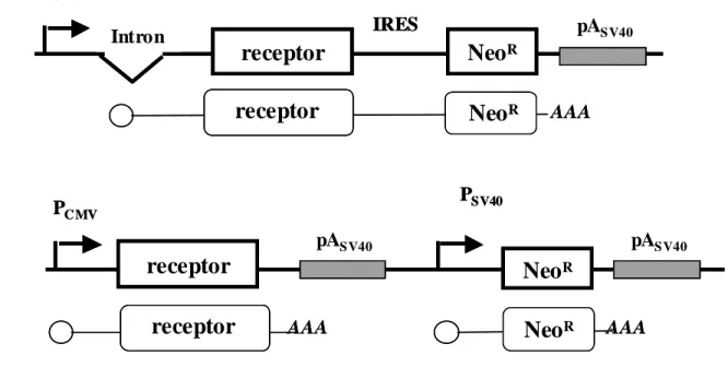
Bicistronic expression vectors with viral or cellular IRESs allow expression of functional NPY5-R and HH4R. cDNAs encoding for the G-protein coupled receptors (GPCR) NPY5-R and HH4R, both flagged with M2 peptide at the C-terminus, were introduced into bicistronic vectors containing IRESs from different origins (Fig. 1A). These vectors contained the receptor cDNA as a first cistron (cap dependent) and the G418 (neo) resistance gene as a second cistron (under the control of the IRES). By doing so, both the receptor and the neo genes are expressed from a single mRNA, which, in principle, guaranties the receptor expression in G418-resistant cells. The IRESs used were I) the EMCV IRES of viral origin and II) the VEGF, FGF1A and FGF2 IRESs of cellular origin. Indeed, most bicistronic vectors described in the literature so far contained the EMCV IRESs, whereas the cellular IRESs have never been used, except for the FGF2 [25].
In parallel, the receptor cDNAs were also introduced into the pcDNA-3 vector, under the control of the CMV promoter, whereas the neo gene is controlled by the SV40 promoter (Fig. 1B). In such a vector, the two genes are encoded by two distinct mRNAs. All these vectors were transfected into Gα16-CHO cells to obtain stable cellular clones in which calcium mobilization is forced by Gα16 protein to the studied receptor [26-27]. Six cell clones expressing each bicistronic vector, and 12 clones expressing the so-called “bigenic” pcDNA-3 derived vector, were analyzed for receptor binding and functionality.
In order to determine NPY5-R or HH4R functional activities for these different clones, we measured the amount of cAMP produced by adenylate cyclase (AC) activity. In presence of forskolin (FK), AC activity leads to cAMP production. The receptor activation by its specific ligand, NPY for NPY5-R and histamine for HH4R, respectively, was expected to decrease the cAMP production due to the AC inhibition by a Gi protein. The results obtained
with NPY5-R revealed six, four, three or two clones expressing an active NPY receptor with the bicistronic vectors containing the EMCV, VEGF, FGF1A or FGF2 IRES, respectively (Fig. 2). In contrast, no functional receptor was detected in any of the twelve cell clones expressing the pcDNA-derived bigenic vector. The receptor activity was also measured for clones expressing the HH4R (Fig. 3). The results were similar: the bicistronic vectors allowed us to obtain, among six clone populations analyzed, six, four, three and one positive clones for the constructs with EMCV, VEGF, FGF1A and FGF2 IRESs, respectively. Six clones expressing the pcDNA-3 derived bigenic vector did not produce any functional HH4R.
These data showed, for NPY5-R and HH4R, striking differences in yield with bicistronic versus bigenic vectors: there was a 100% yield for EMCV IRES, 67% for VEGF IRES, 50% for FGF1A IRES and 13 to 25% for FGF2 IRES, while the yield was null for the two promoters-containing vectors.
The bicistronic vectors allow a higher receptor mRNA expression than the bigenic vector. In an attempt to explain the differences of active receptor expression between IRES-containing bicistronic vectors and two-promoters-containing bigenic vector, NPY5R amplicon was measured by quantitative RT PCR (Fig. 4). The data show that the NPY5R mRNA copy number per ng of 18S RNA was comprised between 550 and 63000 for clones expressing the bicistronic vectors (producing functional receptor or not), whereas it was comprised between 0 and 450 for clones expressing the bigenic pcDNA-3 derived vectors. Thus, a first explanation for the absence of positive clones with the bigenic vector was a loss of expression of NPY5R amplicon, due to either interference between the two promoters or counter-selection of the receptor-expressing clones.
However, another parameter probably also influenced the receptor activity in the different clones: with the bicistronic vectors, there was no correlation between the level of
NPY5R mRNA expression and receptor activity (Fig. 4). The clones expressing inactive receptor could be separated into two groups: the first one comprising clones 4 and 5 with FGF1A IRES and clone 6 with FGF2 IRES which express less than 1000 copies of NPY5R mRNA per ng of 18S RNA. In these clones, the absence of active receptor probably resulted from a poor expression of the receptor. In contrast, a second group of inactive clones expressed NPY5R mRNA at a level comparable to that of the active clones, comprised between 2860 and 15780 copies. The receptor inactivity for these clones cannot be explained by a lack of mRNA expression.
Absence of functional receptor may result from both poor expression and incorrect targeting. To understand the reasons of receptor inactivity, immuno-cytofluorescence experiments were performed with some of the active and inactive clones, using the anti-flagM2 antibody. As shown in Fig. 5, the signal obtained from clones expressing the pcDNA3-derived bigenic plasmid was not superior to naïve Gα16-CHO cells, whereas clones expressing functional receptors from bicistronic vectors provided a significant signal (Fig. 5A). A selection of active or inactive clones expressing the bicistronic vector with VEGF IRES, all positive for NPY5R mRNA quantification (4800 to 10800 copies), were then analyzed. For the three active clones 7, 23 and 24, the NPY5-R was homogeneously detected in all the cells (Fig. 5B), but the receptor seemed localized in the perinuclear granules. In contrast, for the inactive clone 11, the receptor expression was heterogeneous: part of the cells showed a very strong staining whereas no staining was detected in the other part of the cell population. In contrast to the active clones, the receptor was homogeneously detected in the cytoplasm of the stained cells. This suggested that, in clones expressing the receptor mRNA but no functional NPY5-R, the receptor was not correctly targeted at the plasma membrane.
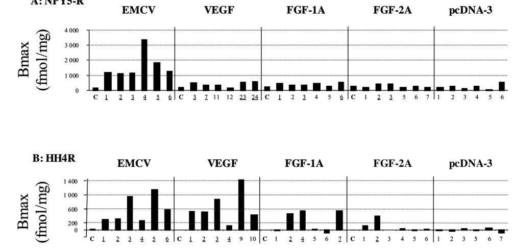
Pharmacological profile of Gα16-CHO transfected cells. The number of binding sites (Bmax)
was estimated for each clone with a saturating concentration of [125I]PYY for the NPY5-R
expressing clones (Fig. 6A). Clones transfected with pcDNA3 led to non functional receptors
in the cAMP assay. [125I]NPY binding to these clones and to the control clones with the IRES
(without NPY5) led to a non specific signal of ca. 300, with the exception of one pcDNA clone (clone 6). A significant cAMP inhibition (factor > 2) and a large binding activity (Bmax > 300) were observed for the six clones obtained with EMCV IRES, for 3 clones with FGF1A (clone 1,3,6), for 4 clones with VEGF (clones 3, 7, 23 and 24) and for 2 clones with FGF2 (clones 2 and 3) IRESs. In the case of EMCV IRES, the clone 4 had the highest Bmax (3369 fmol/ mg protein). For the clones expressing the HH4R, binding experiments were performed
with [3H]-histamine (Fig. 6B). The best clones presented a Bmax between 122 to 1147
fmol/mg protein. Furthermore, 6 clones were positive in terms of cAMP production, for EMCV IRES, 3 for FGF1A (clones 2,4 and 7), 4 for VEGF (clones 1,2,3 and 4) and 1 for FGF2 IRES (clone 2). Surprisingly, the highest Bmax was obtained with the VEGF IRES (clone 9). Further pharmacological characterization was performed on the NPY5-R clone giving the higher Bmax (EMCV IRES, clone 4, Fig. 7). The Ki of different receptor reference ligands were measured and compared with the Ki obtained using the same ligands in transiently transfected COS-7 cells (Table I). Results show that the Ki’s of the different ligands were similar to that obtained in transiently transfected cells, confirming that this clone is fully usable for pharmacological studies.
Long term expression and activity of NPY5-R in stable transfectants. The cell line stability was tested with two clones expressing the transgenes with EMCV or VEGF IRES (Fig 8A). The results did not show any decrease in receptor activity after 49 and 51 passages, for EMCV and VEGF IRESs, respectively. In addition, the Bmax was measured up to 35
passages and did not reveal any loss of the binding capacity. These data show that bicistronic vectors allow a stable expression of transgenes under selection pressure, using both viral and cellular IRESs. Similar results were obtained with H4 receptor (data not shown).
DISCUSSION
The present study shows that bicistronic vectors, containing viral or cellular IRESs, allow the stable expression of functional receptors: NPY5-R and HH4R, and are thus vectors of choice to express active GPCRs with a high yield of positive clones. This study is the first one to compare bicistronic vectors (with IRESs) and bigenic vectors (with two promoters) and leads to a clear conclusion in favor of IRES-containing vectors. These experiments also demonstrate that cellular IRESs provide biotechnological tools, in addition to the widely used EMCV IRES, to express genes of interest in stable cell lines.
The expression of genes of interest often requires the co-expression of at least two proteins. In most cases, the gene of interest is expressed with a selection gene to introduce a selection pressure on the positive clones. Another requirement of gene co-expression is the necessity to express the two sub-units of a hetero-dimeric protein, as for IL-12 [23]. Classically, the vectors used to express two genes simultaneously contain two promoters, each gene being controlled by a distinct transcription unit. Such vectors were successfully used in many cases, which explain their common use in cell transfection, including in our laboratory [4-14]. However, the literature mostly reports successful experiments and is silent about the numerous failures encountered with such vectors, including in our laboratory. Indeed, a promoter interference phenomenon may occur and one has no guarantee of co-expression of both transgenes. Furthermore, the presence of the resistance gene on a transcription unit distinct from that expressing the gene of interest cannot ensure that the latter will be expressed by resistant cells. In particular, if the gene of interest has an inhibitory effect on cell proliferation or any other toxic effect, the selection pressure on a separate transcription unit is not able to prevent the counter-selection of the gene of interest, leading to resistant clones expressing only the resistance gene. In such a case, IRES-containing vectors may solve the
problem, as the two genes of a bicistronic vector are controlled by the same transcription unit. Our data demonstrate the advantage of the bicistronic vector in the case of two different GPCRs, the neuropeptide Y receptor 5 and the histamine receptor 4, for which our ‘standard’ procedure, successful in the past in numerous cases, was not able to provide us with stable cellular clones.
The reasons of pcDNA-3 derived vector inability to express significant amounts of receptor may be discussed. This vector has two transcription units controlled by CMV and SV40 promoters, respectively. This is not the only difference with the bicistronic vector: in both vectors the gene of interest is controlled by the CMV promoter, however in our bicistronic vector, we have introduced a small intron upstream from the first cistron coding sequence, since introns have been described for their ability to ensure higher transgene expression [28]. Thus, we cannot rule out a participation of this intron in the better expression of the transgenes in addition to the selection pressure due to the bicistronic cassette. However, although it may participate in the null yield of the bigenic vector in producing positive clones, the absence of intron cannot explain the strong difference of receptor mRNA expression between the two kinds of vectors. Indeed the neo resistance gene mRNA is expressed with a comparable efficiency in the two cases, with or without an intron, for the bicistronic or bigenic vectors, respectively (data not shown). The most evident reason of the bicistronic vector success is the expression of both the receptor and the neo-resistance proteins from a single transcription unit, which prevents a counter-selection of receptor expression alone.
A striking feature is the apparent correlation between the different yields of positive clones obtained with the four IRESs and their respective efficiency in transient transfection experiments (data not shown). Indeed, we observed a 100% positive clones yield for EMCV IRES, 67% for VEGF IRES, 50% for FGF1A IRES and 13 to 25% for FGF2 IRES, whereas the IRES activities reported in CHO transiently transfected cells for these IRESs are 83, 33, 5
and 12 arbitrary units, respectively (C. Allera-Moreau and A.C. Prats, unpublished results) [29]. Thus, one should expect that poor translation of neo gene under the control of weak IRES should induce a higher bicistronic mRNA expression, to allow sufficient resistance of the cells to G418. Consequently, a higher expression of the receptor, translated by the cap-dependent mechanism from the same mRNA, should be observed. In contrast, we obtained less positive clones with the weak IRESs, in correlation with a lower mRNA expression (Figs. 2 and 4). Among several possible explanations, we hypothesized that the IRES may influence the mRNA stability, or that IRES activity may differ in stable versus transient transfections.
Using the present system, we also showed the functionality of the NYP5 receptor, since the pharmacological data gathered with this system compare well with those reported on the other stable system available in the literature [15] or with our own data obtained, painfully, with the transiently expressed cells [4, 27, 30,31].
Our present data validated the use of bicistronic vectors to establish stable cell lines expressing counter-selected transgenes. Bicistronic vectors have been successfully used in previous reports, in particular for the expression of different FGF2 isoforms [32] or of the two interleukin 12 subunits together with the neo gene in a retrovirus vector [23]. More recently, a bicistronic adenovirus co-expressing VEGF and angiopoïetin 1 has shown a synergistic effect to produce functional and leak-resistant blood vessels by a myoblast-based cell therapy [33]. The present study is the first one to provide a direct comparison of the advantage of IRES-containing vectors versus a two promoter-IRES-containing vector. In addition, most previously used bi- or tri-cistronic vectors contain the EMCV IRES exclusively, probably because commercial bicistronic vectors always bear the EMCV IRES. However, a set of other IRESs have been discovered and could be used to design new vectors. Among them, we can point out the retroviral IRESs which were used to construct retroviral vectors efficiently expressing reporter genes [34,35]. As regards cellular IRESs, we have recently shown that the FGF2
IRES provides better results than the EMCV IRES to co-express co-stimulatory molecules in a retroviral vector [25]. In contrast, the present study shows that the FGF2 IRES, although able to produce stable clones with an acceptable yield, is the less efficient among the four IRESs tested. These data show that the cellular IRESs provide new tools to design bi- or multicistronic vectors, but the optimal IRES to be chosen may vary with the vector (plasmid, virus…), the cells to be transfected or the subtype of receptors.
ACKNOWLEDGMENTS. The authors are grateful to Jean-Pierre Galizzi (2), Marianne
Rodriguez (2) and Hervé Prats (1) for numerous helpful discussions and initial input as well as to Marie-Claire Gensac (1), Laurent Monbrun (1), Nadine Nagel (2) and Christelle Macia (2) for technical assistance. This work was supported by a grant of Institut de Recherches Servier to Camille Allera-Moreau (1).
REFERENCES.
[1]. Jablonowski J.A., Carruthers N.I., Thurmond R.L., The histamine H4 receptor and potential therapeutic uses for H4 ligands, Mini Rev. Med. Chem. 4 (2004) 993-1000.
[2] Duhault J., Boulanger M., Chamorro S., Boutin J.A., Della Zuana O., Douillet E., Fauchere J.L., Feletou M., Germain M., Husson B., Vega A.M., Renard P., Tisserand F., Food intake regulation in rodents: Y5 or Y1 NPY receptors or both?, Can. J. Physiol. Pharmacol. 78 (2000) 173-185.
[3] Spinazzi R., Andreis P.G., Nussdorfer G.G.., Neuropeptide-Y and Y-receptors in the autocrine-paracrine regulation of adrenal gland under physiological and pathophysiological conditions, Int. J. Mol. Med. 15 (2005) 3-13.
[4] Beauverger P., Rodriguez M., Nicolas J.P., Audinot V., Lamamy V., Dromaint S., Nagel N., Macia C., Leopold O., Galizzi J.P., Caignard D.H., Aldana I., Monge A., Chomarat P., Boutin J.A., Functional characterization of human neuropeptide Y receptor subtype five specific antagonists using a luciferase reporter gene assay, Cell. Signal. 17 (2005) 489-496.
[5] Cogé F., Guenin S.P., Renouard-Try A., Rique H., Ouvry C., Richard N., Beauverger P., Nicolas J.P., Galizzi J.P., Boutin J.A., Canet E., Truncated human a1A-adrenoceptors inhibited [3H]-prazosin binding to a 1a-receptor, Biochem. J. 343 (1999) 231-239.
[6] Audinot V., Fabry N., Beauverger P., Nicolas J.P., Newman-Tancredi A., Bornencin F.,
binding at human a2A, a 2B and a 2C adrenoceptors, Cell. Signal. 14 (2002) 829-837.
[7] Nosjean O., Nicolas J.P., Klupsch F., Delagrange P., Canet E., Boutin J.A., Comparative pharmacological studies at the melatonin receptors MT1, MT2 and MT3/QR2, Biochem. Pharamacol. 61 (2000) 1369-1379.
[8] Audinot V., Mailliet F., Lahaye-Brasseur C., Bonnaud A., Le Gall A., Amossé C., Dromaint S., Rodriguez M., Nagel N., Galizzi J.P., Malpaux B., Guillaumet G., Lesieur D., Lefoulon F., Renard P., Delagrange P., Boutin J.A., New selective ligands of human cloned melatonin MT1 and MT2 receptors, Naunyn-Schmiedeberg’s Arch. Pharmacol. 367 (2003) 553-561.
[9] Audinot V., Beauverger P., Lahaye C., Supply T., Rodriguez M., Ouvry C., Lamamy V., Imbert J., Nahon J.L., Galizzi J.P., Canet E., Levens N., Fauchère J.L., Boutin J.A., Structure-activity relationship studies of MCH-related peptide ligands of SLC-1, the human melanin-concentrating hormone receptor, J. Biol. Chem. 276 (2001) 13554-13562.
[10] Rodriguez M., Beauverger P., Naime I., Rique H., Ouvry C., Souchaud S., Dromaint S., Supply T., Audinot V., Boutin J.A., Galizzi J.P., Cloning and molecular characterization of the novel melanin-concentrating hormone receptor (MCH2), Mol. Pharmacol. 60 (2001) 632-639.
[11] Cussac D., Newman-Tancredi A., Nicolas J.P., Boutin J.A., Millan M.J., Antagonist properties of the novel antipsychotic, S 16924, at cloned, human serotonin 5-HT2C receptors: a parallel phosphatidylinositol and calcium accumulation comparison with clozapine and haloperidol, Naunyn-Schmiedeberg’s Arch. Pharmacol. 361 (2000) 549-554.
[12] Cogé F., Guenin-LeChevalier S.P., Audinot V., Renouard-Try A., Macia C., Beauverger P., Nagel N., Rique H., Boutin J.A., Galizzi J.P., Genomic organization and characterization of splice variants of the human histamine H3 receptor, Biochem. J. 355 (2001) 279-288.
[13]. Ferry G., Mozo J., Ubeaud C., Berger S., Beauverger P., Bertrand M., Mesangeau C., Delagrange P., Boutin J.A., Characterization and regulation of a CHO cell line stably expressing the human serotonin N-acetyltransferase (EC 2.3.1.87), Cell. Mol. Life Sci. 59 (2002) 1395-1405.
[14] Mozo J., Ferry G., Studeny A., Rodriguez M., Boutin J.A., Bouillaud F., Expression of UCP3 in CHO cells does not cause uncoupling but controls mitochondrial activity in the presence of glucose, Biochem. J. 393 (2006) 431-440.
[15] Moser C., Bernhardt G., Michel J., Schwarz H., Buschauer A., Cloning and functional expression of the NPY Y5 receptor in human endometrial cancer (HEC-1B) cells, Can. J. Physiol. Pharmacol. 78 ( 2000) 134-142.
[16] Pelletier J., Sonenberg N., Internal initiation of translation of eukaryotic mRNA directed by a sequence derived from poliovirus RNA, Nature 334 (1988) 320-325.
[17] Jackson R.J., mRNA translation. Initiation without an end, Nature 353 (1991) 14-15.
[18] Macejak D.G., Sarnow P., Internal initiation of translation mediated by the 5' leader of a cellular mRNA, Nature 353 (1991) 90-94.
[19] Vagner S., Gensac M.C., Maret A., Bayard F., Amalric F., Prats H., Prats A.C., Alternative translation of human fibroblast growth factor 2 mRNA occurs by internal entry of ribosomes, Mol. Cell. Biol. 15 (1995) 35-44.
[20] Kozak M., The scanning model for translation: an update, J. Cell. Biol. 108 (1989) 229-241.
[21] Jang S.K., Krausslich H.G., Nicklin M.J., Duke G.M., Palmenberg A.C., Wimmer E., A segment of the 5' nontranslated region of encephalomyocarditis virus RNA directs internal entry of ribosomes during in vitro translation, J. Virol. 62 (1988) 2636-2643.
[22] Vagner S., Galy B., Pyronnet S., Irresistible IRES. Attracting the translation machinery to internal ribosome entry sites, EMBO Rep. 2 (2001) 893-898.
[23] Tahara H., Zitvogel L., Storkus W.J., Zeh H.J., McKinney T.G., Schreiber R.D., Gubler U., Robbins P.D., Lotze M.T., Effective eradication of established murine tumors with IL-12 gene therapy using a polycistronic retroviral vector, J. Immunol. 154 (1995) 6466-6474.
[24] Prats A.C., Prats H., Translational control of gene expression: role of IRESs and consequences for cell transformation and angiogenesis, Prog. Nucleic Acid Res. Mol. Biol. 72 (2002) 367-413.
[25] Douin V., Bornes S., Creancier L., Rochaix P., Favre G., Prats A.C., Couderc B., Use and comparison of different internal ribosomal entry sites (IRES) in tricistronic retroviral vectors,
BMC Biotechnol. 4 (2004) 16.
[26] Morse K.L., Behan J., Laz T.M., West R.E., Greenfeder S.A., Anthes J.C., Umland S., Wan Y., Hipkin R.W., Gonsiorek W., Shin N., Gustafson E.L., Qiao X., Wang S., Hedrick J.A., Greene J., Bayne M., Monsma F.J., Cloning and characterization of a novel human histamine receptor, J. Pharmacol. Exp. Ther. 296 (2001) 1058-1066.
[27] Rodriguez M., Audinot V., Dromaint S., Macia C., Lamamy V., Beauverger P., Rique H., Imbert J., Nicolas J.P., Boutin J.A., Galizzi J.P., Molecular identification of the long isoform of the human neuropeptide Y Y5 receptor and pharmacological comparison with the short Y5 receptor isoform, Biochem. J. 369 (2003) 667-673.
[28] Yew N.S., Wysokenski D.M., Wang K.X., Ziegler R.J., Marshall J., McNeilly D., Cherry M., Osburn W., Cheng S.H., Optimization of plasmid vectors for high-level expression in lung epithelial cells, Hum. Gene Ther. 8 (1997) 575-584.
[29] Martineau Y., Le Bec C., Monbrun L., Allo V., Chiu I.M., Danos O., Moine H., Prats H., Prats A.C., Internal ribosome entry site structural motifs conserved among mammalian fibroblast growth factor 1 alternatively spliced mRNAs, Mol. Cell. Biol. 24 (2004) 7622-7635.
[30] Henlin J.M., Boutin J.A., Duchene-Roger F., Desmet-Beaufort C., Nicolas J.P., Levens N., Fauchère J.L., Parallel synthesis and pharmacological screening of non-peptide ligands of the neuropeptide Y receptor subtype Y5, J. Peptide Sci. 57 (2001) 419-427.
[31] Guery S., Rival Y., Wermuth C.G., Renard P., Boutin J.A., A convenient 3-step
synthesis of 3-acetamido-6-arylpyridazines directed to novel Y5 receptor antagonist, Chem. Pharm. Bull. 50 (2002) 363-639.
[32] Arnaud E., Touriol C., Boutonnet C., Gensac M.C., Vagner S., Prats H., Prats A.C., A new 34-kilodalton isoform of human fibroblast growth factor 2 is cap dependently synthesized by using a non-AUG start codon and behaves as a survival factor, Mol. Cell. Biol. 19 (1999) 505-514.
[33] Niagara M.I., Haider H., Ye L., Koh V.S., Lim Y.T., Poh K.K., Ge R., Sim E.K., Autologous skeletal myoblasts transduced with a new adenoviral bicistronic vector for treatment of hind limb ischemia, J. Vasc. Surg. 40 (2004) 774-785.
[34] Derrington E.A., Lopez-Lastra M., Chapel-Fernandez S., Cosset F.L., Belin M.F., Rudkin B.B., Darlix J.L., Retroviral vectors for the expression of two genes in human multipotent neural precursors and their differentiated neuronal and glial progeny, Hum. Gene Ther. 10 (1999) 1129-1138.
[35] Torrent C., Berlioz C., Darlix J.L., Stable MLV-VL30 dicistronic retroviral vectors with a VL30 or MoMLV sequence promoting both packaging of genomic RNA and expression of the 3' cistron, Hum. Gene Ther. 7 (1996) 603-612.
[36] Huez I., Creancier L., Audigier S., Gensac M.C., Prats A.C., Prats H., Two independent internal ribosome entry sites are involved in translation initiation of vascular endothelial growth factor mRNA. Mol. Cell. Biol. 18 (1998) 6178-6190.
LEGENDS FOR FIGURES.
Figure 1 : Bicistronic and bigenic expression vectors for the production of NPY5-R and
HH4R receptors.
Scheme of the bicistronic vector. The bicistronic cassette, under the control of the cytomegalovirus promoter (PCMV), contains the NPY5-R or HH4R coding sequence as a first cistron (cap-dependent) and the neomycine-resistance gene as a second cistron (IRES-dependent). A small intron is present in the 5’ end of the cassette [36]. The bicistronic mRNA is represented with the polyA tail.
Scheme of the bigenic vector. Bigenic vectors lead to the obtention of two mRNA. The first mRNA is under the transcriptional control of the constitutive PCMV, the second one under the transcriptional control of the constitutive PSV40, For maintenance and amplification in Escherichia coli, all vectors contain β-lactamase gene (amp) conferring resistance to ampicillin.
Figure 2 : Measurement of NPY5-R activity of clones expressing bicistronic and bigenic
vectors.
Gα16-CHO cells were transfected with bicistronic or bigenic vectors described in Fig. 1, coding for the NPY5-R. Receptor activity was measured for six clones expressing each bicistronic vector, and 12 clones expressing the bigenic vector, by cAMP radioactive immuno-assay (see Math. & Meth.). A: EMCV IRES, B: VEGF IRES, C: FGF-1A IRES, D: FGF-2 IRES and E: control (No IRES). The legend of the x axis is: C for Gα16-CHO cells transfected with a control vector witch do not contain receptor gene, the numbers refer to different clones. Clones were treated either by forskolin (open bars), or with forskolin and neuropeptide Y (dark bars). Cell Bar Chart was obtained with the Statwiew software.
Grouping variable : Gα16-CHO transfected cells. Error bars : standard error. * : p < 0,05. ** : p< 0,01. *** : p< 0,001.
Figure 3 : Measurement of HH4R activity of clones expressing bicistronic and bigenic
vectors.
Gα16-CHO cells were transfected as in Fig. 2 with bicistronic or bigenic vectors described in Fig. 1, coding for the HH4R. Receptor activity was measured for six clones expressing each bicistronic vector, and 6 clones expressing the bigenic vector, by cAMP radioactive immuno-assay (see Math. & Meth.). A: EMCV IRES, B: VEGF IRES, C: FGF-1A IRES, D: FGF-2 IRES and E: control (No IRES). The legend of the x axis is: C for Gα16-CHO cells transfected with a control vector witch do not contain receptor gene, the numbers refer to different clones. Clones were treated either by forskolin (open bars) or by forskolin + Histidine (dark bars). Cell Bar Chart was obtained with the Statwiew software. Grouping variable : Gα16-CHO transfected cells. Error bars : standard error. * : p < 0,05. ** : p< 0,01. *** : p< 0,001.
Figure 4 : NPY5R mRNA expression in Gα16-CHO transfected cells.
The copy number of NPY5R amplicon per ng of RNA was quantified using real-time RT PCR, for the clones presented in Fig. 2 (see Math. & Meth.). The underlined numbers correspond to the clones expressing active NPY5-R. For each clone, the IRES present in the bicistronic vector is indicated.
Figure 5 : NPY5-R expression and localisation by immunohistofluorescence.
Anti M2 flag antibody was used as primary antibody to detect the cytoplasmic C-terminal end of NPY5-R. (A) Immunofluorescent staining of NPY5-R in clones expressing bicistronic
versus bigenic vectors. (B) Immunofluorescent staining of functional versus inactive receptors in clones expressing the bicistronic construct with VEGF IRES.
Figure 6 : Binding assay of [125I] PYYh or [3H] Histamine on membrane preparation of
Gα16-CHO transfected cells.
The number of binding sites (Bmax) was determined for the different clones presented in Figs. 2 and 3. (A) Y5 cell clones. (B) H4 cell clones. The underlined numbers correspond to the clones expressing active receptor.
Figure 7 : Binding characterization of NPY5-R.
Saturation binding experiments with [125I]-PYY at NPY5-R expressed in Gα16-CHO cells
stably transfected with EMCV IRES - clone 4. Inset: Scatchard reprentation.
Figure 8 : Long term stability of cell lines expressing the NPY5-R from bicistronic
constructs.
NPY5-R activity and binding sites number was measured in cell clones expressing bicistronic vectors after up to 45 passages. (A) Receptor activity. On the left: Gα16-CHO cell clone 4 with EMCV IRES, on the right : Gα16-CHO cell clone 3 with VEGF IRES. FK : forskolin, NPY : Y neuropeptide. Cell Bar Chart was obtained with the Statwiew software. Grouping variable : Gα16-CHO transfected cells. Split by: FK/FK+NPY. Error bars : standard error. * : p < 0,05. ** : p< 0,01. *** : p< 0,001. (B) Bmax of Gα16-CHO cell clone with EMCV, FGF1A or VEGF IRES.
Table 1 : Comparative molecular pharmacology of stably and transiently expressed NPY 5 receptor
Y5 Gα16-CHO cells Y5 COS7 cells
EMCV IRES (transient)
Reference compounds Ki (nM) Ki (nM) mean ± sem (n) mean ± sem (n)
NPY (h,r) 1.5 ± 0.3 (5) 0.64 ± 0.18 (3)
Leu31, Pro34 NPY (h,r) 1.2 ± 1 (2) 3.1 ± 1.5 (3)
PYY (h) 1.5 ± 0.1 (4) 2.1 ± 1.2 (2) NPY (13-36) (h,r) 41 ± 12 (4) 45 ± 5 (2) PYY (3-36) (h) 2.5 ± 1.4 (3) 3.1 ± 1.5 (2) PP (h) 3.4 ± 1.1 (3) 2.3 ± 0.5 (2) D-Trp32 NPY (h,r) 21 ± 3 (4) 22 ± 8 (2) C2 NPY 121 ± 18 (4) 48 ± 1 (2)
Figure 1 Intron IRES PC MV
DNA
mRNA
AAAreceptor
A
B
PC MV PSV40mRNA
DNA
Neo
RNeo
RNeo
Rreceptor
AAA
Neo
R AAApASV40 pASV40 pASV40
receptor
receptor
Intron IRES PC MVDNA
mRNA
AAAreceptor
A
B
PC MV PSV40mRNA
DNA
Neo
RNeo
RNeo
Rreceptor
AAA
Neo
R AAApASV40 pASV40 pASV40 pASV40 pApASV40SV40
receptor
receptor
receptor
receptor
Figure 2 0 10 20 30 C 1 2 3 4 5 6 * ** ** ** *** *** 0 10 20 30 C 1 2 3 4 5 6 0 4 8 12 16 11 12 23 24 C * ** 3 7 * *** 0 4 8 12 16 11 12 23 24 C 3 7 0 10 20 30 0 10 20 30 4 10 11 12 1 2 3 5 6 7 8 9
C
e
ll
c
A
MP
P
ro
d
u
c
it
o
n
(n
M)
0 4 8 12 16 20 C 1 2 3 4 5 6 ** * * 0 4 8 12 16 20 C 1 2 3 4 5 6 20 30 40 1 3 5 7 0 10 20 30 40 C 1 2 3 6 *** ** 7A
B
C
D
E
0 10 20 30 C 1 2 3 4 5 6 * ** ** ** *** *** 0 10 20 30 C 1 2 3 4 5 6 0 4 8 12 16 11 12 23 24 C * ** 3 7 * *** 0 4 8 12 16 11 12 23 24 C 3 7 0 10 20 30 0 10 20 30 4 10 11 12 1 2 3 5 6 7 8 9 0 10 20 30 0 10 20 30 4 10 11 12 1 2 3 5 6 7 8 9C
e
ll
c
A
MP
P
ro
d
u
c
it
o
n
(n
M)
0 4 8 12 16 20 C 1 2 3 4 5 6 ** * * 0 4 8 12 16 20 C 1 2 3 4 5 6 20 30 40 1 3 5 7 0 10 20 30 40 C 1 2 3 6 *** ** 7 0 4 8 12 16 20 C 1 2 3 4 5 6 ** * * 0 4 8 12 16 20 C 1 2 3 4 5 6 20 30 40 1 3 5 7 0 10 20 30 40 C 1 2 3 6 *** ** 7A
B
C
D
E
Figure 3 0 4 8 12 16 1 2 3 5 6 7 0 2 4 6 8 *** ****** *** ** ** C 1 2 3 4 5 6 0 2 4 6 8 C 1 2 3 4 5 6 0 4 8 12 16 * *** ** * C 1 2 3 4 9 10 0 4 8 12 16 C 1 2 3 4 9 10 C 1 2 4 5 6 7 ** 0 1 2 3 4 * ** 0 1 2 3 4 C 1 2 4 5 6 7 4 8 ** C 1 2 3 4 5 6 0 4 8 12 C 1 2 3 4 5 6
C
ell
cA
M
P
P
roduc
ti
on (n
M
)
A
B
D
C
E
0 4 8 12 16 1 2 3 5 6 7 0 4 8 12 16 1 2 3 5 6 7 0 2 4 6 8 *** ****** *** ** ** C 1 2 3 4 5 6 0 2 4 6 8 C 1 2 3 4 5 6 0 4 8 12 16 * *** ** * C 1 2 3 4 9 10 0 4 8 12 16 C 1 2 3 4 9 10 0 2 4 6 8 *** ****** *** ** ** C 1 2 3 4 5 6 0 2 4 6 8 C 1 2 3 4 5 6 0 4 8 12 16 * *** ** * C 1 2 3 4 9 10 0 4 8 12 16 C 1 2 3 4 9 10 C 1 2 4 5 6 7 ** 0 1 2 3 4 * ** 0 1 2 3 4 C 1 2 4 5 6 7 4 8 ** C 1 2 3 4 5 6 0 4 8 12 C 1 2 3 4 5 6 C 1 2 4 5 6 7 ** 0 1 2 3 4 * ** 0 1 2 3 4 C 1 2 4 5 6 7 4 8 ** C 1 2 3 4 5 6 0 4 8 12 C 1 2 3 4 5 6C
ell
cA
M
P
P
roduc
ti
on (n
M
)
A
B
D
C
E
Figure 4
C
o
py
numbe
r o
f Y
5
a
m
pl
ic
o
n
(1
0
-3
/ n
g
RNA)
2. 5. 10 -2 10 .9 2. 4 11 .8 63 .0 8. 2 12 .9 10 -3 5. 9 10 .8 6. 6 4. 8 5.2 0 3. 6 15 .8 1. 4 1. 0 0. 5 1. 5 5. 10 -3 2. 9 2. 8 5. 4 3. 1 0. 8 3. 9 0. 4 0. 3 0.2 0. 1 0 0 5 10 15 60 C 1 2 3 4 5 6 C 3 7 11 23 24 C 1 2 3 4 5 6 C 1 2 3 5 6 7 2 3 4 5 6EMCV
VEGF
FGF-1A
FGF-2
pcDNA3
IRES
2. 5. 10 -2 10 .9 2. 4 11 .8 63 .0 8. 2 12 .9 10 -3 5. 9 10 .8 6. 6 4. 8 5.2 0 3. 6 15 .8 1. 4 1. 0 0. 5 1. 5 5. 10 -3 2. 9 2. 8 5. 4 3. 1 0. 8 3. 9 0. 4 0. 3 0.2 0. 1 0 0 5 10 15 60 C 1 2 3 4 5 6 C 3 7 11 23 24 C 1 2 3 4 5 6 C 1 2 3 5 6 7 2 3 4 5 6EMCV
VEGF
FGF-1A
FGF-2
pcDNA3
Figure 6
A: NPY5-R
0 1 000 2 000 3 000 4 000 C 1 2 3 4 5 6 C 3 7 11 12 23 24 C 1 2 3 5 6 7 1 2 3 4 5 6EMCV
VEGF
FGF-2A
pcDNA-3
Bm
ax
(f
m
ol/m
g)
C 1 2 3 4 5 6FGF-1A
B: HH4R
0 200 600 1 000 1 400 C 1 2 3 4 5 6 C 1 2 3 4 9 10 C 1 2 3 4 5 6 1 2 3 5 6 7EMCV
VEGF
FGF-2A
pcDNA-3
C 1 2 4 5 6 7
FGF-1A
Bm
ax
(f
m
ol/m
g)
Figure 7
0
1
2
3
4
5
6
0
100
200
300
400
[
125I]-PYY (n M)
Sp
ec
if
ic
bo
un
d
(f
m
ol
/m
g)
0 100 200 300 400
0
100
200
300
400
500
Bound (fmol/mg) B ound (f m o l/m g ) / Free (n M)0
1
2
3
4
5
6
0
100
200
300
400
[
125I]-PYY (n M)
Sp
ec
if
ic
bo
un
d
(f
m
ol
/m
g)
0
1
2
3
4
5
6
0
100
200
300
400
[
125I]-PYY (n M)
[
125I]-PYY (n M)
Sp
ec
if
ic
bo
un
d
(f
m
ol
/m
g)
0 100 200 300 400
0
100
200
300
400
500
Bound (fmol/mg) B ound (f m o l/m g ) / Free (n M)0 100 200 300 400
0
100
200
300
400
500
Bound (fmol/mg) B ound (f m o l/m g ) / Free (n M)Figure 8 0 1 2 3 4 5 6 7 8 C el l Me an fo r nM A MP c 15 20 25 30 35 40 45 *** ** *** *** *** *** ** 0 2 4 6 8 10 12 14 16 18 21 26 31 36 41 46 51 *** *** ****** *** *** *** EMCV 0 1 2 3 4 5 6 7 8 C el l Me an fo r nM A MP c 15 20 25 30 35 40 45 *** ** *** *** *** *** ** VEGF 0 2 4 6 8 10 12 14 16 18 21 26 31 36 41 46 51 *** *** ****** *** *** ***
Cell passage number
A
Cell passage number FK FK + NPY
B
1 10 100 1000 10000 10 15 20 25 30 35 Cell passage number EMCV VEGF Bm ax (f m ol /m g) FGF-1A 0 1 2 3 4 5 6 7 8 C el l Me an fo r nM A MP c 15 20 25 30 35 40 45 *** ** *** *** *** *** ** 0 2 4 6 8 10 12 14 16 18 21 26 31 36 41 46 51 *** *** ****** *** *** *** EMCV 0 1 2 3 4 5 6 7 8 C el l Me an fo r nM A MP c 15 20 25 30 35 40 45 *** ** *** *** *** *** ** VEGF 0 2 4 6 8 10 12 14 16 18 21 26 31 36 41 46 51 *** *** ****** *** *** ***Cell passage number
A
Cell passage number FK FK + NPY