Publisher’s version / Version de l'éditeur:
Cell Biology International, 40, 1, pp. 83-90, 2015-09-08
READ THESE TERMS AND CONDITIONS CAREFULLY BEFORE USING THIS WEBSITE. https://nrc-publications.canada.ca/eng/copyright
Vous avez des questions? Nous pouvons vous aider. Pour communiquer directement avec un auteur, consultez la
première page de la revue dans laquelle son article a été publié afin de trouver ses coordonnées. Si vous n’arrivez pas à les repérer, communiquez avec nous à PublicationsArchive-ArchivesPublications@nrc-cnrc.gc.ca.
Questions? Contact the NRC Publications Archive team at
PublicationsArchive-ArchivesPublications@nrc-cnrc.gc.ca. If you wish to email the authors directly, please see the first page of the publication for their contact information.
This publication could be one of several versions: author’s original, accepted manuscript or the publisher’s version. / La version de cette publication peut être l’une des suivantes : la version prépublication de l’auteur, la version acceptée du manuscrit ou la version de l’éditeur.
For the publisher’s version, please access the DOI link below./ Pour consulter la version de l’éditeur, utilisez le lien DOI ci-dessous.
https://doi.org/10.1002/cbin.10541
Access and use of this website and the material on it are subject to the Terms and Conditions set forth at Evidence of a role for S. cerevisiae α-arrestin Art1 (Ldb19) in mating projection and zygote formations
Choudhary, Pooja; Loewen, Michele C.
https://publications-cnrc.canada.ca/fra/droits
L’accès à ce site Web et l’utilisation de son contenu sont assujettis aux conditions présentées dans le site LISEZ CES CONDITIONS ATTENTIVEMENT AVANT D’UTILISER CE SITE WEB.
NRC Publications Record / Notice d'Archives des publications de CNRC:
https://nrc-publications.canada.ca/eng/view/object/?id=9dcc437a-f913-412c-a182-835f6690785e https://publications-cnrc.canada.ca/fra/voir/objet/?id=9dcc437a-f913-412c-a182-835f6690785e
Evidence of a role for S. cerevisiae
α-arrestin Art1 (Ldb19) in mating
projection and zygote formations
Pooja Choudhary1 & Michele C. Loewen1,2
1
Department of Biochemistry, University of Saskatchewan, 107 Wiggins Rd., Saskatoon, SK, S7N 5E5, Canada
2
National Research Council of Canada, 110 Gymnasium Place, Saskatoon, SK., S7N 0W9, Canada
To whom correspondence should be addressed: Michele Loewen, National Research Council of Canada, 110 Gymnasium Place, Saskatoon, SK., S7N 0W9, Canada. Tel: (306) 975-6823, Email: michele.loewen@nrc.ca
Short Title: A role for yeast Arrestin-1(Ldb19) in mating
ABSTRACT
The discovery of arrestin-mediated biased signalling mechanisms for mammalian G-protein coupled receptors (GPCRs) has revolutionized the field over the last decade. Now, with the recent demonstration of a role for α-arrestins in internalization of the yeast pheromone GPCR, Ste2p, the possibility of arrestin-mediated alternate GPCR functionalities in yeast also follows. Here, the effects of knockout and complementation of yeast α-arrestin expression during mating are reported. While minor effects on classical pheromone-related signalling are noted for a few arrestins, much stronger effects were observed downstream of cell cycle arrest, in particular linking Ldb19 (Art1) to mediation of zygote formation. Subsequent phenotypic observations linked this activity to more pronounced projection formation in an Art1 complemented noncuple α-arrestin knockout line, compared to the knockout-line alone, or either of the Art3 or Art6 complemented lines. Together with the observation of ligand-stimulated localization of Art1-GFP to the mating projection, a possible role for this arrestin-like protein in projection formation is supported. While leaving the full mechanism of alternate Art1 functionality to be elucidated, together these findings implicate Art1 in selective regulation of mating events downstream of receptor internalization and cell cycle arrest, leading to schmoo and ultimately zygote formation.
1. INTRODUCTION
Recent reports have highlighted the existence of important alternate G-protein coupled receptor (GPCR)–linked signaling functionalities for mammalian β-arrestins (reviewed in (Kenakin and Christopoulos, 2013; Reiter et al., 2012)). Arrestins, better known for their role in mediating GPCR internalization and recycling/degradation, have now been shown, under certain circumstances, to mediate GPCR signal transduction by linking to RAF/ERK, RhoA, PP2A and PI3K associated pathways. These alternate functionalities are linked to many of the known side effects associated with GPCR-related pharmaceuticals today.
In Saccharomyces cerevisiae (S. cerevisiae), the GPCRs Ste2p and Ste3p, are known to mediate mating responses through binding of opposite mating type pheromones (Xue et al., 2008; Ladds et al., 2005). The classical Ste2p signal transduction pathway, like the mammalian systems, includes G-protein activation and stimulation of MAPK cascades, culminating in cell cycle arrest (Figure 1). With opposite mating types arrested at the same cell cycle stage, mating events are subsequently initiated including projection formation and agglutination, followed by cell fusion (plasmogamy and karyogamy) to form a diploid zygote.
Interestingly, a review of the literature highlights evidence supporting the possibility of alternate functionalities for Ste2p in mating events downstream of cell cycle arrest. Reports going back decades include, demonstrations of roles for pheromone gradients in projection formation (Segall, 1993; Barkai et al., 1998), a need for high concentrations of pheromone in prezygote cell fusion (Brizzio et al., 1996; Elia and Marsh, 1996) and observations of
localization of the receptor to the mating projection (Jackson et al., 1991). As well, inhibition of mating projection formation and subsequent mating events in Ste2p null yeast, that otherwise maintain strong G-protein mediated cell cycle arrest functionalities (Schrick et al., 1997;
Whiteway et al., 1990) has also been reported. More recently selective site directed mutants of Ste2p have been shown to yield reduced mating efficiency, but with essentially normal cell cycle arrest (Shi et al., 2007, 2009). While direct evidence is still lacking due to difficulties addressing the redundancy associated with a single receptor mediating two temporally distinct steps in the same process, the possibility of alternate GPCR-linked signaling in yeast can no longer be ignored.
This is particularly true in light of the recent demonstration of a direct interaction between yeast arrestin-like proteins and Ste2p in internalization events (Alvaro et al., 2014). A family of 10 arrestin-like proteins, termed α-arrestins is known to be expressed in S. cerevisiae. Reports have indicated that these α-arrestins are involved in the ubiquitination and endocytosis of various yeast membrane proteins (Lin et al., 2008; Nikko et al., 2008; Nikko and Pelham, 2009; Hatakeyama et al., 2010; O’Donnell, 2012; Becuwe et al., 2012; Merhi and Andre, 2012). Most recently, this was demonstrated for Ste2p, with Art1 (Ldb19), Art4 (Rod1) and Art7 (Rog3) all being implicated in modulation of receptor internalization (Alvaro et al., 2014).
In this report, the possibility of a role for α-arrestins in mediating alternate mating-related functionalities downstream of cell cycle arrest is investigated. Deletional-analyses highlight moderate, but statistically relevant decreases in zygote formation, with expression of Art1
rescuing this mating defect. Phenotypic characterization highlights recovery of more pronounced projection formation in Art1 complemented cells. Localization analyses further demonstrated selective sequestration of this same arrestin to the projection at one hour post-ligand induction. While highlighting alternate functionalities for Art1, the mechanism by which Art1 mediates projection and zygote formation remains to be elucidated.
1. MATERIALS AND METHODS
2.1 Chemicals. All chemical were obtained from Sigma-Aldrich unless otherwise noted below. CSM-His-Ura, CSM-Leu-Ura and CSM-His-Leu-Ura were obtained from MP Biomedicals, USA. Bacto-Agar, Bacto-Tryptone and Bacto–Yeast Extract were from BD, Canada. CSM-Lys-Met was obtained from Sunrise Science products, USA. Mutagenic primers were obtained from Integrated DNA Technologies Inc. (USA).
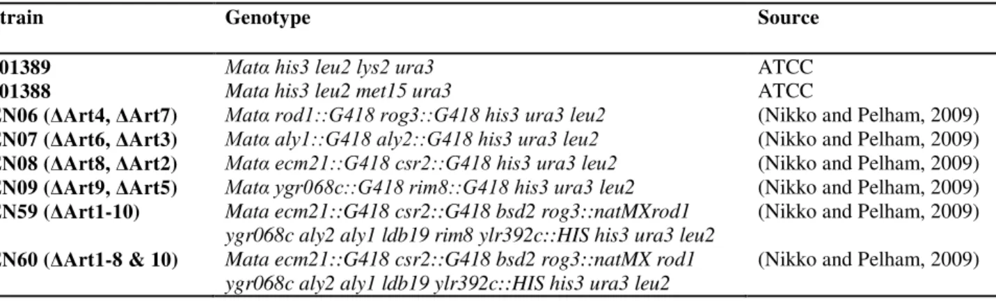
2.2 Strains and plasmids. Escherichia coli strain DH5α and Top10 cells were used as hosts to propagate and amplify plasmids for different experiments. All yeast strains used in this study are described in the Table 1 with their relevant genotype and source. Arrestin expression plasmids were based on the YCplac 111CEN LEU2 or YCplac33CEN URA3 vectors, with individual arrestins expressed from their own promoters as described elsewhere (Nikko and Pelham, 2009). Plasmid pSB234, containing the FUS1-lacZ β galactosidase reporter was used to measure MAPK signal transduction activity of the receptor (Jin et al., 2008). Target arrestin genes were
recombined into the pAG426Gal-GFP (Addgene; 14203, Cambridge, MA, USA) vectors according to Gateway specifications, for fluorescent localization studies. Recombination in pAG426Gal-GFP, yielded arrestin-GFP fusions with the GFP added at the 3’end of the coding sequence. All arrestin containing expression plasmids were transformed into arrestin knockout yeast strains by the LiAc/PEG mediated transformation method (Gietz et al., 1992). Positive transformants were selected by growth on -Leu or -Ura media.
2.3 FUS1-lacZ gene induction assay. Pheromone-stimulated signaling activity was assessed using the FUS1-lacZ gene induction assay as described previously (Kippert, 1995). Yeast strains, were grown (5 mL cultures) overnight, in CSM (complete synthetic media) minimal media, lacking uracil, or leucine and uracil, in 2 % glucose. The following morning cells were
transferred into an equal volume of rich media (YPD with 2 % glucose). After 3-4 hours growth in the rich media, Mat α cultures of EN06, EN07, EN08 and EN09, were incubated with equal amounts of Mat a (201388) as a source of a-factor. Mat a strains EN59 and EN60 were treated with the 1μM α-factor (Zymo Research). Untreated cultures were used as controls. After two hours of incubation with or without pheromone, yeast cells were centrifuged and re-suspended in 300 μl Z-buffer (60mM Na2HPO4, 40mM NaH2PO4, 10mM KCL, 1m Mg2SO4) and the optical
density (OD) measured at 600 nm. All samples were then frozen in liquid nitrogen and stored at -20 °C. For analysis, samples were thawed, incubated at 30 °C for 30 minutes and diluted with the addition of 700 μl Z-buffer containing β mercaptoethanol + 0.2 % sarcosyl to permeabilize the samples. O-nitrophenyl-β-D-galactopyranoside (ONPG, 4 mg/ml) was added and the mixture was incubated at 30 °C until a yellow color developed ( ~ 2-3 h). Subsequently, 400 μl of 1.5 M Na2CO3 was added to stop the reaction. The samples were then centrifuged (10 minutes at
18,000 x g) and the OD at 420 nm measured. The relative β-galactosidase activity was calculated using β galactosidase units = OD420×1000/t×v×OD600, where t is time and v is volume. All
experiments were repeated twice with three replicates on each occasion.
2.4 Quantitative mating efficiency assay. Strains were grown overnight in minimal CSM media for plasmid selection, but containing 2 % glucose. These were transferred the following morning to rich media for 3-4 h. EN cells (8×10⁷ cells) were then mixed with (mated against) the parent WT (2×10⁷ cells) Mat α strain 201389 or Mat a strain 201388, to initiate mating, and then filtered using 0.22 μm filter paper. Equal quantities of each strain were independently filtered to serve as negative controls. The filter papers were placed on rich media plates and incubated for 3 h at 30 °C. Collected cells were subsequently washed and sonicated in a water bath for 30
were plated on CSM minimal media plates lacking both uracil and leucine, or lysine and methionine, or histidine and uracil (as necessary) for 48 hours at 30 °C. The number of single zygote colonies was then quantified and the mating efficiencies calculated according to the method described previously (Sprague, 1991).
2.5 Localization of arrestin by confocal microscopy. EN60 cells expressing GFP tagged arrestins or GFP alone, were cultured overnight in 2 % glucose, uracil minimal media to maintain vectors. Cells were washed with 10 mM Tris, 1 mM EDTA, pH 7.4 (TE buffer), and sterile water and re-suspended in an equal volume of rich induction media (YPD with 2 % galactose and 1 % raffinose). After 4-5 h of growth cells were treated (or not) with 2.5 μM α-factor ligand for different time periods. The cells were mounted on microscope slides with 1.5 μl of cell suspension mixed with 25 μl of hard mounting media and covered with a coverslip. Cells were fixed in 3.7% final concentration of formaldehyde solution (F-79, Fisher Scientific) for 1 h at room temperature before mounting in hard-set media. The slides were left undisturbed overnight at room temperature and examined by confocal microscopy (LEICA SP5, 63x, oil immersion lens), with excitation at 488 nm wavelength and emission monitored at 493-582 nm wavelength. For each arrestin, 3-20 cells were imaged, looking at the top and bottom surfaces of each cell in focus to determine z-thickness. Subsequently, 2D images (16 bit, 512x512,
24.6x24.6 micron, both brightfield and epifluorescent) were taken at the centre of the yeast cell in focus with the same zoom factor (10), power and gain settings (750) across all images. Fluorescence was assessed using Image J software (Schneider et al., 2012).
2.6 Statistics. All statistical analyses were performed using GraphPad Prism 6 (Graph pad, California, USA) software. All data are reported as mean ± SEM. Treatment groups were compared using either t-test or analysis of variance after assumptions of normality and equal
variance were met for all data analyzed. In case of proportionate data, arcsine transformation was completed to normalize the data before applying statistical tests. Differences between multiple groups for different endpoints were tested using Fischer’s least significant difference and probabilities ≤0.001 were considered significant.
2. RESULTS
In order to determine whether α-arrestins modulate alternate functionalities downstream of cell cycle arrest, a series of yeast arrestin knockout strains were comparatively tested for both classical pheromone-stimulated signal transduction and zygote formation.
2.1 Effect of arrestin knockout and complementation on pheromone stimulated signal
transduction. To evaluate effects on the classical pheromone-stimulated signal transduction, six strains were analyzed, including four Mat α double arrestin knockouts and two Mat a lines comprising noncuple (EN60, all knocked-out except Art9) and decuple (EN59) knockouts (Table 1; (Nikko and Pelham, 2009)), using the classical Ste2p-linked FUS1-lacZ gene induction assay (Kippert, 1995). The double knockout lines were evaluated based on previous results showing additive effects for the paired sister arrestins (Nikko et al., 2008; Alvarez, 2008). Unfortunately the double Art1+Art10 knockout line was not available for this study, however both Art1 and Art10 are knocked-out in the noncuple and decuple knockout lines, and considered therein. When transformed with the FUS1-lacZ reporter vector and tested for ligand stimulated signal transduction (Figure 2A), a number of very weak effects were detected. However, in our hands, only the effect recorded for the EN07 line, which included knockout of Art3/Art6, and the
noncuple knockout EN60 line, showed statistically relevant reductions in activity. These results are reflected in the complementation experiments, where each of the arrestins was expressed one at a time in the noncuple knockout (EN60). The EN60 cell line, although not entirely healthy in its own right was selected over the decuple EN59 line, due to EN59 cells being unhealthy and very slow to proliferate, likely associated with impact of no expressing arrestins on the general health of the cell (Nikko and Pelham, 2009)). Here, while there seems to be a general effect of arrestin expression to stimulate signal transduction above that of the noncuple knockout, again possibly associated with general cellular fitness, the most potent effects above this background are associated with Art1 and Art6 (Figure 2B). These results are consistent with the knockout results and emphasize that in the EN07 line, it is likely loss of Art6, and not Art 3 that is mediating the observed decrease in signaling events. Unfortunately, it is not possible to do a similar comparison for Art1, lacking the double Art1/Art10 knockout line, but on their own the complementation experiment highlights a weak effect of Art1 in pheromone-induced signal transduction. Overall the effects observed upon deletion of, or complementation by arrestins on FUS1-LacZ induction are all very weak, but at least partly consistent with a previous more in depth report (Alvaro et al., 2014), and are not investigated further here.
2.2 Effect of arrestin knockout and complementation on zygote formation. In contrast, when the same knockouts were tested for effect on mating events, looking at diploid zygote number, significantly more potent effects were observed. In particular EN06, EN07 and EN09 showed between 38 % and 47 % decreases in mating, with the noncuple (EN60) and decuple (EN59) lines demonstrating evidence of possible additive effects, with 67 % and 56 % reductions in mating respectively (Figure 3A). Obviously in the case of EN07, some of the reduction in
zygote formation is likely attributable to reduced signaling activity as observed in Figure 2A. However it is unlikely that all of the reduced zygote formation reported here is attributable to loss of signaling, as the degree of signaling loss is relatively weak compared to the zygote formation effect. It is also notable here, that the noncuple line has, albeit not an additive effect, but at least a more pronounced effect on zygote formation than any of the double knockouts, implying possible redundant roles for multiple arrestins in zygote formation. Art2 and Art8, which appear to have no effect on zygote formation, would be excluded from this list.
Interestingly, in the complementation experiment (Figure 3B), while Art1 expression yielded a significant recovery of mating activity, showing a 35% increase over the EN60 knockdown level, none of the other single arrestins yielded any significant effect. This highlights a possible
selective role for Art1 as a key component in downstream mating events related to zygote formation. However, it should be noted that Art1 only led to a partial recovery of activity, and consistent with the knockdown experiments, other arrestins (including Art 3, Art4, Art5, Art6, Art7, Art10) may collectively contribute to zygote formation in native cells, but their individual contribution is below the sensitivity of the currently applied complementation assay, or
dependent on the presence of other arrestins.
2.3 Phenotypic characterizations and localization of arrestins upon ligand-stimulation. To further investigate the role of the α-arrestins in mating, select complemented EN60 cell lines were characterized phenotypically. Under the condition of ligand-stimulated induction in the absence of an opposing mating cell (i.e. with the addition of α-factor directly to the
complemented cells) no evidence of any defined projection formation was observed for the EN60 cell line, or either of the Art3-GFP or Art6-GFP complemented lines (Figure 4). This observation is somewhat inconsistent with the observed low levels of zygote formation reported
for these same lines (Figure 3). However this discrepancy is likely explained by the presence of healthy opposite mating yeast cells, producing normal projections and secreting targeted
gradients of pheromone in the zygote formation assay (discussed further below). In contrast to Art3-GFP and Art6-GFP, cells complemented with Art1-GFP showed a recovery of projection formation upon ligand stimulation (Figure 4), despite just the bulk addition of pheromone. This recovery of projection formation for Art1-GFP is consistent with the observed recovery of zygote formation (Figure 3).
Subsequently, localization was evaluated with respect to Ste2p ligand stimulation, by
observation of fluorescence emitted from the overexpressed GFP-tagged arrestins in the EN60 line. The functional relevance of GFP-tagged Art1, as well as other GST-tagged and Myc-tagged yeast arrestin-like proteins has been demonstrated previously (Lin et al., 2008; Hatakeyama et al., 2010). At time 0 prior to pheromone stimulation, the GFP-tagged arrestins were evenly dispersed throughout the cytoplasm (Figure 4). This is at least partially as expected based on a previous report, where GFP tagged Art1 was shown to localize to both the cytoplasm and the trans golgi network (Lin et al., 2008). The difference with respect to the golgi may be related to the background of the EN60 cell line (Table 1), compared to the cell lines used in this earlier study (SEY6210 and BY4742), which were not modified to eliminate expression of all the arrestins. As mentioned above the EN60 cells are not in the best of health (Nikko & Pelham, 2009) No changes were observed at 10 minutes post-pheromone stimulation, when the Ste2p would be internalized (Figure 4). However, by 30 minutes post-ligand induction, Art1-GFP is starting to co-localize with internal organelles, possibly to the trans golgi network, consistent with its documented role in internalization (Lin et al., 2008; Alvaro et al., 2014). Interestingly, by one hour post-stimulation, strong localization to the mating projection itself is observed
(Figure 4). This is in contrast to the GFP alone control, which remained localized to the cytosol throughout. Art3-GFP and Art6-GFP showed no sequestration to internal organelles upon ligand stimulation and of course with no phenotypic evidence of a projection formation, could be expected to localize to the projection. Thus not only does Art1-GFP selectively localize to the projection formation, it appears that it is essential for projection formation in the first place, in contrast to Art3 and Art6.
3. DISCUSSION
Mammalian arrestins have been shown to mediate endocytosis of GPCRs through ubiquitination and direct mediation of signaling pathways (DeWire et al., 2008; Goodman et al., 1996; Luttrell and Gesty-Palmer, 2010). Indeed β-arrestin-biased signaling by GPCRs is a now a
well-documented mechanism of signal transduction linked to alternate GPCR functionalities (Reiter et al., 2012; Kenakin and Christopoulos, 2013). With the recent demonstration of a role for α-arrestins in the internalization of Ste2p (Alvaro et al., 2014), the possibility of alternate arrestin-functionalities in yeast pheromone mating is raised.
Here, overall, while a systematic evaluation of arrestin knockout lines demonstrates some very weak modulation of classical pheromone-induced signaling activities, generally consistent with a previous report (Alvaro et al., 2014), significantly stronger effects on downstream mating (zygote formation) events are reported, linked primarily to Art1. This is supported by
phenotypic observations associating Art1 with projection formation, as well as localization studies, which demonstrated not only internalization of Art1-GFP upon ligand stimulation, but very distinctive and selective association of the Art1p-GFP with the mating projection at one hour post-ligand stimulation (Figure 4). This latter observation is consistent with previous
reports that have demonstrated high-densities of Ste2p-GFP also localizing to the mating projection subsequent to cell cycle arrest (Jackson et al., 1991), as well as another report that shows a direct interaction between Ste2p and Art1 (Alvaro et al., 2014). Whether the interaction between Ste2p and Art1 is responsible for the localization of Art1 to the projection remains to be determined.
Interestingly, the later report highlights interactions and roles for not only Art1, but also Art4 and Art7 in Ste2p internalization. All three of these arrestins were shown to bind to the C-terminal tail of Ste2p and to be required for internalization. However, while Art4 and Art7 were implicated in acting directly on the pheromone receptor in its ligand-occupied conformation effecting de-sensitisation, Art1 (Ldb19) action was proposed to be different, functioning mainly in basal turnover with an enigmatic mechanism of action (Alvaro et al., 2014). Thus, while both Art1 and Art4 were shown to mediate their effects through recruitment of Rsp5 to the receptor (Alvaro et al., 2014), the findings reported herein further emphasize that Art1 mediates distinct functional effects in downstream yeast mating events.
Toward further deciphering the functional roles for Art1 in mating, it is interesting to note that there is no evidence of schmoo formation in the Art3-GFP and Art6-GFP complemented EN60 cultures, nor in the GFP-only expressing EN60 line when observed microscopically (Figure 4). This is in contrast to what one might expect based on the zygote formation data (Figure 3), where the EN60 cell line itself, as well as the Art3 and Art6 complemented EN60 cell lines, showed some low levels (up to 20 %) of relative zygote formation. This discrepancy is likely explained by the presence of healthy opposite mating yeast cells with good mating projection formations and secreting targeted gradients of pheromone in the zygote formation assay. This would lead to a higher level of mating events, as compared to what would be expected when the same cells are
simply treated with bulk pheromone. At the same time, this observation suggests that the lower level of mating activity observed in the EN60 cell line is at least in part related to loss of projection formation, and shows that projection formation is not recovered by expression of either Art3-GFP or Art6-GFP. That neither Art3-GFP, or Art6-GFP, expression contributed to recovery of projection formation is consistent with a previous report concluding that these arrestins likely do not play any role in yeast mating responses (Alvaro et al., 2014), most likely related to differences in secondary and tertiary structures (Lim et al., 2008) On the other hand, projection formation is recovered in the Art1-GFP expressing EN60 line, emphasizing that not only does Art1 localize to the projection, but that Art1 may play a selective role specifically in chemotactic growth or projection formation. Interestingly, receptor internalization has been previously proposed to contribute to the distribution of the receptor during chemotropic growth that yields the projection (Suchkov et al., 2010). The results reported herein, support the
possibility that Art1 may play a role in mediating this mechanism. Whether the localization of Art1 to the projection and its role in projection formation is infact linked to Ste2p remains to be determined.
Conclusion: While the relevance to interactions with Ste2p and other mating machinery remains to be investigated, the results reported here present evidence of two novel effects of Art1 in downstream mating. These effects are independent of classical pheromone-mediated signaling and emphasizes a role in events associated with projection formation and zygote formation. Whether such a role for Art1 in mating events downstream of classical cell cycle arrest and receptor internalization is related to arrestin-mediated alternate signal transduction remains to be evaluated.
REFERENCES
Alvarez, C.E. (2008). On the origins of arrestin and rhodopsin. BMC Evol. Biol. 8: 222.
Alvaro, C.G., O’Donnell, A.F., Prosser, D.C., Augustine, A. a, Goldman, A., Brodsky, J.L., Cyert, M.S., Wendland, B., and Thorner, J. (2014). Specific α-arrestins negatively
regulate Saccharomyces cerevisiae pheromone response by down-modulating the G-protein coupled receptor Ste2. Mol. Cell. Biol.
Barkai, N., Rose, M.D., and Wingreen, N.S. (1998). Protease helps yeast find mating partners.
Nature 396: 422–423.
Becuwe, M., Vieira, N., Lara, D., Gomes-Rezende, J., Soares-Cunha, C., Casal, M.,
Haguenauer-Tsapis, R., Vincent, O., Paiva, S., and Léon, S. (2012). A molecular switch
on an arrestin-like protein relays glucose signaling to transporter endocytosis. J. Cell Biol.
196: 247–259.
Brizzio, V., Gammie, A.E., Nijbroek, G., Michaelis, S., and Rose, M.D. (1996). Cell fusion
during yeast mating requires high levels of a-factor mating pheromone. J. Cell Biol. 135: 1727–1739.
DeWire, S.M., Kim, J., Whalen, E.J., Ahn, S., Chen, M., and Lefkowitz, R.J. (2008).
Beta-arrestin-mediated signaling regulates protein synthesis. J. Biol. Chem. 283: 10611–10620.
Elia, L. and Marsh, L. (1996). Role of the ABC transporter Ste6 in cell fusion during yeast
conjugation. J. Cell Biol. 135: 741–751.
Gietz, D., St Jean, A., Woods, R.A., and Schiestl, R.H. (1992). Improved method for high
efficiency transformation of intact yeast cells. Nucl Acid Res 20: 1425.
Goodman, O.B., Krupnick, J.G., Santini, F., Gurevich, V. V, Penn, R.B., Gagnon, A.W., Keen, J.H., and Benovic, J.L. (1996). Beta-arrestin acts as a clathrin adaptor in
endocytosis of the beta2-adrenergic receptor. Nature 383: 447–450.
Hatakeyama, R., Kamiya, M., Takahara, T., and Maeda, T. (2010). Endocytosis of the
aspartic acid/glutamic acid transporter Dip5 is triggered by substrate-dependent recruitment of the Rsp5 ubiquitin ligase via the arrestin-like protein Aly2. Mol. Cell. Biol. 30: 5598– 5607.
Jackson, C.L., Konopka, J.B., and Hartwell, L.H. (1991). S. cerevisiae alpha pheromone
receptors activate a novel signal transduction pathway for mating partner discrimination. Cell 67: 389–402.
Jin, H., McCaffery, J.M., and Grote, E. (2008). Ergosterol promotes pheromone signaling and
Kenakin, T. and Christopoulos, A. (2013). Signalling bias in new drug discovery: detection,
quantification and therapeutic impact. Nat. Rev. Drug Discov. 12: 205–16.
Kippert, F. (1995). A rapid permeabilization procedure for accurate quantitative determination
of beta-galactosidase activity in yeast cells. FEMS Microbiol. Lett. 128: 201–206.
Ladds, G., Goddard, A., and Davey, J. (2005). Functional analysis of heterologous GPCR
signalling pathways in yeast. Trends Biotechnol. 23: 367–373.
Lin, C.H., MacGurn, J.A., Chu, T., Stefan, C.J., and Emr, S.D. (2008). Arrestin-Related
Ubiquitin-Ligase Adaptors Regulate Endocytosis and Protein Turnover at the Cell Surface. Cell 135: 714–725.
Luttrell, L.M. and Gesty-Palmer, D. (2010). Beyond desensitization: physiological relevance
of arrestin-dependent signaling. Pharmacol. Rev. 62: 305–330.
Merhi, A. and Andre, B. (2012). Internal Amino Acids Promote Gap1 Permease Ubiquitylation
via TORC1/Npr1/14-3-3-Dependent Control of the Bul Arrestin-Like Adaptors. Mol. Cell. Biol. 32: 4510–4522.
Nikko, E. and Pelham, H.R.B. (2009). Arrestin-mediated endocytosis of yeast plasma
membrane transporters. Traffic 10: 1856–1867.
Nikko, E., Sullivan, J.A., and Pelham, H.R.B. (2008). Arrestin-like proteins mediate
ubiquitination and endocytosis of the yeast metal transporter Smf1. EMBO Rep. 9: 1216– 1221.
O’Donnell, A.F. (2012). The Running of the Buls: Control of Permease Trafficking by
-Arrestins Bul1 and Bul2. Mol. Cell. Biol. 32: 4506–4509.
Reiter, E., Ahn, S., Shukla, A.K., and Lefkowitz, R.J. (2012). Molecular mechanism of
β-arrestin-biased agonism at seven-transmembrane receptors. Annu. Rev. Pharmacol. Toxicol.
52: 179–97.
Schneider, C.A., Rasband, W.S., and Eliceiri, K.W. (2012). NIH Image to ImageJ: 25 years of
image analysis. Nat. Methods 9: 671–675.
Schrick, K., Garvik, B., and Hartwell, L.H. (1997). Mating in Saccharomyces cerevisiae: the
role of the pheromone signal transduction pathway in the chemotropic response to pheromone. Genetics 147: 19–32.
Segall, J.E. (1993). Polarization of yeast cells in spatial gradients of alpha mating factor. Proc.
Shi, C., Kaminskyj, S., Caldwell, S., and Loewen, M.C. (2007). A role for a complex between
activated G protein-coupled receptors in yeast cellular mating. Proc. Natl. Acad. Sci. U. S. A. 104: 5395–5400.
Shi, C., Kendall, S.C., Grote, E., Kaminskyj, S., and Loewen, M.C. (2009). N-terminal
residues of the yeast pheromone receptor, Ste2p, mediate mating events independently of G1-arrest signaling. J. Cell. Biochem. 107: 630–8.
Suchkov, DV., DeFlorio, R., Draper, E., Ismael, A., Sukumar, M., Arkowitz, R., Stone, DE. (2010). Polarization of the yeast pheromone receptor requires its internalizationbut not actin-dependent secretion. Mol. Biol. Cell 21: 1737-1752.
Whiteway, M., Hougan, L., and Thomas, D.Y. (1990). Overexpression of the STE4 gene leads
to mating response in haploid Saccharomyces cerevisiae. Mol. Cell. Biol. 10: 217–222.
Xue, C., Hsueh, Y.-P., and Heitman, J. (2008). Magnificent seven: roles of G protein-coupled
receptors in extracellular sensing in fungi. FEMS Microbiol. Rev. 32: 1010–1032.
ACKNOWLEDGEMENTS
We are grateful to Dr. Hugh R.B. Pelham, of the MRC laboratory of Molecular Biology, Cambridge UK, for providing arrestin knockout strains and arrestin expression plasmids.
Addgene yeast expression plasmids were prepared in the laboratory of Susan Lindquist, (Howard Hughes Medical Institute) and deposited in the non-profit plasmid repository. This work was funded by a Natural Science and Engineering Research Council - Discovery Grant (# 261683-06/2012) to M.C.L., and the National Research Council of Canada. P.C. planned, carried out and interpreted all the experiments and provided figures for the paper. M.C.L planned and interpreted experiments and wrote the paper.
FIGURE LEGENDS:
FIGURE 2. Characterization of pheromone induced signal transduction in arrestin
knockout yeast lines. All yeast lines were transformed with the pSB234 for evaluation of
FUS1-lacZ induction. (A) Pheromone-stimulated signalling in arrestin knockout lines. Mat α cultures of double arrestin knockouts EN06, EN07, EN08 and EN09, were treated with equal amounts of Mat a cells (strain 201388) as a source of a-factor and assayed for β-galactosaidase activity. Mat a cultures of the decuple EN59 and noncuple EN60 were incubated with the 1μM α-factor and assayed for β-galactosaidase activity. Values are representative of two independent experiments. Comparisons between mutant values with those of WT were made using a one-way ANOVA test. Stars denotes significant differences to WT with P<0.001. (B) Pheromone-stimulated signalling in complemented arrestin knockout lines. The ligand-stimulated induction of pheromone induced signaling in arrestin knockout line EN60, transformed with the indicated arrestin expression vectors, was determined by evaluation of induction of the FUS1-lacZ reporter gene in the presence of α-factor. Values (mean±SE) are representative of two independent
experiments with three replicates in each experiment. Mean values that share one or more of the same letters are not significantly different (P<0.001).
FIGURE 3. Characterization of mating efficiencies in arrestin knockout lines. (A) Relative
diploid zygote formation of arrestin knockouts. Arrestin knockouts were mated against Mat a or Mat α yeast strains (201388/201389) and zygote growth selected on minimal media plates. Values are representative of six to eight independent experiments. Comparisons between mutant values with those of WT were made using a one-way ANOVA test. Stars denotes significant differences to WT with P<0.001. (B) Relative diploid zygote formation of complemented arrestin knockouts. EN60 yeast strains transformed with plasmid expressing the indicated arrestin protein
were mated against Mat α 201389 strain and diploid zygotes were selected on minimal media plates. Values are representative of six to eight independent experiments. Mean values that share on or more of the same letters are not significantly different (P<0.001).
FIGURE 4. Localization of Art1-GFP, Art3-GFP and Art-6 GFP in response to α-factor ligand stimulation. Plasmids expressing arrestin-GFP or GFP alone, were transformed into the
noncuple arrestin null EN60 yeast strain. The localization of arrestin-GFPs was monitored in the absence (0 minutes) and presence of 2.5 μM α-factor ligand at the indicated times after ligand addition, by confocal fluorescent microscopy.
TABLES:
Table 1: Description and source of yeast strains used in this study.
Strain Genotype Source
201389 Matα his3 leu2 lys2 ura3 ATCC
201388 Mata his3 leu2 met15 ura3 ATCC
EN06 (ΔArt4, ΔArt7) Matα rod1::G418 rog3::G418 his3 ura3 leu2 (Nikko and Pelham, 2009)
EN07 (ΔArt6, ΔArt3) Matα aly1::G418 aly2::G418 his3 ura3 leu2 (Nikko and Pelham, 2009)
EN08 (ΔArt8, ΔArt2) Matα ecm21::G418 csr2::G418 his3 ura3 leu2 (Nikko and Pelham, 2009)
EN09 (ΔArt9, ΔArt5) Matα ygr068c::G418 rim8::G418 his3 ura3 leu2 (Nikko and Pelham, 2009)
EN59 (ΔArt1-10) Mata ecm21::G418 csr2::G418 bsd2 rog3::natMXrod1
ygr068c aly2 aly1 ldb19 rim8 ylr392c::HIS his3 ura3 leu2
(Nikko and Pelham, 2009)
EN60 (ΔArt1-8 & 10) Mata ecm21::G418 csr2::G418 bsd2 rog3::natMX rod1
ygr068c aly2 aly1 ldb19 ylr392c::HIS his3 ura3 leu2