Absorption, conjugation and excretion of the flavanones, naringenin and
hesperetin from a-rhamnosidase-treated orange juice in human subjects
Lea Bredsdorff
1, Inge Lise F. Nielsen
4, Salka E. Rasmussen
2, Claus Cornett
3, Denis Barron
4,
Florilene Bouisset
4, Elizabeth Offord
4and Gary Williamson
4,5*
1Technical University of Denmark, National Food Institute, Mørkhøj Bygade 19, DK-2860 Søborg, Denmark 2NovoNordisk A/S, Novo Nordisk Park 1, DK-2760 Ma˚løv, Denmark
3Faculty of Pharmaceutical Sciences, University of Copenhagen, Universitetsparken 2, DK-2100 København Ø, Denmark 4Nestle´ Research Center, Vers-chez-les-Blanc, 1000 Lausanne 26, Switzerland
5School of Food Science and Nutrition, University of Leeds, Leeds LS2 9JT, UK
(Received 29 September 2009 – Revised 1 December 2009 – Accepted 4 December 2009 – First published online 26 January 2010)
We have determined the absorption, conjugation and excretion of naringenin-7-O-rutinoside (narirutin) compared to the corresponding glucoside in an orange juice matrix in human subjects. Healthy volunteers (eight men and eight women), in a double blind, randomised, crossover study, consumed orange juice with (1) natural content of naringenin-7-O-rutinoside; (2) a-rhamnosidase-treated to yield narin-genin-7-O-glucoside. Blood was sampled at twelve time points and three fractions of urine were collected over 24 h. The area under the plasma – time curve of naringenin from (2) a-rhamnosidase-treated orange juice was increased about 4-fold (P, 0·0001), peak plasma concen-tration (Cmax) was 5·4-fold higher (P, 0·0001) and Tmaxwas decreased from 311 to 92 min (P¼ 0·002) compared to untreated orange juice (1),
indicating a change in absorption site from the colon to the small intestine. Furthermore, the amount in urine was increased from 7 to 47 % (P, 0·0001) of the dose after consumption of the a-rhamnosidase-treated orange juice (2). All urine samples contained both naringenin-7- and -40-O-glucuronides. In addition, to examine the effect of dose and a-rhamnosidase treatment on hesperetin conjugate profiles, a further treat-ment where (3) orange juice fortified with three times the original content of hesperetin-7-O-rutinoside was used. Five hesperetin metabolites (30-O-glucuronide; 7-O-glucuronide; 5,7-O-diglucuronide; 30,7-O-diglucuronide; 30-O-sulphate) were present after all treatments (1 – 3), with the same profile of the conjugates. The present data show that bioavailability of naringenin is increased by conversion from rutinoside to glucoside, but the profile of the conjugates of flavanones formed and excreted in urine is neither affected by the absorption site nor by a 3-fold change in dose.
Naringenin: Hesperetin: Bioavailability: Metabolism: Citrus fruits
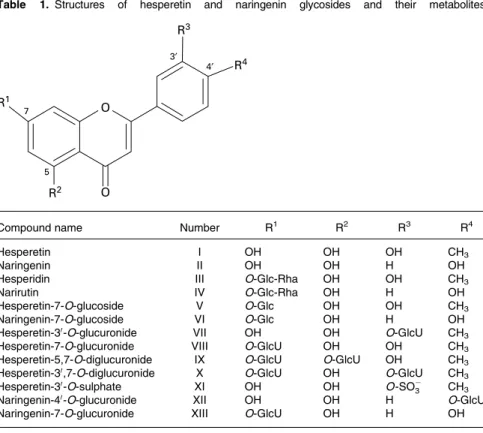
Flavanones, hesperetin and naringenin, are abundant in citrus fruits and constitute a major part of the total flavonoid intake in many European countries(1,2). Studies on hesperetin and naringenin have shown several health-protective properties such as antioxidant and anti-inflammatory action and prevention of bone loss in vivo(3 – 7). Hesperetin and naringenin are naturally present in citrus fruits as their respective 7-O-rutinosides, hesperidin and narirutin(8)(Table 1). By enzymatic treatment with a-rhamnosidase, the rutinoside moiety (rhamnose-glucose) can be hydrolysed to yield hesperetin-7-O-glucoside and naringenin-hesperetin-7-O-glucoside, respectively(9). The sugar moiety is a major determinant of the absorption site and bioavailability of the flavonoid since flavonoid monoglucoside bioavailability is several fold higher than flavonoid rutinosides(9 – 11). Flavonoid monoglucosides are absorbed in the small intestine after hydrolysis by lactase phloridzin hydrolase(12,13) or cytosolic b-glucosidases(14,15), whereas flavonoid rutinosides must reach the microflora
in the colon before they can be hydrolysed and absorbed. Both small intestine and colonic epithelium can metabolise flavonoids via phase 2 conjugation by addition of glucuronic acid or sulphate groups(16) However, the possibly different roles of these two tissues are not clear for flavonoid conju-gation, although it has been recently reported for phenolic acids(17). The endogenously produced metabolites are at least in part responsible for the systemic effects of flavo-noids, and knowledge about the nature of the metabolites produced is therefore crucial for further studies on putative health effects. The removal of the rhamnose sugar gives an ideal system for studying the effect of small intestine and colon absorption on the metabolic pathways. We demon-strated that the bioavailability of hesperetin was increased when hesperidin in orange juice was enzymatically trans-formed into hesperetin-7-O-glucoside(9). Here, we further investigate if this enzymatic treatment, as a tool to investigate the absorption mechanism of hesperidin, could also affect the
* Corresponding author: Gary Williamson, fax þ 44 113 343 2982, email g.williamson@leeds.ac.uk Abbreviations:AUC, area under the curve; DAD, diode array detector.
bioavailability of naringenin and identify the major hesperetin and naringenin metabolites excreted in urine after absorption targeted either to the small intestine or colon.
Materials and methods Study design
The study was conducted as previously described(9). Sixteen healthy volunteers (eight men and eight women) completed a double blind, placebo controlled, randomised, three-treatment crossover study. After an overnight fast, the subjects were randomly assigned to one of the following three treatments of orange juice: (1) orange juice naturally containing 2 and 0·83 mg/kg body weight hesperidin and narirutin, respectively; (2) a-rhamnosidase-treated orange juice providing 1·52 and 0·52 mg/kg body weight hesperetin-7-O-glucoside and naringenin-hesperetin-7-O-glucoside, respectively; (3) hesperidin-fortified orange juice providing 6 mg/kg body weight total hesperidin, for metabolite identification (all values are based on the dose of aglycone equivalents). Samples of blood (4·5 ml) were collected at 0·5, 1, 1·5, 2, 3, 4, 5, 6, 7, 8, 9 and 10 h after consumption. Urine was collected just before treatment and in three different fractions: 0 – 5; 5 – 10; 10 – 24 h. Treatments were separated by no less than a 3-d washout period. The present study was conducted according to the guidelines laid down in the Declaration of Helsinki and all procedures involving human subjects/patients were approved by the Ethical Committee of Nestle´ (Lausanne, Switzerland; protocol 03.15MET). Written consent was obtained from all subjects.
Chemicals
Orange juice was obtained from Nestle´ (Bangkok, Thailand). Hesperidinase (67 000 units/g) was from Amano Enzyme Co., Nagoya, Japan. The isotopically labelled internal standards, 13C3 daidzein and 13C3 O-desmethylangolensin, were purchased from the School of Chemistry, University of St Andrews, St Andrews, UK. The naringenin-7-O-glucoside
and narirutin standards were both purchased from
Extrasynthese, Genay, France. Purity of the compounds was determined to be 88 – 90 % using weight and spectrophotometry. The flavanone standards, hesperetin and naringenin, were obtained from Sigma Chemicals Co., St Louis, MO, USA. The enzymes used for enzymatic hydrolysis of meta-bolites for calibration curves were b-glucuronidase expressed in Escherichia coli (0·14 mmol/min per mg; Roche Diagnostics, Mannheim, Germany) and Aerobacter aerogenes sulphatase (0·0025 mmol/min per mg protein; Sigma-Aldrich). All other reagents and solvents were HPLC grade.
Naringenin analysis in orange juice, plasma and urine Naringenin-7-O-glucoside and narirutin were extracted from orange juice essentially as described previously(18)but without hydrolysis. Aliquots of 5 ml juice were centrifuged (3000 rpm, 10 min) and applied to Bond Elute columns (2 g; Varian, Middelburg, The Netherlands). The pellet was subsequently extracted twice with 5 ml 1 % aqueous formic acid and three times with 5 ml methanol. The total volume after elution with methanol was 30 ml. Aliquots of 200 ml were evaporated, reconstituted (0·5 % aqueous acetic acid, 10 % acetonitrile) Table 1. Structures of hesperetin and naringenin glycosides and their metabolites
3′ 4′ 5 7 O O R3 R4 R1 R2
Compound name Number R1 R2 R3 R4
Hesperetin I OH OH OH CH3
Naringenin II OH OH H OH
Hesperidin III O-Glc-Rha OH OH CH3
Narirutin IV O-Glc-Rha OH H OH
Hesperetin-7-O-glucoside V O-Glc OH OH CH3
Naringenin-7-O-glucoside VI O-Glc OH H OH
Hesperetin-30-O-glucuronide VII OH OH O-GlcU CH
3
Hesperetin-7-O-glucuronide VIII O-GlcU OH OH CH3
Hesperetin-5,7-O-diglucuronide IX O-GlcU O-GlcU OH CH3
Hesperetin-30,7-O-diglucuronide X O-GlcU OH O-GlcU CH
3
Hesperetin-30-O-sulphate XI OH OH O-SO2
3 CH3
Naringenin-40-O-glucuronide XII OH OH H O-GlcU
Naringenin-7-O-glucuronide XIII O-GlcU OH H OH
and analysed on the HPLC – UV – MS system with diode array detector (DAD) as described previously(19). The method of analysis was as reported(9) with minor modifications. Eluents: A, 0·5 % aqueous acetic acid, 10 mM-ammonium acetate (v/v); B, acetonitrile. Negative electrospray ionisation mass spectra were obtained in selected ion monitoring mode. Conditions for electrospray ionisation: gas temperature, 3508C; drying gas, 12 litres/min; nebuliser pressure, 414 kpa; capillary voltage (negative), 3500 V. Calibration curves for naringenin-7-O-glucoside and narirutin were obtained by spiking orange juice (1) (natural content of hesperetin and naringenin rutinosides) with 3·5, 7, 14 and 21 mM
-naringenin-7-O-glucoside standard and (2) a-rhamnosidase-treated orange juice with 10, 20, 30 and 40 mM-narirutin
as standard, respectively. The spiked juice samples were extracted and analysed exactly the same way as the samples. Analysis of hesperetin in orange juice and the concentration of naringenin aglycone in urine and plasma were determined as previously described for hesperetin(9).
Identification of hesperetin and naringenin metabolites in urine
Initially, the identity of metabolites was established on the basis of their MS spectra. Where possible, the exact sites of conjugation were subsequently determined by 1H NMR data after isolation (by collecting fractions directly from the HPLC), differential spectrophotometry and by comparison of their HPLC retention times with authentic synthetic standards. The chemical synthesis of hesperetin conjugates will be published elsewhere.
NMR spectroscopy. NMR spectra were acquired on
a Bruker AV 400 WB spectrometer (Bruker, Rheinstetten, Germany) operating at 400·13 MHz for 1H. A 1 mm triple-resonance inverse probe was used (1H observe, 13C, 77Se decouple). Samples were dissolved min 10 ml d6-dimethyl sulphoxide and transferred to 1 mm NMR tubes.
Typical parameters: 32 768 points were acquired in the time domain with a sweep width of 6000 Hz. The 908 pulse was 5·7 ms. For suppression of the signal from residual water from the freeze-drying process and from d6-dimethyl sulph-oxide, a 1D-noesypresat (Bruker pulse programme library) pulse sequence was used with a pre-saturation time of 1 s, mixing time of 0·3 s and decoupler power level of 70 dB. Receiver gain and number of acquired transients were adjusted according to sample amount and amount of residual formate (from the HPLC mobile phase). Typically, thirty-two transi-ents were acquired for standards and between 2048 and 32 900 for samples.
Spectra were zero-filled to 131 072 complex data points and processed with an exponential multiplication corres-ponding to an increase in line width of 1 Hz. Spectra were interpreted by comparison with authentic non-metabolised standards.
Identification of hesperetin-30-O-sulphate. The
hesperetin-sulphate in urine samples did not respond well to
enzymatic hydrolysis with sulphatase and it was not possi-ble to get a clear spectrum with NMR analysis. Instead, its identity as a hesperetin monosulphate was confirmed by high-resolution LC – quadrupole time-of-flight (Bruker Daltonics, Bremen, Germany) analysis. The monoisotopic
mass of hesperetin-sulphate (382·0359 Da) was detected with a mean error of 1·4 parts per million, and information on the position of conjugation was determined indirectly by differen-tial pH spectrophotometry(20). Urine samples were analysed on HPLC – DAD as described earlier. However, to obtain pH 6 and 8, a constant delivery of 0·055 and 0·1 ml/min 1M-Tris base, respectively, was applied to the gradient post-column. UV detection was carried out at 280 and 330 nm, with peak scanning between 210 and 600 nm (2 nm steps).
Analysis and quantification of hesperetin and naringenin metabolites in urine
Urine samples were filtered through a 0·2 mm filter and portioned into aliquots of 200 ml. An internal standard mix (10 ml, 13C3 daidzein and 13C3 O-desmethylangolensin, 25 ng/ml dimethyl sulphoxide) was added before adjusting to approximately pH 3 with 10 % aqueous acetic acid. A total volume of 220 ml was analysed on HPLC – DAD – MS. UV detection was carried out at 280 nm with peak scanning between 210 and 600 nm (2 nm steps). UV quantification was preferred due to tendencies to ion suppression in MS, and metabolites were quantified on the basis of their UV absorption unless otherwise stated.
Hesperetin-7-O-glucuronide and hesperetin-30 -O-glucuro-nide were quantified with calibration curves of their respective authentic synthetic standards (chemical synthesis to be reported elsewhere). For hesperetin-sulphate-glucuronide estimation, urine samples were evaporated and reconstituted in solvent (pure water adjusted to pH 5 with acetic acid). Calibration curves were made by diluting 1:1 in solvent four times in duplicate, resulting in two calibration curves with four concentrations (100, 50, 25 and 12·5 %). To the samples in one calibration curve were added 3 ml b-glucuronidase, 20 ml sulphatase and 5 ml internal standard (20 ng 13C daidzein/ml dimethyl sulphoxide), and to the samples in the other calibration curve were added 23 ml solvent and 5 ml internal standard. All samples were left at 378C for 1 h. Before analyses on HPLC – DAD – MS, all samples were added to 25 ml 0·5 % acetic acid and 25 ml methanol and centrifuged (10 000 rpm, 5 min). Hesperetin aglycone calibration curves were analysed along with the hydrolysed and unhydrolysed calibration curves for estimation of hesperetin-sulphate-glucuronide.
Calibration curves for naringenin-7- and -40-O-glucuronide were obtained similar to hesperetin-sulphate-glucuronide as described earlier, except no sulphatase was added, and naringenin aglycone calibration curves were analysed along with the hydrolysed and unhydrolysed calibration curves. Due to co-eluting compounds, naringenin-7- and -40-O-glucuronide could not be quantified on the basis of UV data but were quantified from MS data.
For analysis of hesperetin-30-O-sulphate, urine was evapor-ated and reconstituted in 0·1M-HCl. Calibration curves were
then made by diluting 1:1 five times in duplicate. For one calibration curve, 80 ml of sodium acetate (2M) was added
resulting in a pH of about 5 and a total volume of 130 ml and left at room temperature for 1 h. For the other calibration curve (total volume 50 ml), samples were heated to 958C for 1 h. Before HPLC – DAD – MS analysis, the heated samples were added to 10 ml of acetonitrile and 40 ml of pure water,
while the samples left at room temperature were added to 20 ml of acetonitrile.
Hesperetin aglycone calibration curves were analysed along with the hydrolysed and unhydrolysed calibration curves. Correlation coefficients of all calibration curves were . 0·99. Statistics
The incremental area under the curve (AUC) was determined by a mixed log-linear trapezoidal model, where the linear rule (trapezoidal) is applied to the range where concentration is ascending and the log-linear rule is applied to the range where concentration is descending. Combination of the two trapezoidal methods enhances the estimation of the AUC in reducing errors inherent to both methods by applying them under the conditions of best estimate. Values below the limit of quantification were set to limit of quantification (plasma naringenin limit of quantification ¼ 0·0037 mmol/l). To compare groups 1 and 2, no adjustment for multiplicity was used and all tests were performed at a level 5 %. AUC, Cmax and Tmax were calculated using Kinetica (Innaphase, Philadelphia, PA, USA); all other statistical analyses were done with SAS software (version 8.2; SAS Institute, Inc., Cary, NC, USA). From 1 week before the intake of the first treatment, subjects were randomly allocated to one of the three different sequences from the Latin square: [A] – [B] – [C]; [B] – [C] – [A]; [C] – [A] – [B]. The randomisation was done on age and with BMI as the balancing factor.
Results
Plasma bioavailability of total naringenin as aglycone (area under the curve)
The AUC for total plasma naringenin (Fig. 1) after subjects consumed the a-rhamnosidase-treated orange juice (2) was about 4-fold higher compared with consumption of the untreated orange juice (1) and Cmax was 5·4-fold higher (Table 2).
Intake and urinary excretion of naringenin (as aglycone equivalents)
The total naringenin excretion over 24 h was calculated by pooling the three fractions of urine collected, and the data
were expressed as a percentage of naringenin intake. The relative urinary excretion of total naringenin of the subjects was significantly higher (6·7-fold) after consuming a-rhamnosidase-treated orange juice (2) than after consump-tion of untreated orange juice (1) (Table 2).
Hesperetin and naringenin metabolites excreted in urine The consumption of orange juice containing the natural untreated dose of hesperidin and narirutin (1), hesperetin-7-O-glucoside and naringenin-hesperetin-7-O-glucoside (2) or high dose of hesperidin (3) resulted in the detection of a total of six urinary metabolites of hesperetin and two metabolites of naringenin by HPLC – MS (Fig. 2). NMR analysis allowed the structural elucidation of four of the six hesperetin and two naringenin metabolites in human urine (Table 3). The identified hesperetin metabolites were: hesperetin-30-O-glucuronide; hesperetin-7-O-glucuronide; hesperetin-5, 7-O-diglucuronide; hesperetin-30,7-O-diglucuronide. The sub-stitution position of hesperetin-30-O-sulphate was identified by differential pH spectrophotometry. In addition, a hesperetin-sulphate-glucuronide was detected, but it was not possible to specify the exact substitution positions. Hesperetin-sulphate-glucuronide was identified by MS fragmentation patterns and by enzymatic hydrolysis with sulphatase and glucuronidase which required both enzymes to generate the aglycone. Hesperetin-7- and -30-O-glucuronides were con-firmed by comparison with synthetic standards. Hesperetin-5,7-O-diglucuronide and hesperetin-7,30-O-diglucuronide were not quantified due to insufficient amounts. The limit of quantification of these diglucuronides could not be accurately determined, since no standards are available. The naringenin metabolites were naringenin-7-O- and -40-O-glucuronide. Hesperetin and naringenin metabolites were quantified in urine collected at the peak excretion time, which was 0 – 5 h for the group consuming a-rhamnosidase-treated juice (2) and 5 – 10 h for (1) and (3) (Fig. 3).
Discussion
We show for the first time that naringenin-7-O-glucoside is more bioavailable than narirutin (naringenin-7-O-rutinoside) in human subjects. This is as judged by the total naringenin AUC, Cmaxand urinary excretion data that were significantly higher for naringenin-7-O-glucoside compared with the natural untreated orange juice containing narirutin. This is in accordance with hesperetin(9) and quercetin(10). Others have studied the bioavailability of naringenin in rats(21)where the urinary naringenin recovery as percentage of dose was 28, 31, and 14 % after aglycone, glucoside or rhamnoglucoside (naringin) intake, respectively. The urinary naringenin recovery as percentage of dose in the present study was 7 and 47 % after narirutin and naringenin-7-O-glucoside intake, respectively. When naringenin was given as an aglycone to human volunteers, the urinary recoveries for naringenin were approximately 6 % of the administered dose, i.e. there was no increase in bioavailability (as observed in rats)(22). It seems therefore that in human subjects naringenin bioavailability is highest when consumed as a monoglucoside. Higher bioavailability of monoglucosides might provide health protective effects in lower doses since
0 0 0·2 0·4 Plasma naringenin ( µ mol/l) 0·6 0·8 1·0 100 200 300 Time (min) 400 500 600
Fig. 1. Plasma concentration v. time curve of total naringenin in healthy human subjects after consumption of two orange juice treatments. O, Natural juice (1); B, a-rhamnosidase-treated juice (2). Values are means with their standard errors, n 16 (1), n 15 (2).
increased bioavailability of hesperetin-7-O-glucoside resulted in the same level of protection against bone loss as compared to higher levels of hesperidin (hesperetin-7-O-rutinoside) in rats (0·25 and 0·5 % of the diet, respectively)(23).
Other studies on bioavailability of naringenin from orange juice have reported values for relative urinary excretion from 1 to 18 % of narirutin intake(11,24,25); the present study on untreated orange juice for naringenin is in the middle of this range. The biochemical pathway accounting for the increased bioavailability of naringenin-7-O-glucoside has been described in several papers. The absorption of flavonoid glucosides in the small intestine is facilitated by the presence of lactase phloridzin hydrolase in the gut lumen brush border and b-glucosidase in the intestinal cells(12,13,26). Both enzymes are capable of hydrolysing flavonoid glucosides but not rutinosides, which are only absorbed after hydrolysis by the colonic microflora.
We have found six hesperetin and two naringenin metabolites in human urine after consumption of (1) natural untreated orange juice; (2) a-rhamnosidase-treated orange juice; (3) orange juice fortified with three times the original content of hesperetin-7-O-rutinoside. We have identified the exact site of conjugation and quantified several hesperetin and both naringenin metabolites. Two recent studies have identified hesperetin and naringenin metabolites in human urine after consumption of orange juice(25,27). Both have found hesperetin-7-O-glucuronide, naringenin-7- and -40 -O-glucuronide. Brett and co-workers have identified hesperetin-30-O-glucuronide and hesperetin-30-O-sulphate as well. In addition to these metabolites, we have further identified hesperetin-5,7-O-diglucuronide and hesperetin-30 ,7-O-diglu-curonide. All quantification of metabolites was based on the corresponding metabolite either as synthetic standard or collected directly from urine with high metabolite concen-tration. This excludes a potential source of error from quantitative estimates based on non-identical metabolites.
In the present study, glucuronides of hesperetin were the most abundant metabolites recovered. This result is in agreement with several other studies(11,25,28,29). We have also found a substantial amount of hesperetin-30-O-sulphate excretion after all treatments. It has been suggested that flavonol sulphate-glucuronides are glucuronidated before sulphation, and the sulphation then occurs in the liver(30,31). In contrast, Jaganath et al.(32)did not find evidence of sulpha-tion of quercetin after intervensulpha-tion with tomato juice containing rutin (quercetin-3-O-rutinoside which is absorbed
in the colon), whereas quercetin-30-O-sulphate is one of the dominating metabolites in plasma from human volunteers fed fried onions (containing quercetin-40-O-glucoside and quercetin-3,40-O-diglucoside which are absorbed in the small intestine)(30,33). In the present study, the amount of hespere-tin-30-O-sulphate in the urine was not affected by the site of absorption indicating that either hesperetin can be sulphated equally by both small intestinal and colonic sulphotransferases or hesperetin sulphation predominantly occurs in the liver after absorption. It has already been shown that liver cells can deconjugate certain quercetin glucuronides and then subsequently sulphate them(31).
The phase 2 enzymes, UDP glucuronosyl transferase and sulphotransferase, catalyse the conjugation of polyphenols with glucuronic acid and sulphate groups, respectively(16,34,35). Both enzymes exist as numerous isoforms and are present in several tissues including the small intestine, colon and liver(36,37). Expression of both enzyme classes shows common characteristics in the human alimentary tract; however, the pattern of the forms expressed differs between various segments and opens up the possibility that different metabolites can be formed according to where in the alimen-tary tract the aglycone is absorbed(36,37). The present study provides no information on the mechanisms involved in con-jugation of hesperetin and naringenin; however, it is evident that following the release of aglycone, hesperetin and narin-genin are subjected to glucuronidation and/or sulphation, and Table 2. Pharmacokinetic measurements for total naringenin in healthy human subjects after the consumption of two orange juice treatments (untreated (1) and a-rhamnosidase-treated (2))
(Mean values and standard deviations)
Untreated orange juice a-Rhamnosidase-treated orange juice
P value
Mean SD Mean SD
n 16 15
Ingested dose (mg/kg body weight) 0·83 0·52
AUC (0 – 600 min, mmol/l £ min) 18 12 70 29 ,0·0001
Cmax(mmol/l) 0·12 0·14 0·77 0·35 ,0·0001
Tmax(min) 311 183 93 166 0·002
Relative urinary excretion (% intake) 7 3 47 17 ,0·0001
AUC, area under the curve; Cmax, peak plasma concentration; Tmax, time to maximum plasma concentration.
X IX XIV XIII VIII VII XI IS1 IS2 XII R elati ve intensity 20 10 30
Retention time (min) 40
Fig. 2. Representative chromatogram of hesperetin and naringenin metabolites in human urine 0 – 5 h after ingestion of a-rhamnosidase-treated orange juice (2). (X) Hesperetin-30,7-O-diglucuronide, (IX) hesperetin-5, 7-O-diglucuronide, (XIV) hesperetin-sulphate-glucuronide, (XIII) naringenin-7-O-glucuronide, (XII) naringenin 40-O-glucuronide, (VIII) hesperetin 7-O-glucuronide, (VII) hesperetin 30-O-glucuronide, (XI) hesperetin 30-O-sulphate, (IS1) internal standard 13C
3 daidzein, (IS2) internal standard 13C3
the pattern of excreted metabolites is not affected by the site (small intestine or colon) of absorption. Furthermore, increasing the dose of hesperidin 3-fold only affected the total concentration but did not have any impact on the profile of the conjugates formed after colonic absorption. Colonic microflora produce lower molecular weight phenolics
that can be absorbed, such as dihydrocinnamic, phenylacetic and benzoic acids. However, these have not been characterised in the present study.
It is generally agreed that conjugate position is important in relation to the biological activity of flavonoids in human subjects, but only a few studies have looked at the biological activities of the conjugates(38). Hesperetin glucu-ronides, but not the hesperetin aglycone, protected against UV-A-induced necrotic cell death(39). Quercetin glucuronides inhibited Cu2þ-induced oxidation of human LDL(40). Xanthine oxidase was inhibited by quercetin conjugates, and the inhibition varied 50 – 800-fold depending on the site of conjugation(41). Hesperetin aglycone protected against oxidative injury to primary cortical neurons whereas hesperetin-7-O-glucuronide did not(42), probably related to the inability of glucuronide metabolites to passively traverse membranes, unless a transporter is present(31). In general, the responses to flavonoid conjugates are weaker than those to the aglycones(38).
In conclusion, we have shown that the bioavailability of naringenin-7-O-glucoside is increased about 4-fold based on AUC compared to narirutin in healthy human subjects consuming orange juice. There was no significant difference between the pattern of metabolites excreted in urine after consumption of orange juice containing either glucosides or rutinosides, demonstrating that metabolism of hesperetin and naringenin after absorption in the small intestine is not significantly different from the metabolism after absorption in colon. In addition, it was shown for hesperidin that a 3-fold increase in dose does not affect the pattern of excreted metabolites. The eight identified metabolites were all phase 2 conjugates, of which seven were structurally elucidated.
Acknowledgements
We would like to thank all the volunteers of the study. We also wish to thank laboratory technician Anni Schou for skilful technical assistance, senior scientist Henrik Frandsen for valuable scientific assistance and Nestle´ Research
60 (a) (b) 50 40 Urine concentration ( µ mol/l) 30 20 10 0
Natural juice (1) α-Rhamnosidase-treated juice (2) Hesperidin-fortified juice (3) Urine concentration ( µ mol/l) 0 0·5 1·0 1·5 2·0 2·5
Natural juice (1) α-Rhamnosidase-treated juice (2)
Hesperidin-fortified juice (3)
Fig. 3. Metabolite excretion in urine as percentage of intake. (a) Hesperetin metabolites. , Hesperetin-7-O-glucuronide; , hesperetin-30-O-glucuronide; A, hesperetin-sulphate-glucuronide; , hesperetin-30-O-sulphate. (b) Narin-genin metabolites. A, NarinNarin-genin-40-O-glucuronide; , naringenin-7-O-glucuronide. Metabolites were measured in urine collected at the peak excretion time, which was 0 – 5 h for the a-rhamnosidase-treated juice (2) and 5 – 10 h for the natural (1) and the fortified juice (3). Means with their standard errors, n 13 (2 and 3), n 15 (1).
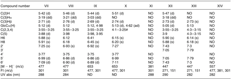
Table 3.1H NMR, LC – MS and UV data of hesperetin and naringenin metabolites excreted in human urine after consumption of three different kinds of orange juice (see Table 1 and Fig. 2)
Compound number VII VIII IX X XI XII XIII XIV
C(2)H 5·42 (d) 5·46 (d) 5·44 (d) 5·51 (d) NO 5·47 (d) NO NO C(3)Ha 3·19 (dd) 3·21 (dd) 3·03 (dd) NO NO 3·18 (dd) NO NO C(3)Hb 2·71 (d) 2·76 (d) 2·69 (d) 2·74 (d) NO 2·73 (d) 2·73 (s) NO GlcCa(H) 5·12 (d) 5·12 5·12, 4·98 5·13 (d), 4·62 (ad) NO 5·05 (d) 5·13 NO C(2,3,4) 3·55 – 3·25 3·55 – 3·25 3·55 – 3·25 4·1 – 3·25# NO 3·55 – 3·25 4·3 – 3·15 NO C(5) 3·88 (d) 3·98 3·98, 3·95 NO 3·9 4·3 – 3·15 NO H6 5·88 (s) 6·12 6·41 6·15 (s) NO 5·90 (s) 6·14 (s) NO H8 5·91 (s) 6·18 6·45 (s) 6·20 (s) NO 5·88 (s) 6·18 (s) NO 20 7·25 (s) 6·93 (s) 6·92 (s) 7·27 NO 7·43 7·3 NO 30 7·05 7·79 40 3·77 3·75 3·75 3·77 NO NO 50 6·99 (d) 6·86 (d) 6·86 (d) 6·99 NO 7·05 7·79 NO 60 7·09 (d) 6·90 (d) 6·89 (d) 7·11 NO 7·43 7·3 NO [M 2 H]2(m/z) 477 477 653 653 381 447 447 557 FI (m/z) 301 301 477, 301 477, 301 301 271, 151 271, 151 477, 381, 301 UV abs (nm) 288 284 ND ND 288 290 282 282
Center, Lausanne, Switzerland, for funding the present study. L. B., S. E. R. and C. C. have no conflicts of interest. I. L. F. N., D. B., F. B., E. O.-C. and G. W. are, or were, full- or part-time employees of the Nestle´ Research Center, Lausanne, Switzerland. I. L. F. N., E. O.-C. and G. W. planned the present study. I. L. F. N. carried out the present study. D. B. provided and characterised hesperetin standards used in the present study. L. B. and S. E. R. analysed the samples, interpreted the data and wrote the first draft of the present paper. F. B. carried out the statistical analysis. C. C. carried out and interpreted the NMR data. G. W., L. B. and I. L. F. N. wrote the manuscript.
References
1. Knekt P, Kumpulainen J, Jarvinen R, et al. (2002) Flavonoid intake and risk of chronic diseases. Am J Clin Nutr 76, 560 – 568.
2. Justesen U, Knuthsen P & Leth T (1997) Determination of plant polyphenols in Danish foodstuffs by HPLC – UV and LC – MS detection. Cancer Lett 114, 165 – 167.
3. Garg A, Garg S, Zaneveld LJ, et al. (2001) Chemistry and pharmacology of the citrus bioflavonoid hesperidin. Phytother Res 15, 655 – 669.
4. Chiba H, Uehara M, Wu J, et al. (2003) Hesperidin, a citrus flavonoid, inhibits bone loss and decreases serum and hepatic lipids in ovariectomized mice. J Nutr 133, 1892 – 1897. 5. Aranganathan S, Selvam JP & Nalini N (2008) Effect of
hesperetin, a citrus flavonoid, on bacterial enzymes and carci-nogen-induced aberrant crypt foci in colon cancer rats: a dose-dependent study. J Pharm Pharmacol 60, 1385 – 1392. 6. Benavente-Garcia O & Castillo J (2008) Update on uses and
properties of citrus flavonoids: new findings in anticancer, cardiovascular, and anti-inflammatory activity. J Agric Food Chem 56, 6185 – 6205.
7. Kim JY, Jung KJ, Choi JS, et al. (2006) Modulation of the age-related nuclear factor-kappaB (NF-kappaB) pathway by hesperetin. Aging Cell 5, 401 – 411.
8. Tomas-Barberen FA & Clifford MN (2000) Flavanones, chalcones and dihydrochalcones – nature, occurrence and dietary burden. J Sci Food Agric 80, 1073 – 1080.
9. Nielsen IL, Chee WS, Poulsen L, et al. (2006) Bioavailability is improved by enzymatic modification of the citrus flavonoid hesperidin in humans: a randomized, double-blind, crossover trial. J Nutr 136, 404 – 408.
10. Hollman PC, Bijsman MN, van Gameren Y, et al. (1999) The sugar moiety is a major determinant of the absorption of dietary flavonoid glycosides in man. Free Radic Res 31, 569 – 573. 11. Manach C, Morand C, Gil-Izquierdo A, et al. (2003)
Bioavail-ability in humans of the flavanones hesperidin and narirutin after the ingestion of two doses of orange juice. Eur J Clin Nutr 57, 235 – 242.
12. Day AJ, Canada FJ, Diaz JC, et al. (2000) Dietary flavonoid and isoflavone glycosides are hydrolysed by the lactase site of lactase phlorizin hydrolase. FEBS Lett 468, 166 – 170. 13. Sesink AL, Arts IC, Faassen-Peters M, et al. (2003) Intestinal
uptake of quercetin-3-glucoside in rats involves hydrolysis by lactase phlorizin hydrolase. J Nutr 133, 773 – 776.
14. Day AJ, DuPont MS, Ridley S, et al. (1998) Deglycosylation of flavonoid and isoflavonoid glycosides by human small intestine and liver b-glucosidase activity. FEBS Lett 436, 71 – 75.
15. Nemeth K, Plumb GW, Berrin JG, et al. (2003) Deglycosylation by small intestinal epithelial cell beta-glucosidases is a critical step in the absorption and metabolism of dietary flavonoid glycosides in humans. Eur J Nutr 42, 29 – 42.
16. Brand W, van der Wel PA, Rein MJ, et al. (2008) Metabolism and transport of the citrus flavonoid hesperetin in Caco-2 cell monolayers. Drug Metab Dispos 36, 1794 – 1802.
17. Poquet L, Clifford MN & Williamson G (2008) Transport and metabolism of ferulic acid through the colonic epithelium. Drug Metab Dispos 36, 190 – 197.
18. Breinholt VM, Svendsen GW, Dragsted LO, et al. (2004) The citrus-derived flavonoid naringenin exerts uterotrophic effects in female mice at human relevant doses. Basic Clin Pharmacol Toxicol 94, 30 – 36.
19. Nielsen SE & Sandstrom B (2003) Simultaneous determination of hydroxycinnamates and catechins in human urine samples by column switching liquid chromatography coupled to atmospheric pressure chemical ionization mass spectrometry. J Chromatogr B Analyt Technol Biomed Life Sci 787, 369 – 379.
20. Xie MX, Xu XY & Wang YD (2005) Interaction between hesperetin and human serum albumin revealed by spectroscopic methods. Biochim Biophys Acta 1724, 215 – 224.
21. Felgines C, Texier O, Morand C, et al. (2000) Bioavailability of the flavanone naringenin and its glycosides in rats. Am J Physiol Gastrointest Liver Physiol 279, G1148 – G1154.
22. Kanaze FI, Bounartzi MI, Georgarakis M, et al. (2007) Pharma-cokinetics of the citrus flavanone aglycones hesperetin and naringenin after single oral administration in human subjects. Eur J Clin Nutr 61, 472 – 477.
23. Habauzit V, Nielsen IL, Gil-Izquierdo A, et al. (2009) Increased bioavailability of hesperetin-7-glucoside compared with hesperidin results in more efficient prevention of bone loss in adult ovariectomised rats. Br J Nutr 102, 976 – 984.
24. Erlund I, Meririnne E, Alfthan G, et al. (2001) Plasma kinetics and urinary excretion of the flavanones naringenin and hesperetin in humans after ingestion of orange juice and grapefruit juice. J Nutr 131, 235 – 241.
25. Mullen W, Archeveque MA, Edwards CA, et al. (2008) Bioavailability and metabolism of orange juice flavanones in humans: impact of a full-fat yogurt. J Agric Food Chem 56, 11157 – 11164.
26. Day AJ, Gee JM, DuPont MS, et al. (2003) Absorption of quercetin-3-glucoside and quercetin-40-glucoside in the rat small intestine: the role of lactase phlorizin hydrolase and the sodium-dependent glucose transporter. Biochem Pharmacol 65, 1199 – 1206.
27. Brett GM, Hollands W, Needs PW, et al. (2008) Absorption, metabolism and excretion of flavanones from single portions of orange fruit and juice and effects of anthropometric variables and contraceptive pill use on flavanone excretion. Br J Nutr 101, 1 – 12.
28. Matsumoto H, Ikoma Y, Sugiura M, et al. (2004) Identification and quantification of the conjugated metabolites derived from orally administered hesperidin in rat plasma. J Agric Food Chem 52, 6653 – 6659.
29. Yamada M, Tanabe F, Arai N, et al. (2006) Bioavailability of glucosyl hesperidin in rats. Biosci Biotechnol Biochem 70, 1386 – 1394.
30. Mullen W, Edwards CA & Crozier A (2006) Absorption, excretion and metabolite profiling of methyl-, glucuronyl-, glucosyl- and sulpho-conjugates of quercetin in human plasma and urine after ingestion of onions. Br J Nutr 96, 107 – 116.
31. O’Leary KA, Day AJ, Needs PW, et al. (2003) Metabolism of quercetin-7- and quercetin-3-glucuronides by an in vitro hepatic model: the role of human b-glucuronidase, sulfotransferase, catechol-O-methyltransferase and multi-resistant protein 2 (MRP2) in flavonoid metabolism. Biochem Pharmacol 65, 479 – 491.
32. Jaganath IB, Mullen W, Edwards CA, et al. (2006) The relative contribution of the small and large intestine to the absorption and metabolism of rutin in man. Free Radic Res 40, 1035 – 1046. 33. Day AJ, Mellon FA, Barron D, et al. (2001) Human metabolism of dietary flavonoids: identification of plasma metabolites of quercetin. Free Radic Res 212, 941 – 952.
34. Silberberg M, Morand C, Mathevon T, et al. (2006) The bioavailability of polyphenols is highly governed by the capacity of the intestine and of the liver to secrete conjugated metabolites. Eur J Nutr 45, 88 – 96.
35. Scalbert A & Williamson G (2000) Dietary intake and bioavailability of polyphenols. J Nutr 130, 2073S – 2085S. 36. Strassburg CP, Nguyen N, Manns MP, et al. (1999)
UDP-glucuronosyltransferase activity in human liver and colon. Gastroenterology 116, 149 – 160.
37. Glatt H, Engelke CE, Pabel U, et al. (2000) Sulfotransferases: genetics and role in toxicology. Toxicol Lett 112–113, 341–348.
38. Williamson G, Barron D, Shimoi K, et al. (2005) In vitro biological properties of flavonoid conjugates found in vivo. Free Radic Res 39, 457 – 469.
39. Proteggente AR, Basu-Modak S, Kuhnle G, et al. (2003) Hesperetin glucuronide, a photoprotective agent arising from flavonoid metabolism in human skin fibroblasts. Photochem Photobiol 78, 256 – 261.
40. Manach C, Morand C, Crespy V, et al. (1998) Quercetin is recovered in human plasma as conjugated derivatives which retain antioxidant properties. FEBS Lett 426, 331 – 336. 41. Day AJ, Bao Y, Morgan MRA, et al. (2000) Conjugation
position of quercetin glucuronides and effect on biological activity. Free Rad Biol Med 29, 1234 – 1243.
42. Vauzour D, Vafeiadou K, Rice-Evans C, et al. (2007) Activation of pro-survival Akt and ERK1/2 signalling pathways underlie the anti-apoptotic effects of flavanones in cortical neurons. J Neurochem 103, 1355 – 1367.