HAL Id: hal-02966846
https://hal.sorbonne-universite.fr/hal-02966846
Submitted on 17 Nov 2020HAL is a multi-disciplinary open access archive for the deposit and dissemination of sci-entific research documents, whether they are pub-lished or not. The documents may come from teaching and research institutions in France or abroad, or from public or private research centers.
L’archive ouverte pluridisciplinaire HAL, est destinée au dépôt et à la diffusion de documents scientifiques de niveau recherche, publiés ou non, émanant des établissements d’enseignement et de recherche français ou étrangers, des laboratoires publics ou privés.
An exploratory study of heterotrophic protists of the
mesopelagic Mediterranean Sea
John Dolan, Maria Ciobanu, Sophie Marro, Laurent Coppola
To cite this version:
John Dolan, Maria Ciobanu, Sophie Marro, Laurent Coppola. An exploratory study of heterotrophic protists of the mesopelagic Mediterranean Sea. ICES Journal of Marine Science, Oxford University Press (OUP), 2019, 76 (3), pp.616-625. �10.1093/icesjms/fsx218�. �hal-02966846�
ICES Journal of Marine Science, accepted. Special Issue: Mesopelagic Resources
An exploratory study of heterotrophic protists of the mesopelagic
Mediterranean Sea
John R. Dolan*, Maria Ciobanu, Sophie Marro, and Laurent Coppola Sorbonne Universités, UPMC Université Paris VI, CNRS UMR 7093, Laboratoire
d'Océanographie de Villefranche-‐sur-‐Mer, Station Zoologique, 06230 Villefranche-‐sur-‐ Mer, France
* corresponding author : dolan@obs-‐vlfr.fr Abstract
Is there a mesopelagic protist fauna composed of species different from that of the overlying surface community? Does the mesopelagic community show seasonal changes in abundances and species composition? We addressed these questions by considering 3 distinct groups in which species identification is relatively unambiguous: tintinnid ciliates, phaeodarian radiolarians, and amphisolenid dinoflagellates. We sampled weekly at 250 m and 30 m depth from January to June a deep-‐water coastal site characterized by seasonal changes in water column structure; notably in winter the mixed layer extends down into mesopelagic depths. We found a deep-‐water community of tintinnid ciliates comprised of forms apparently restricted to deep waters and species also found in the surface layer. This latter group was dominant during the winter mixis period when tintinnid concentrations were highest and subsequently declined with water column stratification. Phaeodarian radiolarians and the amphisolenid dinoflagellates were regularly found in deep samples but were largely absent from surface water samples and showed distinct patterns in the mesopelagic. Phaeodarian radiolarians declined with water column mixing and then increased in concentration with water
column stratification while amphisolenid dinoflagellates concentrations showed no pattern but species composition varied. We conclude that for all three protists groups there appear to be both distinct mesopelagic forms and seasonal patterns.
Keywords: amphisoleniaceae, deep sea, microplankton, phaeodarea, tintinnida
Introduction
Recent reviews have stated that there is an urgent need to improve our understanding of the biology of the mesopelagic (e.g. St. John et al., 2016; Costello and Breyer, 2017). Primarily this is because of questions with regard to the standing stocks of deep-‐water fish (e.g., Irigoien et al., 2013; Sutton, 2013) and as well uncertainties with regard to fluxes of carbon into the bathypelagic and consequently the possible effects of climate change (e.g. Sanders et al., 2016). The food web of the mesopelagic is often depicted as a simple system (compared to the surface layer) in which the inputs are sedimenting particles (aggregates of marine snow, phytoplankton flocs and zooplankton fecal pellets) exploited and re-‐packaged by resident or vertically migrating metazoan zooplankton and fish (e.g. Burd et al., 2010; Robinson et al., 2010). Microzooplankton, thought to dominate consumption of primary producers in the surface layer (e.g. Calbet and
Landry, 2004), if not entirely absent in depictions of mesopelagic food webs (e.g. Burd et
al., 2010), are relegated to a minor role of consumers of free-‐living prokaryotes (e.g.,
Giering et al., 2014). This is despite the fact that that microzooplankton in the
mesopelagic are known to be a diverse assemblage of both protists and metazoans of distinct ecologies whose aggregate respiration may rival that of the metazoan
zooplankton (e.g. Gowing et al., 2003). Microzooplankton in the mesopelagic are thought to be the source of small fecal pellets (minipellets) whose flux may exceed that of
metazoan fecal flux (Gowing and Silver, 1985). A surprising variety of mesopelagic consumers prey upon microzooplankton ranging from copepods (Sano et al. 2013), myctophid fishes (Conley and Hopkins, 2004), large tunicates (Hopcroft and Robinson, 1999) to midwater polychaetes (Uttal and Buck, 1996). Close interactions between metazoan consumers and microzooplankton have been proposed as "gardening by metazoans" through fragmenting detrital particles, promoting microbial populations (Mayor et al.,2014).
In recent years a good deal of data on mesopelagic protists has become available through genomic surveys and studies employing flow cytometric analyses (e.g.,
reviewed in Edgcomb 2016). These studies suggest that in the mesopelagic protists are diverse, abundant and distinct from the surface layer assemblages. However, studies of temporal variability of different groups of heterotrophic protists in the mesopelagic appear to be quite limited. To our knowledge they concern only the Arabian Sea and the North West Mediterranean Sea. The studies have given contrasting results, likely reflecting differences in the dynamics of the systems examined. Gowing et al. (2003) sampled several stations in the Arabian Sea through 4 cruises during different
monsoonal seasons in 1995. The upper mesopelagic (200-‐250 m depth) populations of protists, grouped as 'dinoflagellates', 'ciliates' or 'sarcodines', were relatively consistent in concentration, varying in abundance only within a factor of two over time covering distinct monsoonal seasons (Gowing et al. 2003 supplementary data table). Our study site in the N. W. Mediterranean Sea is clearly distinct in terms of some fauna as well dynamics compared to the Arabian Sea.
Indeed, the Mediterranean mesopelagic may be distinct from most other systems. With regard to the biological component, the taxonomic composition of fish and
richness (Sutton et al., 2017). However, mesopelagic fish biomass appears to be
comparatively high based on the characteristics of the deep-‐scattering layer, from which fish biomass has been modeled (Proud et al., 2017). With regard to microbes, the
metabolic activity of prokaryotes appears to be higher for mesopelagic Mediterranean populations compared to those found at similar depths in other systems, related
possibly to the relatively warm temperature of the deep waters, 13°C (Luna et al., 2012). Also, the abundances of typical bacterivores, heterotrophic nanoflagellates, appear to be low relative to abundances of prokaryotes compared to other systems perhaps because of high ciliate concentrations (Aristegui et al., 2009).
The Mediterrean Sea also differs with regard to water column dynamics. The N.W. Mediterranean is a site of winter deep-‐water formation. In the winter the surface mixed layer extends below 200 m into the mesopelagic and occasionally down to depths of over 2000 m in the Gulf of Lyons (e.g. Herrmann et al., 2008; Houpert et al., 2016). In contrast to clear seasonal changes in water column structure, vertical export flux to the mesopelagic (measured using sediment traps at 200 m) show highly variable fluxes without any clear seasonal pattern or relationship with mixed layer depth (e.g., Heimbürger et al., 2013). Overall then, it perhaps not surprising that the microbial community of the mesopelagic in the N.W. Mediterranean may be more variable than that of the Arabian Sea. Data are limited to those from only 3 studies but worth reviewing in some detail.
Tanaka and Rassoulzadegan (2002), through bi-‐monthly sampling, examined changes in the vertical profiles of 'bacteria', 'heterotrophic nanoflagellate's and 'ciliates' in a deep site in the open N.W. Mediterranean Sea. They found large seasonal changes in upper mesopelagic (200-‐300 m depth) concentrations of 'ciliates' and 'heterotrophic nanoflagellates', ranging well over an order of magnitude between dates while
concentrations of 'bacteria' varied less, within a factor of 3. For all 3 groups, the highest concentrations corresponded with the period of winter water column mixis.
Subsequently, Winter et al. (2009a,b) sampling at the same site and depth using bi-‐ monthly sampling lfound similar seasonal variability in prokaryotic abundances
(varying by a factor of 3) and documented seasonal shifts in community composition of prokaryotes. In a more recent study, Weinbauer et al. (2013), through monthly
sampling at a coastal deep water site in the NW Mediterranean (the site examined here) over one year, documented seasonal changes in the composition of the mesopelagic prokaryotic community and the abundances of heterotrophic nanoflagellates. They found marked changes in the mesopelagic assemblages. The shifts corresponded with seasonal changes in water column structure; in particular winter water column mixis that preceded marked shifts in the relative abundances of archea and eubacteria as well increases in the abundances of heterotrophic nanoflagellates. However, they reported a pronounced peak in both prokaryotes and heterotrophic flagellate abundance at the beginning of the stratified period. Similar to the findings of Tanaka and Rassoulzadegan, throughout an annual cycle prokaryote abundance varied less, only by a factor of 2, while their presumed predators, heterotrophic nanoflagellates, varied by over an order of magnitude (Weinbauer et al., 20123, Fig. 4). Based on the limited data available protists appear to be a highly dynamic component of the mesopelagic in the N.W. Mediterranean. However, data on species compositions is completely lacking as well as short-‐term (< month) variability.
Here we build on the study by Weinbauer et al. (2013) using a more intensive sampling at the same deep-‐water coastal site, sampling at weekly intervals both a surface layer depth and a mesopelagic depth. To examine possible changes in species composition, we focused on distinct groups of protists in which species identifications
(based on gross morphology) can be made unambiguously using light microscopy. In this exploratory study we set out to address two questions: Is there a mesopelagic protist fauna distinct in terms of species composition from that of the overlying surface? Is the mesopelagic protist community dynamic, does it show seasonal changes in species compositions and concentrations?
The three groups of protists we investigated are each phylogenetically coherent but are of distinct ecologies. In common are morphologies allowing sampling using fine mesh nets and relatively unambiguous species identifications: tintinnid ciliates,
phaeodarian radiolarians, and amphisolenid dinoflagellates. Tintinnid ciliates are
grazers on primarily nano-‐sized prey items (2-‐20 µm in length) and are characterized by the possession of a (more or less) species-‐specific lorica or shell into which the ciliate cell may contract. The biology and ecology of tintinnid ciliates is relatively well known (e.g. Dolan et al., 2013). Tintinnids are thought to be largely restricted to the euphotic zone of the oceans although some species are most abundant in the mesopelagic at least in the Adriatic Sea (Krsinic, 1998). Pheodarian radiolarians of the order Phaeogromidae (e.g. Challengarids) are generally considered to be deep-‐water residents (Anderson 1983; Kling and Boltovskoy, 1999; Nakamura and Suzuki, 2015) largely feeding on sinking organic aggregates (Gowing and Bentham, 1994; Nothing and Gowing, 1991). Heterotrophic dinoflagellates of the family Amphisoleniaceae group species of the enigmatic genera Amphisolenia and Triplosolenia, species of which lack chloroplasts but host endosymbionts. Amphisolenia can harbor both prokaryotic and eukaryotic
phototrophs but nothing is known with regard to their role in the nutrition of the host cell (Gaines and Elbrachter, 1987). Triplosolenia have been included in lists of "shade species" found in deep, poorly lit waters (Taylor and Pollingerher, 1987). Triplosolenia have never been found to contain either chloroplasts or food vacuoles (Gaines and
Elbrachter, 1987) and like Amphisolenia may contain endosymbiotic cyanobacteria (Saldarriaga and Taylor, 2017).
Material and Methods Study Site
Sampling at 250 m and 30 m depth was conducted at 'Point C', a standard sampling site approximately 1 Km offshore near the entrance of the Bay of Villefranche (43°51' 00"N, 07° 19' 00"E). At the site water column profiles of temperature, salinity conductivity and oxygen of the 300 m depth water column are obtained weekly as part of a French national network of marine sites, SOMLIT ( http://somlit.epoc.u-‐bordeaux1.fr/fr/). Our sampling at Point C was conducted on the same day or within one day of the CTD water column profiling. At this site, as in most of the N.W. Mediterranean Sea, there are large seasonal changes in the depth of the surface mixed layer. During winter (Jan. -‐ March) the mixed layer extends down to the mesopelagic depths (> 200 m) with deep water (> 500 m) formation in areas such as the Gulf of Lions (e.g., D'Ortenzio et al., 2005). The deep-‐water site, Point C, is located about 700 m from shallower (80 m depth) main coastal sampling station "Point B". At this main station, hydrology and biology have been well studied. It is characterized by winter mixing of the water column and annual average water column concentrations of chlorophyll of about 0.3 µg chlorophyll a l-‐1
with pronounced maxima in spring and autumn contrasting with low concentrations in the summer (e.g. Bustillos-‐Guzman et al., 1995; Mostajir et al., 1995).
Sampling and sample processing
Sampling was conducted at weekly intervals from January 9 to June 1 2017. Water samples were obtained using multiple casts of a 30 liter Niskin Bottle. The Niskin bottle water was gently emptied into plankton net (20 µm mesh plankton net with a 250 ml
collector) placed in a 30-‐liter container. We first sampled at 250 m depth, concentrating material from 120 -‐ 240 l. Water volumes varied as sea conditions did not always permit several casts and volumes sampled were increased during the stratified period when organismal concentrations declined. Elapsed time between bottle casts at 250 m depth was about 5 minutes. Net material was preserved with Lugol's fixative (2% final conc.). The net was then thoroughly rinsed and sampling at 30 m depth, material from 90 to 120 l was collected (time between bottle casts was about 2 minutes). From the first bottle casts water samples were also taken for counts of heterotrophic prokaryotes and eukaryotes beginning in late February. Here only data from samples from 250 m are considered.
Prokaryotes and heterotrophic protists cell counts using flow cytometer
Beginning in late February, samples were taken for flow cytometric analysis. Total cell abundances for heterotrophic prokaryotes and eukaryotes were determined by flow cytometer on 4 ml pre-‐filtered seawater (water passed through the 20 µm net plankton net) preserved onboard with a mix of glutaraldehyde (0.25% final concentration, EMS)/Pluronic F68 (0.01% final concentration, Sigma-‐Aldrich) for 15 min at 4°C, in the dark, then flash frozen in liquid nitrogen and stored at -‐80°C until analysis (Mari et al., 2014). All samples were analyzed with a FACS Calibur flow cytometer (BD Biosciences, San Jose, CA, USA) equipped with a blue laser emitting at 488 nm at a maximum flow rate of 75μl min-‐1. For prokaryotes, 0,5 ml undiluted samples were stained with SYBR
Green I (5X final concentration, Invitrogen) and incubated in the dark at room
temperature for 10 min. The flow rate varied between 45 µl min–1 (medium) and 75 µl
min–1 (high) and acquisition time was 60 sec. Their analysis was based on their
signature in a side scatter (SSC, related to cell structure) plot versus green fluorescence (FL1) plot (Gasol and Del Giorgio, 2000). 1 µm fluorescent latex beads (Polyscience Inc.,
Europe) were always used as internal standards. For heterotrophic protists, subsamples of 1 ml from the same thawed sample were stained with SYBR Green I (1X final
concentration, Invitrogen) and incubated in the dark, at room temperature for 10min. A mix of fluorescent beads of different sizes (0.5, 1.0, 3.0, and 10.0 µm, Polyscience Inc., Europe) was used as internal standards. Heterotrophic protists populations were distinguished from large bacteria on an SSC versus FL1 plot as described by Christaki et
al., 2011. In our case, the flow rate was established at about 75 µl min-‐1 (high) and the
data acquisition was achieved for 12 min (see Fig. 1 in Supplementary files: cytometric methods). Pure cultures of the heterotrophic flagellate Amastigomonas sp. were used to validate the heterotrophic protists gate on the FL1 versus SSC plot. Undiluted, unstained natural seawater samples were used to better visualize and exclude the autotrophs from the FL3 versus SSC gate. Negative controls represented by 0.2 µm daily filtered seawater plus SYBR Green and the mix of beads were systematically run and never showed any event in the gates of interest. All data were analyzed with CellQuest™Pro (BD) software and concentrations expressed as cells ml-‐1.
Heterotrophic microplankton cell counts using light microscopy
The plankton net material was concentrated through sedimentation in graduated cylinders with a supernate of about 200 ml removed through gentle siphoning. For each sample, multiple aliquots of (1-‐3 ml) of the concentrated material were examined using an inverted microscope (Olympus IX71 equipped with DIC optics, a DP71 camera and CellSense image analysis software). Generally material from at least 100 liters from 250 m depth, or 20 liters from 30m depth, was examined for each sample. Species
identifications of the three groups considered here were made using a variety of
taxonomic works. Those concerning tintinnid ciliates included Abboud-‐Abi Saab (2008), Balech (1959) Jörgensen (1924) Kofoid and Campbell (1929, 1939) Krsinic (2010). For
the phaeogromida phaeodarian radiolarians (i.e., Challengerids, Medusettids, Lirellids) the works consulted were Kling and Boltovskoy (1999), Nakamura and Suzuki (2015) and Borgert (1906; 1911). For the Amphisolenid dinoflagellates (species of Amphisolenia and Triplosolenia) works consulted were Kofoid (1906), Kofoid and Skogsberg (1928) and Taylor (1976).
Categorization of Species
Species were assigned to several (some non-‐exclusive) categories of occurrence: "rare", "present", "common", "deep water species", "surface water species" and "entire water column species" using the following criteria. Occasional or "rare" species were those found on only one or two dates (≤ 10% of sampling dates). These species were not further categorized. Species found on at least 3 dates were categorized as "present" and further subdivided.
Species found on at least 3 dates in deep water samples were categorized as "deep water species" if fulfilling both the conditions 1) not found on more on than 2 dates in surface samples and 2) if found in the surface sample, the concentration did not exceed trace (i.e., more than 1 cell found). Among deep-‐water species, "common species" were those found on 10 or more dates (≥ 50 of samples.)
Species found on at least 3 dates in surface water samples were categorized as surface water species if fulfilling both thee conditions 1) not found on more on than 2 dates in deep water samples and if found in a deep water sample, the concentration did not exceed trace (i.e., more than 1 cell found). Among surface water species, "common species" were those found on 10 or more dates (≥ 50 of samples.)
Species found in both deep-‐water samples and surface water samples on 3 or more dates, and in greater than trace concentrations, were categorized as "entire water column species". We assumed, but did not regularly take samples to prove, that species
found at both 30 m and 250 m likely occurred throughout the water column. Complete count data are furnished as a supplementary date file.
Stratification index and Mixed Layer Depth estimations
A stratification index was used to characterize the structure of the water column. The index was calculated as the difference in potential density between 10 m and 300 m using the salinity and temperature profiles obtained by the Seabird SBE25 CTD
following Behrenfeld et al., (2006), Dave and Lozier (2010) Lozier et al., (2011). When the difference in potential density is smaller than 0.125 kg m3, the upper 300 m can be
considered as non-‐stratified (De Boyer Montegut et al., 2004).
The Mixed Layer Depth (MLD) was estimated based on potential density profiles, calculated from pressure, temperature and salinity data. For each potential density profile, the MLD corresponds to the depth where the difference between the potential density at the reference depth (10 dbar) and the measured potential density is higher than the threshold of 0.03 kg m3, which is used in the Mediterranean Sea (D'Ortenzio et al., 2005; de Boyer Montégut, 2004).
Results
Water Column Structure
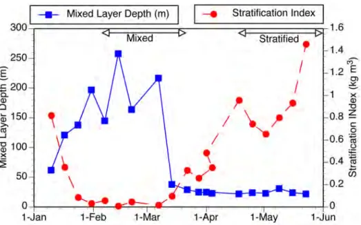
Seasonal changes in stratification and the depth of the surface mixed layer (Fig. 1) were typical for the site. In early January the mixed layer reached 150 m depth and
subsequently deepened to nearly 250 m by early February. Water column stratification began in late March and by mid-‐April the water column was strongly stratified. The surface mixed layer was restricted to the top 25 m of the water column from early April on and it remained shallow through the end of the sampling period in late June. Based upon the stratification index, the mixed period (index < 0.125) included sampling from
January 30 to March 9 (n = 6 dates) and strongly stratified water column (index > 0.6) encompassed sampling from April 18 to June 1 (n = 7 dates). Simple T-‐tests were used to test for significant differences in organismal concentrations comparing mixed and
stratified periods.
Fig. 1. Temporal changes in water column structure: two contrasting periods of "mixed" and "stratified" are indicated based on large differences in the stratification index.
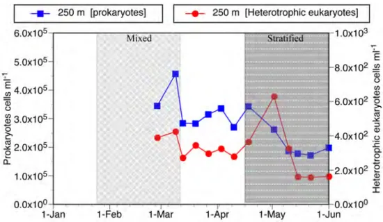
Heterotrophic prokaryotes (bacteria) and Eukaryotes (pico and nanoflagellates) At 250 depth heterotrophic prokaryote (bacteria & archeabacteria) abundance was highest during the mixed period, declining from a peak abundance of 4.6 x 10⁶ cells ml-‐1
to about 2 x 10⁵ cells ml-‐1 during the stratified period, thus varying only by about a
factor of 2 to 3 (Fig. 2). Irregular shifts in abundance characterized the heterotrophic eukaryote population. During the mixed period abundance was relatively high ranging from 390 -‐ 420 cells ml-‐1. During the transitional period between the stratified and
mixed periods concentrations were about 300 cells ml-‐1. Subsequently, the
period to a peak of about 630 cells ml-‐1 and subsequently declined to low levels of about
160 cells ml-‐1. Overall, the heterotrophic eukaryotes concentrations varied by a factor of
4, considerably more than the prokaryotes.
Fig. 2. Temporal changes in concentrations of heterotrophic prokaryotes and eukaryotes at 250 m during the study period. For water column characteristics distinguishing 'mixed' from 'stratified' periods indicated see Fig. 1.
Tintinnid Ciliates
A large number of tintinnid species were encountered (see supplementary Table 1). Out of a total of 74 species, 53 were found in the deep-‐water samples (250 m) and 58 in the surface samples (30 m). However, some were found only once or twice in either deep water or surface samples (≤ 10% of dates). These numbered 18 out of 58 in the surface samples and 7 out of 53 in the deep-‐water samples. Leaving aside these rare occurrence species yields then a pool of 46 for the deep-‐water samples and 40 in the surface
samples. The 46 species found in deep waters on at least 3 dates can be divided into 17 deep-‐water species, and those also found in the surface samples, 29 species. Of the 34
species in surface samples found on at least 3 dates, the surface water species numbered 12. Out of the total species pool, the species found on at least 3 dates were putatively 17 deep-‐water forms, 22 occurring throughout the water column and 12 surface water species.
Relatively few species occurred commonly, defined here as found on at least 50% of the sampling dates. In the deep water, 20 species were common and in surface layer samples only 10 species were common. In the deep-‐water samples, the 20 commonly occurring species were composed of two sets: 9 deep-‐water species and 11 species also found in surface samples. This latter group of forms found both in the deep water and surface samples, was 8 of the 10 common species of the surface layer samples. Thus, common deep-‐water tintinnids were a mixed assemblage of deep-‐water species and most of the common surface layer forms. The 9 common deep-‐water species includes 5 apparently described forms of basically two size-‐groups (see fig. 3).
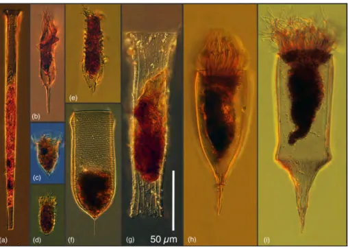
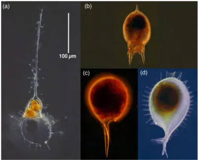
Fig. 3. Common forms of tintinnid ciliates found in deep waters, absent or nearly absent from surface waters: (a) an undescribed Salpingella species, (b) an undescribed Albatrossiella species, (c) an undescribed Ormosella species, (d) a small undescribed Amphorellopsis species, (e) an undescribed 'ringed' Amphorellopsis species, (f) Favella aciculifera, (g) Daturella striata, (h) Xystonellopsis scyphium, (i) Parundella messinensis. Note that there are two basic groups
presumably feding on different sized prey items: forms with small oral opening diameters of 10-‐ 20 µm (a-‐e) and forms with markedly larger oral opening diameters of 40-‐50 µm (f-‐g).
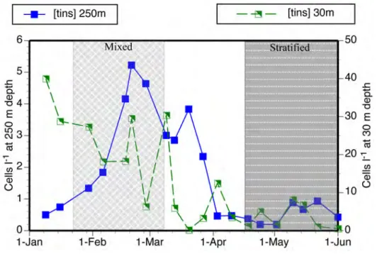
Temporal changes in concentrations differed considerably between surface and deep layer populations (fig. 4). The surface population showed much higher variability compared to the deep-‐water population. Partly this reflected a bloom of salps that corresponded with the near disappearance of tintinnids in the surface water samples in March. Within the deep water population, changes in concentrations largely reflected concentrations of surface and entire water column species in deep waters, which increased markedly with the depth of the mixed layer, compared to shifts in the
significantly (p< 0.006) higher (3.3 cells l-‐1) during the period of mixis compared to the
stratified period (1.4 cells l-‐1). The species richness of deep-‐water forms was relatively
constant with several forms present regardless of cell abundance while the numbers of entire water column and surface species showed a distinct peak during the mixed period (Fig. 6). Unlike the Phaeogromid radiolarians and Amphisolenid dinoflagellates (see below), there was no clear pattern among any deep-‐water tintinnid species of presence vs. absence during the mixed compared to stratified periods.
Fig. 4. Temporal changes in the concentrations of tintinnids at 250 m and 30 m during the study period. Note the different scales for populations at 250 m and those found at 30m. For water column characteristics distinguishing 'mixed' from 'stratified' periods indicated see Fig. 1.
Fig. 5. Temporal changes in the concentrations of tintinnids at 250 m distinguishing two groups-‐ those found only (or nearly only) in the deep water samples, 'Deep spp' from other forms, found mainly or exclusively in the surface water samples, "Entire Water Column & Surface ssp'. Note the different scales for the two groups of species. For water column characteristics
distinguishing 'mixed' from 'stratified' periods indicated see Fig. 1.
Fig. 6. Species richness of the deep-‐water tintinnid assemblage. Temporal changes in the numbers of tintinnid species at 250 m distinguishing two groups-‐ those found only (or nearly only) in the deep water samples, 'Deep spp' from other forms, found mainly or exclusively in the surface water samples, "Entire Water Column & Surface ssp'. Note the relatively invariant number of deep water species.
Phaeogromid Radiolarians
A total of 9 phaeogromid species were found in the samples from 250 m, and none were encountered in the samples from 30 m (see supplemental table and data file for details). In the samples from 250 m, 7 species were found on at least 3 dates: and 4 occurred commonly (Fig. 7). The lowest concentrations were recorded from the beginning of the mixed period and increased over time to peak at the start of the stratified period (Fig. 8). Consequently, the average concentration in the mixed period (0.05 cells l-‐1) compared to
the average during the stratified period (0.14 cells l-‐1) was significantly (p< 0.015)
lower. The higher concentrations of the stratified period reflected mainly the concentrations of species which were not encountered during the mixed period or present in trace concentrations, but consistently found in the stratified period:
Challengeranium diodon, Challengeron willemoseii, Euphysetta lucani and Euphysetta pusilla. Species richness was highest during the stratified period (Fig. 8).
Fig. 7. Common forms of phaeogromid radiolarians found in deep waters: (a) Medusetta parthenopaea, (b) Challengeranium diodon, (c) Challengeria xiphodon, (d) Challengeron willemoesii.
Fig. 8. Temporal changes in the concentrations and species richness (numbers of species) of phaeogromid radiolarians at 250 m. Note that changes in concentrations and species richness are similar. For water column characteristics distinguishing 'mixed' from 'stratified' periods indicated see Fig. 1.
Amphisolenid Dinoflagellates
Only 4 amphisolenid species were encountered, 3 species of Amphisolenia (A. bidentata,
A. extensa, A. globulosa) and Triplosolenia bicornis. They were occasionally encountered
in surface water samples (4 of 20 dates) in trace concentrations but with exception of the late January sampling, consistently present at 250 m. Two species were found commonly, Amphisolenia globulosa and Triplosolenia bicornis (Fig. 9). There was no significant difference between concentrations in the mixed period vs. the stratified period, both averaged 0.06 cells l-‐1. However, there was a marked shift in the
composition of the assemblage from the mixed period with relatively abundant
Amphisolenia and an absence of Triplosolenia to the stratified period dominated by Triplosolenia bicornis (Fig 10).
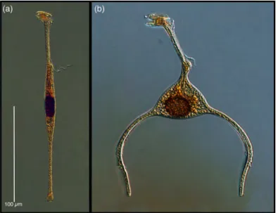
Fig. 9. Common species of amphisolenid dinoflagellates found in deep waters: (a) Amphisolenia globulosa, and (b) Triposolenia bicornis.
Fig. 10. Temporal changes in the concentrations of species of Amphisolenia and the
concentration of Triposolenia bicornis at 250 m. Note the distinct temporal trends of the two genera. For water column characteristics distinguishing 'mixed' from 'stratified' periods indicated see Fig. 1.
Discussion
We found clear, consistent and large differences between the surface layer and
mesopelagic layer populations both in terms of abundances and species compositions. The differences are unlikely to be due to sampling variability. A previous a study of sampling variabilty, with regard to tintinnids conducted at a nearby site, using sample sizes considerably smaller than those employed in the present study (10 l) found the major differences among replicate samplings to be the number and identity of rare species (Dolan & Stoeck, 2011).
The seasonal changes we documented in water column structure and
mesopelagic abundances of heterotrophic prokaryotes and eukaryotes largely conform to previous studies in terms of magnitudes. Both Tanaka & Rassoulzadegan (2002) and Winter et al. (2009a,b) investigated populations at the open water Dyfamed site in the
NW Mediterranean while Weinbauer et al. (2013) reported on samples from our study site. We found prokaryote abundances to range between 1.7 -‐ 4.6 x 10⁵ cells ml-‐1 and
heterotrophic eukaryotes concentrations ranging from 157 -‐ 629 cells ml-‐1 (Fig. 2).
Tanaka & Rassoulzadegan found prokaryote abundances at 300 m to vary between 1 -‐ 4 x 10⁵ cells ml-‐1 from January to May while heterotrophic nanoflagellate concentrations
varied between 40 and 200 cells ml-‐1. Winter et al. (2009b) reported mesopelagic
prokaryote abundances of 1 -‐ 2.1 x 10⁵ cells ml-‐1 between February and June. For the
period between March and June, Weinbauer et al. (2013) reported prokaryote abundance of 2.5 -‐ 3.2 x 10⁵ cells ml-‐1 and the heterotrophic nanoflagellate
concentration from 120 to 250 cells ml-‐1. Despite largely identical seasonal changes in
water column structure, the periods of peak abundance varied among the previous studies and compared to our findings. For example Tanaka & Rassoulzadegan (2002) and Winter et al. (2009b), both reporting on samples from the open water Dyfamed site, reported peak prokaryote abundances in February and May respectively. Weinbauer et
al. (2013), sampling at the same site as the present report, found peak prokaryote and
heterotrophic nanoflagellate abundance in May. We found peak prokaryote and
eukaryote abundances in March and May, respectively (Fig. 2). However, these apparent differences may be due to the relatively coarse sampling of bi-‐monthly to monthly
previously employed.
To our knowledge, our study is the first to address weekly temporal changes among mesopelagic protists and document changes in the abundances of individual species. All three groups we examined contained species absent from surface water samples. All three groups showed marked changes in either species compositions, or concentrations, or both over the study period that covered contrasting water column conditions. In the assemblage of tintinnid ciliates 9 species were found commonly in the
deep-‐water samples. The deep-‐sea tintinnids were a minority of the tintinnid
assemblage during the period of water column mixis when surface water species were found at depth but dominated the assemblage in the stratified period. Peak abundance occurred at the beginning of the mixed period. The phaeogromid radiolarians,
completely absent from the surface samples, showed increased abundances and changes in species richness comparing the mixed and stratified periods. They showed peak abundance at the beginning of the stratified period. Among the amphisolenid dinoflagellates peak abundances were recorded in the middle of the mixed period. Species of Amphisolenia were found during the mixed period and Triplosolenia bicornata, absent during the mixed period, dominated numbers during the stratified period.
The protist species we found that appear to be mainly mesopelagic residents includes both forms suspected to be deep-‐water species and those known to inhabit deep-‐water layer. For example, among the Amphisolenid dinoflagellates, Triplosolenia
bicornis was originally described from plankton net tow material gathered from 250 -‐to
50 depth, "rarely from surface hauls" (Kofoid 1906) and Jorgensen (1923) described it as almost exclusively from deep water in the Mediterranean and missing from winter samples, similar to our findings. Amphisolenia species, while known from surface water samples (e.g. Gomez et al. 2011), are most abundant in sub-‐surface waters (100-‐200) but occur in down to 400 m depth (Tarangkoon et al., 2010). With regard to
phaeogromid radiolarians, the species we denoted as common in the mesopelagic are also known from the mesopelagic of the Adriatic Sea (Krsinic & Krsinic, 2012) and the western and central Pacific Ocean (Yamashita et al., 2002). Similar to our findings in the NW Mediterranean, in the Adriatic the radiolarians are very rarely found in the surface layer (Krsinic & Krsinic, 2010).
On the other hand the deep-‐water tintinnid fauna we encountered may be unique to our study site. The 17 tintinnid species we found to be common or present at 250 m (thus absent or rare in surface samples) differed not only in number (8 species) but also identity of those described as deep water forms in the Adriatic Sea (Krsinic, 1998). In common with the Adriatic list of Krsinic (1998), we found Parundella lohmanni, P.
messinensis, Xystonellopsis scyphium and Favella aciculifera. However of the 5 other
species reported as Adriatic deep-‐water forms we found 1 in both surface and deep water samples (Ormosella trachleum), 2 are common in in the Bay of Villefranche,
Amphorides teragona and Cyttarocylis ampulla (Dolan, 2017), and 1 is not known to date
from the study area (Xystonellopsis cymatica). However, it should be noted that the studies in the Adriatic employed a different sampling method, vertical tows of a 53 µm mesh net thus sampling large volumes of water (several m3) but with a net size unlikely
to catch the small forms we commonly found (Fig. 3).
Concentrations of all three groups varied by over an order of magnitude but quite asynchronously. Tintinnid ciliates, phaeogromid radiolarians and amphisolenid
dinoflagellates, are of overall similar sizes and were found in similar concentrations. They would appear to represent roughly similar food items for consumers of
microplankton so their distinct abundance patterns argues against strong top-‐down control by grazers since this would presumably have yielded similar abundance patterns for the three groups. By default the distinct abundance patterns support a hypothesis of variability in group-‐specific resources and/or variability in group-‐specific mortality. While we know little about sources of mortality, the three groups in principle exploit very different food resources.
Tintinnid ciliates graze on suspended food items with optimal prey size related to the lorica opening diameter. The average optimal prey size is about 0.25 lorica oral
diameter (Dolan, 2010). The tintinnids common in the mesopelagic were of two groups in terms of lorica oral diameter (fig. 3) those with diameters of about 10-‐20 µm and forms with diameters of about 40-‐50 µm. The mesopelagic tintinnid ciliates presumably relied on prey of about 4 and 11 µm diameter respectively, thus nano-‐plankton-‐size prey such as heterotrophic nanoflagellates. The phaeogromid radiolarians are thought to feed on sinking organic aggregates (Gowing ,1986). A study of the food vacuole content of several species including species recorded in our study (Challengeria xiphodon,
Challengeron willemoesii, Euphysetta pusilla), found the major food items to be bacteria
and eukaryotic algae presumably from ingesting suspended organic aggregates (Gowing and Bentham, 1994). Simple correlation analysis revealed no significant relationship with prokaryotic or eukaryotic heterotrophs enumerated using flow cytometry with tintinnid or radiolarian abundances. However, it should be noted that we have a relatively small number of data points for the flow cytometry counts (n = 13). With regard to amphosolenid dinoflagellate nutrition, virtually nothing is known. They are known to harbor endosymbiotic phototrophs, some with both prokaryotic and
eukaryotic endosymbionts; presumably the dinoflagellate host cell profit from the presence of endosymbionts but their role in the nutrition of the host cell has not been examined (Daugbjerg et al., 2013). However, based on the Sechi disk depths, which varied between 8 and 21 m (data not shown), the euphotic zone reached a maximum depth of about 40 m arguing against a role for endosymbiont photosynthesis in the populations at 250 m depth. The possibility that amphisolenid dinoflagellates below the photic zone digest their symbionts for nutrition can not excluded. Overall, our study admittedly raises many more questions than the two we set out to answer. For example, the factors controlling the abundance of the mesopelagic protists considered here
clearly remain to be revealed and the possibility that the morpho-‐species considered may or may not be genetically distinct merits attention.
Conclusions
We examined short-‐term temporal changes in the species composition and abundance of 3 distinct groups of mesopelagic protists in the N.W Mediterranean Sea over the period corresponding to large changes in water column structure. All three groups examined contained species absent from surface water samples. All three groups showed marked changes in either species compositions, or concentrations, or both over the study period. We established for all three protists groups (tintinnid ciliates, phaeogromid radiolarians, and amphisolenid dinoflagellates) that there appear to be specific mesopelagic forms and the three groups display distinct seasonal patterns of abundance and species
composition. We conclude that the mesopelagic protist assemblage is clearly diverse and dynamic at least with regard to groups in which species can be identified using light microscopy.
Supplementary data
Supplementary materials, tables summarizing species occurrences (Supplementary Tables File) and count data for each date (Supplementary Data File) are available at the ICESJMS online version of the article.
Acknowledgements
We thank the skipper, Jean-‐Yves Carval, and crew members Pierre Cohen and Julien Garde, of the vessel ‘Sagitta 3’ for ship operations and aid in sampling. Water column profile data used to calculate mixed layer depth and the stratification index for Point C were kindly supplied by the SOMLIT coastal observation program. Sabine Agatha and
Sylvia Angerer kindly commented on some species identifications. However, the responsibility for all species identification, for better or for worse, remains with the authors.
Funding
This work was funded by the French Agence National de la Recherche through the AncesStram project coordinated by David Moreira, Université Paris-‐Sud.
References
Abboud-‐Abi Saab, M. 2008. Tintinnids of the Lebanese Coastal Waters (Eastern Mediterranean ). CNRS-‐Lebanon/UNEP/MAP/RAC/SPA, Lebanon, 192 pp.
Anderson, O.R. 1983. Radiolaria. Springer-‐Verlag, 355 pp.
Aristegui, J., Gasol, J. M., Duarte, C. M., Herndld, G. J. 2009. Microbial oceanography of the dark ocean's pelagic realm. Limnology and Oceanography, 54: 1501-‐1529.
Balech, E. 1959. Tintinnoinea del Mediterraneo. Trabajos del Instituto Español de Oceanografia, 28: 1-‐88.
Behrenfeld, M. J., T O'Malley, R., Siegel, D. A., McClain, C. R., Sarmiento, J. L., Feldman, G. C.C, Milligan, et al. 2006. Climate-‐driven trends in contemporary ocean productivity. Nature, 444: 752-‐755.
Borgert, A. 1906. Die Tripyleen Radiolarian der Plankton-‐Expedition. Medusettidae. Ergebnisee Plankton-‐Expedition der Humboldt-‐Stiftung, 3(4): 133-‐193.
Borgert, A. 1911. Die Tripyleen Radiolarian der Plankton-‐Expedition. Challengeridae. Ergebnisee Plankton-‐Expedition der Humboldt-‐Stiftung, 3(11): 148-‐536.
Burd, A. B., Hansell, D. A., Steinberg, D. K., Anderson, T. R., Arístegui, J., Baltar, F., Beaupré, S.R., et al. 2010. Assessing the apparent imbalance between geochemical and biochemical indicators of meso-‐and bathypelagic biological activity: What the@ $♯! is wrong with present calculations of carbon budgets?. Deep Sea Research Part II: Topical Studies in Oceanography, 57: 1557-‐1571.
Bustillos-‐Guzmán, J., Claustre, H., Marty, J. C. 1995. Specific phytoplankton signatures and their relationship to hydrographic conditions in the coastal northwestern Mediterranean Sea. Marine Ecology Progress Series, 124: 247-‐258.