HAL Id: hal-02950695
https://hal-amu.archives-ouvertes.fr/hal-02950695
Submitted on 28 Sep 2020
HAL is a multi-disciplinary open access
archive for the deposit and dissemination of sci-entific research documents, whether they are pub-lished or not. The documents may come from teaching and research institutions in France or abroad, or from public or private research centers.
L’archive ouverte pluridisciplinaire HAL, est destinée au dépôt et à la diffusion de documents scientifiques de niveau recherche, publiés ou non, émanant des établissements d’enseignement et de recherche français ou étrangers, des laboratoires publics ou privés.
derivatives against Mycobacterium abscessus
Abdeldjalil Madani, Ivy Mallick, Alexandre Guy, Céline Crauste, Thierry
Durand, Patrick Fourquet, Stéphane Audebert, Luc Camoin, Stéphane
Canaan, Jean François Cavalier
To cite this version:
Abdeldjalil Madani, Ivy Mallick, Alexandre Guy, Céline Crauste, Thierry Durand, et al.. Dissecting the antibacterial activity of oxadiazolone-core derivatives against Mycobacterium abscessus. PLoS ONE, Public Library of Science, 2020, 15 (9), pp.e0238178. �10.1371/journal.pone.0238178�. �hal-02950695�
RESEARCH ARTICLE
Dissecting the antibacterial activity of
oxadiazolone-core derivatives against
Mycobacterium abscessus
Abdeldjalil Madani1☯, Ivy Mallick1,2☯, Alexandre Guy3, Ce´line Crauste3, Thierry Durand3, Patrick Fourquet4, Ste´phane Audebert4, Luc CamoinID4, Ste´phane Canaan1, Jean
Franc¸ois CavalierID1*
1 Aix-Marseille Univ., CNRS, LISM, Institut de Microbiologie de la Me´diterrane´e FR3479, Marseille, France, 2 IHU Me´ diterrane´e Infection, Aix-Marseille Univ., Marseille, France, 3 IBMM, Univ Montpellier, CNRS, ENSCM, Montpellier, France, 4 Aix-Marseille Univ, INSERM, CNRS, Institut Paoli-Calmettes, CRCM, Marseille Prote´omique, Marseille, France
☯These authors contributed equally to this work. *[email protected]
Abstract
Mycobacterium abscessus (M. abscessus), a rapidly growing mycobacterium, is an emer-gent opportunistic pathogen responsible for chronic bronchopulmonary infections in individ-uals with respiratory diseases such as cystic fibrosis. Most treatments of M. abscessus pulmonary infections are poorly effective due to the intrinsic resistance of this bacteria against a broad range of antibiotics including anti-tuberculosis agents. Consequently, the number of drugs that are efficient against M. abscessus remains limited. In this context, 19 oxadiazolone (OX) derivatives have been investigated for their antibacterial activity against both the rough (R) and smooth (S) variants of M. abscessus. Several OXs impair extracellu-lar M. abscessus growth with moderated minimal inhibitory concentrations (MIC), or act intracellularly by inhibiting M. abscessus growth inside infected macrophages with MIC val-ues similar to those of imipenem. Such promising results prompted us to identify the poten-tial target enzymes of the sole extra and intracellular inhibitor of M. abscessus growth, i.e., compound iBpPPOX, via activity-based protein profiling combined with mass spectrometry. This approach led to the identification of 21 potential protein candidates being mostly involved in M. abscessus lipid metabolism and/or in cell wall biosynthesis. Among them, the Ag85C protein has been confirmed as a vulnerable target of iBpPPOX. This study clearly emphasizes the potential of the OX derivatives to inhibit the extracellular and/or intracellular growth of M. abscessus by targeting various enzymes potentially involved in many physio-logical processes of this most drug-resistant mycobacterial species.
Introduction
Non-tuberculous mycobacteria (NTM) are naturally-occurring bacterial species mostly found in soil and water that do not cause tuberculosis or leprosy [1]. NTM are opportunistic
a1111111111 a1111111111 a1111111111 a1111111111 a1111111111 OPEN ACCESS
Citation: Madani A, Mallick I, Guy A, Crauste C, Durand T, Fourquet P, et al. (2020) Dissecting the antibacterial activity of oxadiazolone-core derivatives against Mycobacterium abscessus. PLoS ONE 15(9): e0238178.https://doi.org/ 10.1371/journal.pone.0238178
Editor: Olivier Neyrolles, Institut de Pharmacologie et de Biologie Structurale, FRANCE
Received: December 18, 2019 Accepted: August 12, 2020 Published: September 18, 2020
Copyright:© 2020 Madani et al. This is an open access article distributed under the terms of the Creative Commons Attribution License, which permits unrestricted use, distribution, and reproduction in any medium, provided the original author and source are credited.
Data Availability Statement: The mass
spectrometry proteomics data have been deposited to the ProteomeXchange Consortium (www. proteomexchange.org) via the PRIDE partner repository with the dataset identifier PXD015680.
Funding: This work was supported by the CNRS, Aix Marseille University and by the grant ANR-19-CE44-0011 from the Agence Nationale de la Recherche (https://anr.fr/). AM was supported by a PhD fellowship from the Association Gre´gory Lemarchal and Vaincre la Mucoviscidose
pathogens able to infect humans with predisposing conditions like cystic fibrosis (CF) or immunosuppression and responsible for wide range of infections like skin infections, pulmo-nary infections or disseminated diseases [2–4]. In the last decades, NTM infections are increas-ing worldwide, the most frequently reported species beincreas-ingMycobacterium avium complex
(MAC) andM. abscessus complex [3,5].
M. abscessus can be isolated from solid medium with either a smooth (S) or a rough (R)
col-ony morphotype [6]. The difference between both morphotypes is related to the presence of glycopeptidolipids (GPLs) in the cell wall of the S variant, while absent in the R one [7]. This latter R strain is also associated with severe and persistent infections [8]. In CF patients, treat-ment ofM. abscessus complex infections requires a multidrug therapy including a daily oral
macrolide (clarithromycin or azythromicin) in conjunction with intravenous amikacin and a β-lactam (imipenem or cefoxitin) [9]. However, almost 60% ofM. abscessus strains could
develop both intrinsic and acquired resistance to currently available antibiotics, including macrolides [4,10]. As a direct consequence, treatment of such infections has become very complicated with very limited alternative options [5,11].
Due to the worldwide increasing incidence and prevalence ofM. abscessus and the inherent
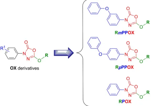
difficulties to manage such resistant pulmonary infections, new active molecules are urgently needed. In this context, we recently investigated the antibacterial activities of 19 oxadiazolone-core (OX) derivatives (Fig 1) against three pathogenic slow-growing mycobacteria:M. mari-num, M. bovis BCG as well as M. tuberculosis H37Rv the etiologic agent of tuberculosis [12]. These OX compounds exhibited not only encouraging minimal inhibitory concentrations (MIC), but above all, they were also found to display a diversity of actions by acting either only on extracellularM. tuberculosis growth, or both intracellularly on infected macrophages as
well as extracellularly on bacterial growth. Remarkably, all OX derivatives exhibited very low
Fig 1. Chemical structure of the OX derivatives. Rm(or p)PPOX nomenclature is as follows: m (or p)P represents themeta (or para)-Phenoxy group when present; P the phenyl group; OX the Oxadiazolone core; and R the alkyl chain
(i.e., M; methyl, E, ethyl; B, butyl; iB, isobutyl; H, hexyl; O, octyl; Eh, 2-ethylhexyl; D, decyl; Do, dodecyl; Be,
benzyloxyethyl; Me, methoxyethyl). Adapted from [12]. https://doi.org/10.1371/journal.pone.0238178.g001 (https://www.vaincrelamuco.org/) (project n
˚RF20160501651). A. M. received a financial support from the IHU Me´diterrane´e Infection (Marseille, France). Proteomics analyses were supported by the Institut Paoli-Calmettes and the Centre de Recherche en Cance´rologie de Marseille. Proteomic analyses were done using the mass spectrometry facility of Marseille Proteomics (marseille-proteomique.univ-amu.fr) supported by IBISA (Infrastructures Biologie Sante´ et
Agronomie), the Cance´ropoˆle PACA, the Provence-Alpes-Coˆte d’Azur Re´gion, the Institut Paoli-Calmettes, and Fonds Europe´en de De´veloppement Re´gional (FEDER). The funders had no role in study design, data collection and analysis, decision to publish, or preparation of the manuscript.
Competing interests: The authors have declared that no competing interests exist.
Abbreviations: ABPP, activity-based protein profiling; AMK, amikacin; CC50, compound
concentration leading to 50% Raw264.7 macrophages toxicity; CF, cystic fibrosis; CFU, colony-forming units; IMP, imipenem; MIC50/
MIC90, minimal inhibitory concentration leading to
50% or 90% bacterial growth inhibition, respectively; NTM, non-tuberculous mycobacteria; OX, oxadiazolone; pFDR, permutation false discovery rate; REMA, resazurin microtiter assay.
toxicity towards host cell macrophages [12]. Of interest, only the iBpPPOX derivative exhib-ited moderate (MIC50= 32.0μM) to quite good (MIC50= 8.5μM) antibacterial activity against
both extracellular and intramacrophagicM. tuberculosis H37Rv, respectively [12]. Following an activity-based protein profiling (ABPP) approach combined with mass spectrometry, 18 putative target(s) of HPOX, a selective inhibitor ofM. tuberculosis extracellular growth, were
identified. All these proteins were (Ser/Cys)-enzymes possessing a catalytic serine or cysteine residue, and involved inM. tuberculosis lipid metabolism and/or in cell wall biosynthesis.
Above all, the results of this study imply that such OX derivatives represent a novel class of multi-target mycobacterial inhibitorsvia the formation of a covalent bond with the catalytic
residue of various mycobacterial (Ser/Cys)-containing enzymes involved in various physiolog-ical processes.
Given all these previous findings, in the present study we have further assessed the antibac-terial activity of these 19 OXs againstM. abscessus growth. The determined MIC revealed that
some OXs were able to inhibitM. abscessus growth in vitro in culture broth medium and/or
intracellularly inside macrophages. In addition, using a similar ABPP assay as previously reported forM. tuberculosis [12], the potential target enzymes of iBpPPOX, the most active inhibitor of extra- and intracellular bacterial growth, were further identified.
Materials and methods
Bacterial strains and growth conditions
M. abscessus CIP104536Twith either a smooth (S) or rough (R) morphotype was grown in Middlebrook 7H9 broth (BD Difco, Le Pont de Claix, France) supplemented with 0.2% glycerol, 0.05% Tween 80 and 0.2% glucose (Sigma-Aldrich, St. Quentin Fallavier, France) (7H9-S).
Chemicals
Clarythromycine and Imipenem mixture w/Cilastatin were purchased from Euromedex (Souf-felweyersheim, France). The Oxadiazolone derivatives were synthesized as previously reported and were at least 98% pure as determined by HPLC analysis [12]. Stock solutions of each inhib-itor (4 mg/mL) were prepared in DMSO and stored at -20 ˚C before use.
Resazurin microtiter assay (REMA) for MIC determination—Extracellular
assay
Susceptibility testing was performed using the Middlebrook 7H9 broth microdilution method. MICs of the OXs were determined in 96-well flat-bottom Nunclon Delta Surface microplates with lid (Thermo-Fisher Scientific, ref. 167008) using the resazurin microtiter assay (REMA) [12–15]. Briefly, log-phase bacteria were diluted to a cell density of 5× 10⁶cells/mL and 100μL of this inoculum was grown in a 96-well plate in the presence of serial dilutions of each
OX compound. After 3–5 days incubation at 37 ˚C, 20μL of a 0.025% (w/v) resazurin solution was added to each well (200μL) and incubation was continued until the appearance of a color change (from blue to pink) in the control well (i.e., bacteria without antibiotics). Fluorescence
of the resazurin metabolite resorufin (λexcitation, 530 nm;λemission, 590 nm) was then measured
[13,16] and the concentration leading to 50% and 90% growth inhibition was defined as the MIC50and MIC90, respectively. SeeS1 Appendixfor detailed protocol.
Intramacrophage killing assay—Intracellular assay
The intracellular growth ofM. abscessus S was assessed following a 24 h exposure of infected
Raw264.7 murine macrophages cell line (American Type Culture Collection TIB-71) to each of the 19 OX compounds at a final concentration of 30μM [17]. To avoid growth of extracellular mycobacteria, cells were extensively washed and treated with amikacin (200μg/mL = 340 μM; 87 × MIC50) prior to treatment with the OX analogs. Imipenem
(IMP; 80μg/mL = 267 μM; 64 × MIC50) was used as positive control for this intracellular
killing assay. In each case, the viability of infected macrophages was checked by addition of trypan blue [18] before cell lysis and plating for CFU count. SeeS1 Appendixfor detailed protocol.
iB
pPPOX target enzymes identification
Activity-Based Protein Profiling (ABPP) for the identification of iBpPPOX target
enzymes. Bacterial suspension ofM. abscessus R in 7H9-S was adjusted at an OD600
corre-sponding to 6×10⁹cells/mL and then incubated with iBpPPOX inhibitor (400 μM final con-centration) or DMSO (control) at 37 ˚C for 2–3 h. under gentle shaking at 75 rpm. Bacteria were then washed 3 times with PBS containing 0.05% Tween 80, resuspended in PBS buffer at a 1:1 (w/v) ratio and then lysed by mechanical disruption on a BioSpec Beadbeater. Both iBpP-POX-treatedM. abscessus and DMSO-control lysate samples (750 μL– 0.75 mg total proteins)
were labeled with 2μM Desthiobiotin-FP probe for 90 min at room temperature. Samples were enriched for biotinylated proteins using 0.8μm Nanolink streptavidin magnetic beads (Solulink), according to the manufacturer’s instructions. The resulting captured biotinylated proteins solution was mixed with 5X Laemmli reducing sample buffer, and heated at 95 ˚C for 5 min. The released denatured proteins were subjected to tryptic digestion, peptide extraction, and LC-MS/MS analysis as described below.
Alternatively,M. abscessus R total lysates (500 μL − 1 mg total proteins) were further
pre-incubated with iBpPPOX (400 μM final concentration) or DMSO as control for 60 min at 37˚C, and then treated with 2μM ActivX Desthiobiotin-FP probe (ThermoFisher Scientific) and processed as described above forM. abscessus R living cells. Detailed protocol regarding
ABPP experiments is given inS1 Appendix.
Mass spectrometry analysis for enzyme identification and quantification
Protein extract were loaded and stacked on a NuPAGE gel (Life Technologies). Stained bands were submitted to an in-gel trypsin digestion [19]. Peptides extracts were reconstituted with 0.1% trifluoroacetic acid in 4% acetonitrile and analyzed by liquid chromatography (LC)-tan-dem mass spectrometry (MS/MS) using Orbitrap Mass Spectrometers (Thermo Electron, Bremen, Germany) online with a nanoLC Ultimate 3000 chromatography system (Dionex, Sunnyvale, CA). Protein identification and quantification were processed using the MaxQuant computational proteomics platform, version 1.5.3.8 [20] using a UniProtM. abscessus ATCC
19977 (Taxon 561007) database (date 2017.02; 4940 entries). The statistical analysis was done with Perseus program (version 1.5.6.0). Differential proteins were detected using a two-sample
t-test at 0.01 and 0.05 permutation-based FDR. Detailed Materials and Methods are given in
S1 Appendix.
The mass spectrometry proteomics data have been deposited to the ProteomeXchange Consortium (www.proteomexchange.org) [21]via the PRIDE partner repository with the
Validation of Ag85C
Mabsby iB
pPPOX
Plasmids and DNA manipulations. All specific oligonucleotides and plasmids used in
this study are listed inS1 Appendix(see S3 and S4 Tables—page S8). All cloned fragments were amplified using purifiedM. abscessus genomic DNA. The mab_0175 gene encoding
Ag85C was amplified by PCR using the specific forward (pMyc::ag85C-F) and reverse
(pMyc::ag85C-R) primers. For the inactivated Ser124Ala mutant ag85CS124Aconstruction, overlap extension PCR (OE-PCR) was used. For the generation of first fragment containing the mutation of the active serine to alanine, primer setspMyc::ag85C-F and pMyc::
ag85CS124A-R were used, the second fragment containing the mutation was generated using
the primer setspMyc::ag85CS124A-F and pMyc::ag85C-R. The two fragments were further
purified, mixed in 1:1 (v/v) ratio and used as template to amplify the complete insert
con-taining the mutation, using the primer pairspMyc::ag85C-F and pMyc::ag85C-R. The
respective PCR products were cloned into pMyC vector, following digestion with NcoI and HindIII, enabling the incorporation of a6His-tag in the C-terminus of the Ag85C or
Ag85CS124Aprotein. Deletion mutantΔmab_0175 (= Δag85C) was obtained by a simple and rapid gene disruption strategy inM. abscessus developed by Viljoen et al. [22].
Ag85C gene was amplified using primer pairs pUX1::Δag85C-F and pUX1::Δag85C-R,
then cloned into pUX1 vector using NheI and BamHI restriction sites by classical cloning. Finally, for complementation strain, themab_0175 gene was amplified using the primer
pairspVV16::ag85C-F and pVV16::ag85C-R, and cloned into pVV16 plasmid in frame with
a6His-tag located in C-terminal and downstream of thehsp60 promoter also containing a
kanamycin resistance cassette using restriction free cloning (SLIC) [23] to generatepVV16:: ag85C. Sequence integrity of each construct was confirmed by DNA sequencing (Eurofins
Genomics). All the constructs were further transformed in electrocompetentM. abscessus S
and R types and selected on respective antibiotic agar plates as described previously [22]. Positive transformants were further grown in 7H9OADCmedium (i.e., 7H9 broth + 0.2%
glycerol + 0.05% Tween 80 + 10% oleic acid, albumin, dextrose, catalase) supplemented with either hygromycin (1000μg/mL; i.e., overexpression and inactivated strains), kanamycin (250μg/mL; i.e., deletion strain) or both antibiotics (1000 μg/mL hygromycin + 250μg/mL kanamycin; i.e., complementation strain), up to OD600of 1. The
overproduc-tion of the recombinant proteins in the overexpression and inactivated strains as well as in the complementation strain was checked by Western blot using the HisProbe™ HRP con-jugate (ThermoFisher Scientific). Regarding the deletion strain, the selection was made based on red fluorescent colonies followed by PCR amplification and sequencing strategy as described in [22].
Functional validation of Ag85C
Mabstarget enzyme
The abovementioned transformed bacteria,i.e., the M. abscessus_pMyc::ag85C overexpressing
strains, the inactivatedM. abscessus_pMyc::ag85CS124Aoverexpressing strains, theM. abscessu-s_Δag85C deletion strains and their complemented counterparts M. abscessuabscessu-s_Δag85C::C were
grown in 7H9OADCmedium supplemented with either hygromycin (1000μg/mL; i.e., overex-pression and inactivated strains), kanamycin (250μg/mL; i.e., deletion strain), or both antibi-otics (1000μg/mL hygromycin + 250 μg/mL kanamycin; i.e., complementation strain) until the OD600reached 2. In the case of the overexpression and inactivated strains, induction was
further done with 0.2% acetamide and the culture was incubated at 37˚C for additional 24 h. Susceptibility testing of each of theM. abscessus mutant strains against various concentrations
Expression and purification of
M. abscessus antigen Ag85C
The plasmid harboring themab_0175 gene was used to transform the M. smegmatis ΔgroEl
expression strain. Transformed bacteria were grown in 7H9 medium containing hygromycin (200μg/mL) until the OD600reached 2.0. Induction was done with 0.2% acetamide and the
culture was further incubated at 37 ˚C for 24 h. One L of bacterial pellets were collected by centrifugation (8,000× g, 4 ˚C, 1 h), re-suspended in 30 mL ice-cold buffer (50 mM Tris pH 8.0 containing 200 mM NaCl), and were broken using a French Pressure cell at 1,100 psi. The lysate was clarified by centrifugation (12,000× g, 4 ˚C, 30 min) prior to purification by nickel affinity chromatography with Ni-NTA sepharose beads and elution with the previous Tris (pH 8.0) buffer containing 500 mM imidazole. Purified protein was concentrated at 1 mg/mL and stored at –80 ˚C [24,25].
In vitro inhibition of pure recombinant M. abscessus Ag85C by iBpPPOX
A 14μM (i.e., 25 μg) concentration of Ag85CMabswas incubated for 1 h in its native form
with increasing molar excess of iBpPPOX (i.e. enzyme/inhibitor molar ratio, E/I = 1:1; 1:5, 1:10, 1:25, 1:50, and 1:75) in a reaction mixture containing 10 mM Tris buffer (pH 8), 150 mM NaCl and 0.1% (w/v) Triton X-100. Each sample was further treated with
10μM ActiveX TAMRA-FP fluorescent probe (ThermoFisher Scientific) for 1 h at room temperature in the darkness. The reaction was stopped by adding 5X Laemmli reducing buffer followed by boiling, and equal amounts of proteins (12μg) were separated by 12% SDS-PAGE. Subsequently, TAMRA FP-labeled proteins were detected by fluorescent gel scanning (TAMRA:λex557 nm,λem583 nm) using the Cy13 filter of a ChemiDoc MP
Imager (Bio-Rad) before staining the gel with Coomassie Brilliant Blue dye. Finally, relative fluorescence quantification of each band was performed using the ImageLab™ software ver-sion 5.0 (Bio-Rad) by taking the labeled Ag85CMabs-TAMRA adduct as 100% absolute
fluo-rescence level.
Mass spectrometry analysis of Ag85C
Mabs-iB
pPPOX complex
Purified Ag85CMabsrecombinant protein (14μM– 100 μg) was further incubated for 1 h in its
native form with iBpPPOX, using an enzyme/inhibitor molar ratio E/I = 1:100 to ensure total inhibition. Samples of the resulting Ag85CMabs-iBpPPOX complex were analysed on a
MAL-DI-TOF-TOF Bruker Ultraflex III spectrometer (Bruker Daltonics, Wissembourg, France) controlled by the Flexcontrol 3.0 package (Build 51), as described previously [24] (seeS1 Appendixfor full details). The total mass of the untreated protein (theoretical Mw = 32,057.83 Da; experimental Mw = 32,048.7 Da) is corresponding to the native enzyme lacking the 36 first N-terminal amino acids (i.e., M1 SVRVKARRVLSALLAAFVMPVSMAAAMTINPA-TAH36) consisting of a Sec signal peptide cleaved at the Ala-X-Ala (i.e., A35-H36-A37) site, as confirmed by N-terminal Edman sequencing [26].
Statistical analysis
Graphpad Prism 5 was used to perform the statistical analyses of the intracellular activity of the OX compounds, and of all susceptibility testing onM. abscessus mutant strains. The
statis-tical analysis related to MIC50Rawwas completed using a Student’st-test. The statistical
signifi-cance of differences in the MIC50or MIC90values between each mutant strain was analyzed by
Results and discussion
In vitro activity of oxadiazolone derivatives against M. abscessus
Drug susceptibility testing of the OX derivatives was assessed against both S and R variants of
M. abscessus, with amikacin (AMK) as standard drug. The corresponding MIC50/MIC90values
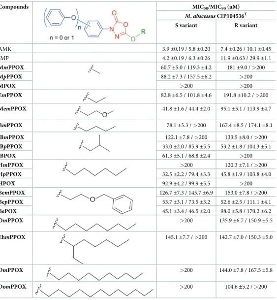
for each OX compound, as determined by the REMA assay [12–16], are reported inTable 1. Among all tested compounds, 14 OXs were able to block the growth ofM. abscessus S variant.
The best growth inhibitors were iBpPPOX (33.0 ±2.0 μM), HpPPOX (32.5 ±2.2 μM),
Mem-PPOX (41.8±1.6 μM) and BePOX (45.1 ±3.4 μM) which displayed interesting MIC50values
(Table 1). In all other cases, MIC50values were indicative either of a moderate (MIC50around
Table 1. Antibacterial activities of the oxadiazolone derivatives againstM. abscessus growth in broth medium using the REMA methoda.
Compounds MIC50/MIC90(μM)
M. abscessus CIP104536T S variant R variant AMK 3.9±0.19 / 5.8 ±0.20 7.4±0.26 / 10.1 ±0.45 IMP 4.2±0.19 / 6.3 ±0.26 11.9±0.63 / 29.9 ±1.1 MmPPOX 60.7±5.0 / 119.3 ±4.2 181±9.0 / >200 MpPPOX 88.2±7.3 / 157.5 ±6.2 >200 MPOX >200 >200 EmPPOX 82.8±6.5 / 101.8 ±4.6 191.8±10.2 / >200 MemPPOX O 41.8±1.6 / 44.4 ±2.0 95.1±5.1 / 113.9 ±4.7 BmPPOX 78.1±5.3 / >200 167.4±8.5 / 174.1 ±8.1 iBmPPOX 122.1±7.8 / >200 133.5±8.0 / >200 iBpPPOX 33.0±2.0 / 85.9 ±5.5 53.2±1.8 / 104.3 ±5.1 iBPOX 61.3±5.1 / 68.8 ±2.4 >200 HmPPOX >200 120.3±7.1 / >200 HpPPOX 32.5±2.2 / 79.4 ±3.3 45.8±1.9 / 103.8 ±4.0 HPOX 92.9±4.2 / 99.9 ±5.5 >200 BemPPOX O 126.7±7.3 / 145.7 ±6.9 153.0±7.8 / >200 BepPPOX 53.7±3.1 / 73.5 ±3.2 52.6±2.5 / 111.1 ±4.1 BePOX 45.1±3.4 / 46.5 ±2.0 98.0±5.8 / 170.2 ±6.2 OmPPOX >200 135.9±6.7 / 150.9 ±5.5 EhmPPOX 145.1±7.7 / >200 142.7±7.0 / 150.3 ±5.0 DmPPOX >200 144.0±7.8 / 167.5 ±5.8 DomPPOX >200 104.6±5.2 / >200
aExperiments were performed as described in Materials and Methods. MIC
50/ MIC90: compound minimal
concentration leading to 50% or 90% of growth inhibition, respectively, as determined by the REMA assay. Values are mean of at least two independent assays performed in triplicate. AMK, amikacin. IMP, imipenem.
53–61μM for MmPPOX, iBPOX, and BepPPOX), a weak (MIC50around 78–93μM for
MpPPOX, EmPPOX, BmPPOX, and HPOX), or a poor (MIC50> 120μM for iBmPPOX,
EhmPPOX, and BemPPOX) antibacterial activity (Table 1). Considering the MIC90values
reached onM. abscessus S, they are up to 2.5-fold greater than the corresponding MIC50;
except for HPOX (MIC50= 92.9±4.2 μM / MIC90= 99.9±5.5 μM), MemPPOX (MIC50=
41.8±1.6 μM / MIC90= 44.4±2.0 μM) and BePOX (MIC50= 45.1±3.4 μM / MIC90= 46.5
±2.0 μM) for which both MICs are in the same order of magnitude (Table 1).
Compared to the S morphotype,M. abscessus R variant was nearly 1.3- to 3.6-times less
sen-sitive to the OX compounds (Table 1); a property already observed for many drugs including AMK [27]. The best inhibitors ofM. abscessus R growth were iBpPPOX (MIC50= 53.2
±1.8 μM / MIC90= 104.3±5.1 μM), HpPPOX (MIC50= 45.8±1.9 μM / MIC90= 103.8
±4.0 μM), and BepPPOX (MIC50= 52.6±2.5 μM / MIC90= 111.1±4.1 μM) which exhibited
similar MIC50and MIC90values, respectively (Table 1). Interestingly, MpPPOX bearing a
short methyl chain has no antibacterial effect as compared to the three abovementioned
para-phenoxyphenyl derivatives. In summary, iBpPPOX, HpPPOX, and BepPPOX all possessing the phenoxy group in apara position as well as bulky ester chains, displayed the best
antibacte-rial activity againstM. abscessus R. No other clear trends or rules in terms of structure-activity
relationships (SAR) have emerged regarding the potency of these oxadiazolone-core com-pounds againstM. abscessus.
It is noteworthy that with MIC50values ranging from 31 to >120μM [12],M. tuberculosis
susceptibility to the OX compounds is similar to that of the S variant ofM. abscessus; iBpP-POX being the best growth inhibitor of both species. The increased tolerance of the
most-viru-lentM. abscessus R variant towards the OX compounds is in line with its high resistance to
classical antibiotics [4] compared toM. tuberculosis; a result that supports M. abscessus R’s
nickname of “antibiotics nightmare” [28].
Intramacrophagic susceptibility of
Mycobacterium abscessus to OX
derivatives
Macrophages, as the primary target, represent the host’s first line of defense but also an impor-tant reservoir of mycobacteria in lungs. From our previous work, the OXs were able to inhibit the growth ofM. tuberculosis inside infected macrophages, and found to be non-toxic for
Raw264.7 murine macrophages cell line with a CC50> 100μM (i.e., compound concentration
leading to 50% cell toxicity) [12]. Considering such properties, we further investigated the abil-ity of OXs to inhibit the intra-macrophagic growth ofM. abscessus.
The intrinsic nature of the R variant is to form bacterial clumps and cords in culture medium with time. As reported by Bernutet al., M. abscessus R cording prevents its
phagocy-tosis by macrophages. Consequently, the strain continues to grow extracellularly, and rapidly induces cell toxicity leading to cell death [29,30]. Such cording characteristic makes macro-phage infection experiments usingM. abscessus R very difficult to handle. Indeed, nearly all
macrophages were lysed at 24 h post-infection withM. abscessus R variant, making it
impossi-ble to quantify the intracellular effect of the OXs. This is, however, not the case withM. absces-sus S for which more homogenous bacterial absces-suspensions can be obtained for macrophages
infection studies [25,31,32].
Therefore, Raw264.7 cells were infected withM. abscessus S at a multiplicity of infection
(MOI) of 10, and then incubated for 24 h with all the OX compounds at a final concentration of 90, 60 and 30μM, or with imipenem (IMP) used as positive drug control. Among the 19 compounds tested, only 3 OXs (i.e., MPOX, MpPPOX, and iBpPPOX) exhibited an
which are weakly and not active against extracellular bacilli, respectively, were however able to significantly decrease the intramacrophagicM. abscessus present 24 h after infection (Fig 2).
MpPPOX displayed a moderate activity against intracellular M. abscessus S (Fig 2) with an approximated MIC50Rawof around 75μM which is 2.6 times higher that of IMP (MIC50Raw=
28.3μM). In contrast, 24 h-treatment with 30–60 μM MPOX led to a 53% reduction in myco-bacteria which increased up to 73.5% at 90μM, a percentage value comparable to the one elic-ited by IMP,i.e., 74.0% reduction following treatment with 60 μM (Fig 2). Remarkably, and as observed previously forM. tuberculosis [12], iBpPPOX was the sole identified inhibitor able to impair extracellular as well as intracellular growth ofM. abscessus. A plateau value
correspond-ing to 58.5±0.8% bacterial killing was indeed reached, whatever the iBpPPOX concentration used (30–90μM) to treat the infected cells.
Such a difference between the intra and extracellular activities has already been reported in our previous works with the OX derivatives [12], as well as with another family of growth inhibitors, the Cyclipostins & Cyclophostin analogs [13,14] acting againstM. tuberculosis and M. abscessus [25]. Similar toM. tuberculosis [12], the intracellular and extracellular inhibition ofM. abscessus growth may probably result from several different mechanisms of action or
penetration of the OX derivatives. The short methyl chain MpPPOX and MPOX display a bet-ter antimycobacbet-terial activity against intramacrophagicM. abscessus than in broth medium.
This clear preference against intracellularly-replicating mycobacteria may imply that the intra-cellular activity and/or the targets of these two compounds might differ from that of OXs act-ing on extracellularly-replicatact-ing bacilli. Several factors may indeed account for these
discrepancies, such as the metabolic status/fitness which varies between extra- and intracellular replicating bacteria. Another hypothesis could be that their corresponding target(s) would be more accessible and/or vulnerable during the intracellular lifestyle ofM. abscessus. A specific
response of the macrophage stimulated by the action of these compounds and leading to bacte-rial clearance cannot, however, be excluded. On the other hand, the iBpPPOX retains a similar Fig 2. Intracellular activity of MpPPOX, MPOX and iBpPPOX as compared to imipenem (IMP). The activity of selected OXs on intracellularM. abscessus was tested in Raw264.7 murine macrophages. Cells were infected at a
multiplicity of infection (MOI) of 10:1 withM. abscessus S variant and treated with various concentrations of each
inhibitor or IMP for 24 h. Then, surviving bacteria were enumerated by plating serial dilutions of macrophage lysates. DMSO-treated infected macrophages (i.e., SC, solvent control) were used as control representing 100% of bacterial
viability. Results are shown as mean± standard error of the mean (SEM) of three independent assays performed in triplicate.��,p-value < 0.01.�,p-value < 0.05. Statistical analysis was done using a Student’s t-test.
activity againstM. abscessus both extracellularly (MIC50= 33.0μM) and inside macrophages
(~59% bacterial clearance at 30μM). Regarding its intracellular antibacterial activity, the pres-ence of a plateau value, whatever the concentration used, might underline a different effect of
iBpPPOX towards infected macrophages compared to MmPPOX and MPOX for which a more classical dose-response has been reached. As mentioned above, one can speculate that the cellular stress caused by the action of iBpPPOX on the infected macrophages might induce a specific stringent response of these host cells, such as possible cell metabolism, therefore lead-ing to bacterial death.
Given the previously determined very low toxicity of the three selected compounds toward Raw264.7 cells with CC50> 100μM [12] similar to AMK (CC50� 150μM) [33], the
selectivity index (SI = CC50/MIC50Raw) of these best intracellular inhibitors onM. abscessus
vs. Raw264.7 cells was thus valued to be in a range from around 1.3 for MpPPOX and up to >3 for iBpPPOX.
From these findings, it can be assumed that the observed inhibitory potency of the OX compoundsi) might result from the inhibition of specific but most likely distinct
mycobacte-rial target enzymes between intramacrophagic-vs. extracellularly-replicating bacilli; or ii) may
reflect differences in the uptake and accumulation of the different compound inside the mac-rophage. Overall, these results suggest that both MpPPOX, MPOX and iBpPPOX would be able to enter the macrophages and arrest bacterial replication without exhibiting significant toxicity for the host cell.
iB
pPPOX inhibit M. abscessus by targeting various serine/cysteine enzymes
Given the previous results obtained with the HPOX on target enzymes identification during
M. tuberculosis in vitro growth in broth medium [12], we thus performed a similar ABPP approach [12,13,34–37] to identify the potential target enzymes impacted by iBpPPOX, the sole extra and intracellular inhibitor ofM. abscessus growth.
The R variant being associated to the most virulent form ofM. abscessus and thus to severe
pulmonary infections [6,28,38]; a crude lysate ofM. abscessus R was, in the first approach,
incubated with the iBpPPOX inhibitor (or DMSO as a control) and then subjected to competi-tive probe labelling/enrichment assay with the ActivX™ Desthiobiotin-FP probe (Thermo-Fisher Scientific), as reported previously in the case ofM. tuberculosis [12,13]. The obtained enriched mixtures were further digested with trypsin, and the resulting peptides were analyzed by liquid chromatography-tandem mass spectrometry (LC-MS/MS) followed by subsequent label free quantification analysis. The proteins also found in the control experiment (i.e.,
DMSO alone for unspecific binding to streptavidin-magnetic beads) were not considered. A panel of 58 distinct protein candidates were then identified with a permutation false discovery rate (pFDR) of 10%, which was reduced to 21 and 11 when applying a pFDR of 5% and 1%, respectively (seeS1 Table).
Since most of the identified proteins were putative inM. abscessus, the corresponding
orthologs inM. tuberculosis H37Rv have been reported to bring more information about their
essentiality, activity and predicted location [39]. Eleven out of 21 identified proteins (at a pFDR of 5%) were (Ser/Cys)-based enzymes, mainly involved in lipid metabolism and cell wall biosynthesis [40,41]. These included the probable serine protease PepD (MAB_1078); the D-amino acid D-aminohydrolase MAB_2605c (i.e., Rv2913c); the probable carboxylesterase
MAB_1919 (i.e., Rv2223c); and the putative β-lactamase MAB_2833 (i.e., Rv1367c) possibly
involved in cell wall biosynthesis. Three members of the lipase family Lip [42], LipH (MAB_2039), LipN (MAB_3270c) and LipI (MAB_2814); three Cutinase-like proteins [41], Cut2 (MAB_3263), Cut3 (MAB_3765) and Cut4 (MAB_3766); and MAB_175 (Ag85C), a
member of the antigen 85 (Ag85) complex [24,43] which catalyzes the biosynthesis of treha-lose dimycolate, triacylglycerol as well as the mycolylation of arabinogalactan, were also uncov-ered with iBpPPOX.
In a second approach, similar ABPP experiments were performed on living bacterial cells in order to take into account the ability of iBpPPOX to penetrate/diffuse through the mycobacterial cell wall. Accordingly,M. abscessus R cells were grown to log phase and
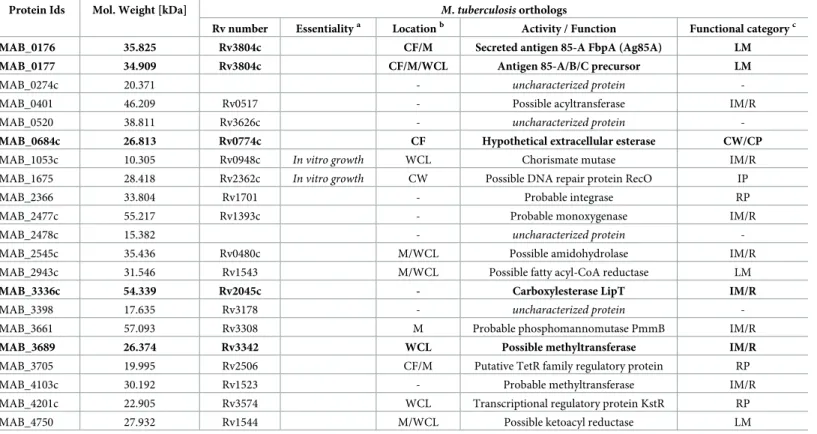
incu-bated with iBpPPOX or DMSO as a control. After cell lysis, the obtained total lysate was processed as described above with ActivX™ Desthiobiotin-FP probe and streptavidin mag-netic beads. Tryptic digestion followed by tandem mass spectrometry analysis led to the identification of 21 protein candidates at a pFDR of 5%, and only 5 at a pFDR of 1% (Table 2andS2 Table).
Although 4 of the identified proteins are only conserved hypotheticals, the remaining 17 ranged in their functional category from intermediary metabolism/respiration (8 proteins), lipid metabolism (4 proteins), regulatory pathways (3 proteins), cell wall/cell processes (1 pro-tein), and information pathways (1 protein). Among them, MAB_1675, the probable DNA repair protein RecO (i.e., Rv2362c), and MAB_1053c (i.e., Rv0948c) a putative chorismate
mutase possibly involved in phenylalanine, tyrosine and tryptophan biosynthesis, are anno-tated as essential enzymes for thein vitro growth of M. tuberculosis [44,45]. In good agreement with our previous work onM. tuberculosis target enzymes [12], several hydrolases were Table 2. iBpPPOX target proteins identified at a pFDR of 1% and 5% in M. abscessus R culture by LC-ESI-MS/MS analysis.
Protein Ids Mol. Weight [kDa] M. tuberculosis orthologs
Rv number Essentialitya Locationb Activity / Function Functional categoryc
MAB_0176 35.825 Rv3804c CF/M Secreted antigen 85-A FbpA (Ag85A) LM
MAB_0177 34.909 Rv3804c CF/M/WCL Antigen 85-A/B/C precursor LM
MAB_0274c 20.371 - uncharacterized protein
-MAB_0401 46.209 Rv0517 - Possible acyltransferase IM/R
MAB_0520 38.811 Rv3626c - uncharacterized protein
-MAB_0684c 26.813 Rv0774c CF Hypothetical extracellular esterase CW/CP
MAB_1053c 10.305 Rv0948c In vitro growth WCL Chorismate mutase IM/R
MAB_1675 28.418 Rv2362c In vitro growth CW Possible DNA repair protein RecO IP
MAB_2366 33.804 Rv1701 - Probable integrase RP
MAB_2477c 55.217 Rv1393c - Probable monoxygenase IM/R
MAB_2478c 15.382 - uncharacterized protein
-MAB_2545c 35.436 Rv0480c M/WCL Possible amidohydrolase IM/R
MAB_2943c 31.546 Rv1543 M/WCL Possible fatty acyl-CoA reductase LM
MAB_3336c 54.339 Rv2045c - Carboxylesterase LipT IM/R
MAB_3398 17.635 Rv3178 - uncharacterized protein
-MAB_3661 57.093 Rv3308 M Probable phosphomannomutase PmmB IM/R
MAB_3689 26.374 Rv3342 WCL Possible methyltransferase IM/R
MAB_3705 19.995 Rv2506 CF/M Putative TetR family regulatory protein RP
MAB_4103c 30.192 Rv1523 - Probable methyltransferase IM/R
MAB_4201c 22.905 Rv3574 WCL Transcriptional regulatory protein KstR RP
MAB_4750 27.932 Rv1544 M/WCL Possible ketoacyl reductase LM
In bold, the 5 proteins identified at a pFDR of 1%.
aFrom [44,45].
bCF: Culture filtrate; CW: Cell wall; M: Membrane fraction; WCL: Whole cell lysate.
cIM/R: intermediary metabolism/respiration; IP: information pathways; CW/CP: cell wall/cell processes; LM: lipid metabolism; RP: regulatory protein.
detected, including one hypothetical extracellular esterase (MAB_2181c), three putative methyltransferases (MAB_3689, MAB_4103c, MAB_0401); the carboxylesterase LipT (MAB_3336c) belonging to the Lip-family members, and the mycolyltransferases MAB_176 (Ag85A) and MAB_177 (Ag85-A/B/C precursor) two members of the Ag85 complex (Table 2 andS2 Table).
It is noteworthy that among these 21 potential hits, only Ag85 proteins were previously detected in the iBpPPOX-treated total lysate (seeS1andS2Tables); thus, implying that nearly 19 proteins had not been detected in the previous treatedM. abscessus total lysate, or at least at
a pFDR � 10%. On the other hand, such result suggests that Antigen 85 proteins may be the first target enzymes encountered and thus inhibited by the OX compounds.
Validation of M. abscessus Ag85C as vulnerable target of iBpPPOX
Knowing the importance of the Ag85 complex in mycobacterial membrane integrity due to its central role in cell envelope biogenesis, and given the fact that inhibiting the Ag85C was found to restrictM. tuberculosis growth [46], we decided to confirm the Ag85CMabs, which shares
nearly 58% amino acid sequence identity with itsM. tuberculosis ortholog and retains the
same conserved catalytic triad (i.e., Ser124-Glu228-His260), as a potential target of the OX compounds.
We thus followed two different strategies: the first one was based on the susceptibility test-ing of variousM. abscessus mutant strains to the iBpPPOX; and the second one relied on the
molecular interaction between the iBpPPOX and the purified recombinant Ag85CMabs.
In the first step, genes encoding either Ag85CMabsor the inactivated Ag85CS124Aprotein
were cloned and overexpressed inM. abscessus S and R variants using the pMyC::ag85C /
pMyC::ag85CS124Ainducible plasmids, where genes were cloned under the control of an acet-amide promoter (Fig 3A). Moreover, a deletion mutant of Ag85CMabsnamedΔag85C was
gen-erated by using a recent one-step single cross-over system with the pUX1 vector [22]; and its complemented counterpartΔag85C::C (Fig 3B) was obtained using the pVV16::ag85C
comple-mentation plasmid which allows the constitutive production of recombinant Ag85CMabsunder
the control of thehsp60 promoter (seeS1 Appendixfor cloning details). In each case, the over-expression/complementation of antigen 85C protein was confirmed by Western blotting as compared to the parental strain (WT) (Fig 3).
In order to examine whether the overexpression, inactivation or deletion/complementation of the Ag85CMabsprotein affect the strain susceptibility to the iBpPPOX compound, their
respective MICs were further determined.
As depicted inTable 3, the overexpression of Ag85CMabsprotein (i.e., M. abscessus
S_pMyc::ag85C and M. abscessus R_pMyc::ag85C) led to a significant increase in MIC50values
by 2.7-fold for both the S (87.3±3.4 μM; p-value <0.01) and R variant (148.2 ±2.1 μM; p-value
<0.01), as well as in MIC90values (>200μM), compared to the respective pMyc vector control
and wild-type strains. These results clearly suggest that Ag85CMabsis responsible for the
decreased susceptibility to the iBpPPOX, thus confirming this protein as one of the targets of our compound.
Regarding the inactivated Ag85CS124AmutantM. abscessus S_pMyc::ag85CS124A, the gene deletion mutantM. abscessus S_Δag85C and its complemented counterpart M. abscessus
S_Δag85C::C, as well as the wild-type M. abscessus S strain, they all responded similarly to
iBpPPOX. In the case of M. abscessus R, although no significant variation was observed in MIC90values (mean MIC90= 111.1±8.4 μM), a slight decrease in MIC50of around 0.89- to
0.58-fold was reached for the inactivated Ag85CS124A(47.5±2.0 μM; p-value <0.05) and the Δag85C (30.9 ±2.1 μM; p-value <0.01) mutants, respectively, compared to the wild-type strain
(53.2±1.8 μM); while complementation of Ag85CMabs(i.e., M. abscessus R_Δag85C::C)
restored the wild-type R phenotype (51.8±3.1 μM—Table 3).
Based on these results, purified Ag85CMabsrecombinant protein [25] was further incubated
with iBpPPOX, using increasing enzyme/inhibitor molar ratio (E/I) ranging from 1:1 to 1:75, and then treated with ActivX TAMRA-FP fluorescent probe, as reported previously [24,25]. Equal amounts of proteins were separated on SDS-PAGE and visualized by Coomassie stain-ing or in-gel fluorescence for TAMRA detection (Fig 4A). Relative fluorescence quantification of each band was done using the ImageLab™ software version 5.0 (Bio-Rad) by taking as 100% absolute fluorescence level, the labeled Ag85CMabs-TAMRA adduct (Fig 4A). As expected,
pre-treating Ag85CMabswith iBpPPOX, resulted in a significant loss in fluorescence intensity by
around 32.8±1.8% (E/I = 1:1 to 1:10), 58.5 ±0.70% (E/I = 1:25), 64.0 ±1.8% (E/I = 1:50) and up Fig 3. Western blot analysis ofM. abscessus-Ag85C-mutant strains: (A) overexpression of active (i.e., Ag85C) or inactivated (i. e., Ag85CS124A) protein; (B) deletion (i.e., Δag85C) and complementation (i.e., Δag85C::C) strains (seeS1 Appendixfile for cloning details). Each overexpressed protein, indicated with a red star, were revealed using HisProbe™ HRP conjugate (Thermo-Fisher Scientific) and compared to theM. abscessus wild-type strain (WT) as well as pure recombinant Ag85CMabsprotein, as
control. In each case, equal amount of whole bacterial cell lysate has been loaded for the overexpression strains (panel A), and for the deletion/complementation strains (panel B), respectively.
to >90% (E/I = 1:75) as compared to the non-treated protein labeled by the TAMRA-FP probe (Fig 4A). This means that the TAMRA-FP probe cannot bind the catalytic serine when the Ag85CMabs-iBpPPOX complex has been formed, as revealed by the significant loss in
fluores-cence emission (Fig 4A).
Table 3. Variation of MIC (μM) of iBpPPOX against M. abscessus-Ag85C-mutant strainsa.
M. abscessus strains MIC50/ MIC90(μM) MIC50/ MIC90ratio mutantvs. WT
M. abscessus S WT 33.0±2.0¶/ 85.9
±5.5┴ 1.0 / 1.0
M. abscessus S_pMyc empty vector 31.9±1.7 / 82.4 ±0.92 0.97 / 0.96
M. abscessus S_pMyc::ag85CS124A 34.4±3.0 / 83.1 ±6.8 1.04 / 0.97
M. abscessus S_Δag85C 33.7±1.9 / 81.5 ±7.4 1.02 / 0.95
M. abscessus S_Δag85C::C 32.6±1.3 / 87.4 ±1.5 0.99 / 1.02 M. abscessus S_pMyc::ag85C 87.3±3.4¶/ >200┴ 2.65 / >3.0
M. abscessus R WT 53.2±1.8‡,†,§/ 104.3±5.1║ 1.0 / 1.0
M. abscessus R_pMyc empty vector 49.9±2.6 / 109.2 ±10.4 0.94 / 1.05
M. abscessus R_pMyc::ag85CS124A 47.5±2.0‡,�/ 119.0±9.6 0.89 / 1.14
M. abscessus R_Δag85C 30.9±2.1†,�,#/ 114.9
±8.2 0.58 / 1.10
M. abscessus R_Δag85C::C 51.8±3.1#/ 108.2±4.6 0.97 / 1.04 M. abscessus R_pMyc::ag85C 148.2±2.1§
/ >200║ 2.78 / >2
aExperiments were performed as described in Materials and Methods. MIC
50/ MIC90: compound minimal
concentration leading to 50% or 90% growth inhibition, respectively. Values are mean of two independent assays performed in triplicate. MIC values with a common symbol are significantly different (‡:p-value<0.05;¶,┴, †, §, ║,�,#
:
p-value<0.01; ANOVA followed by Fisher’s test).
https://doi.org/10.1371/journal.pone.0238178.t003
Fig 4. Inhibition of the Ag85CMabsby iBpPPOX. (A) Ag85CMabswas pre-treated with iBpPPOX (i.e. enzyme/inhibitor molar ratio of 1:1 to 1:75),
incubated with ActiveX TAMRA-FP, separated by 12% SDS-PAGE, and visualized by Coomassie blue staining (upper panel) or in-gel fluorescence
visualization (middle panel). The merged image is shown in the lower panel. Untreated protein (i.e., no TAMRA-FP and no iBpPPOX) was used as
control. No TAMRA-FP labeling is detected in the presence of inactivated heat-treated Ag85CMabs. TAMRA labeling of Ag85CMabsis impaired in the
Ag85CMabs-iBpPPOX adducts, as evidenced by the loss of fluorescence in the iBpPPOX lanes, presumably resulting from the covalent binding of
iBpPPOX to the catalytic serine as previously observed [24,25]. TAMRA-labeled Ag85CMabswas detected by fluorescent gel scanning (λex557 nm,λem
583 nm) using the Cy13 filter of a ChemiDoc MP Imager (Bio-Rad) before staining of the gel with Coomassie Brilliant Blue dye. Relative fluorescence quantification of each band was performed using the ImageLab™ software version 5.0 (Bio-Rad) by taking as 100% absolute fluorescence level the labeled Ag85CMabs-TAMRA adduct. (B) Global mass modification of Ag85CMabspre-incubated with iBpPPOX, at an enzyme/inhibitor molar ratio of
1:100 to ensure total inhibition, as determined using an MALDI-TOF-TOF mass spectrometer in linear mode. (C) Mechanism of inhibition of Ag85CMabsby the oxadiazolone iBpPPOX, based on mass spectrometry analysis. a.u., arbitrary units.
MALDI-TOF mass spectrometry was further used to confirm the (covalent) nature of the inhibition. Sample of the Ag85CMabs-iBpPPOX (E/I = 1:100) complex was subjected to
MAL-DI-TOF mass spectrometry analyses. Mass increment of +305.3 Da was then observed within the global mass of the inhibited Ag85CMabsas compared with the untreated protein (Fig 4B);
whereas no changes in the global mass were observed with the inactivated heat-treated protein. Such result is thus consistent with the formation of a covalent enzyme-inhibitor adduct, as the reaction between the catalytic Ser124 and iBpPPOX is expected to yield a mass increase of +326 Da; and also, in agreement with the mechanism of action of such OX derivatives [42]. All these findings conclusively indicate that pure recombinant Ag85CMabsprotein is covalently
modified by the iBpPPOX derivative (Fig 4C), in good agreement with the known classical mechanism of action of such OX compounds as previously demonstrated using pure lipolytic enzymes [12,42].
Taken together, thein vitro inhibitory experiments conducted with iBpPPOX on pure
recombinant Ag85CMabsprotein (Fig 4), as well as the statistically significant increased
resis-tance levels when overexpressing the Ag85CMabsprotein inM. abscessus S and R variants
(Table 3), thus confirm the assertion that this enzyme is an effective target of iBpPPOX.
Conclusion
As already highlighted in the case ofM. tuberculosis [12], our series of oxadiazolone-core OX derivatives are able to impair different metabolic pathways during either extracellular and/or intracellular bacterial growthvia the inhibition of various (Ser/Cys)-based enzymes, therefore
resulting inM. abscessus death. Although the efficiency of these OX molecules could not be
considered as sufficient enough to obtain powerful anti-mycobacterial agents, they may how-ever represent attractive tools for deciphering the lipid metabolism inM. abscessus and/or in M. tuberculosis. We have indeed reported that the MmPPOX compound was able to prevent
intracytoplasmic lipid inclusion (ILI) catabolismin vivo in M. bovis BCG infected murine
bone-marrow-derived macrophages (mBMDM) [47–49]; as well asin vitro under carbon
excess and nitrogen-deprived conditions allowing ILI biosynthesis and hydrolysis inM. absces-sus [50]. Taken together, all these findings support that the OX derivatives are able to abolish the activity of several (Ser/Cys)-containing enzymes involved in mycobacterial lipid metabo-lism and/or in cell wall biosynthesis. This is the case of the Ag85 complex proteins which are essential players in the biosynthesis of lipids from mycobacterial membrane as well as in intra-cellular lipid metabolism, but also of proteins belonging to the hormone-sensitive lipase (HSL) family member proteins (i.e., Lip-HSL) [42], including LipY the major Lip-HSL lipase involved in mycobacterial lipid catabolism [49–52]. Therefore, the respective effects of these OX com-pounds against lipid-poorvs. lipid-rich bacteria deserve to be investigated in more details.
More especially, deciphering how the presence of intracytoplasmic lipid inclusions (ILI) in lipid-rich bacteria can actively contribute to substantially enhanced mycobacterial virulence and pathogenesis as compared to lipid-poor strains, as reported recently [50], will provide major insights for understanding the general development of mycobacterial-related diseases. Such experiments are currently underway, and will be reported in due course.
Supporting information
S1 Appendix. Detailed protocols regarding the MIC determination, targets identification and mass spectrometry analysis of Ag85CMabs; as well as the list of plasmids and primers
used in this study.
S1 Fig. Uncropped and unadjusted image for Western Blotting ofFig 3. Each overexpressed
protein was revealed using the HisProbe™ HRP conjugate (ThermoFisher Scientific) and com-pared to theM. abscessus wild type strain as well as the pure recombinant Ag85CMabsprotein. (TIF)
S2 Fig. Uncropped and unadjusted images for SDS-PAGE gel ofFig 4A. SDS-PAGE gel
visualized by Coomassie blue staining (upper panel) or by in-gel fluorescence visualization
(middle panel). Superimposition of both images is reported in the lower panel. Molecular
weights were derived from the Unstained Protein Molecular Weight Marker (Euromedex). (TIF)
S1 Table. iBpPPOX target proteins identified in M. abscessus R total lysate by LC-ESI-MS/ MS analysis. Only positive hits with a pFDR of 1%, 5% and 10% are reported.
(XLSX)
S2 Table. iBpPPOX target proteins identified in M. abscessus R culture cell by LC-ESI-MS/ MS analysis. Only positive hits with a pFDR of 1% and 5% are reported.
(XLSX)
Acknowledgments
Authors would like to thank Dr. R. Lebrun and P. Mansuelle at the Proteomics platform of the Institut de Microbiologie de la Me´diterrane´e FR3479 (Marseille, France) for N-Terminal Edman sequencing.
Author Contributions
Conceptualization: Ste´phane Canaan, Jean Franc¸ois Cavalier.
Data curation: Abdeldjalil Madani, Ivy Mallick, Patrick Fourquet, Ste´phane Audebert, Luc
Camoin.
Formal analysis: Ste´phane Audebert, Luc Camoin, Ste´phane Canaan, Jean Franc¸ois Cavalier. Funding acquisition: Jean Franc¸ois Cavalier.
Investigation: Abdeldjalil Madani, Ivy Mallick, Patrick Fourquet, Ste´phane Audebert, Luc
Camoin.
Project administration: Jean Franc¸ois Cavalier.
Resources: Alexandre Guy, Ce´line Crauste, Thierry Durand. Supervision: Jean Franc¸ois Cavalier.
Validation: Ste´phane Canaan, Jean Franc¸ois Cavalier. Visualization: Jean Franc¸ois Cavalier.
Writing – original draft: Abdeldjalil Madani, Ivy Mallick.
Writing – review & editing: Ce´line Crauste, Thierry Durand, Ste´phane Audebert, Luc
Camoin, Ste´phane Canaan, Jean Franc¸ois Cavalier.
References
1. Porvaznik I, Solovic I, Mokry J. Non-Tuberculous Mycobacteria: Classification, Diagnostics, and Ther-apy. Adv Exp Med Biol. 2017; 944:19–25. Epub 2016/11/09.https://doi.org/10.1007/5584_2016_45 PMID:27826888.
2. Claeys TA, Robinson RT. The many lives of nontuberculous mycobacteria. J Bacteriol. 2018. Epub 2018/02/28.https://doi.org/10.1128/JB.00739-17PMID:29483164.
3. Lee MR, Sheng WH, Hung CC, Yu CJ, Lee LN, Hsueh PR. Mycobacterium abscessus Complex Infec-tions in Humans. Emerg Infect Dis. 2015; 21(9):1638–46. Epub 2015/08/22.https://doi.org/10.3201/ 2109.141634PMID:26295364.
4. Luthra S, Rominski A, Sander P. The Role of Antibiotic-Target-Modifying and Antibiotic-Modifying Enzymes in Mycobacterium abscessus Drug Resistance. Front Microbiol. 2018; 9:2179. Epub 2018/09/ 28.https://doi.org/10.3389/fmicb.2018.02179PMID:30258428.
5. Brown-Elliott BA, Nash KA, Wallace RJ Jr. Antimicrobial susceptibility testing, drug resistance mecha-nisms, and therapy of infections with nontuberculous mycobacteria. Clin Microbiol Rev. 2012; 25 (3):545–82. Epub 2012/07/06.https://doi.org/10.1128/CMR.05030-11PMID:22763637.
6. Catherinot E, Clarissou J, Etienne G, Ripoll F, Emile JF, Daffe M, et al. Hypervirulence of a rough variant of the Mycobacterium abscessus type strain. Infect Immun. 2007; 75(2):1055–8. Epub 2006/12/06. https://doi.org/10.1128/IAI.00835-06PMID:17145951.
7. Howard ST, Rhoades E, Recht J, Pang X, Alsup A, Kolter R, et al. Spontaneous reversion of Mycobac-terium abscessus from a smooth to a rough morphotype is associated with reduced expression of glyco-peptidolipid and reacquisition of an invasive phenotype. Microbiology. 2006; 152(Pt 6):1581–90. Epub 2006/06/01.https://doi.org/10.1099/mic.0.28625-0PMID:16735722.
8. Pawlik A, Garnier G, Orgeur M, Tong P, Lohan A, Le Chevalier F, et al. Identification and characteriza-tion of the genetic changes responsible for the characteristic smooth-to-rough morphotype alteracharacteriza-tions of clinically persistent Mycobacterium abscessus. Mol Microbiol. 2013; 90(3):612–29. Epub 2013/09/ 04.https://doi.org/10.1111/mmi.12387PMID:23998761.
9. Floto RA, Olivier KN, Saiman L, Daley CL, Herrmann JL, Nick JA, et al. US Cystic Fibrosis Foundation and European Cystic Fibrosis Society consensus recommendations for the management of non-tuber-culous mycobacteria in individuals with cystic fibrosis: executive summary. Thorax. 2016; 71(1):88–90. Epub 2015/12/19.https://doi.org/10.1136/thoraxjnl-2015-207983PMID:26678435.
10. Nash KA, Brown-Elliott BA, Wallace RJ Jr. A novel gene, erm(41), confers inducible macrolide resis-tance to clinical isolates of Mycobacterium abscessus but is absent from Mycobacterium chelonae. Anti-microb Agents Chemother. 2009; 53(4):1367–76. Epub 2009/01/28.https://doi.org/10.1128/AAC. 01275-08PMID:19171799.
11. Bastian S, Veziris N, Roux AL, Brossier F, Gaillard JL, Jarlier V, et al. Assessment of clarithromycin sus-ceptibility in strains belonging to the Mycobacterium abscessus group by erm(41) and rrl sequencing. Antimicrob Agents Chemother. 2011; 55(2):775–81. Epub 2010/12/08.https://doi.org/10.1128/AAC. 00861-10PMID:21135185.
12. Nguyen PC, Delorme V, Be´narouche A, Guy A, Landry V, Audebert S, et al. Oxadiazolone derivatives, new promising multi-target inhibitors against M. tuberculosis. Bioorg Chem. 2018; 81:414–24.https:// doi.org/10.1016/j.bioorg.2018.08.025PMID:30212765
13. Nguyen PC, Delorme V, Be´narouche A, Martin BP, Paudel R, Gnawali GR, et al. Cyclipostins and Cyclophostin analogs as promising compounds in the fight against tuberculosis. Scientific Reports. 2017; 7(1):11751.https://doi.org/10.1038/s41598-017-11843-4PMID:28924204
14. Nguyen PC, Madani A, Santucci P, Martin BP, Paudel RR, Delattre S, et al. Cyclophostin and Cyclipos-tins analogs, new promising molecules to treat mycobacterial-related diseases. Int J Antimicrob Agents. 2018; 51:651–4.https://doi.org/10.1016/j.ijantimicag.2017.12.001PMID:29241819
15. Palomino JC, Martin A, Camacho M, Guerra H, Swings J, Portaels F. Resazurin microtiter assay plate: simple and inexpensive method for detection of drug resistance in Mycobacterium tuberculosis. Antimi-crob Agents Chemother. 2002; 46(8):2720–2. Epub 2002/07/18.https://doi.org/10.1128/aac.46.8. 2720-2722.2002PMID:12121966.
16. Rybniker J, Vocat A, Sala C, Busso P, Pojer F, Benjak A, et al. Lansoprazole is an antituberculous pro-drug targeting cytochrome bc1. Nat Commun. 2015; 6:7659. Epub 2015/07/15.https://doi.org/10.1038/ ncomms8659PMID:26158909.
17. Rodrigues Felix C, Gupta R, Geden S, Roberts J, Winder P, Pomponi SA, et al. Selective Killing of Dor-mant Mycobacterium tuberculosis by Marine Natural Products. Antimicrob Agents Chemother. 2017; 61 (8):e00743–17.https://doi.org/10.1128/aac.00743-17PMID:28607021
18. Strober W. Trypan blue exclusion test of cell viability. Curr Protoc Immunol. 2001; Appendix 3(1):Appen-dix 3B. Epub 2008/04/25.https://doi.org/10.1002/0471142735.ima03bs21PMID:18432654.
19. Shevchenko A, Wilm M, Vorm O, Mann M. Mass spectrometric sequencing of proteins silver-stained polyacrylamide gels. Anal Chem. 1996; 68(5):850–8. Epub 1996/03/01.https://doi.org/10.1021/ ac950914hPMID:8779443.
20. Cox J, Mann M. MaxQuant enables high peptide identification rates, individualized p.p.b.-range mass accuracies and proteome-wide protein quantification. Nat Biotechnol. 2008; 26(12):1367–72. Epub 2008/11/26.https://doi.org/10.1038/nbt.1511PMID:19029910.
21. Vizcaino JA, Deutsch EW, Wang R, Csordas A, Reisinger F, Rios D, et al. ProteomeXchange provides globally coordinated proteomics data submission and dissemination. Nat Biotechnol. 2014; 32(3):223– 6. Epub 2014/04/15.https://doi.org/10.1038/nbt.2839PMID:24727771.
22. Viljoen A, Gutierrez AV, Dupont C, Ghigo E, Kremer L. A Simple and Rapid Gene Disruption Strategy in Mycobacterium abscessus: On the Design and Application of Glycopeptidolipid Mutants. Front Cell Infect Microbiol. 2018; 8:69. Epub 2018/03/30.https://doi.org/10.3389/fcimb.2018.00069PMID: 29594066.
23. Jeong J-Y, Yim H-S, Ryu J-Y, Lee HS, Lee J-H, Seen D-S, et al. One-Step Sequence- and Ligation-Independent Cloning as a Rapid and Versatile Cloning Method for Functional Genomics Studies. Appl Environ Microbiol. 2012; 78(15):5440–3.https://doi.org/10.1128/AEM.00844-12PMID:22610439
24. Viljoen A, Richard M, Nguyen PC, Fourquet P, Camoin L, Paudal RR, et al. Cyclipostins and Cyclophos-tin analogs inhibit the antigen 85C from Mycobacterium tuberculosis both in vitro and in vivo. J Biol Chem. 2018; 293(8):2755–69.https://doi.org/10.1074/jbc.RA117.000760PMID:29301937
25. Madani A, Ridenour JN, Martin BP, Paudel RR, Abdul Basir A, Le Moigne V, et al. Cyclipostins and Cyclophostin Analogues as Multitarget Inhibitors That Impair Growth of Mycobacterium abscessus. ACS Infect Dis. 2019; 5(9):1597–608. Epub 2019/07/13.https://doi.org/10.1021/acsinfecdis.9b00172 PMID:31299146.
26. Hawke D, Yuan P. S-Pyridylethylation of cystine residues. Applied Biosystems Bulletin 28. 1987: Applied Biosystems, Foster City, CA.
27. Singh S, Bouzinbi N, Chaturvedi V, Godreuil S, Kremer L. In vitro evaluation of a new drug combination against clinical isolates belonging to the Mycobacterium abscessus complex. Clin Microbiol Infect. 2014; 20(12):O1124–O7.https://doi.org/10.1111/1469-0691.12780PMID:25185732
28. Nessar R, Cambau E, Reyrat JM, Murray A, Gicquel B. Mycobacterium abscessus: a new antibiotic nightmare. J Antimicrob Chemother. 2012; 67(4):810–8.https://doi.org/10.1093/jac/dkr578PMID: 22290346
29. Bernut A, Herrmann JL, Kissa K, Dubremetz JF, Gaillard JL, Lutfalla G, et al. Mycobacterium abscessus cording prevents phagocytosis and promotes abscess formation. Proceedings of the National Academy of Sciences of the United States of America. 2014; 111(10):E943–52. Epub 2014/02/26.https://doi.org/ 10.1073/pnas.1321390111PMID:24567393.
30. Johansen MD, Herrmann JL, Kremer L. Non-tuberculous mycobacteria and the rise of Mycobacterium abscessus. Nat Rev Microbiol. 2020. Epub 2020/02/23.https://doi.org/10.1038/s41579-020-0331-1 PMID:32086501.
31. Lefebvre AL, Le Moigne V, Bernut A, Veckerle C, Compain F, Herrmann JL, et al. Inhibition of the beta-Lactamase BlaMabby Avibactam Improves the In Vitro and In Vivo Efficacy of Imipenem against
Myco-bacterium abscessus. Antimicrob Agents Chemother. 2017; 61(4):e02440–16. Epub 2017/01/18.
https://doi.org/10.1128/AAC.02440-16PMID:28096155.
32. Le Run E, Arthur M, Mainardi JL. In Vitro and Intracellular Activity of Imipenem Combined with Tedizolid, Rifabutin, and Avibactam against Mycobacterium abscessus. Antimicrob Agents Chemother. 2019; 63 (4):e01915–18. Epub 2019/02/13.https://doi.org/10.1128/AAC.01915-18PMID:30745387.
33. Christophe T, Jackson M, Jeon HK, Fenistein D, Contreras-Dominguez M, Kim J, et al. High content screening identifies decaprenyl-phosphoribose 2’ epimerase as a target for intracellular antimycobac-terial inhibitors. PLoS Pathog. 2009; 5(10):e1000645. Epub 2009/10/31.https://doi.org/10.1371/journal. ppat.1000645PMID:19876393.
34. Ravindran MS, Rao SP, Cheng X, Shukla A, Cazenave-Gassiot A, Yao SQ, et al. Targeting Lipid Ester-ases in Mycobacteria Grown Under Different Physiological Conditions Using Activity-based Profiling with Tetrahydrolipstatin (THL). Mol Cell Proteomics. 2014; 13(2):435–48.https://doi.org/10.1074/mcp. M113.029942PMID:24345785
35. Tallman KR, Levine SR, Beatty KE. Small Molecule Probes Reveal Esterases with Persistent Activity in Dormant and Reactivating Mycobacterium tuberculosis. ACS Infect Dis. 2016; 2(12):936–44. Epub 2016/10/04.https://doi.org/10.1021/acsinfecdis.6b00135PMID:27690385.
36. Lehmann J, Cheng TY, Aggarwal A, Park AS, Zeiler E, Raju RM, et al. An Antibacterial beta-Lactone Kills Mycobacterium tuberculosis by Disrupting Mycolic Acid Biosynthesis. Angew Chem Int Ed Engl. 2018; 57(1):348–53. Epub 2017/10/27.https://doi.org/10.1002/anie.201709365PMID:29067779.
37. Lehmann J, Vomacka J, Esser K, Nodwell M, Kolbe K, Ramer P, et al. Human lysosomal acid lipase inhibitor lalistat impairs Mycobacterium tuberculosis growth by targeting bacterial hydrolases. Med-ChemComm. 2016; 7:1797–801.https://doi.org/10.1039/c6md00231e.
38. Catherinot E, Roux AL, Macheras E, Hubert D, Matmar M, Dannhoffer L, et al. Acute respiratory failure involving an R variant of Mycobacterium abscessus. J Clin Microbiol. 2009; 47(1):271–4. Epub 2008/ 11/21.https://doi.org/10.1128/JCM.01478-08PMID:19020061.
39. Koonin EV. Orthologs, Paralogs, and Evolutionary Genomics. Annu Rev Genet. 2005; 39(1):309–38. https://doi.org/10.1146/annurev.genet.39.073003.114725PMID:16285863.
40. Dedieu L, Serveau-Avesque C, Kremer L, Canaan S. Mycobacterial lipolytic enzymes: a gold mine for tuberculosis research. Biochimie. 2013; 95(1):66–73.https://doi.org/10.1016/j.biochi.2012.07.008 PMID:22819994.
41. Johnson G. The alpha/beta Hydrolase Fold Proteins of Mycobacterium tuberculosis, with Reference to their Contribution to Virulence. Curr Protein Pept Sci. 2017; 18(3):190–210. Epub 2016/08/03.https:// doi.org/10.2174/1389203717666160729093515PMID:27480283.
42. Delorme V, Diomande´ SV, Dedieu L, Cavalier J-F, Carrière F, Kremer L, et al. MmPPOX Inhibits
Myco-bacterium tuberculosis Lipolytic Enzymes Belonging to the Hormone-Sensitive Lipase Family and Alters
Mycobacterial Growth. PLoS ONE. 2012; 7(9):e46493.https://doi.org/10.1371/journal.pone.0046493 PMID:23029536
43. Sacchettini JC, Ronning DR. The mycobacterial antigens 85 complex—from structure to function: response. Trends Microbiol. 2000; 8(10):441. Epub 2001/02/24.https://doi.org/10.1016/S0966-842X (00)01843-6PMID:11203233.
44. Griffin JE, Gawronski JD, Dejesus MA, Ioerger TR, Akerley BJ, Sassetti CM. High-resolution phenotypic profiling defines genes essential for mycobacterial growth and cholesterol catabolism. PLoS Pathog. 2011; 7(9):e1002251. Epub 2011/10/08.https://doi.org/10.1371/journal.ppat.1002251PMID: 21980284.
45. Sassetti CM, Boyd DH, Rubin EJ. Genes required for mycobacterial growth defined by high density mutagenesis. Mol Microbiol. 2003; 48(1):77–84.https://doi.org/10.1046/j.1365-2958.2003.03425.x PMID:12657046
46. Warrier T, Tropis M, Werngren J, Diehl A, Gengenbacher M, Schlegel B, et al. Antigen 85C inhibition restricts Mycobacterium tuberculosis growth through disruption of cord factor biosynthesis. Antimicrob Agents Chemother. 2012; 56(4):1735–43. Epub 2012/02/01.https://doi.org/10.1128/AAC.05742-11 PMID:22290959.
47. Caire-Brandli I, Papadopoulos A, Malaga W, Marais D, Canaan S, Thilo L, et al. Reversible lipid accu-mulation and associated division arrest of Mycobacterium avium in lipoprotein-induced foamy macro-phages may resemble key events during latency and reactivation of tuberculosis. Infect Immun. 2014; 82(2):476–90.https://doi.org/10.1128/IAI.01196-13PMID:24478064.
48. Santucci P, Bouzid F, Smichi N, Poncin I, Kremer L, De Chastellier C, et al. Experimental Models of Foamy Macrophages and Approaches for Dissecting the Mechanisms of Lipid Accumulation and Con-sumption during Dormancy and Reactivation of Tuberculosis. Front Cell Infect Microbiol. 2016; 6:122. Epub 2016/10/25.https://doi.org/10.3389/fcimb.2016.00122PMID:27774438.
49. Santucci P, Diomande´ S, Poncin I, Alibaud L, Viljoen A, Kremer L, et al. Delineating the physiological roles of the PE and catalytic domain of LipY in lipid consumption in mycobacteria-infected foamy macro-phages. Infect Immun. 2018; 86(9):e00394–18.https://doi.org/10.1128/iai.00394-18PMID:29986895
50. Santucci P, Johansen MD, Point V, Poncin I, Viljoen A, Cavalier J-F, et al. Nitrogen deprivation induces triacylglycerol accumulation, drug tolerance and hypervirulence in mycobacteria. Scientific Reports. 2019; 9(1):8667.https://doi.org/10.1038/s41598-019-45164-5PMID:31209261
51. Deb C, Daniel J, Sirakova TD, Abomoelak B, Dubey VS, Kolattukudy PE. A novel lipase belonging to the hormone-sensitive lipase family induced under starvation to utilize stored triacylglycerol in Mycobac-terium tuberculosis. J Biol Chem. 2006; 281(7):3866–75. Epub 2005/12/16.https://doi.org/10.1074/jbc. M505556200PMID:16354661.
52. Mishra KC, de Chastellier C, Narayana Y, Bifani P, Brown AK, Besra GS, et al. Functional role of the PE domain and immunogenicity of the Mycobacterium tuberculosis triacylglycerol hydrolase LipY. Infect Immun. 2008; 76(1):127–40. Epub 2007/10/17.https://doi.org/10.1128/IAI.00410-07PMID:17938218.