COMMENTARY
Peroxisome proliferator-activated receptor
γ coactivator 1β
(PGC-1
β) improves skeletal muscle mitochondrial
function and insulin sensitivity
C. Handschin
Received: 14 February 2011 / Accepted: 10 March 2011 / Published online: 2 April 2011 # Springer-Verlag 2011
Abstract Proteins belonging to the peroxisome proliferator-activated receptor γ coactivator 1 (PGC-1) family are key regulators of cellular energy homeostasis in a number of oxidative tissues, including skeletal muscle. While the regulation and function of PGC-1α seems central to muscle fibre plasticity in endurance exercise, the role of PGC-1β in this tissue is less clear. Wright et al. (Diabetologia, DOI:
10.1007/s00125-011-2068-x) provide evidence for a protec-tive effect of moderately elevated PGC-1β in electroporated rat skeletal muscle against high-fat-diet-induced insulin resistance, at least in part by promoting the oxidation of long chain acyl-CoA entities and the elimination of reactive oxygen species. These data provide important insights into the biological role of PGC-1β in skeletal muscle and imply novel therapeutic avenues for improving peripheral insulin sensitivity.
Keywords Insulin resistance . Long-chain acyl-CoA . PGC-1β . Reactive oxygen species . Skeletal muscle Abbreviation
PGC Peroxisome proliferator-activated receptor γ coactivator
Adequate skeletal muscle oxidative capacity and mitochon-drial function are crucial for maintaining systemic glucose and insulin levels and, accordingly, the regulation of these processes is tightly controlled. In recent years, the three members of the peroxisome proliferator-activated receptorγ coactivator 1 (PGC-1) family of transcriptional coactiva-tors have emerged as central regulacoactiva-tors of energy homeostasis in muscle and other tissues. Pgc-1α (also known as Ppargc1a) gene expression and the levels and activity of the protein are affected by almost all of the signalling pathways that are activated in the contracting muscle fibres [1]. In turn, PGC-1α controls the adaptive
changes elicited by endurance training [1]. In contrast to the more detailed information available on PGC-1α, much less is known about PGC-1β and the PGC-1-related coactivator. While relative level of physical activity is the dominant regulator of Pgc-1α gene expression in skeletal muscle, transcriptional control of Pgc-1β (also known as Ppargc1b) seems less straightforward, and different effects of exercise, denervation, dietary restriction, bariatric surgery, obesity and insulin stimulation on muscle Pgc-1β mRNA levels have been described, some with conflicting results in different experimental settings (summarised in references [1–4]). The elegant study by Wright et al. published in this issue of Diabetologia [2] sheds more light on the important role of PGC-1β in muscle physiology. Pgc-1β was over-expressed in electroporated tibialis cranialis and extensor digitorum longus muscles of low-fat- or high-fat-fed rats (Fig. 1). Despite the very moderate increase in PGC-1β
levels, a significant elevation in mitochondrial oxidative
C. Handschin (*)
Biozentrum, Division of Pharmacology/Neurobiology, University of Basel, Klingelbergstrasse 50/70, 4056 Basel, Switzerland e-mail: [email protected] Diabetologia (2011) 54:1270–1272 DOI 10.1007/s00125-011-2135-3
capacity, reactive oxidative species detoxification and lipid metabolism ensued. Importantly though, ectopic expression of PGC-1β in the high-fat-fed rats also boosted insulin sensitivity. While a causal link between reduced insulin resistance and the observed induction of long-chain acyl-CoA metabolism and antioxidant defence by PGC-1β was not examined, these long-chain acyl-CoA and reactive oxygen species are likely to contribute to the development of muscle insulin resistance [5].
Dysregulation of PGC-1α and PGC-1β gene expression levels has been found in the skeletal muscle of type 2 diabetic patients and individuals with impaired glucose tolerance, at least in some populations [6, 7]. Studies in skeletal muscle-specific knockout mice for Pgc-1α or Pgc-1β alone failed to show a direct involvement of genetic ablation of the individual coactivators in the aetiology of insulin resistance in this tissue [8–10]. The consequence of a Pgc-1α/Pgc-1β muscle-specific double knockout ap-proach remains unknown. In contrast, the data provided by Wright et al. [2] suggest a protective role for PGC-1β
against high-fat-diet-induced insulin resistance in skeletal
muscle. Similarly, the moderate and acute modulation of electroporated PGC-1α in muscle resulted in improvement of insulin sensitivity in the muscle of lean and obese Zucker rats [11]. Interestingly, the exact opposite observation, i.e. a more rapidly progressing insulin resistance, was reported for high-fat-fed muscle-specific Pgc-1α transgenic mice [12]. The electroporation-based approach used by Wright et al. [2] to increase PGC-1β levels in individual muscles
differs from the use of muscle-specific transgenic animals, which produces a stronger, chronic elevation of PGC-1β [13]. Thus, potentially confounding effects caused by the strong transgenic expression—such as the strong inhibition of Pgc-1α gene expression in the Pgc-1β muscle-specific transgenic animals [13] that could contribute to the muscle fibre-type switch observed in these mice—were not encountered by Wright et al. Taking into consideration their findings and the previous work by Bonen et al. [11], Wright et al. propose that a moderate acute elevation of PGC-1α or PGC-1β restores insulin sensitivity, at least in glycolytic muscles in rats. Accordingly, modulation of PGC-1α and PGC-1β gene expression might be an attractive novel therapeutic option against type 2 diabetes. However, designing pharmacological interventions aimed at the PGC-1 coactivators is not a trivial task [3]. First, the chemical entities that selectively induce PGC-1α or PGC-1β gene expression in skeletal muscle remain elusive. Second, pharmacological methods that alter PGC-1 levels must achieve a concentration within a therapeutically benefi-cial window in order to avoid the detrimental effects associated with inadequate or excessive PGC-1 levels [3]. Finally, some paradoxical findings about the PGC-1 coactivators in muscle remain unexplained. For example, in the present study [2], Pgc-1β expression and oxidative
metabolism are already increased as a consequence of the high-fat diet, and it is unclear how further elevation of PGC-1β levels exerts the observed amelioration of insulin sensitivity. Moreover, while ectopic PGC-1β lowers the long-chain acyl-CoA pool in muscle, the high-fat-diet-induced elevation of intramuscular ceramide and diacyl-glycerol, both strongly implicated in causing insulin resistance [5], is unaffected. It is also unclear how the chronic vs acute and moderate vs strong expression of Pgc-1α and Pgc-1β differ mechanistically and, at least in the case of Pgc-1α, result in diametrically opposite outcomes in terms of peripheral insulin sensitivity. Whether this is also true for Pgc-1β is unclear, since the effect of the transgenic expression of Pgc-1β in muscle on insulin resistance has not yet been elucidated [13]. Therefore, more studies are needed to fundamentally understand the regulation and function of the PGC-1 proteins in skeletal muscle before an appropriate drug-targeting strategy aimed at these coactivators can be attempted.
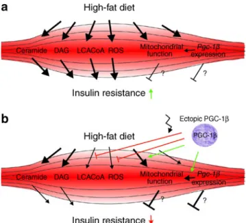
Fig. 1 Overexpression of Pgc-1β in glycolytic rat skeletal muscles improves insulin sensitivity. a A high-fat diet promotes the increase in intramuscular levels of ceramide, diacylglycerol (DAG), long-chain acyl CoA entities (LCACoA) and reactive oxygen species (ROS), all of which contribute to the development of insulin resistance. The reason for the increase in mitochondrial function and Pgc-1β expression in the muscle of high-fat-fed rats remains unclear. It is conceivable that the resulting boost in fatty acid oxidation is an adaptive compensatory, yet inadequate, mechanism to deal with the lipid overload. b Ectopic expression of Pgc-1β in electroporated glycolytic rat muscles results in increased mitochondrial function, fatty acid metabolism and antioxidant defence. As a consequence, LCACoA levels are lowered. Despite the inability of ectopic PGC-1β to reduce intramuscular ceramide and DAG, an improvement in insulin sensitivity is achieved
Acknowledgements The research in our laboratory is supported by the Swiss National Science Foundation (SNF), the Muscular Dystro-phy Association USA (MDA), the Association Française contre les Myopathies (AFM), the Swiss Society for Research on Muscle Diseases (SSEM), the Gebert-Rüf Foundation‘Rare Diseases’ Program (GRS), the Swiss Initiative in Systems Biology (SystemsX.ch), the United Mitochondrial Disease Foundation (UMDF), the Roche Foundation, the Swiss Diabetes Association, the SwissLife ‘Jubi-läumsstiftung für Volksgesundheit und medizinische Forschung’ and the University of Basel.
Duality of interest statement The author declares that there is no duality of interest associated with this manuscript.
References
1. Handschin C (2010) Regulation of skeletal muscle cell plasticity by the peroxisome proliferator-activated receptor γ coactivator 1α. J Recept Signal Transduct Res 30:376–384
2. Wright LE, Brandon AE, Hoy AJ et al (2011) Amelioration of lipid-induced insulin resistance in rat skeletal muscle by overexpression of Pgc-1β involves reductions in long-chain acyl-CoA levels and oxidative stress. Diabetologia. doi:10.1007/s00125-011-2068-x 3. Handschin C (2009) The biology of PGC-1α and its therapeutic
potential. Trends Pharmacol Sci 30:322–329
4. Handschin C (2009) PGC-1α in muscle links metabolism to inflammation. Clin Exp Pharmacol Physiol 36:1139–1143 5. Samuel VT, Petersen KF, Shulman GI (2010) Lipid-induced insulin
resistance: unravelling the mechanism. Lancet 375:2267–2277
6. Patti ME, Butte AJ, Crunkhorn S et al (2003) Coordinated reduction of genes of oxidative metabolism in humans with insulin resistance and diabetes: potential role of PGC1 and NRF1. Proc Natl Acad Sci USA 100:8466–8471
7. Mootha VK, Lindgren CM, Eriksson KF et al (2003) PGC-1α-responsive genes involved in oxidative phosphorylation are coordinately downregulated in human diabetes. Nat Genet 34:267–273
8. Handschin C, Choi CS, Chin S et al (2007) Abnormal glucose homeostasis in skeletal muscle-specific PGC-1α knockout mice reveals skeletal muscle-pancreatic beta cell crosstalk. J Clin Investig 117:3463–3474
9. Zechner C, Lai L, Zechner JF et al (2010) Total skeletal muscle PGC-1 deficiency uncouples mitochondrial derangements from fiber type determination and insulin sensitivity. Cell Metab 12:633–642
10. Handschin C, Chin S, Li P et al (2007) Skeletal muscle fiber-type switching, exercise intolerance, and myopathy in PGC-1α muscle-specific knock-out animals. J Biol Chem 282:30014–30021 11. Benton CR, Holloway GP, Han XX et al (2010) Increased levels
of peroxisome proliferator-activated receptor gamma, coactivator 1 alpha (PGC-1α) improve lipid utilisation, insulin signalling and glucose transport in skeletal muscle of lean and insulin-resistant obese Zucker rats. Diabetologia 53:2008–2019
12. Choi CS, Befroy DE, Codella R et al (2008) Paradoxical effects of increased expression of PGC-1α on muscle mitochondrial function and insulin-stimulated muscle glucose metabolism. Proc Natl Acad Sci USA 105:19926–19931
13. Arany Z, Lebrasseur N, Morris C et al (2007) The transcriptional coactivator PGC-1β drives the formation of oxidative type IIX fibers in skeletal muscle. Cell Metab 5:35–46