HAL Id: hal-02146273
https://hal.archives-ouvertes.fr/hal-02146273
Submitted on 12 Nov 2020
HAL is a multi-disciplinary open access
archive for the deposit and dissemination of
sci-entific research documents, whether they are
pub-lished or not. The documents may come from
teaching and research institutions in France or
abroad, or from public or private research centers.
L’archive ouverte pluridisciplinaire HAL, est
destinée au dépôt et à la diffusion de documents
scientifiques de niveau recherche, publiés ou non,
émanant des établissements d’enseignement et de
recherche français ou étrangers, des laboratoires
publics ou privés.
Distributed under a Creative Commons Attribution| 4.0 International License
and accumulation in uninfected cells
Christophe Chopard, Phuoc Bao Viet Tong, Petra Toth, Malvina Schatz,
Hocine Yezid, Solène Debaisieux, Clément Mettling, Antoine Gross, Martine
Pugnière, Annie Tu, et al.
To cite this version:
Christophe Chopard, Phuoc Bao Viet Tong, Petra Toth, Malvina Schatz, Hocine Yezid, et al..
Cy-clophilin A enables specific HIV-1 Tat palmitoylation and accumulation in uninfected cells. Nature
Communications, Nature Publishing Group, 2018, 9 (1), pp.2251. �10.1038/s41467-018-04674-y�.
�hal-02146273�
Cyclophilin A enables speci
fic HIV-1 Tat
palmitoylation and accumulation in uninfected cells
Christophe Chopard
1
, Phuoc Bao Viet Tong
1
, Petra Tóth
2
, Malvina Schatz
1
, Hocine Yezid
1
, Solène Debaisieux
1
,
Clément Mettling
3
, Antoine Gross
1
, Martine Pugnière
4
, Annie Tu
2
, Jean-Marc Strub
5
, Jean-Michel Mesnard
1
,
Nicolas Vitale
2,6
& Bruno Beaumelle
1
Most HIV-1 Tat is unconventionally secreted by infected cells following Tat interaction with
phosphatidylinositol (4,5) bisphosphate (PI(4,5)P
2) at the plasma membrane. Extracellular
Tat is endocytosed by uninfected cells before escaping from endosomes to reach the cytosol
and bind PI(4,5)P
2. It is not clear whether and how incoming Tat concentrates in uninfected
cells. Here we show that, in uninfected cells, the S-acyl transferase DHHC-20 together with
the prolylisomerases cyclophilin A (CypA) and FKBP12 palmitoylate Tat on Cys31 thereby
increasing Tat af
finity for PI(4,5)P
2. In infected cells, CypA is bound by HIV-1 Gag, resulting in
its encapsidation and CypA depletion from cells. Because of the lack of this essential cofactor,
Tat is not palmitoylated in infected cells but strongly secreted. Hence, Tat palmitoylation
speci
fically takes place in uninfected cells. Moreover, palmitoylation is required for Tat to
accumulate at the plasma membrane and affect PI(4,5)P
2-dependent membrane traf
fic such
as phagocytosis and neurosecretion.
DOI: 10.1038/s41467-018-04674-y
OPEN
1IRIM, UMR 9004, Université de Montpellier-CNRS, 1919 Route de Mende, 34293 Montpellier, France.2INCI, UPR 3212 CNRS, 5 rue Blaise Pascal, 67084
Strasbourg, France.3IGH, UPR 1142 CNRS, 141 Rue de la Cardonille, 34396 Montpellier, France.4IRCM, INSERM U 1194, 208 Rue des Apothicaires, 34298
Montpellier, France.5CNRS, IPHC UMR 7178, Université de Strasbourg, 67000 Strasbourg, France.6INSERM, 75654 Paris Cedex 13, France.
Correspondence and requests for materials should be addressed to P.Tót. (email:petra.toth@inci-cnrs.unistra.fr) or to B.B. (email:bruno.beaumelle@irim.cnrs.fr)
123456789
H
IV-1 Tat enables robust transcription from HIV-1 LTR.
This small basic protein is strictly required for
viral gene expression and HIV-1 virion production
1. But
Tat can also be secreted by infected cells using an unconventional
pathway
2. This secretion is based on the strong and
specific interaction of Tat with phosphatidylinositol (4,5)
bisphosphate (PI(4,5)P
2), a phosphoinositide that is specifically
concentrated on the inner leaflet of the plasma membrane
3and
enables Tat recruitment at this level. Tat export is very
active since ~2/3 of Tat are secreted by infected T-cells
4.
Con-sistently, a Tat concentration in the nanomolar range has been
detected in the sera of HIV-1 infected patients
5–7. Circulating Tat
acts as a viral toxin. Tat is endocytosed by most cell types
8and,
once in the endosome, low pH triggers unmasking of Trp11,
enabling membrane insertion that culminates with
Hsp90-assisted Tat translocation to the cytosol
9,10. Incoming Tat
induces a variety of cell responses
11. Indeed, Tat is able to modify
the expression of cellular genes
12, some of them being involved in
cell transformation and leading to the development of HIV-1
associated cancers
13. Tat is also a key regulator of HIV-1
latency
14.
Palmitoylation (or S-acylation) is the thioester linkage of a
palmitate (the most abundant fatty acid) to a cysteine, resulting in
membrane tethering. In mammals, a family of 23 protein acyl
transferases that share a conserved DHHC sequence in their
active site has been identified
15.
HIV-1 infected patients suffer from defects in phagocytosis
16and cardiac repolarization
17. They also present various
neuro-cognitive disorders
18. We accordingly showed that, in target
cells such as macrophages, neurons and myocytes, incoming
Tat binds to PI(4,5)P
2and severely inhibits cell machineries
that rely on protein recruitment by this phosphoinositide, i.e.,
phagocytosis, neurosecretion and key cardiac potassium
chan-nels
19. To this end, Tat prevents cdc42 recruitment at the
phagocytic cup in macrophages thereby inhibiting
phagocy-tosis
20. In neuroendocrine cells, Tat impairs the recruitment of
annexin-2 to the exocytic sites, resulting in neurosecretion
inhibition
21. In myocytes, Tat accelerates hERG and KCNE1/
KCNQ1 deactivation, thereby increasing action potential
duration
22.
Intriguingly, especially in the phagocytosis case, minute
doses of Tat (~0.2 nM) only were necessary to be effective. This
observation raises two questions. How can such small doses of
Tat be inhibitory while plenty of PI(4,5)P
2(~ 10 µM
23) is
present within cells? And how is it possible for Tat to perturb
PI(4,5)P
2mediated protein recruitment while it should quickly
abandon PI(4,5)P
2to cross the plasma membrane for
secretion?
We here propose a response to both issues: Tat is palmitoylated
in target cells, such as T-cells, macrophages and neurons.
We found that Tat is specifically palmitoylated on Cys31 by
the S-acyl transferase DHHC-20. Tat palmitoylation prevents
Tat secretion and enables Tat accumulation on PI(4,5)P
2at the plasma membrane thereby allowing this viral toxin to
severely interfere with PI(4,5)P
2-dependent membrane traffic.
This result in turn raises the question of how can infected
T-cells secrete Tat so actively. Indeed, it is difficult to reconcile
the efficiency of this export with Tat palmitoylation that
should prevent it. In fact, the viral Gag protein interacts
with cyclophilin A (CypA), resulting in its encapsidation
24.
We found that HIV-1 budding essentially depletes cells in
CypA and, because CypA is required for Tat palmitoylation, this
process is thereby inhibited in infected cells. HIV-1 thus
uses an elaborate mechanism to efficiently ensure both Tat
secretion by infected T-cells and Tat retention on PI(4,5)P
2in
uninfected cells.
Results
Incoming HIV-1 Tat is palmitoylated in various cell types. We
used His
6-tagged Tat and the click chemistry technique
25to
examine whether exogenous Tat can be palmitoylated in various
cell lines, i.e., human T-cells (Jurkat), macrophages (RAW 264.7)
and neurosecretory cells (PC12 cells). To this end, cells were
incubated with Tat-His
6and 17-octadecanoic acid (17-ODYA), a
palmitate analog with a terminal alkyne group. Tat became
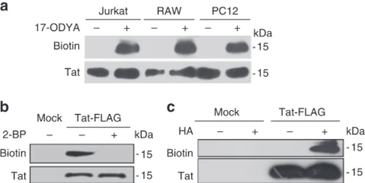
labeled with 17-ODYA in all these cell lines (Fig.
1
a), indicating
that most cell types are able to palmitoylate incoming Tat.
Neurons are an important target for HIV-1 Tat
21and to study
in detail Tat palmitoylation, we transiently transfected Tat-FLAG
in the neurosecretory cell line PC12 and detected palmitoylation
using both the click-chemistry approach
25and the acyl-biotin
exchange technique (ABE) that is based on the replacement of the
protein-acyl chain by a biotin. This exchange relies on the use of
hydroxylamine that selectively cleaves thioester bonds
26. Both the
click chemistry (Fig.
1
b) and the ABE (Fig.
1
c) techniques showed
that
transfected
Tat
is
palmitoylated.
Addition
of
2-bromopalmitate (2-BP), a well-established palmitoylation
inhi-bitor
27prevented Tat palmitoylation (Fig.
1
b). The sensitivity of
Tat-acyl chain bond to hydroxylamine (Fig.
1
c) showed that this
bond is a thioester bond
26and thus that the acyl chain is attached
to at least one of the seven Cys of Tat.
Tat is specifically palmitoylated on Cys31. Tat Cys are located in
position 22, 25, 27, 30, 31, 34, and 37 and, to identify the
pal-mitoylated Cys, we individually mutated each of them in Ser. A
double mutant Tat-C30S-C31S was also generated because, in the
case of Cys doublets, the mutation of one of the Cys can
some-times aberrantly lead to palmitoylation of the remaining Cys
28.
We then examined the capacity of these mutants to be
palmi-toylated in PC12 cells. All mutants were expressed to similar
levels, but those devoid of Cys31 only failed to be palmitoylated
(Fig.
2
a), indicating that Tat is palmitoylated on Cys31. Mass
spectrometric analysis of immunoprecipitated Tat-FLAG using
nano LC-MS/MS confirmed that Tat is palmitoylated on Cys31
(Supplementary Table
1
).
15 15 Tat-FLAG Tat + – 15 15 Mock + – + – kDa 15 15 kDa Tat – + – + – + – Biotin 2-BP Tat Biotin HA Tat-FLAG Mock Biotin 17-ODYA Jurkat RAW PC12a
b
c
kDaFig. 1 Tat is palmitoylated in T-cells, macrophages and neuron precursors. a T-cells (Jurkat), macrophages (RAW 264.7) or neuron precursors (PC12 cells) were labeled overnight with 17-ODYA and Tat-His6in lipid-free medium before cell lysis, Tat-His6purification, biotin labeling of 17-ODYA using click chemistry and SDS-PAGE. The blot wasfirst incubated with avidin-peroxidase to detect biotin, then with anti-Tat antibodies.b PC12 cells were transfected with Tat-FLAG before overnight labeling with 17-ODYA, anti-FLAG immunoprecipitation, and click chemistry. When indicated 100µM 2-bromopalmitate (2-BP) was present during labeling with 17-ODYA.c PC12 cells were transfected with Tat-Flag before anti-Flag immunoprecipitation and acyl-biotin exchange. Hydroxylamine (HA) treatment is used to remove the acyl chain and enables to replace it with biotin for palmitoylation detection. Representative data fromn = 3 experiments
To examine whether the C31S mutation affected Tat structure
we
first assessed the capacity of Tat-C31S to transactivate a
co-transfected LTR-driven luciferase. The results showed that
transfected Tat-C31S displayed native Tat transactivation
capa-city (Supplementary Fig.
1
), indicating that the C31S mutation
does not affect Tat structure and reactivity. When the same
experiment was performed using extracellular recombinant
Tat-C31S, no difference was observed either. Hence, the C31S
mutation did not impair the capacity of Tat to enter cells. We also
examined, using liposomes and isothermal calorimetry (ITC)
4,
whether this mutation affects Tat affinity for PI(4,5)P
2and found
that Tat-C31S, with a Kd of 8 ± 5 nM (mean ± SEM, n
= 3) has
essentially the same affinity for PI(4,5)P
2as WT Tat (Kd
= 55
± 20 nM; Supplementary Fig.
2
). Collectively, these results show
that the C31S mutation prevents palmitoylation but does not
significantly affect Tat folding or transactivation activity. They are
consistent with the observation that HIV-1 clade C present in
India and that has a Tat-31S is essentially as virulent as other
clades regarding its capacity to multiply in peripheral blood
mononuclear cells (PBMCs)
29.
We then examined the subcellular localization of Tat Cys
mutants in PC12 cells. All mutants except C31S and
Tat-C30S-C31S localized to the plasma membrane, while a large
fraction (~60%) of Tat-C31S and Tat-C30S-C31S accumulated in
the cytosol. Similar data were obtained when palmitoylation was
prevented using 2-BP (Fig.
2
b). This observation not only
indicated that Tat palmitoylation is required for its stable
association with the plasma membrane, but also that a large
fraction of Tat is palmitoylated in PC12 cells. Similar results were
obtained using primary hippocampal neurons (Supplementary
Fig.
3
). Altogether, this
first set of data indicates that, in
neuroendocrine cells as well as primary neurons, a significant
pool of Tat is palmitoylated on Cys31 and that this modification
is important for plasma membrane localization. We examined
next whether incoming Tat-C31S became palmitoylated in Jurkat
cells. This was not the case (Supplementary Fig.
4
) and, since
Tat-C31S is not affected in its internalization pathway
(Supplemen-tary Fig.
1
) this result confirms that Cys31 is the only
palmitoylated residue in Tat, whether Tat-producer (T-cells) or
Tat-target cells (neuroendocrine cells) are used.
Tat palmitoylation requires PI(4,5)P
2binding. Tat-W11Y
poorly binds PI(4,5)P
24, while showing native transactivation
activity, indicating that this mutation does not affect Tat
con-formation
9. This mutant, as observed before in T-cells
4, is
cyto-solic and/or nuclear in PC12 cells (Fig.
2
b) and Tat-W11Y failed
to be palmitoylated (Fig.
2
a). Hence, PI(4,5)P
2binding appears as
a prerequisite for Tat palmitoylation, indicating that this
mod-ification takes place at the plasma membrane.
Tat palmitoylation is performed by DHHC-20. DHHC enzymes
are membrane proteins that can acylate proteins once the
sub-strate has reached the appropriate membrane
30. In agreement
HA 22S 25S 27S 30S 31S 30/31S 34S 37S W11Y + – – + – + – + – + – + – + – + – + WT 22S 34S - 15 - 15 kDa
**
*
Biotin Tat Merge F-actin Tat 25S 27S 30S 31S 30/31S 37S W11Y 2-BP Tat/F-actin colocalization 0.0 0.2 0.4 0.6 0.8 WT 22S 25S 27S 30S 31S 30/31S34S 37Sa
b
c
Fig. 2 Tat palmitoylation takes place on Cys31 and requires PI(4,5)P2binding.a PC12 cells were transfected with the indicated FLAG-CXXS or Tat-W11Y mutant before anti-FLAG immunoprecipitation, acyl-biotin exchange (HA, hydroxylamine), western blot and biotin then Tat detection. Images from two blots were assembled. Films with the same exposure time were used and the dashed line marks the separation between them.b Tat-transfected PC12 cells were processed for detection of F-actin usingfluorescent phalloidin and Tat using immunofluorescence. When indicated, cells transfected with WT Tat were treated with 100µM 2-BP for 5 h before fixation. Representative median confocal sections are shown. Bar, 5 µm. c plasma membrane localization of Tat-mutants was evaluated by quantifying Tat/F-actin colocalization using confocal images fromn = 10 cells and Mander’s coefficient calculation (mean ± SEM). The significance of differences with WT Tat was assessed using one-way ANOVA (*p < 0.05; **p < 0.01). Similar microscopy data were obtained using primary neurons (Supplementary Fig.3)
with previous observations
31we found that, among the 23 DHHC
proteins, DHHC-5 and DHHC-20 only localized to the plasma
membrane in PC12 cells (Supplementary Fig.
5
). Since Tat is
palmitoylated at this level, we
first examined using an
over-expression approach which of DHHC-5 or DHHC-20 favors Tat
palmitoylation, using DHHC-21 as control. We did not use PC12
cells for this experiment because Tat palmitoylation is already
very efficient in this cell type (Fig.
2
b). We used HEK 293 T cells
that were cotransfected with Tat and myc-tagged human
DHHC-5, -20 or -21. Exogenous DHHC-5 and DHHC-20 were expressed
to the same level while DHHC-21 expression was ~3-fold lower
(Fig.
3
b). Tat palmitoylation was assessed using the acyl-RAC
technique, a variation of the ABE technique that enables easier
quantification. Flotillin-2 which is known to be stably
palmitoy-lated was used as positive control
32. The efficiency of
overexpressed-Tat palmitoylation in HEK cells (~15%) was not
affected by cotransfection with DHHC-5 or DHHC-21, while it
reached 65% upon DHHC-20 coexpression (Fig.
3
a). This result
indicated that Tat is palmitoylated by DHHC-20. To confirm this
finding, we used siRNAs against rat DHHC-20 that silenced its
expression by ~80% in PC12 cells according to qRT-PCR
quan-tification (Supplementary Fig.
6
). Silencing DHHC-20 led to a
complete inhibition of Tat palmitoylation, while the control
siRNA had no effect (Fig.
3
c). Hence, Tat palmitoylation is
performed by DHHC-20, an enzyme that is expressed in T-cells,
macrophages and PC12 cells (Fig.
3
d for DHHC-20 and
Sup-plementary Fig.
7
for all DHHCs). In agreement with previous
observations, DHHC localization can be cell type-dependent
15,
and in fact DHHC-20 seems to be the only DHHC that localizes
to the plasma membrane in primary T-cells, while
DHHC-5-EGFP was observed at the Golgi apparatus in these cells
(Sup-plementary Fig.
8
).
Tat palmitoylation increases its affinity for PI(4,5)P
2. We then
assessed whether palmitoylation affects Tat affinity for PI(4,5)P
2.
To this end, we obtained a Tat devoid of Cys except Cys31
(Tat-C31 only termed Tat-(Tat-C31O) that was labeled with palmitate. We
had to use this strategy because it was not possible to specifically
label Cys31 with palmitate when the other Cys are present.
MALDI-TOF/TOF analysis indicated that Tat-C31O was acylated
by a single palmitate with an efficiency of 47 ± 8%
(Supple-mentary Fig.
9
). Using PI(4,5)P
2-containing liposomes and
Sur-face Plasmon Resonance we observed that the affinity for PI(4,5)
P
2of Tat-C31O (Kd
= 0.31 ± 0.02 nM) was the same as that of
native Tat
4, while Tat-C31O-Palm showed a Kd of 0.18 ± 0.02
nM, suggesting that palmitoylation increases Tat affinity for PI
(4,5)P
2. Since only half of Tat-C31O was palmitoylated these
DHHC-5 DHHC-20 DHHC-21 UC BC UH BH 15 15 15 15 kDa 48 kDa 0 50
***
100 Vector Vector DHHC-5 DHHC-20DHHC-21 DHHC-5 DHHC-20 DHHC-21 Flotillin-2 0.00001 0.0001 0.001 0.01 Primary T-cells Jurkat PC12 MDMacDHHC-20/ GADPH mRNA level 75 37 31 α-Flotillin-2 Vector Transfection Input ( α -myc) Tat + S-acylation (%) Biotin HA – + – + kDa 15 15 Tat DHHC-20 Control siRNA α-Tat
a
b
c
d
Fig. 3 Tat palmitoylation is performed by DHHC-20. a HEK 293 T cells were cotransfected (1/5) with the indicated myc-tagged human DHHC and Tat. Tat palmitoylation was then assessed using the acyl-RAC technique, UC unbound control, BC bound control, UH unbound hydroxylamine, BH bound hydroxylamine. Palmitoylated Tat is present in the BH fraction. Palmitoylation was calculated as BH/(BH+ UH)-BC/(BC + UC), and flotillin-2 was used as a positive control. The graph shows mean ± SEM ofn = 3 independent experiments, ***p < 0.001 (one-way ANOVA). b DHHC-myc overexpression level was assessed using anti-myc western blot. DHHC-5, -20, and -21 migrated at 75, 37 and 31 kDa, respectively. Transfection efficiency was 60–70%. c PC12 Cells were cotransfected with Tat-FLAG and the indicated siRNA before detecting Tat palmitoylation using acyl-biotin exchange. The efficiency of siRNA-mediated silencing is shown in Supplementary Fig.6.d The RNA from monocyte-derived macrophages (MDMac), PC12, Jurkat or human primary CD4+ T-cells was extracted before quantification of DHHC-20 and GAPDH mRNAs using qRT-PCR. Results are expressed as DHHC-20/GAPDH ratio. Results for all DHHCs are shown in Supplementary Fig.7. Mean ± SEM,n = 3 independent experiments
results indicate that palmitoylation increases Tat affinity for PI
(4,5)P
2by ~5-fold.
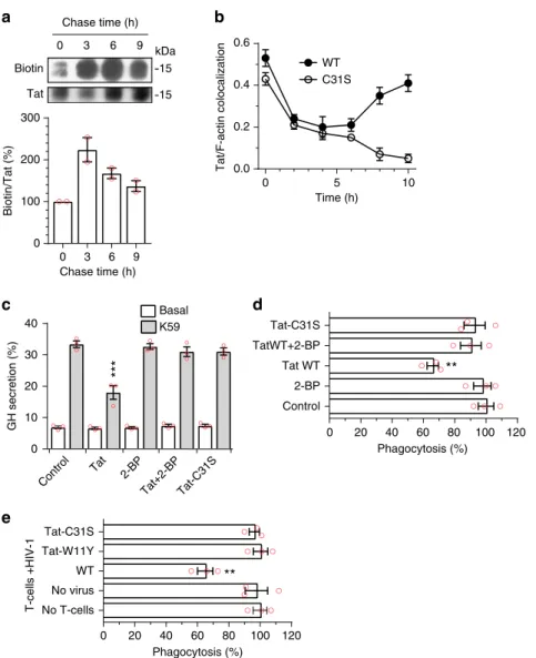
Tat palmitoylation is a stable modi
fication. To examine the
stability of Tat palmitoylation, PC12 cells were labeled overnight
with Tat-His
6and 17-ODYA before a chase. Tat palmitoylation
showed a half-life of ~7.3 h (R
2= 0.985; Fig.
4
a), indicating that
this is a stable modification.
Palmitoylation enables Tat accumulation at the plasma
mem-brane. We then assessed the effect of Tat palmitoylation on the
plasma membrane recruitment of incoming Tat. To this end cells
were labeled with recombinant Tat at 4 °C before washing and
chasing at 37 °C. Under these conditions, Tat is
first observed at
the plasma membrane, then in endocytic vesicles from which it
escapes to reach the cytosol, and it is
finally recruited at the
plasma membrane by PI(4,5)P
221. This relocalization to the
plasma membrane can be followed using Tat/F-actin
colocaliza-tion, and is clearly observed for WT Tat at late chase time points
(8–10 h; Fig.
4
b; representative images are in Supplementary
Fig.
10
). In sharp contrast, Tat-C31S is not recruited to the
plasma membrane after 8–10 h of chase. In fact, Tat-C31S
dis-appears from the cell at late time points. These results obtained
Biotin kDa 15 15 Tat 0 3 6 9 Basal
***
**
**
0 20 40 60 80 100 120 Control 2-BP Tat WT TatWT+2-BP Tat-C31S 0 3 6 9 0 100 200 300 0 20 40 60 80 100 120 No T-cells No virus WT Tat-W11Y Tat-C31S Phagocytosis (%) 0 10 20 30 40 K59 Chase time (h) Biotin/Tat (%) Chase time (h) GH secretion (%) T-cells +HIV-1 Control Tat 2-BP Tat+2-BP Tat-C31S Phagocytosis (%)a
b
c
d
e
10 WT C31S 0 5 Tat/F-actin colocalization 0.6 0.4 0.2 0.0 Time (h)Fig. 4 Palmitoylation is a stable modification that enables Tat accumulation at the plasma membrane and Tat-mediated inhibition of PI(4,5)P2-dependent membrane traffic. a PC12 cells were labeled overnight with Tat-His6and 17-ODYA before chasing for the indicated times, cell lysis, Tat-His6purification and click chemistry. Results from a representative experiment are shown. The graph (mean ± SEM of 2 independent experiments) shows Tat palmitoylation efficiency as the biotin/Tat ratio (% of the t0 value) as a function of time. The increase of labeling during the first 3 h of chase is probably due to residual intracellular 17-ODYA.b PC12 cells were labeled with Tat (WT or C31S) at 4 °C before washing, chasing for the indicated times and Tat staining by immunofluorescence with F-actin labeling using fluorescent phalloidin. Tat membrane localization was evaluated by quantifying Tat/F-actin colocalization using confocal images from 50 <n < 100 cells and Mander’s coefficient calculation. Mean ± SEM. Representative images are in Supplementary Fig.10.c PC12 cells were transfected with human growth hormone (GH) then treated for 5 h with 100µM 2-BP, 20 nM Tat WT or Tat-C31S as indicated. GH secretion was then triggered using 59 mM K+(K59) and quantified by ELISA. Mean ± SEM (n = 3). d Macrophages (MDMs) were treated for 3 h with 100 µM 2-BP, 5 nM Tat or Tat-C31S as indicated before assaying phagocytosis of IgG-coated 3 µm latex beads. Extracellular beads were stained before cell fixation and examination using a fluorescence microscope. e CD4 + T cells were purified, stimulated then infected with a T-tropic (NL4.3) virus, bearing Tat-WT, -W11Y or -C31S as indicated, then added to Transwells into wells containing autologous MDMs. FcγR-mediated phagocytosis by MDMs was assayed after 8 days of co-culture. At this time 20–35% of T cells and 0% of macrophages were infected. Data in panels d and e are mean ± SEM of three independent experiments (countingn > 100 cells for each). The significance of differences with controls was assessed using ANOVA, one-way (d, e) or two-way (c) (***p < 0.001; **p < 0.01)
with incoming Tat are consistent with data from Tat-transfected
cells (Fig.
2
b) and indicate that Tat palmitoylation enables stable
Tat association with PI(4,5)P
2.
Palmitoylated Tat inhibits PI(4,5)P
2-dependent traffic. If Tat
palmitoylation is required for stable Tat association with PI(4,5)
P
2it would be needed for Tat to affect PI(4,5)P
2-dependent
membrane traffic. We examined this issue using both 2-BP
treatment and Tat-C31S that cannot be palmitoylated (Fig.
2
a and
Supplementary Fig.
4
). We
first assessed whether palmitoylation
is involved in Tat capacity to inhibit neurosecretion. To this end,
PC12 cells were transfected with human growth hormone (GH)
before treatment with extracellular Tat. In this assay, GH is
incorporated in secretion granules that, upon stimulation, fuse
with the plasma membrane in a process that recapitulates
neu-romediator secretion, a PI(4,5)P
2-dependent process
21. Tat
strongly inhibited GH release (−40%), as observed before
21, while
2-BP had no effect. Both treatment with 2-BP and the C31S
mutation prevented Tat inhibitory effect on neurosecretion
(Fig.
4
c). Identical data were obtained using primary chromaffin
neurorendocrine cells (Supplementary Fig.
11
). These data showed
that palmitoylation is required for Tat to inhibit neurosecretion.
We also examined whether Tat palmitoylation is needed for
Tat to inhibit FcγR-mediated phagocytosis. Tat inhibits this key
phagocytic process by preventing the PI(4,5)P
2-mediated
recruit-ment of cdc42 to the phagocytic cup
20. Monocyte-derived human
primary macrophages (MDMs) were treated with Tat, Tat-C31S
or 2-BP before assaying the phagocytosis of IgG-coated latex
beads. Tat inhibited phagocytosis by ~ 35% in this assay whereas
2-BP had no significant effect. Either 2-BP or the C31S mutation
blocked Tat inhibitory effect on phagocytosis (Fig.
4
d). It was
important to validate these
findings in the context of infection. To
this end, primary CD4
+T-cells were infected with T-tropic
viruses bearing either Tat WT, Tat-W11Y or Tat-C31S that were
then added on autologous MDMs using a Transwell
configura-tion. After 8 days, macrophage FcγR-mediated phagocytic activity
was assayed. At this time, T-cells are efficiently infected, while
macrophages remain uninfected. Tat secretion by T-cells into the
medium is responsible for phagocytosis inhibition
20and the
W11Y mutation that impairs both Tat secretion
4and entry into
cells
9prevents Tat from affecting phagocytosis
20. The C31S
mutation also prevented Tat effect on phagocytosis in this
coculture assay (Fig.
4
e). Since Tat-C31S is actively secreted (Fig.
5
a) and efficiently enters cells (Supplementary Fig.
1
) these results
confirmed, in primary cells, that Tat palmitoylation enables it to
perturb phagocytosis.
Altogether, these data showed that Tat palmitoylation is
needed for Tat to inhibit PI(4,5)P
2-dependent membrane traffic
processes such as neurosecretion and phagocytosis.
Tat palmitoylation inhibits its secretion. Since palmitoylation
increases Tat affinity for PI(4,5)P
2and stabilizes Tat association
with the plasma membrane (Fig.
4
b), it should affect Tat
secre-tion. We examined Tat secretion in two cell types, T-cells (Jurkat,
producer cells) and PC12 (target cells), using different assays
33.
Tat secretion by Jurkat cells (~15% in 6 h
4) was weakly inhibited
in the presence of palmitate (−20%), and strongly enhanced by
2-BP (+120%) or by introduction of the C31S mutation (+60%)
(Fig.
5
a). Similar data were obtained using PC12 cells (Fig.
5
b).
Hence, although Tat-C31S is more cytosolic than WT Tat
(Fig.
2
b; Supplementary Fig.
3
), it is more efficiently secreted.
This apparent discrepancy is most likely due to the fact that
palmitoylation stabilizes Tat association with the plasma
mem-brane. Tat secretion and palmitoylation are thus concurrent
mechanisms that apparently take place similarly in target and
producer cells. This was intriguing since Tat secretion by infected
cells is very active with ~2/3 of Tat being secreted
4, suggesting
that Tat palmitoylation is prevented in infected cells.
Cyclophilin A and FKBP12 are required for Tat
palmitoyla-tion. In the search for cell proteins that might regulate Tat
pal-mitoylation, we focused on prolylisomerases, i.e., proteins of the
immunophilin family
34. Indeed, although most proteins have
<5% of Pro residues
35, Tat owns ~10 % of Pro residues. Four of
them are located in the N-terminal region (in positions 6, 10, 14,
and 18) and highly conserved among viral isolates
8, indicating
that they are probably involved in Tat structure and/or biological
activity.
We envisioned that prolylisomerases could be involved in Tat
palmitoylation, as shown earlier for Ras
36. We
first used a
pharmacological approach to examine the potential implication
of the prolylisomerases CypA and FKBP12 in Tat palmitoylation.
It can be seen in Fig.
6
a that FK506 and rapamycin that inhibit
FKBP12
36, and cyclosporin A (CSA) that inhibits CypA
37,
strongly impaired Tat palmitoylation in PC12 cells. Accordingly,
these inhibitors displace Tat from the membrane just as efficiently
as 2-BP (Supplementary Fig.
12
a). A cotransfected EGFP chimera
of PH-PLCδ, an established ligand for PI(4,5)P
2, remained at the
plasma membrane whatever the drug, showing that these
inhibitors do not affect PI(4,5)P
2availability (Supplementary
Fig.
12
a). These drugs strongly favored Tat secretion (by 2- to
6-fold; Fig.
6
b), confirming that secretion and palmitoylation are
15 15 kDa
***
***
Jurkats PC12**
*
0 100 200 300 WT WT+palmitate WT+2-BP C31STat secretion (% of control)
0 20 40 60 WT 11Y C31S Tat Tat secretion (%) Medium Cell 11Y WT 31S
a
b
Fig. 5 Tat palmitoylation inhibits its secretion. a Jurkat cells were transfected with Tat or Tat-C31S before treatment for 3 h with 50µM 2-BP or palmitate as indicated and assaying Tat secretion by ELISA (Mean ± SEM,n = 3 independent experiments performed in triplicate). b PC12 cells were treated similarly before assaying Tat secretion using
immunoprecipitation and Western blotting. Results from a representative experiment are shown together with the quantification (Mean ± SEM, n = 3). The significance of differences with controls was assessed using one-way ANOVA (***p < 0.001; **p < 0.01; *p < 0.05)
concurrent processes. These inhibitors also prevented Tat from
affecting neurosecretion (Fig.
6
c) and phagocytosis (Fig.
6
d).
Collectively, these pharmacological data indicated that the activity
of both CypA and FKBP12 prolylisomerases is required for Tat
palmitoylation. To confirm this point, we used siRNAs that
efficiently inhibited the expression of CypA and FKBP12 (Fig.
6
e).
These siRNAs efficiently blocked Tat palmitoylation (Fig.
6
f)
confirming that both CypA and FKBP12 are required for Tat
palmitoylation. Conversely, silencing CypA or FKBP12 in Jurkat
cells (Supplementary Fig.
12
b) increased Tat secretion by 40–60%
(Fig.
6
g). These results further confirmed that inhibition of Tat
palmitoylation promotes its secretion by T cells and that CypA
and FKBP12 are required for Tat palmitoylation.
Tat interacts with CypA and DHHC-20. We then examined
whether Tat could interact with the proteins involved in its
pal-mitoylation, i.e., CypA and DHHC-20. Following Tat-FLAG
transfection into HEK 293 T cells, we indeed found that CypA
and DHHC-20, but not DHHC-5, co-immunoprecipitated with
Tat (Fig.
7
a). These results confirm that DHHC-20 is involved in
Tat palmitoylation. CypA interacted less efficiently with
non-palmitoylable Tat (Tat-C31S). To confirm these findings, we used
GST pull down experiments. Both DHHC-5 and DHHC-20 could
be recovered from cell extracts by GST-CypA (but not by GST),
indicating that CypA interacts with these enzymes even in the
absence of Tat (Fig.
7
b). When Tat-transfected cells were used,
Tat could be pulled-down by GST-CypA. Moreover, in the
pre-sence of Tat, ~4-fold more DHHC-20 was recovered by
GST-CypA, indicating the existence of a Tat-CypA−DHHC-20
com-plex. The level of DHHC-5 pulled-down by GST-CypA was
insensitive to the presence of Tat, confirming that DHHC-5 does
not significantly interact with Tat. Altogether these results
sup-port the existence of a Tat palmitoylation complex containing
Tat, CypA and DHHC-20.
Tat interacts with FKBP12. Although FKBP12 could be
immu-noprecipited by Tat (Supplementary Fig.
13
), GST-FKBP12 did
not enable to pull-down Tat. We concluded that the interaction of
Tat with FKBP12 is weaker than that with CypA. CypA was thus
a better candidate to be used by the virus to modulate Tat
kDa 13 55 Ctrl CypA FKBP12 + – – + – + kDa 15 15 HA Tat Biotin Tubulin Ctrl CypA kDa 17 55
***
***
***
***
***
*
*
**
0 50 100 Solvent Rapa CSA FK506Tat palmitoylation (% of control)
0 200 400 600 800 Solvent
Rapa CSA FK506
Tat secretion (% of control)
0 50 100 150 Control
CypA FKBP12
Tat secretion (% of control)
siRNA Mock 0 10 20 30
Solvent FK506 CSA Rapa Tat 0 20 40 60 80 100 120
Solvent FK506 CSA Rapa Mock Tat Net GH secretion (%) Phagocytosis (%)
***
**
siRNA FKBP12 Tubulin siRNA FKBP12 Ctrl siRNA CypAa
b
c
d
e
f
g
Fig. 6 Cyclophilin A and FKBP12 are required for Tat palmitoylation and Tat-mediated inhibition of PI(4,5)P2-dependent membrane traffic. a PC12 cells were transfected with Tat-FLAG before treatment for 6 h with 1µM FK506, 2 µM CSA, 1 µM rapamycin or solvent. Palmitoylation was then assayed using acyl-biotin exchange before band quantification. Results are expressed as biotin/Tat intensity ratio (% of control). b Jurkat T-cells were transfected with Tat before adding drugs for 6 h and assaying Tat secretion by ELISA.c PC12 cells were transfected with GH before drug treatment for 6 h and GH secretion assay.d Human MDMs were treated with the drug for 5 h before assaying phagocytosis of IgG-coated beads. e PC12 cells were transfected with the indicated siRNA before lysis then anti-CypA or anti-FKBP12 then anti-αtubulin western blot. f PC12 cells were cotransfected with the indicated siRNA and Tat-FLAG before detecting Tat palmitoylation using acyl-biotin exchange. (HA, hydroxylamine).g Jurkat cells were cotransfected with Tat and the indicated siRNA (silencing efficiency is shown in Supplementary Fig.12b) and Tat secretion was assayed after 48 h. Data are mean ± SEM,n = 3 independent experiments. The significance of differences with controls was assessed using ANOVA, one-way (a, b, g) or two-way (c, d) (***p < 0.001; **p < 0.01; *p < 0.05)
palmitoylation. Moreover, while CypA is well known to be
encapsidated by the virus (200 CypA /virion
38), FKBP12 was
found to be either not
39or weakly encapsidated (25 FKBP12/
virion)
40and we thus focused on CypA.
Gag inhibits Tat palmitoylation by exporting CypA. HIV-1 Gag
binds both PI(4,5)P
241and CypA
38through its matrix (MA) and
capsid (CA) protein domain, respectively. Gag was therefore
among HIV-1 proteins the best candidate to regulate Tat
pal-mitoylation by modulating PI(4,5)P
2and/or CypA availability for
Tat. Consistently, when Tat and Gag were coexpressed in PC12
cells, Tat palmitoylation was abrogated (Fig.
8
a). We
first
examined whether Gag could prevent Tat palmitoylation by
inhibiting Tat recruitment to the plasma membrane, i.e., by
dis-placing Tat from PI(4,5)P
2. As observed before
4, when primary
CD4
+T-cells were infected by HIV-1 (NL4.3), both Tat and Gag
localize to the plasma membrane of infected T-cells (Fig.
8
b).
Hence, Gag does not prevent Tat palmitoylation by impairing Tat
recruitment to the plasma membrane. Gag and Tat are both
displaced to the cytosol in the presence of neomycin, a cationic
antibiotic that tightly binds PI(4,5)P
242, thereby confirming the
key role of this phosphoinositide in the localization of these
HIV-1 proteins to the plasma membrane of infected T-cells (Fig.
8
b).
Since Gag is unable to displace Tat from PI(4,5)P
2, we
suspected that Gag prevented Tat palmitoylation by affecting
CypA availability. Gag could either prevent Tat binding to CypA,
i.e., act by a titration effect and/or deplete cells from CypA due to
encapsidation i.e., act by a depletion effect.
To discriminate between the titration and depletion effects, we
prepared Gag mutants with the G89V mutation in CA to inhibit
CypA binding
43or the K22A and R27A mutations in MA to
impair PI(4,5)P
2binding
44. The capacity of the CA-G89V
mutation
to
prevent
Gag-CypA
interaction
has
been
validated earlier
43. We found that the MA-22/27A mutations
prevented Gag recruitment to the T-cell plasma membrane (a PI
(4,5)P
2-dependent process, Fig.
8
b) while the CA-G89V mutation
had no effect (Supplementary Fig.
14
a). As expected, upon
transient transfection the MA-22/27A mutations severely
inhib-ited (by 40–50%) the formation of virus-like particles (VLPs) by
T-cells while the CA-G89V mutation did not significantly affect
the production of VLPs, although they are devoid of CypA
(Supplementary Fig.
14
b). When Gag-MA-22/27A or
Gag-CA-G89V were cotransfected with Tat they did not significantly affect
Tat palmitoylation (Fig.
8
c). In the case of the CA-G89V mutant,
this result indicates that CypA binding is needed for Gag to
interfere with Tat palmitoylation, but this could be by a
titration or a depletion effect. The fact that the MA-22/27A
mutant that binds CypA but barely form VLPs (Supplementary
Fig.
14
b) does not affect palmitoylation thus indicates that VLP
formation is required for Gag to shut off Tat palmitoylation.
Altogether these data showed that Gag inhibits Tat palmitoylation
by exporting CypA into VLPs and thus acts by depletion and not
by titration.
15 37 75 18 37 75 18 kDaVector Tat WT Tat-W11YTat-C31S
15 75 37 24 45 kDa 37 75 pCi Tat DHHC-20 0 100 200 300 400 500 DHHC-5 Tat DHHC-20 DHHC-5 CypA DHHC-20 DHHC-5 CypA Input DHHC-20 DHHC-5 GST Tat Input DHHC-20 DHHC-5 Tat pCi DHHC pull-down/input (%)
***
GST-CypA GSTpCi Tat pCi Tat
a
b
Fig. 7 Tat interacts with CypA and DHHC-20. a HEK 293 T cells were transfected with an empty vector or Tat-FLAG (WT, 31 S, or 11Y). Cells were lysed 48 h after transfection before anti-FLAG immunoprecipitation and western blots against CypA, DHHC-5 and DHHC-20.b Cells were transfected with an empty (pCi) or Tat vector. GST or GST-CypA was added to cell extracts for GST pull-down before western blots. The graph shows the quantification of the DHHC pulled-down/input intensity ratio, setting the empty vector ratio to 100%. Representative data (mean ± SEM,n = 3 independent experiments) are shown.***p < 0.001 (Student’s t-test between Tat and vector)
HIV-1 budding depletes cells in CypA. CypA is an abundant
protein present at 5–10 µM in the cytosol
37. Hence, it was not
obvious that the budding process could be sufficient to remove all
CypA from the infected cell. We
first examined this point using
Western blotting. When Jurkat T-cells were transiently
trans-fected with WT Gag, CypA level dropped by ~80% (Fig.
8
d).
Since transfection efficiency was also ~80%, this result indicated
that WT Gag efficiently depletes T-cells in CypA. This was not
the case when mutants such as Gag-CA-22/27 A (strongly
affec-ted in budding) or Gag-CA-G89V (unable to bind CypA) were
used (Fig.
8
d). This result was confirmed by immunofluorescence
(Fig.
9
a). Moreover, when cells were infected with pseudotyped
HIV-1, infected cells showed after 24–30 h a strong depletion in
their CypA content, while a virus unable to bind CypA
(HIV-1-CA-G89V) did not affect CypA level (Fig.
9
a). Altogether these
data indicated that HIV-1 budding or transient transfection by
Gag depletes cells of CypA.
Tat is not palmitoylated in HIV-1 infected cells. If our working
model is correct, in infected T-cells, WT Tat secretion should be
almost as efficient as that of Tat-C31S because Gag inhibits Tat
palmitoylation by promoting CypA export. This was indeed the
case (Fig.
9
b), although Tat-C31S tends to be slightly more
effi-ciently secreted. This is probably because Tat-C31S, contrary to
WT Tat, can be secreted before all CypA has been exported from
the cell by virions. When a virus unable to bind CypA
(HIV-1-CA-G89V) was used, Tat secretion was strongly inhibited
(−50%), confirming that Gag inhibits Tat palmitoylation by
exporting CypA. The negative control, Tat-W11Y that poorly
binds PI(4,5)P
2was not significantly secreted as observed before
4.
It was important to check that Tat is palmitoylated in
HIV-1-CA-G89V infected cells to confirm our results in the context of
infection, and to check that Gag, through its interaction with
CypA, is the main viral regulator of Tat palmitoylation. To
perform this experiment we had to circumvent a technical issue.
Indeed, to follow Tat palmitoylation it is necessary to
immuno-precipitate Tat and, since anti-Tat antibodies do not allow
quantitative immunoprecipitation, Tat has to be tagged. This can
only be done on its C-terminal side
45, and is not possible in the
virus sequence because the three reading frames of Tat, Rev and
Env overlap in this part of the sequence
46. We thus chose to
inactivate the tat gene in pNL4.3 and to cotransfect the resulting
pNL4.3ΔTat or pNL4.3-CA-G89VΔTat with Tat-FLAG (WT or
C31S) into Jurkat T-cells. This cotransfection allows for virus
production (Supplementary Fig.
15
). Tat palmitoylation was only
observed when WT Tat, but not Tat-C31S, was cotransfected with
pNL4.3-CA-G89VΔTat but not pNL4.3ΔTat (Fig.
9
c) confirming,
in infected cells, that Tat is indeed specifically palmitoylated on
Mock Gag kDa 15 15
a
Tat HA – + – + – + – + – +c
kDa 15 15 Tatb
WT 22/27A G89V Tubulin kDa 18 55 55 Gag 0 50 100 Vector WT MA-22/27A CA-G89V CypA/tubulin (%) Gag CypA***
Vector Gag Biotin CA-G89V MA-22/27A GagWT Vector Mock Tat+ +Neomycin ControlGag WGA Merge Biotin
Tat
Vector Tat+
d
Fig. 8 HIV-1 Gag inhibits Tat palmitoylation by exporting Cyclophilin A. a PC12 cells were cotransfected with Tat-Flag and Gag (1/1 ratio) or an empty vector as indicated. Cells were then labeled overnight with 17-ODYA before assaying Tat palmitoylation using click chemistry.b Human primary CD4+ T-cells were infected with HIV-1 (NL4.3). After 24 h T-cells were processed for immunofluorescence detection of Tat and Gag, using fluorescent WGA to localize the plasma membrane and the TGN. When indicated, cells were pretreated with 5 mM neomycin for 1 h beforefixation. Bar, 5 µm. c PC12 cells were cotransfected (1/1) with Tat-FLAG and a Gag mutant or an empty vector as indicated before assaying Tat palmitoylation using ABE. Gag-MA-22/27A and Gag-CA-G89V are mutated within their PI(4,5)P2- and CypA-binding motif, respectively.d Jurkat T-cells were transfected with the indicated Gag mutant using nucleofection. Transfection efficiency was ~80%. After 24 h, cells were lysed for Western blots. Triplicate blots were used for the quantification. Data are mean ± SEM (n = 3) and were analyzed using one-way ANOVA (***p < 0.001)
Cys31 in a CypA-dependent manner. Moreover, the efficiency of
WT Tat palmitoylation was essentially the same when
cotrans-fected with pNL4.3-CA-G89VΔTat or with an empty vector.
Since pNL4.3-CA-G89V encapsidates essentially as little FKBP12
as WT pNL4.3 (Supplementary Fig.
16
), this result indicates that
FKBP12 encapsidation is not involved in the inhibition of Tat
palmitoylation observed in infected cells. More generally, this
result shows that Gag-mediated CypA export is the main
mechanism responsible for Tat palmitoylation inhibition in
HIV-1 infected cells. Figure
10
is a schematic illustration of the
results obtained in this study.
Discussion
Most membrane traffic involving the plasma membrane uses PI
(4,5)P
2-mediated protein recruitment at this level
47.
Here we showed that Tat palmitoylation is required for Tat to
interfere with PI(4,5)P
2-dependent membrane traffic. Indeed,
palmitoylation stabilizes the association of Tat with PI(4,5)P
2-membranes and prevents Tat secretion, while a non palmitoylable
Tat (i.e., Tat-C31S) rapidly leaves the cell following PI(4,5)P
2binding. Different cell types, such as T-cells, macrophages or
neurosecretory cells can palmitoylate Tat. This result is consistent
with the observation that Tat-palmitoylating enzyme, DHHC-20,
is well expressed in most cell types
31(Supplementary Fig.
7
). Tat
palmitoylation is a stable modification with a t
1/2of 7.3 h, similar
to the one of calnexin, a stably S-acylated protein
48. Some
bac-terial virulence factors are also S-acylated by target cells. As it is
the case for Tat, palmitoylation of these effectors is a stable
modification that is required for durable association with the
plasma membrane
49.
Tat is recognized as a major determinant of HIV-1
neuro-pathogenesis
18, and we identified Cys31 as the palmitoylated
residue in Tat. Interestingly, this residue is replaced by a Ser in
the Indian HIV-1 subtype C, a clade associated with a much
lower neurotoxicity compared to other subtypes that owns a
palmitoylable Tat
50. Several studies identified Tat-Cys31 as a key
residue responsible for the neurotoxicity of HIV-1 subtype B.
Indeed, when applied onto primary neurons, Tat from HIV-1C
showed several defects that include lower induction of
chemo-kines, apoptosis induction, weaker mitochondrial membrane
depolarization
51and, when injected into mice, lower cognitive
dysfunction
50compared to Tat from HIV-1B. The dicysteine
motif (C30–C31) is critical for these effects of exogenous Tat on
human primary neurons and both Tat-C30S and Tat-C31S failed
to elicit neurotoxicity
51. To examine whether Tat palmitoylation
could be involved in Tat deleterious effects on neuron activity we
monitored whether exogenous Tat-C30S and Tat-C31S could be
palmitoylated. Exogenous Tat-C31S is not palmitoylated since
C31 is the palmitoylation site (Supplementary Fig.
4
for Jurkat
and 17a for PC12 cells), but we also observed that exogenous
Tat-C30S is not palmitoylated (Supplementary Fig.
17
a). This might
sound puzzling since transfected Tat-C30S is palmitoylated (Fig.
2
), but recombinant Tat-C30S weakly binds to cells (57% of WT
affinity, Supplementary Fig.
17
b). Moreover, after overnight
HIV-1 Gag CA-G89V WT MA-22/27A CA-G89V WT kDa 15 15 WT 31S – WT WT Biotin
***
***
0 50 100 150 C31S+G89V CA-G89V Tat-C31S Tat-W11Y WT***
**
0 50 100 Gag-MA-22/27A Gag-CA-G89V Gag-WT HIV-CA-G89V HIV-WT Cyclophilin A (% of control) Merge WGA CypA p24Tat secretion (% of control)
HIV-1 Tat CA-89V ΔTat-virus Tat
a
b
c
Fig. 9 Encapsidation-mediated depletion of cyclophilin A by HIV-1 inhibits Tat palmitoylation and enables strong Tat secretion.a Jurkat T-cells were infected with pseudotyped HIV-1 (NL4.3 WT or CA-G89V) or transfected with the indicated Gag mutant. After 24 h, cells werefixed for CypA and p24 staining by immunofluorescence. WGA was used to label the plasma membrane and the TGN. Representative median optical sections are shown (bar, 10µm) together with the quantification from 20 < n < 30 cells. Representative results ofn = 3 experiments. b Primary human CD4+ T-cells were infected with a pseudotyped NL4.3 bearing either a mutation in Tat (TatW11Y, unable to bind PI(4,5)P2or Tat-C31S, non-palmitoylable) or/ and in CA (G89V, unable to bind CypA). Tat secretion was assayed by ELISA after 24 h. Results are expressed as percentage of Tat secretion by cells infected by the WT virus. Data (mean ± SEM,n = 3) were analyzed using one-way ANOVA (***p < 0.001; **p < 0,01). c Jurkat T cells were cotransfected (ratio 1/4) with Tat-FLAG or Tat-C31S-FLAG and an empty vector, pNL4.3ΔTat virus WT) or pNL4.3CA-G89V-ΔTat (ΔTat-virus CA-G89V) as indicated. Palmitoylation was then assayed using ABE before anti-Tat western blot. Representative experiment ofn = 3
labeling exogenous Tat-C30S is not detectable in the cytosol, even
on overexposed western (supplementary Fig.
17
a). Recombinant
Tat-C30S apparently suffers from a conformational problem.
Indeed, it does not
firmly bind to the heparin-agarose column
used during Tat purification, indicating that Tat basic domain is
not properly exposed in this mutant. The fact that both
exo-genous Tat-C30S and Tat-C31S failed to be palmitoylated
indi-cates that the requirement for Tat dicysteine motif to elicit
neurotoxicity on primary neurons might be due to the need for
Tat to be palmitoylated and thereby become resident on PI(4,5)P
2to trigger neuronal toxicity. It is indeed well established that PI
(4,5)P
2is a key lipid for neuron biology
52.Since we found that
Tat inhibits both neurosecretion
21and key potassium ion
chan-nels
22, it is thus tempting to speculate that the low
neuropatho-genesis of the Indian clade C involves the inability of its Tat to
become palmitoylated. Alternatively, since the dicysteine motif of
Tat is also present in several chemokines
53, the absence of
chemokine-like activity of Indian clade C Tat
54might also be
implicated in its lower neurotoxicity.
We found that both CypA and FKBP12 are required for Tat
palmitoylation. In the case of Tat, immunophilins are required for
palmitoylation, while FKBP12 was found to promote H-Ras
depalmitoylation
36. These chaperones thus regulate
palmitoyla-tion in a substrate-dependent manner. As Tat transmembrane
transport requires unfolding
55, it is possible that prolylisomerases
inhibit Tat secretion by favoring or inducing Tat folding, in such
a way that DHHC-20 has easy access to Tat-Cys31. Accordingly,
we found that both CypA and FKBP12 interact with Tat,
although Tat- FKBP12 interaction seems to be weaker and not
involved in Tat palmitoylation regulation by HIV-1. We also
obtained evidence for a Tat-CypA-DHHC-20 complex. Both the
organization and the stoichiometry of the complex components
are unclear. Indeed, CypA has a single catalytic domain that
should not allow it to have more than one partner at the same
time.
Why does Tat palmitoylation require both FKBP12 and CypA?
The specificity of immunophilins is known to be rather low
37.
There is nevertheless some substrate specificity that is, at least for
CypA and FKBP12, largely due to the residue before the Pro.
Hence, CypA prefers small residues, with the GP motif being the
most efficient
56. This motif is present in HIV-1 capsid protein
(CA-Gly89-Pro90)
38. FKBP12 prefers large hydrophobic residues
such as Leu or Phe before the Pro
57. Hence, there is minimum
specificity overlap between CypA and FKBP12. It was therefore
not surprising that both chaperones were needed for Tat
palmi-toylation. Regarding the possible target prolines for CypA in the
palmitoylation complex, both Tat (Gly83-Pro84) and DHHC-20
(Gly253-Pro254) own a GP motif. Whether CypA binding to
these Pro residues is required for Tat palmitoylation remains to
be established. More generally, the Pro residues that need to
interact with CypA or FKBP12 to insure robust Tat
palmitoyla-tion by the palmitoylapalmitoyla-tion complex await identificapalmitoyla-tion.
During HIV-1 assembly, CypA directly binds CA-Pro90 and is
thereby encapsidated with an efficiency of ~ 200 copies of CypA/
virion
38. We found that CypA encapsidation by HIV-1 depletes
T-cells of CypA. This
finding is consistent with known
con-centration of CypA in the cytosol, i.e., ~0.25% of cytosolic
pro-teins
37. This corresponds to ~10
7molecules/cell and we
accordingly measured using recombinant human CypA as
stan-dard and a semi quantitative Western blot assay ~6.10
6mole-cules/Jurkat T-cell. Since HIV-1 maximum production rate is
approximately 5 × 10
4virions/24 h
58, this means that HIV-1 can
daily exports ~10
7CypA molecules, i.e., approximately twice the
cellular stock of CypA. The impact of CypA removal on cell
viability is likely moderate. Indeed, a CypA-deficient Jurkat cell
line is only weakly affected in growth rate
24, and a CypA-deficient
mice is fully viable
59.
Gag-mediated export of CypA strongly favors efficient Tat
secretion that will be maximum at late stages of the viral cycle
when budding events will have exported all cellular CypA. Hence,
this regulatory system enables the virus to keep Tat intracellular
during the early stages of the viral cycle when Tat is needed to
boost viral transcription while permitting strong Tat secretion at
late stages when Tat is more abundant. In target cells, i.e.,
non-infected cells in which extracellular Tat enters by endocytosis
before translocation to the cytosol, Gag is absent and Tat is
HIV genes Nucleus Infected T-cell + Endocytosis Uninfected cells PIP2 PIP2 Tat Palm Tat Tat Tat Tat Secretion CypA Endosome Cell genes Nucleus + Tat HIV-1 Gag
Fig. 10 Schematic representation of the results. Neosynthesized Tat enters the nucleus to ensure efficient transcription of viral genes, but most (~60%) of Tat is secreted by infected cells using an unconventional mechanism based on the recruitment of Tat by PI(4,5)P2at the plasma membrane. Gag multimerization drives virus assembly at the plasma membrane. Because Gag binds CypA, virus budding depletes cells of CypA thereby inhibiting Tat palmitoylation specifically and only in infected cells. Secreted Tat can enter target cells (such as macrophages, neurons, myocytes, etc) by endocytosis before translocation to the cytosol from endosomes. Tat can then go to the nucleus to affect the transcription of cellular genes, but most of it is recruited by PI(4,5)P2at the plasma membrane, before CypA-dependent palmitoylation. Tat palmitoylation inhibits its secretion and enables cumulative effects of minute Tat doses that will efficiently impair PI(4,5)P2-dependent cell activities such as, depending on cell types, phagocytosis, neurosecretion and ion transport (not depicted)
efficiently palmitoylated, locking it on PI(4,5)P
2, reducing its
secretion and enabling cumulative effects of Tat doses. Hence,
minute Tat concentration can efficiently interfere with PI(4,5)P
2-dependent processes. This PI(4,5)P
2-targeting toxin is likely
involved in the development of (i) opportunistic infections, due to
its capacity to inhibit phagocytosis
20, (ii) HIV-1 associated
neu-rological disorders due to Tat ability to impair neurotransmitter
secretion
21and (iii) cardiac disorders linked to Tat inhibition of
key cardiac ion channels
22. These defects have been documented
in HIV-1 infected patients
20–22. The effects of Tat on other PI
(4,5)P
2-dependent machineries remains to be demonstrated but
available data indicate that palmitoylation enables Tat to act as a
wide range PI(4,5)P
2-toxin affecting most of the numerous
pro-cesses in which this phosphoinositide is involved.
Methods
Materials. Antibodies were obtained as follows. Polyclonal rabbit Tat anti-bodies (a kind gift of Dr E. Loret, Marseille), monoclonal anti-Tat (SantaCruz sc-65912 or 65913), goat anti-p24 antibodies (Serotec 4999-9007), anti-CypA (sc134910), anti-FKBP12 (Pierce PA1-026A), anti-MAP2 (a generous gift of Pr. Klosen, INCI Strasbourg), anti-DHHC-5 (Sigma HPA014670), anti-DHHC-20 (Sigma SAB 4501054), anti-flotillin-2 (sc-28320) and anti-αtubulin (Sigma T5168) were obtained as indicated. The specificity of anti-CypA antibodies was checked using Jurkat cells deficient for CypA24. Anti-FLAG M2 antibodies and anti-FLAG M2 Magnetic beads were from Sigma. Kc57-RD1 anti-p24 antibody for FACS analysis was from Beckman-Coulter. Anti-24 antibodies for p24 ELISA (ARP410 and ARP454) were from the NIBSC/CFAR bank (England). Secondary antibodies were all from Jackson Immunoresearch. Antibodies were used at ~1 µg/ml for immunofluorescence and ~30 ng/ml for Western blots. Wheat Germ Agglutinin (WGA),fluorescent phalloidins, Opti-MEM, Lipofectamine 2000 and biotin-azide were from Life technologies. Cyclosporin A (CSA), rapamycin, and FK506 were from Merck-Millipore. 17-ODYA was from Cayman Chemical, EZ-link®HPDP-Biotin from Thermo Scientific and PEImax from Tebu-bio. Protease inhibitor cocktail (Complete) was from Roche. Tris(2-carboxyethyl)phosphine (TCEP), tris [(1-benzyl-1H-1,2,3-triazol-4-yl)methyl]amine (TBTA), ExtrAvidin-conjugated horseradish peroxidase, palmitoyl chloride, most chemicals, oligos for qRT-PCR and Mission siRNAs were from Sigma. Fetal bovine serum (Life technologies) was delipidated by extensive dialysis against PBS at 4 °C. The pBI-Tat expression vector has been described before4. A FLAG epitope (Asp-Tyr-Lys-Asp-Asp-Asp-Asp-Lys) was attached to Tat C-terminus by PCR. Tat Cys to Ser mutants were generated using Quickchange (Agilent Technologies) and mutants were entirely sequenced. Mouse DHHC GFP-expression vectors and human Myc-tagged DHHC plasmids were from Christophe Lamaze and Laurence Abrami, respectively. To prepare solutions containing fatty acids (palmitate or 2-BP), they werefirst added (1 mM in DMSO) to 5% defatted BSA then to delipidated serum and incubated 3 min at 37 ° C before adding to cells.
Recombinant proteins. Recombinant Tat (WT, C31S, W11Y or His6) was pro-duced and purified from transfected E. coli, as described10. Tat devoid of all Cys except C31 (TatC31O) was synthesized by Proteogenix (Schiltigheim, France). Its identity and purity (>96%) was verified by mass spectrometry (Supplementary Fig.
9). It was palmitoylated using palmitoyl chloride (Sigma) essentially as indicated60. Briefly, 1.65 mg of Tat-C31O (170 nmoles) was dissolved in 100 µl trifluoro acetic acid (TFA) and half of it was reacted for 10 min at RT with (or without for the control protein) a 10-molar excess of palmitoyl chloride. The reaction was quen-ched with ethanol before injection of Tat-C31O and Tat-C31O-Palm on a C4 HPLC column and elution with a gradient of acetonitrile in water. TFA (0.1%) was added to solvents10. The purified proteins were then aliquoted, dried under vacuum (Eppendorf concentrator plus) and stored at−80 °C. MALDI-TOF analysis was performed by the IRCM facility (Montpellier, France) using sinapinic acid (10 mg/ ml in 50% acetonitrile/TFA 0.1%) as a matrix and a 4800 Plus MALDI-TOF/TOF Proteomics Analyser (ABSciex).
GST-FKBP12 and GST-CypA (kindly provided by Mark Phillips and Phillipe Gallay, respectively) were produced in E. coli BL21, purified on glutathione-agarose as described61and stored at−80 °C. These constructions have been validated in previous studies36,62. Recombinant HIV-1 capsid protein (p24) was purified from transfected E.coli, as described63.
Cells and transfections. All cell lines were obtained from the ATCC and culti-vated following their recommendations. Cell lines were routinely screened (every 1–2 months) for mycoplasma contamination using MycoAlert (Lonza). PC12 cells (ATCC CRL-1721) were seeded at 70–80% confluence in a T75 flask the day before transfection. One hour before transfection the medium was replaced by Opti-MEM medium (Life Technologies). Cells were then transfected with DNA or cotrans-fected with DNA and siRNAs using Lipofectamine 2000 or 3000 and the protocol described by the manufacturer. After 5 h, the transfection medium was replaced by
complete medium. When indicated cells were treated for 5 h with 100 µM 2-BP, 2 µM CSA, 1 µM rapamycin or 1 µM FK506.
Jurkat cells (ATCC TIB-152) were transfected by electroporation10, or using nucleofection and the protocol of the manufacturer (Amaxa). HEK 293 T cells (ATCC CRL-11268) were transfected using PEImax as described64.
For the preparation of human monocytes and T-cells, human blood was obtained from the local blood bank (Etablissement Français du Sang, Montpellier) according to the French rules and the agreement 21/PLER/MTP/CNR11/2013-049 between EFS and IRIM. PBMCs were isolated by separation on Ficoll-Hypaque (Eurobio). Monocytes and CD4+T-cells were then purified using CD14 microbeads (Miltenyi Biotec) and a CD4 easysep negative selection kit (Stemcell), respectively. Monocytes were differentiated to macrophages by cultivation for 6–8 days in the presence of 50 ng/ml Macrophage colony-stimulating factor (Immunotools), before phagocytosis assays. CD4+ T-cells were activated using phytohemagglutinin (1μg/ml) for 24 h then interleukin-2 (50 U/ml) for 6 days before nucleofection (as recommended by the manufacturer) or infection by HIV-1. Chromaffin cells were isolated from fresh bovine adrenal glands and maintained in primary culture as described previously21.
New-born wild type C57BL/6 mice pups were obtained from the INCI animal house that purchases adult mice from Charles River Laboratories (Saint-Germain-Nuelles, France). The breading was carried out in house under controlled conditions (Authorization No. A67-2018-38). For neuronal cultures new born pups (P0) were sacrificed respecting regular ethical statement—Authorization No. AL/ 01/26/11/12—according to the French Ministry of Research and ethic commission (CREMEAS). Neurons were transfected with a Tat (WT or C31S) vector on the day of culture by the Neon electroporation system (Invitrogen) using 1 µg of DNA for 1 million cells in 100 µl of electroporation buffer. Neurons (30 µl) were then plated onto coverslips in 24-well plates and werefixed after 24 h either directly or following 4 h incubation with 50 µM palmitate or 2-BP. Similar data were obtained following neuron transfection with Lipofectamine 2000.
Transactivation assays. PC12 cells were cotransfected with a Tat vector, a vector expressing a Firefly gene under the control of an LTR promoter, a plasmid with a Renilla gene under a CMV promoter (both described before4) and a human cyclin T1 vector (provided by Dr R. Kiernan, Montpellier). The latter was required because Cyclin T1 is needed for Tat transactivation and rodent cyclin T1 does not support transactivation65. When Extracellular Tat was tested, cells were not transfected with Tat but treated with 200 nM recombinant Tat for 24 h. Luciferase activities were then assayed and transactivation is expressed as the Firefly/Renilla ratio (%)10.
Infections. The quickchange method was used to introduce Tat-C31S,ΔTat (M1T, D5G, L8STOP), and CA-G89V mutations in the NL4-3 virus that owns a 101 residues Tat protein4. The amplified regions were entirely sequenced. To produce pseudotyped virions HEK 293 T cells were transfected with pNL4.3 and pCMV-VSV-G vector (Addgene) using a 5:1 ratio. After 48 h the supernatant was har-vested,filtered onto 0.22 µm filters and ultracentrifugated on 20% sucrose at 125 000 × g for 2 h at 4 °C66. Viruses were stored at−80 °C in aliquots. Virus titers were determined using Jurkat cells stained with kc57-RD1 and analyzed by FACS. Infections were performed using MOI of 0.2-1. For palmitoylation experiments Jurkat T cells were transfected with pNL4.3ΔTat and pBi-Tat-FLAG using a 4/1 ratio.
p24 ELISA. The protocol 3 of Aalto Bio Reagents Ltd (Dublin, Ireland) was fol-lowed with minor modifications. White 96-plates were coated with a mixture of three sheep anti-Gag antibodies (ARP410) at 5 µg/ml. The plate was then washed with TBS before saturation with BSA (3% in TBS), then incubated with samples and p24 standards (both diluted in TBS containing 0.1% Empigen BB (Sigma)). A biotinylated monoclonal anti-p24 antibody (ARP454) was then added, andfinally extravidin-peroxidase. Peroxidase activity was assayed using Luminata forte and a Berthold luminometer.
Click chemistry. The original method was used25with slight modifications. For transfected Tat-FLAG labeling with 17-ODYA, 4 h after transfection the medium was changed to medium with delipidated serum (delipidated medium) to starve cells. Cells were then washed with PBS before overnight incubation in delipidated medium containing 100 µM of 17-ODYA previously complexed to 10% fatty acid-free BSA. Cells were then washed once in ice-cold PBS, harvested using a cell scraper, collected by centrifugation (400 g, 5 min, 4 °C) and lysed in lysis buffer [150 mM NaCl, 50 mM citric acid, 1% Triton X-100, protease inhibitors, 10 mM N-ethylmaleimide (NEM), 0.5% 3-[(3-Cholamidopropyl)dimethylammonio]-1-pro-panesulfonate (CHAPS)]. The lysate was clarified by centrifugation (20000 × g, for 20 min at 4 °C). 10 µL of anti-FLAG magnetic beads were then added to clarified lysates before incubation at 4 °C for 1 h with end-over-end rotation. Magnetic beads were washed three times in lysis buffer then once in TC buffer (150 mM NaCl, 50 mM Tris-HCl, 1% TX-100, pH 7.4).
When using recombinant Tat-His6, this protocol was modified as follows. 50 nM Tat-His6was added together with 100 µM 17-ODYA and Tat-His6was purified from cell lysates using Ni-NTA agarose beads (Qiagen). Beads werefinally