HAL Id: pasteur-02552658
https://hal-pasteur.archives-ouvertes.fr/pasteur-02552658
Submitted on 23 Apr 2020
HAL is a multi-disciplinary open access
archive for the deposit and dissemination of
sci-entific research documents, whether they are
pub-lished or not. The documents may come from
teaching and research institutions in France or
abroad, or from public or private research centers.
L’archive ouverte pluridisciplinaire HAL, est
destinée au dépôt et à la diffusion de documents
scientifiques de niveau recherche, publiés ou non,
émanant des établissements d’enseignement et de
recherche français ou étrangers, des laboratoires
publics ou privés.
Distributed under a Creative Commons Attribution| 4.0 International License
Chromosome 1 Responsible for Mouse Embryonic Death
Magalie Vatin, Gaétan Burgio, Gilles Renault, Paul Laissue, Virginie Firlej,
Françoise Mondon, Xavier Montagutelli, Daniel Vaiman, Catherine Serres,
Ahmed Ziyyat
To cite this version:
Magalie Vatin, Gaétan Burgio, Gilles Renault, Paul Laissue, Virginie Firlej, et al.. Refined Mapping of
a Quantitative Trait Locus on Chromosome 1 Responsible for Mouse Embryonic Death. PLoS ONE,
Public Library of Science, 2012, 7 (8), pp.e43356. �10.1371/journal.pone.0043356�. �pasteur-02552658�
Chromosome 1 Responsible for Mouse Embryonic Death
Magalie Vatin1, Gaetan Burgio2,3, Gilles Renault1, Paul Laissue4, Virginie Firlej1, Franc¸oise Mondon1, Xavier Montagutelli2, Daniel Vaiman1, Catherine Serres1, Ahmed Ziyyat1*1 Universite´ Paris Descartes, Institut Cochin Inserm U1016 CNRS UMR 8104, Paris, France, 2 Institut Pasteur, Unite´ de Ge´ne´tique des Mammife`res, Paris, France, 3 Department of Genetics, Menzies Research Institute, University of Tasmania, Hobart, Australia, 4 Unidad de Gene´tica, Facultad de Medicina, Universidad Del Rosario, Bogota, Colombia
Abstract
Recurrent spontaneous abortion (RSA) is defined as the loss of three or more consecutive pregnancies during the first trimester of embryonic intrauterine development. This kind of human infertility is frequent among the general population since it affects 1 to 5% of women. In half of the cases the etiology remains unelucidated. In the present study, we used interspecific recombinant congenic mouse strains (IRCS) in the aim to identify genes responsible for embryonic lethality. Applying a cartographic approach using a genotype/phenotype association, we identified a minimal QTL region, of about 6 Mb on chromosome 1, responsible for a high rate of embryonic death (,30%). Genetic analysis suggests that the observed phenotype is linked to uterine dysfunction. Transcriptomic analysis of the uterine tissue revealed a preferential deregulation of genes of this region compared to the rest of the genome. Some genes from the QTL region are associated with VEGF signaling, mTOR signaling and ubiquitine/proteasome-protein degradation pathways. This work may contribute to elucidate the molecular basis of a multifactorial and complex human disorder as RSA.
Citation: Vatin M, Burgio G, Renault G, Laissue P, Firlej V, et al. (2012) Refined Mapping of a Quantitative Trait Locus on Chromosome 1 Responsible for Mouse Embryonic Death. PLoS ONE 7(8): e43356. doi:10.1371/journal.pone.0043356
Editor: Shizufumi Ebihara, Nagoya University, Japan
Received October 19, 2011; Accepted July 23, 2012; Published August 16, 2012
Copyright: ß 2012 Vatin et al. This is an open-access article distributed under the terms of the Creative Commons Attribution License, which permits unrestricted use, distribution, and reproduction in any medium, provided the original author and source are credited.
Funding: MV is a PhD student funded by the French Ministry of Research (Doctoral School Gc2ID, Universite´ Paris-Descartes). The funders had no role in study design, data collection and analysis, decision to publish, or preparation of the manuscript.
Competing Interests: The authors have declared that no competing interests exist. * E-mail: ahmed.ziyyat@parisdescartes.fr
Introduction
Embryonic development in mammals begins from the female and male interaction which leads to the oocyte fertilization. After 5 to 6 cell divisions inside the zona pellucida, the blastocyst undergoes its development conducing to the implantation in the uterine tissue. The external cells of the blastocyst develop into the placenta, a pivotal organ which allows immune tolerance, bidirectional foeto-maternal exchanges and crucial synthesis of gestational hormones [1]. All these biological processes are required for the survival of every mammalian species, and logically, they underlie a high level of complexity. Dysfunctions in these processes can lead to infertility. In humans it is a considerable public health problem, affecting up to 15% of couples. Due to the number of factors involved in a successful reproductive process, the mechanistics of infertility are far to being completely understood.
At present, although hundreds of mutant mouse models with reproductive phenotypes have been generated [2] and substantial progress has been made in the identification of genetic causes of human infertility, more than 70% of the cases are still considered as idiopathic [3]. Among these, recurrent spontaneous abortion (RSA) (defined by the occurrence of at least three successive pregnancy losses) affects one to five percent of couples [4]. This pathology can be the result of chromosomal anomalies [5], maternal and fetal structural abnormalities [6,7], thrombophilic disorders [8] and autoimmune disorders such as the
antiphospho-lipid syndrome [9]. However, in fifty percent of the cases the etiology remains unknown [10,11]. Up to now, RSA genetic causes have already been explored with variable degrees of success. For instance, in 2006, Kaare et al. analyzed the entire open reading frame of the Amnionless gene (AMN) in patients affected by RSA but no causal mutations could be identified [12]. More recently, the study of Mercier et al. described a statistical association between the p.Val617Phe mutation of the Janus kinase 2 protein and RSA [13]. All in all, the intrinsic difficulty to genetically dissect mammalian reproductive phenotypes, in which hundreds of genes interact into subtle regulatory networks, has not permitted to identify etiological molecular factors that could explain a significant proportion of infertility cases.
In recent years, in order to overcome these constraints we created an original mouse model of interspecific recombinant congenic strains (IRCS) which permit to localize chromosomal regions associated with complex phenotypes (Quantitative Trait Loci or QTL) [14]. This model is composed of 53 strains of mice which harbor, on average, 2% of Mus spretus SEG/Pas genome fixed at homozygous state on Mus Musculus C57Bl6/J (B6) genomic background. Using IRCS animals we have previously shown that 3 QTL of embryonic lethality mapped on a unique spretus fragment in 3 strains, 66H-MMU13, 66H-MMU1 and 135E. The first, Led1 in 66H-MMU13 strain on the MMU13 (,2.6 Mb) comprised between the rs120693734 and D13Mit47 polymorphic genetic markers. The second, Led2 in 66H-MMU1 was analyzed in the present study and the third, Led3 located on MMU19 in
135E strain encompassing a unique Spretus fragment of 8 Mb located between D19Mit49 and D19Mit137 markers. The 66H-MMU1 strain, which encompasses a unique spretus chromosomal fragment located on MMU1 is affected by high levels of embryonic death (24.6%). This strain encompasses a QTL of embryonic lethality (named Led2) spreading on 32 Mb and containing 215 genes (143 annotated and 72 predicted) [15].
Here, we present a thorough genetic dissection of Led2. For this purpose, we created 15 substrains from 66H-MMU1 animals, which encompass distinct overlapping spretus fragments. Using in vivo high frequency ultrasonography to follow the embryonic development, we used an approach of type ‘‘phenotype/genotype association’’ to refine this QTL of embryonic death. We identified, into the Led2 QTL, one region of approximately 6 Mb, Led2minA, which has a main effect on the rate of embryonic death. In addition, we pointed out a second region, Led2minB, which could also have a small effect on the phenotype, although statistically not demonstrated.
Materials and Methods Ethics Statement
Procedures for handling and experimentation were conducted in accordance with the policies of the Paris Descartes University, the Cochin Institute and the Guidelines for Biomedical Research Involving Animals. The experiments were approved by the departmental veterinary services of Paris (approval number: A75 14-02).
Animals
The 66H-MMU1 strain was created at the Pasteur Institute (Paris) by successive crosses of the two parental species Mus musculus (C57BL6/J) and Mus spretus SEG/Pas (originating initially from Spain). The design of these crosses was reported in a previous work [16]. For this study, 15 new recombinant substrains were generated by backcrosses of 66H-MMU1 with C57BL6/J mice. After weaning, 4 weeks aged mice were maintained in an animal facility of the Cochin Institute (Paris) at normal temperature (21– 23uC) and 14 h light/10 h dark photoperiods with free access to water and food.
Microsatellite Genotyping
DNA was extracted from mouse tail fragments by a standard procedure. Eight new microsatellites located on MMU1 (D1Mit439, D1Mit183, D1Mit44, D1Mit383, D1Mit8, D1Mit384, D1Mit255, D1Mit438) were genotyped in order to precise the boundaries of the spretus segment present in the 66H-MMU1 genome. Primer microsatellites were retrieved from the Mouse Genome Informatic website (MGI) website of the Jackson Laboratory (www.informatics.jax.org). PCRs were performed using Taq DNA Polymerase (New England Biolabs). PCR products were loaded in a 2% Nusieve, 2% agarose gel (Cambrex Bio Science Rockland, Inc).
Phenotyping: Ultrasonographic Examinations
The gestation was obtained by crossing each IRCS female with a C57/BL6 male. Each female was used one time to collect phenotypic data from the primo-gestation. For each group one female per gestation and the number of animals studied is always .4. C57/BL6 females from the control group were crossed with C57/BL6 males. Substrain’s phenotyping was carried out at the small animal imaging facility of the Cochin Institute using high frequency ultrasonography (VEVO 770, Visulasonics, Toronto, Canada). Eight to 12 weeks IRCS females were mated with
C57BL/6J males, during a period of 2.5 days. Then, female mice were anesthetized with 1.5% of isoflurane in order to achieve ultrasound examination (Minerve Veterinary Equipment, France). Briefly, a chemical hair remover was used to eliminate abdominal hair. Ultrasonographic contact gel was used to ensure contact between the skin surface and the transducer. Body temperature, electrocardiographic and respiratory profiles were monitored using ultrasound device’s integrated heating pad and monitoring device (THM150, Indus Instruments, Webster, TX, USA). Examinations were performed using 2 different high frequency probes depending on the size of the embryos: a 60 MHz transducer for early stages of development (RMV708) and a 40 MHz (RMV704) transducer for late developmental stages.
In order to follow the gestation in vivo, three ultrasonographic examinations were performed at three time points (between E7 and E14). During each examination, we assessed the number of implanted embryos in each uterine horn as well as their status (alive or dead) was assessed. For each gestation, the embryonic lethality rate was calculated as the number of resorbed embryos in both horns reported to the total number of implanted embryos.
RNA Extraction and Gene Expression Arrays
Females from IRCS and B6 strains were crossed with B6 males. Each pregnant female was subjected to ultrasonographic exami-nations in order to precisely determine the embryonic develop-mental stage. Female mice were euthanized by cervical dislocation and tissues were taken at E12.5. Total RNA of uterine tissue from six mice of the IRC substrain of interest was extracted using TRIzol Reagent (Invitrogen, Carlsbad, CA, USA) in accordance with the manufacturer’s instructions. Similarly, six B6 were used to extract total RNA. In order to duplicate microarray hybridizations of samples from uterine tissues, two pools of three RNA extractions were created for both IRCS and B6 animals. One microgram of RNA from IRC substrains of interest and the B6 controls was used for hybridization on a NimbleGen expression array. cDNA synthesis, DNA end-labeling, hybridization, scan-ning, and data normalization were performed at the genomic/ transcriptomic platform of the Cochin institute. The average fluorescence values for each transcript were collected chromosome per chromosome for each analyzed strain. To evaluate gene expression modifications in IRC strains, these fluorescence levels (considered as expression values) were divided gene per gene by the corresponding ones from B6 which were taken as a reference. The results of the gene expression were deposited at GEO (NCBI) with the accession reference GSE32460.
cDNA Synthesis by Reverse-transcription and Quantitative RT-PCR
After RNA preparation, the total RNA was treated with DNase I (Invitrogen Life Technologies) for 10 min at room temperature followed by inactivation with EDTA (Sigma-Aldrich). Total uterus RNA was reverse transcribed to obtain cDNA using M-MLV Reverse Transcriptase (Invitrogen, Carlsbad, CA, USA) following manufacturer’s protocols.
Quantitative PCR was carried out using fast SYBR Green Master Mix (Applied Biosystems) and a real time PCR system (Light Cycler 1.5, Roche Diagnostics, Division Applied Sciences, Meylan, France) according to standard PCR conditions. To validate the primers used in qRT-PCR, four pairs of primers were tested for each gene and four housekeeping genes were also tested to choose the reference gene (Table S1 in supplemental data). For quantitative calculations, values were normalized to mouse Cyclophiline A expression. Primer sequences are listed in Table S1.
Statistical Analysis
Results are expressed as mean6SEM calculated from the variation between individual female. The statistical significance of the differences observed between the mean values of IRCS and the control group (C57BL/6J) samples was evaluated by t-test using the Bonferroni-corrected levels. As we used 7 substrains, a p value ,0.007 (0.05/7) was considered as significant. Statistically significant results are labeled as follows in all figures: *: p,0.05; **: p,0.01; ***: p,0.001.
Results
Creation of 66H-MMU1 Substrains
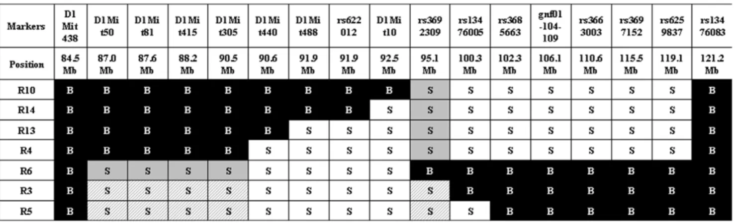
We started our study using the 66H-MMU1 strain which harbors a high rate of embryonic lethality (24.6%) caused by the Led2 QTL [15]. This QTL of ,32 Mb, which was initially delimited by D1Mit50 (87.0 Mb) and rs6259837 (119.1 Mb) markers located on chromosome 1, corresponds to a spretus fragment carried by the 66H-MMU1 substrain. However, a uncertainty of ,6.4 Mb existed at the proximal boundary of this QTL since the interval comprised between D1Mit134 (80.6 Mb) and D1Mit50 (87.0 Mb) markers corresponds to this distance and the breakpoint is located somewhere between these two markers. Indeed, D1Mit134 and D1Mit50 allele markers are of B6 and spretus natures respectively (http://www.pasteur.fr/recherche/ unites/Gfons/ircs/ircshome.htm). In an attempt to precise the position of the breakpoint, we genotyped 8 novel markers located on this region that permitted to reduce the recombination region to a 2.5 Mb interval comprised between 84.5 Mb and 87 Mb (markers D1Mit438 and D1Mit50 respectively, see Figure 1). Then, we initiated a fine mapping approach using fifteen recombinant substrain issued from 66H-MMU1 animals. In each of these strains, a crossing-over fragmented the original DNA region of spretus origin that was initially present in the 66H-MMU1 strain (Figure 1). Among 15 starting strains, seven survived and were available for our study (recombinants, R3, R4, R5, R6, R10, R13 and R14).
In vivo Phenotyping
Females of the different recombinant substrains were crossed with B6 males and their gestation was followed up using in vivo
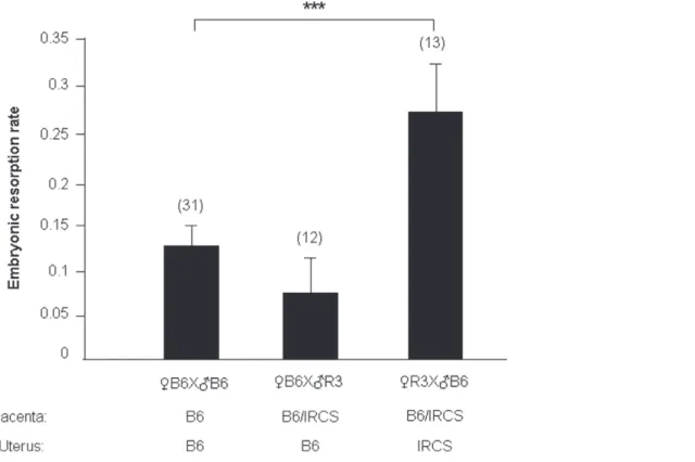
ultrasonography as previously described [15]. Animals obtained from these crosses have a placenta which is heterozygous for all the genes located on the fragment of spretus origin (B6/SEG), while the uterus was homozygous SEG/SEG for the same spretus fragment. The control group was obtained by crossing males and females of the B6 strain. A total of 97 gestations (31 and 66 of B6 and IRCS types respectively) were analyzed. For each gestation, we counted the number of implanted and resorbed embryos during three ultrasonographic examinations. There was no correlation between embryonic death and the position inside the womb, which suggests that the death of one embryo did not have deleterious repercussions on the contiguously implanted structures (Figure 2). We noted a strong variability in the percentage of embryonic death between the different substrains (Table 1), while apparently the number of implanted embryo was not significantly different. The strains R4, R6, R10, R13 and R14 (group 1) presented a percentage of embryonic death that was not statistically different from that of B6 control animals (1064%, 1965%, 963%, 1265% and 1668% for the five strains, respectively, versus 1262% in the B6 parent). Note that within this group, R6 is distinguished by the highest rate of embryonic death. By contrast, R3 and R5 strains (group 2) presented a percentage of embryonic death (2765% and 2966% respectively) significantly higher than B6 control at p = 0.0013 and p = 0.0045 respectively (Table 1). Since the mean number of implanted embryos was not different between the substrains compared to the B6 control (Table 1), we deduced that the increase in embryonic death observed for R3 and R5 IRCS was caused by post-implantation events.
QTL Fine Mapping
In order to refine Led2 localization, we realized an analysis by genotype/phenotype segregation. R3 and R5 strains (which exhibit the phenotype) shared a large spretus region (.84.5 Mb to 90.5 Mb) with the R6 strain (which does not display the embryonic resorption phenotype) and the rest (until ,100.3 Mb) is also shared with the other strains (R4, R10, R13 and R14) which are not affected. This configuration suggests that two spretus regions, shared by R3 and R5 strains and not present together in the other strains, seem to be necessary to explain the apparition of the phenotype in R3 and R5. We defined a first spretus subfragment called Led2minA which encompasses D1Mit50 to
Figure 1. Genomic structure of the IRCS mice in the region of interest of chromosome 1. The map presents the genomic background, spretus or musculus, of the 7 recombinant substrains (Rc) in the chromosomal region corresponding to Led2 QTL. Recombinant strains were generated at the Pasteur institute (Paris) from 66HMMU1 strain by recombination events inside the MMU1 spretus segment. These strains have been genotyped using 24 polymorph markers. Marker positions are given in megabase pairs (Mb). ‘‘S’’ corresponds to the marker in a spretus homozygous form and ‘‘B’’ to the marker in a musculus (B6) homozygous form. The two minimal spretus regions (Led2minA with main effect and Led2minB with probable weak effect) responsible for the phenotype of interest are highlighted in gray and in gray hatched when coexisting in the same substrain. doi:10.1371/journal.pone.0043356.g001
D1Mit305 region (.84.5 Mb to ,90.5 Mb) and a second region called Led2minB located at the rs3692309 marker (.92.5 Mb to ,100.3 Mb) (see gray boxes in Figure 1). When these two spretus regions (Led2min) are separated as in R6 (that contains Led2minA only) or in R4, R10, R13 or R14 (Led2minB only) the phenotype of embryonic death is absent. The presence of the two spretus regions seems indispensable to permit the manifestation of the phenotype, it’s the case for R3 and R5 (gray hatched boxes in Figure 1). To statistically prove the presence of these two QTLs, Led2minA and Led2minB each one responsible for a part of the effect on the phenotype, and an eventual epistatic interaction between these two QTLs able to increase the embryonic death, we compared several recombinants among themselves, R6 bearing Led2minA, R4 bearing Led2minB and R3 (or R5) bearing these two spretus regions.
The results of statistical t-tests are shown in Figure 3. When we compared the mean rate of embryonic death between R4 and R3 (or R5), the statistical result (significant difference at P#0.01) proved the presence of Led2minA QTL. By contrast, the comparison between R6 and R3 (or R5), did not statistically indicate the presence of Led2minB QTL. However the embryonic death rates of R3 and R5 both have a tendency to be higher than that of R6 (Figure 1) suggesting a possible very small effect of Led2minB on the phenotype. In the same way, the difference in embryonic death rate between R4 (Led2minB only) and R3 (or R5) was 17%–19% which is comparable to 15%–17% difference between B6 (with no spretus regions) and R3 (or R5) also suggesting a nil or very small effect of Led2minB. In consequence this result did not support the presence of an epistatic interaction
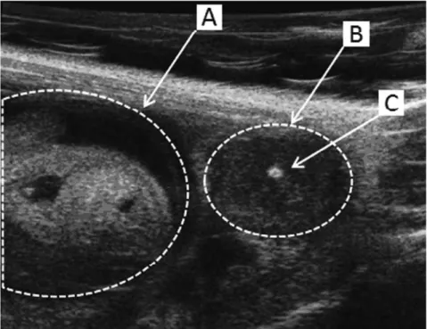
Figure 2. Ultrasound biomicroscopicin vivoobservation of the embryonic development. During the gestation of B6 and IRC mice the embryonic development was followed up by an in vivo ultrasonic method. The viability of developing embryo (A) was assessed by the presence of heartbeats and a positive umbilical cord Doppler. Dead embryos (B) displayed a central highly echogenic zone (C) corresponding to the embryonic resorption.
doi:10.1371/journal.pone.0043356.g002
Table 1. Statistical analysis of the embryonic resorption phenotype.
Strains Number of gestations Implanted embryos; Mean± SEM (p value)
Embryonic resorption rate; Mean± SEM (p value) B6 31 7.9060.41 12%62 R3 13 7.4660.50 (p = 0.2717; NS) 27%±5 (p = 0.0013) R4 15 7.2060.71 (p = 0.1845; NS) 10%64 (p = 0.3021; NS) R5 4 9.5060.50 (p = 0.0932; NS) 29%±6 (p = 0.0045) R6 6 8.5060.99 (p = 0.2848; NS) 19%65 (p = 0.1158; NS) R10 8 9.1260.51 (p = 0.0828; NS) 9%63 (p = 0.2188; NS) R13 12 8.8360.45 (p = 0.1045; NS) 12%65 (p = 0.4349; NS) R14 8 8.7560.70 (p = 0.1759; NS) 16%68 ((p = 0.2351; NS) Comparison between IRCs and B6 control using t-test with Bonferroni-corrected level.
(NS: non-significant).
between Led2minA and Led2minB regions, but a Led2minB additive effect could be revealed by increasing sample size of this ‘‘QTL’’ representative strains. For this raison, the genes present in these two regions, Led2minA and Led2minB are listed in Table 2.
Is Placenta or Uterus Responsible for the Led2min Effect?
Since R3 and R5 mice did not harbor any obvious develop-mental anomaly or pathology, excepting for some embryonic death, it was reasonable to suspect that placental and/or uterine dysfunctions could be responsible for the embryonic lethality increase. Thus, we initiated a genetic approach in order to identify in which of these two organs dysfunction could be related with the phenotype. In the previous set of experimental crosses, IRCS females were mated with B6 males. Genetically, this permitted the co-existence of heterozygous foeto-placental alleles (B6/SEG) and homozygous uterine alleles (SEG/SEG) within the same genomic region (spretus fragment). Conversely, we performed reverse crosses (RB6 6 =IRCS), giving a heterozygous placenta for the genes of the fragment, but a B6 homozygous uterus. In this situation, the would-be disorders ought to find their origin exclusively from a placental-fetal/embryonic defect, caused by the spretus state of the MMU1 fragment, but not from a B6 womb defect. In this optic we realized the cross RB6 with =R3 (IRCS group2). We observed that the mean of embryonic resorption rate (6SEM) was 0.0760.04 and not significantly different from the control (RB6 6 =B6: 0.1260.02; p = 0.118) whereas the inverse crossing, leading also to a heterozygous foeto-placental complex implanted in homozygous spretus uterus (R3), produced a significantly higher embryonic resorption rate (0.2760.05, p = 0.001) (Figure 4). From this last observation, we concluded that a uterine dysfunction is very likely at the basis of the observed phenotype.
Assessing of Differentially Expressed Genes in Uterus
We analyzed the expression level of uterine genes in pregnant R3 females compared to those from B6 control animals at E12.5, an important time point when most resorption occurred during our study. For this purpose, we hybridized cDNA synthesized from RNA uterine tissue to Nimblegen mouse microarrays. The Nimblegen arrays interrogated a total of 25,631 mouse transcripts. Gene expression levels were quantified by fluorescence intensity
assessing and ranged from 20 to .50,000 arbitrary units of fluorescence (AUF) (mean value ,5,250 AUF). These results were highly reproducible since they showed strong correlations between experimental duplicates (r = 0.967 for B6 and 0.983 for R3). Thus, for subsequent analysis, we took the average of both values for each transcript. We first focused on transcripts with fluorescence levels higher than 100 AUF, we assumed that values under this threshold were very close to background signals and, with this threshold, we selected 18,085 transcripts (70.6% of the total). We considered a gene as differentially expressed if a two-fold difference of expression (up or down) was observed. Consequently, 3,436 (19% of the expressed uterine genes) transcripts were modified in R3 uterus when compared to those expressed in B6 (Table 3). A similar number of repressed and induced genes was observed (10.9% and 8.1%, respectively). This deregulation was identified over all the genome. However a significantly higher proportion of genes were deregulated when only the MMU1 spretus fragment or the two Led2min regions were considered (Table 3).
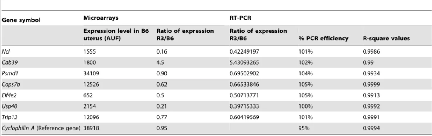
In order to validate the differential gene expression obtained by the microarray analysis, we checked 7 genes of the Led2minA QTL region by quantitative RT-PCR. As shown in Table 4 we obtained a very good agreement between microarray and qPCR results.
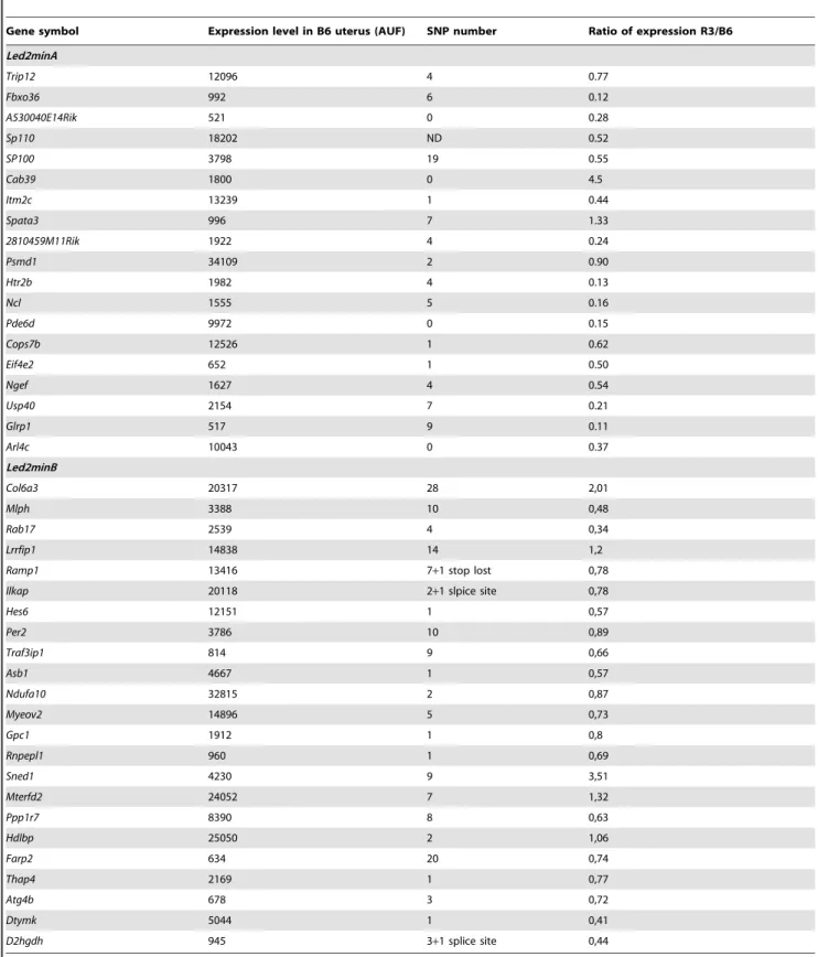
Considering that the uterine dysfunction can take its origin from a deregulation of the gene expression or/and non-synonymous coding polymorphisms accumulated during independent evolutive processes of Mus musculus and Mus spretus species, we listed the genes of the Led2min QTL corresponding to these criteria and thus potentially involved in embryonic resorption. Finally we consid-ered the transcripts with an expression level .500 AUF and either exhibiting a deregulation (R3/B6 expressional ratio .2 or ,0.5) and the presence of non synonymous polymorphisms between Mus musculus and Mus spretus (provided by SANGER database: http:// www.sanger.ac.uk/) (Table 5).
Then, we searched whether deregulated transcripts could be grouped into functional clusters using DAVID database [17], considering 1758 transcripts with a threshold of .500 AUF in the expression level. This analysis led to the identification of five functional groups of genes and signaling pathways that were deregulated, such as ribosome protein genes (p value: 0.00054), endocytosis process (p value: 0.0027), VEGF (vascular endothelial
Figure 3. Statistical comparisons between embryonic death rates of IRCS. The mean of embryonic death rate (6SEM) for (n) gestations is presented for four substrains containing different regions of spretus origin: R6 (containing Led2minA only), R3 and R5 (containing Led2minA and Led2minB), and R4 (Led2minB only).
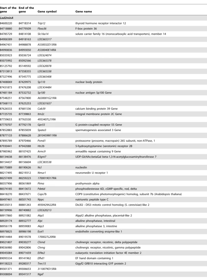
Table 2. Gene list in the minimal Led2min region.
Start of the gene
End of the
gene Gene symbol Gene name Led2minA
84600220 84718314 Trip12 thyroid hormone receptor interactor 12 84718880 84779509 Fbxo36 F-box protein 36
84785729 84814108 Slc16a14 solute carrier family 16 (monocarboxylic acid transporters), member 14 84906589 84918163 LOC665317 84967431 84988878 A530032D15Rik 84990836 84993050 A530040E14Rik 85035923 85036724 LOC624074 85075992 85092566 LOC665378 85125792 85149592 LOC620078 87313813 87358355 LOC665338 87327496 87345775 LOC665408
87408069 87429975 Sp110 nuclear body protein 87431873 87476208 LOC434484
87481184 87532732 Sp100 nuclear antigen Sp100 Gene 87548251 87567800 A630001G21Rik
87568115 87625253 LOC631657
87626533 87681536 Cab39 calcium binding protein 39 Gene 87725735 87739863 Itm2c integral membrane protein 2C Gene 87759653 87763530 4933407L21Rik
87770707 87792178 Gpr55 G protein-coupled receptor 55 Gene 87852883 87855839 Spata3 spermatogenesis associated 3 Gene 87877133 87886628 2810459M11Rik
87895789 87970466 Psmd1 proteasome (prosome, macropain) 26S subunit, non-ATPase, 1 87930441 87942088 Htr2b 5-hydroxytryptamine (serotonin) receptor 2B
87985962 88107425 Armc9 armadillo repeat containing 9 Gene
88134638 88138476 B3gnt7 UDP-GlcNAc:betaGal beta-1,3-N-acetylglucosaminyltransferase 7 88154437 88156604 LOC383538
88175889 88190626 Ncl nucleolin
88217495 88219312 Nmur1 neuromedin U receptor 1 88257499 88259223 1700019O17Rik
88357906 88361869 Ptma prothymosin alpha
88374185 88413672 Pde6d phosphodiesterase 6D, cGMP-specific, rod, delta
88418270 88437671 Cops7b COP9 (constitutive photomorphogenic) homolog, subunit 7b (Arabidopsis thaliana) 88497461 88501743 Nppc natriuretic peptide type C
88535013 88881263 4930429A22Rik Dis3l2 : DIS3 mitotic control homolog (S. cerevisiae)-like 2 88739906 88740882 LOC620213
88917860 88921082 Akp5 Alppl2 alkaline phosphatase, placental-like 2 88929174 88932777 Alpi alkaline phosphatase, intestinal
88956178 88959083 Akp3 alkaline phosphatase 3, intestine 88978825 88986198 Ecel1 endothelin converting enzyme-like 1 89014484 89019578 1700027L20Rik
89021807 89030277 Chrnd cholinergic receptor, nicotinic, delta polypeptide 89036980 89042806 Chrng cholinergic receptor, nicotinic, gamma polypeptide 89045084 89071659 Eif4e2 eukaryotic translation initiation factor 4E member 2 89095534 89141962 Efhd1 EF hand domain containing 1
89158223 89280317 Tnrc15 Gigyf2 GRB10 interacting GYF protein 2 89301371 89306653 3110079O15Rik
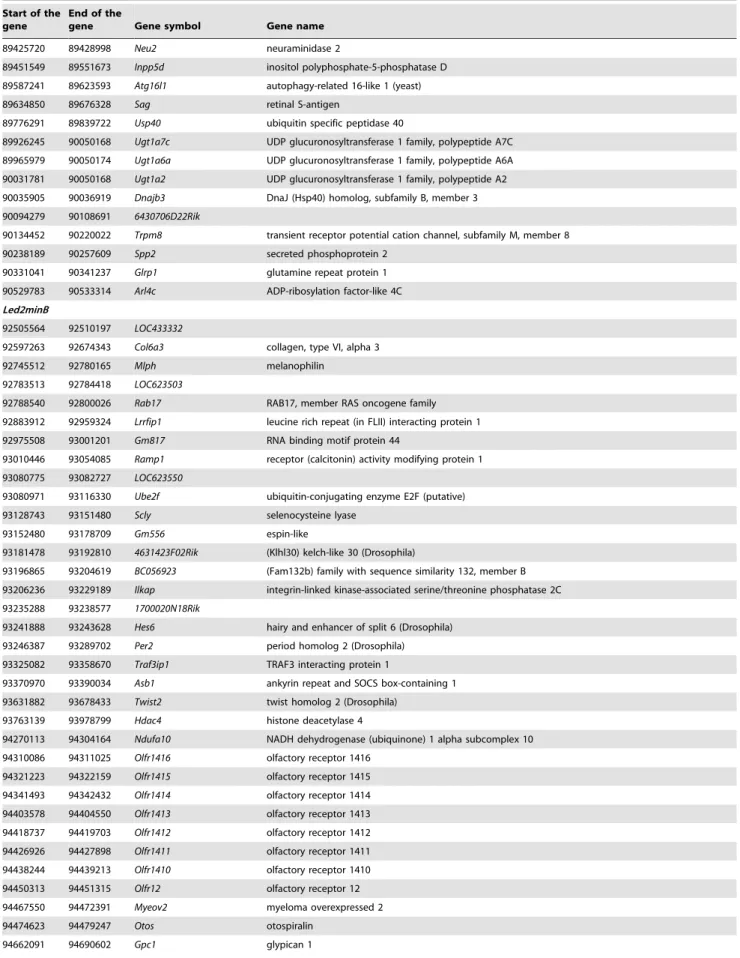
Table 2. Cont.
Start of the gene
End of the
gene Gene symbol Gene name 89425720 89428998 Neu2 neuraminidase 2
89451549 89551673 Inpp5d inositol polyphosphate-5-phosphatase D 89587241 89623593 Atg16l1 autophagy-related 16-like 1 (yeast) 89634850 89676328 Sag retinal S-antigen
89776291 89839722 Usp40 ubiquitin specific peptidase 40
89926245 90050168 Ugt1a7c UDP glucuronosyltransferase 1 family, polypeptide A7C 89965979 90050174 Ugt1a6a UDP glucuronosyltransferase 1 family, polypeptide A6A 90031781 90050168 Ugt1a2 UDP glucuronosyltransferase 1 family, polypeptide A2 90035905 90036919 Dnajb3 DnaJ (Hsp40) homolog, subfamily B, member 3 90094279 90108691 6430706D22Rik
90134452 90220022 Trpm8 transient receptor potential cation channel, subfamily M, member 8 90238189 90257609 Spp2 secreted phosphoprotein 2
90331041 90341237 Glrp1 glutamine repeat protein 1 90529783 90533314 Arl4c ADP-ribosylation factor-like 4C Led2minB
92505564 92510197 LOC433332
92597263 92674343 Col6a3 collagen, type VI, alpha 3 92745512 92780165 Mlph melanophilin
92783513 92784418 LOC623503
92788540 92800026 Rab17 RAB17, member RAS oncogene family 92883912 92959324 Lrrfip1 leucine rich repeat (in FLII) interacting protein 1 92975508 93001201 Gm817 RNA binding motif protein 44
93010446 93054085 Ramp1 receptor (calcitonin) activity modifying protein 1 93080775 93082727 LOC623550
93080971 93116330 Ube2f ubiquitin-conjugating enzyme E2F (putative) 93128743 93151480 Scly selenocysteine lyase
93152480 93178709 Gm556 espin-like
93181478 93192810 4631423F02Rik (Klhl30) kelch-like 30 (Drosophila)
93196865 93204619 BC056923 (Fam132b) family with sequence similarity 132, member B 93206236 93229189 Ilkap integrin-linked kinase-associated serine/threonine phosphatase 2C 93235288 93238577 1700020N18Rik
93241888 93243628 Hes6 hairy and enhancer of split 6 (Drosophila) 93246387 93289702 Per2 period homolog 2 (Drosophila) 93325082 93358670 Traf3ip1 TRAF3 interacting protein 1
93370970 93390034 Asb1 ankyrin repeat and SOCS box-containing 1 93631882 93678433 Twist2 twist homolog 2 (Drosophila)
93763139 93978799 Hdac4 histone deacetylase 4
94270113 94304164 Ndufa10 NADH dehydrogenase (ubiquinone) 1 alpha subcomplex 10 94310086 94311025 Olfr1416 olfactory receptor 1416
94321223 94322159 Olfr1415 olfactory receptor 1415 94341493 94342432 Olfr1414 olfactory receptor 1414 94403578 94404550 Olfr1413 olfactory receptor 1413 94418737 94419703 Olfr1412 olfactory receptor 1412 94426926 94427898 Olfr1411 olfactory receptor 1411 94438244 94439213 Olfr1410 olfactory receptor 1410 94450313 94451315 Olfr12 olfactory receptor 12 94467550 94472391 Myeov2 myeloma overexpressed 2 94474623 94479247 Otos otospiralin
Table 2. Cont.
Start of the gene
End of the
gene Gene symbol Gene name
94700534 94733312 Ankmy1 ankyrin repeat and MYND domain containing 1 94737394 94739026 0710001B24Rik (Dusp28) dual specificity phosphatase 28 94741810 94750986 Rnpepl1 arginyl aminopeptidase (aminopeptidase B)-like 1 94764813 94778354 Capn10 calpain 10
94809526 94815938 Gpr35 G protein-coupled receptor 35 94836739 94842672 Aqp12 aquaporin 12
94848683 94932228 Kif1a kinesin family member 1A 94965650 94988099 Agxt alanine-glyoxylate aminotransferase 94981761 94991354 2310007B03Rik
94999130 95061480 E030010N08Rik
95066246 95131471 Sned1 sushi, nidogen and EGF-like domains 1 95129616 95136276 Mterfd2 MTERF domain containing 2
95139842 95167980 Pask PAS domain containing serine/threonine kinase 95174050 95198024 Ppp1r7 protein phosphatase 1, regulatory (inhibitor) subunit 7 95204301 95233908 Tmem16g Ano7: anoctamin 7
95236345 95309214 Hdlbp high density lipoprotein (HDL) binding protein 95252098 95252952 LOC621682
95309449 95340136 sept-02 septin 2
95342530 95452188 Farp2 FERM, RhoGEF and pleckstrin domain protein 2 95452309 95466056 Stk25 serine/threonine kinase 25 (yeast)
95516099 95526168 Bok BCL2-related ovarian killer protein 95535796 95585244 Thap4 THAP domain containing 4 95585438 95619935 Atg4b autophagy-related 4B (yeast) 95630900 95632281 Dtymk deoxythymidylate kinase
95634370 95652507 Ing5 inhibitor of growth family, member 5 95655698 95682580 D2hgdh D-2-hydroxyglutarate dehydrogenase 95691749 95706900 Gal3st2 galactose-3-O-sulfotransferase 2 95792483 95816522 LOC666009
95820914 95841947 LOC619597
95850898 95858740 Neu4 sialidase 4
95868710 95882959 Pdcd1 programmed cell death 1
97144149 97165471 2310044D20Rik Fam174a : family with sequence similarity 174, member A 97418087 97498000 St8sia4 ST8 alpha-N-acetyl-neuraminide alpha-2,8-sialyltransferase 4 98475880 98476840 Gm1833 predicted gene 1833
98648136 98702541 Slco4c1 solute carrier organic anion transporter family, member 4C1 98702644 98708307 9530060I07
98736581 98827966 Slco6b1 solute carrier organic anion transporter family, member 6b1 98889854 98958709 Slco6c1 solute carrier organic anion transporter family, member 6b1 98993265 99132680 LOC634331
99176834 99309408 LOC634346
99474308 99492382 D1Ertd622e DNA segment, Chr 1, ERATO Doi 622, expressed 99536564 99591955 Hisppd1 Ppip5k2: diphosphoinositol pentakisphosphate kinase 2 99600605 99623375 4930429M06Rik Gin1: gypsy retrotransposon integrase 1
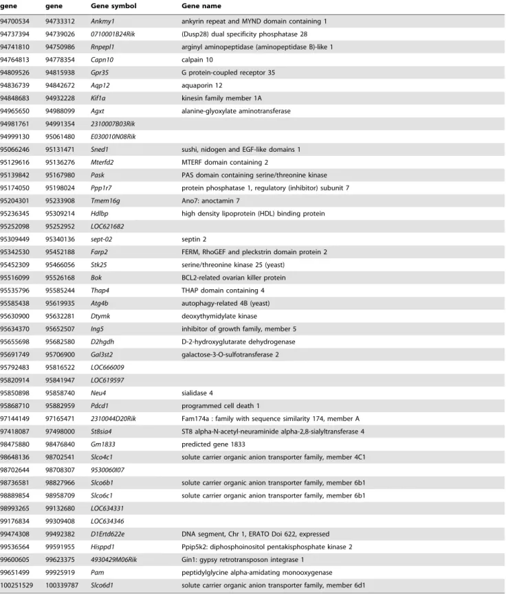
99651499 99925919 Pam peptidylglycine alpha-amidating monooxygenase 100251529 100339787 Slco6d1 solute carrier organic anion transporter family, member 6d1 doi:10.1371/journal.pone.0043356.t002
growth factor) signaling (p value: 0.0078), chemokine interactions (p value: 0.011) and mTOR (mammalian Target Of Rapamycin) signaling pathway (p value: 0.014).
Discussion
In the reproductive processes, as in others, hundreds of genes interact into subtle regulatory networks, and this complexity does not permit to easily identify the molecular factors of dysfunctions leading to infertility cases. Moreover, when our interest is turned towards the human clinic, the study of factors involved in reproductive defects is particularly challenging due to obvious ethical constraints, which rends obligatory the use of animal models. However although hundreds of mutant mouse models with infertility/hypofertility phenotypes have been generated [2], the genetic causes of infertilities are far from being elucidated in their whole [3]. This is the case for the RSA pathology which affects a non negligible percentage of the population (1 to 5%) and for which the genetic origin(s) is still little documented. In the aim to identify new genes responsible for embryonic lethality, we used
a mouse model of interspecific recombinant congenic strains (IRCS). Although during the gestation/pregnancy development, mice and humans do not establish exactly the same system of placentation, similarities strong enough between these two species exist, making the mouse model useful to identify genes involved in humans. Indeed, in several mouse models involving the comple-ment system [18,19], it has been clearly shown that there is a continuum between embryo resorption and placental diseases, since the same mice have these resorption and preeclampsia-like symptoms [20]. It is also known that C3 defects are clearly linked with VEGF defects, thus inducing defective placentation, leading in the most extreme cases to embryonic death. Therefore, mouse models of embryo resorption via known deregulations of complement system are proved to be suitable models of the human continuum placental vascular disease-spontaneous resorp-tion. A well studied mouse model of immunologically mediated peri-implantation pregnancy loss that shares features with human recurrent miscarriage is derivated from DBA/2-mated CBA/J mice (CBA/J 6DBA/2) [21,22,23]. Indeed embryos derived from mating CBA/J females with DBA/2 males showed an increased
Figure 4. Embryonic resorption rate in function of the type of crossing realized with IRCS and B6 mice. The results of different crosses (RIRCS 6 =B6, RB6 6 =IRCS and RB6 6 =B6) are presented as the average (6SEM) of the embryonic resorption rate for (n) gestations.
doi:10.1371/journal.pone.0043356.g004
Table 3. Number of transcripts modified in uterus of R3 IRCS compared to C57BL6/J.
Genomic region Expressed Transcripts (.100 AUF) Deregulated transcripts (2-fold threshold) Total genome 18,085 3,436 (19%)
Spretus fragment in MMU1 148 53 (36%) Led2minA (84.5–90.5 Mb) 55 24 (44%) Led2minB (95.1 Mb to ,100.3 Mb) 25 8 (32%) doi:10.1371/journal.pone.0043356.t003
frequency of resorption (29.466.5%), more than three times greater than that seen within these and other strains or strain combinations (CBA/J 6 CBA/J: 8.965.1%; CBA/J 6 BALB/c: 8.265.6%; DBA/2 6 DBA/2: 8.566.6%; n = 6–32 mice/group; CBA/J 6 DBA/2 vs. others, p,0.01) [18]. Spontaneous resorption in the CBA/J 6 DBA/2 model is attributed to NK cells, macrophages, and Th1-type cytokines. and represent a rejection of the semiallogeneic fetoplacental [24]. Murine resorp-tions are characterized by focal necrosis at the junction of the fetal trophoblast with decidua, an infiltrate of polymorphonuclear leukocytes (with some lymphocytic cells) at sites of necrosis and along the walls of large vessels in decidua, and by thrombosis and hemorrhage [25,26,27,28]. Infiltration begins on day 6.5 of gestation, 2 days after implantation has occurred, and abortions begin after day 8.5 of pregnancy [24,28].
In the present work we used a mouse model including interspecific recombinant congenic strains (IRCS). The originality of this whole model is based on the presence of a small homozygous fragments of Mus spretus genome fixed on a Mus musculus B6 genetic background [16]. Thus, a strain differs of each other and from the B6 parental strain by the spretus segments. Mus musculus and Mus spretus diverged ,2 million years ago meaning that the association of their two genomes has the potential to lead to genetic incompatibilities [14]. Using this model, in past studies, we were able to localize various QTL modulating male and female reproductive processes. We identified a QTL responsible for ,25% of the embryonic resorptions present in the IRCS-66H-MMU1 strain containing a solely chromosomal fragment of spretus origin located on MMU1. This QTL was called Led2 and has been mapped to an interval of 32 Mb which contains 215 genes [15]. The aim of the present study was to redefine this region and to identify candidate genes potentially involved in embryonic lethality.
To accomplish the fine mapping of this QTL, we generated recombinant substrains from 66H-MMU1 by backcrosses, each of them presenting a unique sub-fragment of the Led2 QTL. Each recombinant substrain females were crossed with B6 males, resulting in a fetus/placenta complex with heterozygous B6/ SEG genes (at the Led2 locus) and uterine homozygous spretus genes (at the Led2 locus). During each gestation, the substrains were phenotyped in vivo by ultrasonography. This non-invasive tech-nology, based upon a high frequency ultrasound device [29] allows in vivo real time high resolution observations of embryonic development [15,30] and resorption (,70mm and ,40mm lateral and axial resolution, respectively) and permits to carry
out longitudinal analysis of gestation. We observed an increase of the embryonic death rate in R3 and R5 substrains (Group 2). The analysis of the genotype/phenotype segregation allowed us to determine two reduced QTL regions (Led2minA and Led2minB) of approximately 6 Mb each, present together in spretus version only in R3 and R5 strains. In the other recombinant substrains which have not the phenotype, the one or the other of the region is present but not the two regions together. So we defined the first reduced spretus region called Led2minA which encompasses D1Mit50 to D1Mit305 region (.84.5 Mb to 90.5 Mb) and the second called Led2minB located at the rs3692309 marker (.92.5 Mb to ,100.3 Mb). Our statistical analysis succeeded in proving the presence of Led2minA QTL responsible for a main effect on embryonic death but it failed it for Led2minB. However, notable differences between the embryonic death rates of certain strains (R6 compared to R3 or R5) led to suppose that this latter region could also have a small effect in the phenotype. Taken together, these data did not support the presence of an epistatic interaction between Led2minA and Led2minB.
Reverse crosses using IRCS Group 2 males and B6 females revealed that the genes expressed at heterozygous state in the placental tissues are not deleterious for the gestation. Therefore, we deduced that the high rate of embryonic death occurring during the gestation resulted from dysfunction of genes expressed in the uterine tissue. This is in accordance with the normal embryonic development observed in group 2 IRCS females. Then, we carried out a microarray analysis searching to identify uterine deregulated genes in IRCS animals from Group 2. Although we observed deregulated genes located in all chromosomes (19%) we noticed that those situated on the spretus fragment were preferen-tially modified (,40%). This concentration of deregulated genes located on the spretus fragment has already been reported in a previous study of our group performed on testis transcriptome [31]. It has been showed that at genomic scale differential SNPs between Mus musculus and Mus spretus are frequent since they appear, in average, every 100 bp. When located on the promoter regions of spretus origin, these nucleotide substitutions could modify the transactivation/transrepression properties of transcription factors of C57BL/6J nature, thus modifying the spretus gene expressions. Additionally, dysfunctions leading to embryonic death could result from non-synonymous coding polymorphisms, accu-mulated during evolution in the spretus genome. These phenomena should be originated from evolution of separated genomic regions that produces transcription factors/DNA (‘‘transcriptomic shock’’) and/or protein-protein (‘‘proteomic shock’’) incompatibilities [32]. Table 4. Microarray validation by RT-QPCR on 7 genes of the QTL region.
Gene symbol Microarrays RT-PCR Expression level in B6
uterus (AUF)
Ratio of expression R3/B6
Ratio of expression
R3/B6 % PCR efficiency R-square values Ncl 1555 0.16 0.42249197 101% 0.9986 Cab39 1800 4.5 5.43093265 102% 0.99 Psmd1 34109 0.90 0.69502902 104% 0.9934 Cops7b 12526 0.62 0.66533846 105% 0.9999 Eif4e2 652 0.5 0.50713771 105% 0.9913 Usp40 2154 0.21 0.39715333 100% 0.9992 Trip12 12096 0.77 0.60419569 101% 0.9991 Cyclophilin A (Reference gene) 38918 0.95 95% 0.9994 doi:10.1371/journal.pone.0043356.t004
Table 5. Genes of Led2min region expressed in uterus (.500 AUF) and displaying a deregulation (R3/B6 ratio) and/or non synonymous SNP.
Gene symbol Expression level in B6 uterus (AUF) SNP number Ratio of expression R3/B6 Led2minA Trip12 12096 4 0.77 Fbxo36 992 6 0.12 A530040E14Rik 521 0 0.28 Sp110 18202 ND 0.52 SP100 3798 19 0.55 Cab39 1800 0 4.5 Itm2c 13239 1 0.44 Spata3 996 7 1.33 2810459M11Rik 1922 4 0.24 Psmd1 34109 2 0.90 Htr2b 1982 4 0.13 Ncl 1555 5 0.16 Pde6d 9972 0 0.15 Cops7b 12526 1 0.62 Eif4e2 652 1 0.50 Ngef 1627 4 0.54 Usp40 2154 7 0.21 Glrp1 517 9 0.11 Arl4c 10043 0 0.37 Led2minB Col6a3 20317 28 2,01 Mlph 3388 10 0,48 Rab17 2539 4 0,34 Lrrfip1 14838 14 1,2
Ramp1 13416 7+1 stop lost 0,78 Ilkap 20118 2+1 slpice site 0,78
Hes6 12151 1 0,57 Per2 3786 10 0,89 Traf3ip1 814 9 0,66 Asb1 4667 1 0,57 Ndufa10 32815 2 0,87 Myeov2 14896 5 0,73 Gpc1 1912 1 0,8 Rnpepl1 960 1 0,69 Sned1 4230 9 3,51 Mterfd2 24052 7 1,32 Ppp1r7 8390 8 0,63 Hdlbp 25050 2 1,06 Farp2 634 20 0,74 Thap4 2169 1 0,77 Atg4b 678 3 0,72 Dtymk 5044 1 0,41 D2hgdh 945 3+1 splice site 0,44 ND: Not determined. doi:10.1371/journal.pone.0043356.t005
Focusing on genes of the Led2minA QTL and applying filters from bioinformatics databases, bibliography and our own results, we propose a selection of 7 genes (Trip12, Cab39, Psmd1, Ncl, Cops7b, Eif4e2 and Usp40) as putative actors of the embryonic death. These genes play a role in VEGF signaling, mTOR signaling and ubiquitine/proteasome-protein degradation path-way. Their effects could be reinforced by a small participation of genes situated on Led2minB region and which could act in the same signaling pathways (Asb1, Traf3ip1, Ramp1 and Col6a3).
Trip12, Psmd1, Cops7b and Usp40 from Led2minA and Asb1 from Led2minB are involved in protein degradation process through the ubiquitin-proteasome pathway. Trip12 exerts a ligase activity related to ubiquitination [33], Usp40 functions as a deubiquitina-tion enzyme in the same degradadeubiquitina-tion pathway [34] and Asb1 is a member of the ankyrin repeat and SOCS box (ASB) family. These family proteins interact with Cul5-Rbx2 to form E3 ubiquitin ligase [35]. Psmd1 is a component of the 26S proteasome. Cops7b is a subunit of the eight-subunit heteromeric Cop9 signalosome complex. Genetic invalidations of some Cop9 subunits have been associated with developmental defects of post-implantation embryos [36,37,38]. Additionally, Usp40 functions as a deubiqui-tination enzyme in the same degradation pathway [34]. Asb1 is a member of the ankyrin repeat and SOCS box (ASB) family. These family proteins interact with Cul5-Rbx2 to form E3 ubiquitin ligase [35].
In the same manner, Led2minA Ncl gene and Led2minB Ramp1 and Col6a3 genes are involved in angiogenesis. Ncl encodes nucleolin and treatment of endothelial cells with anti-nucleolin antibody induces apoptosis of these cells [39]. Moreover, nucleolin associates with VEGF-C62 and can be potentially involved in epithelial cell adhesion and proliferation [39,40]. Concerning Ramp1 gene, RAMP1 (receptor activity modifying protein) forms a functional receptor for CALCA (Calcitonin gene-related peptide) which is a proangiogenic growth factor in the human placental development and plays a critical role in embryonic development and fetal growth [41]. Concerning the Collagen typeVI a3 gene, COL IV is a main endometrial extracellular matrix component, and an abnormal increased deposition of collagen might impair uterine function, possibly by interfering with vascularization or retarding remodeling events at implantation [42].
Finally, Cab39 (also called Mo25) and Eif4e2 from Led2minA and Traf3ip1 from Led2minB, participate in the mTOR signaling pathway, a regulatory step of protein synthesis and growth. Cab39 effect has been described upstream of mTOR activation while Eif4e2 is a downstream signaling target involved in translation initiation. Interestingly, Mtor genetic disruption in mice leads to early embryonic death [43]. Homozygous Traf3ip1 (Tumor
necrosis factor alpha receptor 3 interacting protein 1) mutant mice are not viable. Traf3ip1 mutant mouse line was generated and the enlarged mutant cell size in culture was associated with elevated basal mTOR pathway activity [44]. Otherwise, mTOR pathway is implicated on the VEGF pathway activation. It is worth noting that the VEGF and mTOR pathways have been identified as significantly deregulated in our functional clustering analysis.
Conclusions
We used an in vivo approach of the embryonic development on a mouse IRCS model to refine a chromosome 1 region (Led2) responsible for embryonic death. The present study succeeded in fine-mapping Led2minA QTL which has a main effect on the embryonic death (about 30%) and pointed out a second region Led2minB which could have a minor effect on the same phenotype. Collecting and analyzing experimental, bioinformatics and liter-ature data on the expression and function of genes present in the two regions (Led2minA and Led2minB), we propose 7 genes from Led2minA that could be related with the phenotype. It appears that the vascularization could be the common denominator at these categories of genes involving angiogenesis and the fluidity of the extracellular matrix. The actual identification of the gene(s) involved in this phenotype will necessarily pass through further molecular approaches. An important outcome of this study is the possibility to evaluate novel promising candidates of RSA in humans [13]. This might contribute to elucidate the molecular basis of this multifactorial and complex human disorder and to propose new diagnostic markers.
Supporting Information
Table S1 Sequences of used real time RT-PCR primers. (DOC)
Acknowledgments
We thank I. Lanctin and J. Chevalier (Pasteur Institute, Paris) for the IRCS breeding and C. Marchiol (Small animal imaging facility of the Cochin Institute, Paris) for their technical assistance. We thank J. Coquet for critical reading of the manuscript.
Author Contributions
Conceived and designed the experiments: DV CS AZ XM. Performed the experiments: MV GB GR FM VF. Analyzed the data: MV DV CS AZ PL. Contributed reagents/materials/analysis tools: MV GR FM VF. Wrote the paper: DV CS AZ MV PL.
References
1. Norris W, Nevers T, Sharma S, Kalkunte S (2011) Review: hCG, preeclampsia and regulatory T cells. Placenta 32 Suppl 2: S182–185.
2. Matzuk MM, Lamb DJ (2008) The biology of infertility: research advances and clinical challenges. Nat Med 14: 1197–1213.
3. Zheng K, Yang F, Wang PJ (2009) Regulation of male fertility by X-linked genes. J Androl 31: 79–85.
4. Rai R, Regan L (2006) Recurrent miscarriage. Lancet 368: 601–611. 5. Stephenson MD, Awartani KA, Robinson WP (2002) Cytogenetic analysis of
miscarriages from couples with recurrent miscarriage: a case-control study. Hum Reprod 17: 446–451.
6. Philipp T, Philipp K, Reiner A, Beer F, Kalousek DK (2003) Embryoscopic and cytogenetic analysis of 233 missed abortions: factors involved in the pathogenesis of developmental defects of early failed pregnancies. Hum Reprod 18: 1724– 1732.
7. Salim R, Regan L, Woelfer B, Backos M, Jurkovic D (2003) A comparative study of the morphology of congenital uterine anomalies in women with and without a history of recurrent first trimester miscarriage. Hum Reprod 18: 162–166.
8. Rey E, Kahn SR, David M, Shrier I (2003) Thrombophilic disorders and fetal loss: a meta-analysis. Lancet 361: 901–908.
9. Levine JS, Branch DW, Rauch J (2002) The antiphospholipid syndrome. N Engl J Med 346: 752–763.
10. Li TC, Makris M, Tomsu M, Tuckerman E, Laird S (2002) Recurrent miscarriage: aetiology, management and prognosis. Hum Reprod Update 8: 463–481.
11. Tulppala M, Palosuo T, Ramsay T, Miettinen A, Salonen R, et al. (1993) A prospective study of 63 couples with a history of recurrent spontaneous abortion: contributing factors and outcome of subsequent pregnancies. Hum Reprod 8: 764–770.
12. Kaare M, Painter JN, Ulander VM, Kaaja R, Aittomaki K (2006) Variations of the Amnionless gene in recurrent spontaneous abortions. Mol Hum Reprod 12: 25–29.
13. Mercier E, Lissalde-Lavigne G, Gris JC (2007) JAK2 V617F mutation in unexplained loss of first pregnancy. N Engl J Med 357: 1984–1985.
14. Benayoun BA, Caburet S, Dipietromaria A, Georges A, D’Haene B, et al. (2010) Functional exploration of the adult ovarian granulosa cell tumor-associated somatic FOXL2 mutation p.Cys134Trp (c.402C.G). PLoS One 5: e8789. 15. Laissue P, Burgio G, l’Hote D, Renault G, Marchiol-Fournigault C, et al. (2009)
Identification of Quantitative Trait Loci responsible for embryonic lethality in mice assessed by ultrasonography. Int J Dev Biol 53: 623–629.
16. Burgio G, Szatanik M, Guenet JL, Arnau MR, Panthier JJ, et al. (2007) Interspecific recombinant congenic strains between C57BL/6 and mice of the Mus spretus species: a powerful tool to dissect genetic control of complex traits. Genetics 177: 2321–2333.
17. Huang da W, Sherman BT, Lempicki RA (2009) Systematic and integrative analysis of large gene lists using DAVID bioinformatics resources. Nat Protoc 4: 44–57.
18. Girardi G, Yarilin D, Thurman JM, Holers VM, Salmon JE (2006) Complement activation induces dysregulation of angiogenic factors and causes fetal rejection and growth restriction. J Exp Med 203: 2165–2175.
19. Singh J, Ahmed A, Girardi G (2011) Role of complement component C1q in the onset of preeclampsia in mice. Hypertension 58: 716–724.
20. Ahmed A, Singh J, Khan Y, Seshan SV, Girardi G (2010) A new mouse model to explore therapies for preeclampsia. PLoS One 5: e13663.
21. Blois S, Tometten M, Kandil J, Hagen E, Klapp BF, et al. (2005) Intercellular adhesion molecule-1/LFA-1 cross talk is a proximate mediator capable of disrupting immune integration and tolerance mechanism at the feto-maternal interface in murine pregnancies. J Immunol 174: 1820–1829.
22. Clark DA, Chaouat G, Arck PC, Mittruecker HW, Levy GA (1998) Cytokine-dependent abortion in CBA 6 DBA/2 mice is mediated by the procoagulant fgl2 prothrombinase [correction of prothombinase]. J Immunol 160: 545–549. 23. Bogdarina I, Murphy HC, Burns SP, Clark AJ (2004) Investigation of the role of
epigenetic modification of the rat glucokinase gene in fetal programming. Life Sci 74: 1407–1415.
24. Clark DA (1991) Controversies in reproductive immunology. Crit Rev Immunol 11: 215–247.
25. Clark DA, Quarrington C, Banwatt D, Manuel J, Fulop G (1994) Spontaneous abortion in immunodeficient SCID mice. Am J Reprod Immunol 32: 15–25. 26. Critchley HO, Kelly RW, Lea RG, Drudy TA, Jones RL, et al. (1996) Sex
steroid regulation of leukocyte traffic in human decidua. Hum Reprod 11: 2257– 2262.
27. Deanesly R (1973) Termination of early pregnancy in rats after ovariectomy is due to immediate collapse of the progesterone-dependent decidua. J Reprod Fertil 35: 183–186.
28. Duclos AJ, Haddad EK, Baines MG (1995) Presence of activated macrophages in a murine model of early embryo loss. Am J Reprod Immunol 33: 354–366. 29. Foster FS, Pavlin CJ, Harasiewicz KA, Christopher DA, Turnbull DH (2000)
Advances in ultrasound biomicroscopy. Ultrasound Med Biol 26: 1–27.
30. Ouyang YQ, Li SJ, Zhang Q, Cai HB, Chen HP (2009) Interactions between inflammatory and oxidative stress in preeclampsia. Hypertens Pregnancy 28: 56–62.
31. Brezillon NM, DaSilva L, L’Hote D, Bernex F, Piquet J, et al. (2008) Rescue of fertility in homozygous mice for the urokinase plasminogen activator transgene by the transplantation of mouse hepatocytes. Cell Transplant 17: 803–812. 32. Sahin M, Greer PL, Lin MZ, Poucher H, Eberhart J, et al. (2005)
Eph-dependent tyrosine phosphorylation of ephexin1 modulates growth cone collapse. Neuron 46: 191–204.
33. Park Y, Yoon SK, Yoon JB (2009) The HECT domain of TRIP12 ubiquitinates substrates of the ubiquitin fusion degradation pathway. J Biol Chem 284: 1540– 1549.
34. Quesada V, Diaz-Perales A, Gutierrez-Fernandez A, Garabaya C, Cal S, et al. (2004) Cloning and enzymatic analysis of 22 novel human ubiquitin-specific proteases. Biochem Biophys Res Commun 314: 54–62.
35. Kohroki J, Nishiyama T, Nakamura T, Masuho Y (2005) ASB proteins interact with Cullin5 and Rbx2 to form E3 ubiquitin ligase complexes. FEBS Lett 579: 6796–6802.
36. Lykke-Andersen K, Schaefer L, Menon S, Deng XW, Miller JB, et al. (2003) Disruption of the COP9 signalosome Csn2 subunit in mice causes deficient cell proliferation, accumulation of p53 and cyclin E, and early embryonic death. Mol Cell Biol 23: 6790–6797.
37. Yan J, Walz K, Nakamura H, Carattini-Rivera S, Zhao Q, et al. (2003) COP9 signalosome subunit 3 is essential for maintenance of cell proliferation in the mouse embryonic epiblast. Mol Cell Biol 23: 6798–6808.
38. Tomoda K, Yoneda-Kato N, Fukumoto A, Yamanaka S, Kato JY (2004) Multiple functions of Jab1 are required for early embryonic development and growth potential in mice. J Biol Chem 279: 43013–43018.
39. Fogal V, Sugahara KN, Ruoslahti E, Christian S (2009) Cell surface nucleolin antagonist causes endothelial cell apoptosis and normalization of tumor vasculature. Angiogenesis 12: 91–100.
40. Wang ZG, Puri TS, Quigg RJ (2010) Characterization of novel VEGF (vascular endothelial growth factor)-C splicing isoforms from mouse. Biochem J 428: 347– 354.
41. Dong YL, Reddy DM, Green KE, Chauhan MS, Wang HQ, et al. (2007) Calcitonin gene-related peptide (CALCA) is a proangiogenic growth factor in the human placental development. Biol Reprod 76: 892–899.
42. Diao H, Aplin JD, Xiao S, Chun J, Li Z, et al. (2011) Altered spatiotemporal expression of collagen types I, III, IV, and VI in Lpar3-deficient peri-implantation mouse uterus. Biol Reprod 84: 255–265.
43. Gangloff YG, Mueller M, Dann SG, Svoboda P, Sticker M, et al. (2004) Disruption of the mouse mTOR gene leads to early postimplantation lethality and prohibits embryonic stem cell development. Mol Cell Biol 24: 9508–9516. 44. Berbari NF, Kin NW, Sharma N, Michaud EJ, Kesterson RA, et al. (2011) Mutations in Traf3ip1 reveal defects in ciliogenesis, embryonic development, and altered cell size regulation. Dev Biol 360: 66–76.
![[PDF] Catalogue de formation bureautique PDF | Cours Bureautique](data:image/gif;base64,R0lGODlhAQABAIAAAP///wAAACH5BAEAAAAALAAAAAABAAEAAAICRAEAOw==)