HAL Id: inserm-02914660
https://www.hal.inserm.fr/inserm-02914660
Submitted on 12 Aug 2020
HAL is a multi-disciplinary open access archive for the deposit and dissemination of sci-entific research documents, whether they are pub-lished or not. The documents may come from teaching and research institutions in France or abroad, or from public or private research centers.
L’archive ouverte pluridisciplinaire HAL, est destinée au dépôt et à la diffusion de documents scientifiques de niveau recherche, publiés ou non, émanant des établissements d’enseignement et de recherche français ou étrangers, des laboratoires publics ou privés.
immunoregulatory function in myeloid dendritic cell
precursors through integrin αv and TGF-β activation
Ailiang Zhang, Helena Païdassi, Adam Lacy-Hulbert, John Savill
To cite this version:
Ailiang Zhang, Helena Païdassi, Adam Lacy-Hulbert, John Savill. Apoptotic cells induce CD103 ex-pression and immunoregulatory function in myeloid dendritic cell precursors through integrin αv and TGF-β activation. PLoS ONE, Public Library of Science, 2020, 15 (7), pp.e0232307. �10.1371/jour-nal.pone.0232307�. �inserm-02914660�
RESEARCH ARTICLE
Apoptotic cells induce CD103 expression and
immunoregulatory function in myeloid
dendritic cell precursors through integrin
α
v
and TGF-β activation
Ailiang Zhang1, Helena Paidassi2, Adam Lacy-Hulbert3, John SavillID1*
1 Medical Research Council Centre for Inflammation Research, Queen’s Medical Research Institute,
University of Edinburgh, Edinburgh BioQuarter, Edinburgh, Scotland, United Kingdom, 2 Inserm U1111, Lyon, France, 3 Benaroya Research Institute at Virginia Mason, Seattle, Washington, United States of America
Abstract
In the mammalian gut CD103+ve myeloid DCs are known to suppress inflammation threat-ened by luminal bacteria, but stimuli driving DC precursor maturation towards this beneficial phenotype are incompletely understood. We isolated CD11+ve DCs from mesenteric lymph nodes (MLNs) of healthy mice; CD103+ve DCs were 8–24 fold more likely than CD103-ve DCs to exhibit extensive of prior phagocytosis of apoptotic intestinal epithelial cells. How-ever, CD103+ve and CD103-ve MLN DCs exhibited similar ex vivo capacity to ingest apo-ptotic cells, indicating that apoapo-ptotic cells might drive immature DC maturation towards the CD103+ve phenotype. When cultured with apoptotic cells, myeloid DC precursors isolated from murine bone marrow and characterised as lineage-ve CD103-ve, displayed enhanced expression of CD103 andβ8 integrin and acquired increased capacity to induce T regulatory lymphocytes (Tregs) after 7d in vitro. However, DC precursors isolated fromαv-tie2 mice
lackingαvintegrins in the myeloid line exhibited reduced binding of apoptotic cells and
com-plete deficiency in the capacity of apoptotic cells and/or latent TGF-β1 to enhance CD103 expression in culture, whereas active TGF-β1 increased DC precursor CD103 expression irrespective ofαvexpression. Fluorescence microscopy revealed clustering ofαvintegrin
chains and latent TGF-β1 at points of contact between DC precursors and apoptotic cells. We conclude that myeloid DC precursors can deployαvintegrin to orchestrate binding of
apoptotic cells, activation of latent TGF-β1 and acquisition of the immunoregulatory CD103 +veβ8+ve DC phenotype. This implies that a hitherto unrecognised consequence of apo-ptotic cell interaction with myeloid phagocytes is programming that prevents inflammation.
Introduction
Mammalian bowel contents are laden with potently pro-inflammatory bacteria and yet the healthy gut is not inflamed. There is now evidence that gut inflammation can be kept in check a1111111111 a1111111111 a1111111111 a1111111111 a1111111111 OPEN ACCESS
Citation: Zhang A, Paidassi H, Lacy-Hulbert A,
Savill J (2020) Apoptotic cells induce CD103 expression and immunoregulatory function in myeloid dendritic cell precursors through integrin αvand TGF-β activation. PLoS ONE 15(7):
e0232307.https://doi.org/10.1371/journal. pone.0232307
Editor: Jean Kanellopoulos, Universite Paris-Sud,
FRANCE
Received: April 8, 2020 Accepted: June 26, 2020 Published: July 15, 2020
Copyright:© 2020 Zhang et al. This is an open access article distributed under the terms of the
Creative Commons Attribution License, which permits unrestricted use, distribution, and reproduction in any medium, provided the original author and source are credited.
Data Availability Statement: All relevant data are
within the manuscript.
Funding: This work was supported by a grant from
the UK Medical Research Council to J.S. (G0802069) and by a grant from the US National Institutes of Health (NIH) to A.L-H. (U19 AI125378). The funders had no role in study design, data collection and analysis, decision to publish, or preparation of the manuscript.
by anti-inflammatory programming of myeloid phagocytes encountering cells dying by apo-ptosis in the gut wall. Elegant work from Blander’s group employed transgenic techniques to induce apoptosis in intestinal epithelial cells (IECs)in vivo and then track their uptake by mye-loid phagocytes. Dendritic cells (DCs) expressing CD103 were observed to ingest apoptotic IECs and respond with two notable patterns of gene transcription; downregulation of inflam-matory genes and suppression of the immune response [1]. Previous studies had reported that CD103+ve myeloid DCs in the gut wall and draining lymphatics directed powerful suppres-sion of immune responses, inducing peripheral T regulatory lymphocytes in draining mesen-teric lymph nodes through mechanisms including TGF-β1 activation [2–6]. However, this body of work had not addressed whether CD103+ve myeloid DCs in the gut had interacted with cells spontaneously undergoing apoptosis in the gut wall. Nevertheless, murine CD103 +ve myeloid DCs in the mouse gut have been considered analogous to OX41-ve DCs in rats, which are known to ingest IECs spontaneously undergoing apoptosis and transport these to T cell areas of mesenteric lymph nodes in a manner suggesting a role in the induction and main-tenance of self-tolerance [7].
There is growing evidence that myeloid DCαvintegrins are crucial for capture of apoptotic
cells and associated anti-inflammatory TGF-β1 activation. Thus, αvβ5 mediates myeloid DC
recognition of apoptotic cells [8–10] andαvβ8 is crucial for binding and activation of latent
TGF-β1 [3,11–13]. When we deletedαvin the myeloid line, knockout mice housed in
conven-tional specific pathogen-free conditions, but known to be colonised byHelicobacter spp., spon-taneously developed ulcerative colitis associated with a reduction in Tregs in the colon and diminishedex vivo capacity of gut-associated myeloid DCs to ingest apoptotic cells efficiently and induce Tregs. Furthermore, there was a reduction in CD103+ve CD11c+ve DCs in mesen-teric lymph nodes of mice lackingαvin the myeloid line, suggesting thatαvmight be necessary
for the development of CD103+ve DCs from putatively CD103-ve precursors, although this possibility was not dissected further [9].
In this study we sought evidence that apoptotic cells arising spontaneously in the healthy mouse gut are instrumental in inducing the CD103+ immunoregulatory phenotype in DCs migrating to mesenteric lymph nodes. In keeping with this hypothesis, we report that in healthy mice, CD103+ve MLN DCs were around 8-fold more likely to exhibit histochemical evidence of prior spontaneous ingestion of apoptotic IECs than CD103-ve MLN DCs, despite the two DC subpopulations having similar capacity for phagocytosis of apoptotic targetsex vivo. In a model system in vitro, co-culture with apoptotic cells promoted maturation of line-age-ve CD103-ve myeloid DC precursors towards an immunoregulatory phenotype, with increased expression of CD103 and theβ8 integrin chain necessary for TGF-β1 activation, cou-pled with increased capacity to induce Tregs. Immunostaining demonstrated thatαvintegrin
and latent TGF-β1 co-localised at points of contact between apoptotic cells and DC precursors. Furthermore, by contrast with wild type, DC precursors prepared from mice defected forαvin
the myeloid line failed to increase CD103 expression when co-cultured with apoptotic cells and/or latent TGF-β1, but did do so when cultured with active TGF-β1.
We conclude that immature myeloid dendritic cell precursors can deployαvintegrins to
capture apoptotic cells and co-ordinately activate latent TGF-β1 with the result that such pre-cursors mature towards the CD103+ve immunoregulatory DC phenotype. Such DCs are known to be crucial in suppression of inflammation in the murine gut [2–6] and the current study confirms that CD103+ve DCs migrating to mesenteric lymph nodes exhibit extensive evidence of prior ingestion of apoptotic cells arising spontaneously in the gut wall. These data suggest that future studies should examine whether tonic suppression of inflammation in the gastrointestinal tract is a newly identified function for clearance of apoptotic cells in addition to those previously described in resolution and repair of established inflammation [14–16].
Competing interests: The authors have declared
Materials and methods
Animals
Female C57BL/6 mice (6–8 weeks old) were purchased from Charles River laboratories UK. Foxp3+eGFP mice and CD45.1 OT-II mice were generously provided by Stephen Anderton’s Group (MRC Centre for Inflammation Research, University of Edinburgh). We have previ-ously demonstrated [9] targeting ofαvdeletion inαv-tie2 mice backcrossed to C57BL/6
back-grounds for 10 generations. In these experiments control mice were wild type genotype from the same litters at 6 to 8 weeks old. All mice were housed in conventional specific and opportu-nistic pathogen-free facilities. All experiments were performed in accordance with the UK Home Office Scientific Procedures Act (1986) with PPL number 60/4434, and under the supervision of the University of Edinburgh Ethical Review Committee.
Isolation of CD103+ve DC and CD103-ve DC from MLNs and
immunofluorescence microscopy
Mesenteric lymph nodes were collected from euthanised naïve female C57BL/6 mice and were digested in RPMI1640 containing DNaseI (20 mg/ml) and Liberase TL (0.33 mg/ml; both from Roche) for 30 minutes and then incubated in PBS/ 2% BSA/ 10 mM EDTA for another 5 minutes. Cells were filtered in PBS/ 2% BSA/ 2 mM EDTA by passing through 40μm cell strainers and CD11+ve cells were enriched by CD11c-MACs beads (Miltenyi Biotec). CD11c+ CD103+ve and CD103-ve dendritic cells were further sorted by flow cytometry [3]. The sorted CD103+ve and CD103-ve DCs cells were used for fluorescent staining.
For apoptotic cell DNA detection in MLN DC, the FACS-sorted CD103+ve and CD103-ve DC were processed by the TUNEL (terminal deoxynucleotidyl transferase–mediated dUTP-biotin nick-end labelling) method using an TACS™TdT In-Situ Apoptosis Detection Kit (R&D) according to the manufacturer’s instructions and nuclei subsequently labelled with DAPI.
For detection of non-specific esterase (NSE), the FACS-sorted single cell suspensions were prepared by cytospin and fixed with 2% paraformaldehyde. NSE reactivity was developed by usingα-naphthyl acetate (Sigma) as substrate. Diff-Quick staining was also performed to show the DC morphology. All images were obtained using an Axioskop microscope (Zeiss Ltd, UK).
Generation of CD103+ve and CD103-ve DCs from myeloid DC precursors
DC precursors were isolated from C57BL/6 mouse bone marrow as described previously [17] with minor changes. Briefly mouse bone marrow cells were harvested by flushing femurs and tibias and passing through a 40μm cell strainer. The cells were stained with a biotin conjugated cocktail of lineage-specific Abs (CD3, CD11c, CD45R, NK1.1, Ter-119, ly6G; from BD biosci-ence) and negatively selected by anti-biotin microbeads (Miltenyi Biotec). These untouched lineage negative cells were CD11c-ve CD103-ve, considered to be DC precursors, and were cultured in 24 well plates at 0.5x10⁶cells/ml in RPMI 1640 supplemented with 2 mM L-gluta-mine, 100 U/ml penicillin, 100μg/ml streptomycin, 10% heat-inactivated FBS, 50μm β-ME and 20 ng/ml GM-CSF (From Invitrogen) or in serum free X-VIVO 15 medium (LONZA) with 20ng/ml GM-CSF. Recombinant human active-TGF-β1 (0.2 ng/ml) and purified human latent TGF-β1 (2 ng/ml) (all R&D Systems) and apoptotic cells were added in at the indicated time points and ratios as described in Figure legends. To block active TGF-β1, cells were cul-tured in presence of anti-TGF-β antibody at 2μg/ml (1D11; R&D Systems). Cells were fed every two to three days with fresh medium and were harvested at day 6 to 8.
Generation of apoptotic cells
Human neutrophils were extracted from peripheral blood of healthy volunteers, as described previously [14]. After overnight aging, the cells were routinely 70–80% apoptotic, assessed by positive Annexin V and negative propidium iodide (PI) staining; <5% were PI+ve. Mouse thy-mus was removed and disaggregated by passing through a 40um cell strainer in RPMI 1640 supplemented with 2 mM L-glutamine, 100U/ml penicillin, and 100g/ml streptomycin, to yield a single-cell suspension. Thymocytes were aged overnight at 4x10⁶/ml in RPMI 1640 sup-plemented medium with 1% FBS and 1μm of Dexamethasone, which yielded a largely annexin V- positive (>85%± 5%) and low PI-positive (<15% ± 5%) cell population [9,12,15].
In vitro assay of binding apoptotic cells
DC precursors from bone marrow were labelled with CM-orange (Invitrogen) and cultured with fluorescently labelled (Cell Tracker Green CMFDA, Invitrogen) apoptotic cells at a ratio of 1:5 (DC:AC) for two or three hours as indicated in figure legends. Cells were harvested and analysed by flow cytometry.
For fluorescence microscopy, cells were fixed with 4% paraformaldehyde and images were obtained using Axioskop microscope (Zeiss Ltd, UK).
Flow cytometry and antibodies
The single cell suspensions were pre-incubated with 5μg/ml blocking antibody against CD16/ CD32 (eBioscience) to reduce nonspecific binding. The cells were stained in PBS containing 0.2% BSA and 0.02% NaN3 with the following antibodies: CD11c-APC (HL3), anti-CD103-PE (M290), B220-PE (all from BD bioscience), MHC II-Brilliant Violet 421 (Biole-gend), anti-CD11b-Per-CP-Cy5.5 (M1/70) and mouse regulatory T cell staining Kit (PE Foxp3 FJK-16s, FITC CD4, APC CD25) (all from ebioscience). FACS data acquisition was performed on a LSRII (BD Bioscience) running FACS Diva software. FlowJo software (Tree Star, Ashland, OR) was used for data analysis. Cell sorting of CD103+ve and CD103-ve DC was performed on a FACSAria flow cytometer (BD Bioscience).
In vitro FoxP3+ve T regulatory cell generation
Lymphocytes were harvested from spleens of FoxP3-GFP mice by physical disruption. CD4 +ve T cells were pre-sorted by magnetic separation (CD4+pre-enrichment kit, Stem Cell Tech-nologies). Naïve CD4+ve FoxP3-ve GFP-ve T cells were further sorted by FACs. CD11c+ve BMDCs were harvested from cultures with apoptotic cell or without as control and resus-pended into 1x10⁶/ml and cultured with naïve CD4+ve FoxP3-ve T cells in serum free X-VIVO 15 medium (LONZA) supplemented with 2 mM L-glutamine, 100 U/ml penicillin, 100μg/ml streptomycin, 50 μM 2-β-mercaptoethanol. Cells were co-cultured in the ratio of 1:2.5 (DC: T cell) in the presence of 0.5μg/ml anti-CD3 antibody (145-2C11; BD Biosciences). Additional cytokines used were recombinant active-TGF-β1 (0.5 ng/ml) and purified human latent TGF-β (2 ng/ml) (R&D Systems) and retinoic acid (100nm, SIGMA). Cells were cul-tured for 5 days and Treg generation were assessed by GFP expression with flow cytometry.
In vitro T regulatory cell functional assay
CD4+ve T cells were isolated from CD45.1 OT-II mouse spleen by negative selection using the CD4+ T-cell isolation kit (Miltenyi Biotec). A biotinylated anti-CD25 antibody was added to the cocktail Ab mixture for depletion of FoxP3+ve CD25+ve T cells. Naïve CD4+ve CD25-ve T cells were labelled with e670 (eFluor1 670, eBioscience) according to the manufacturer’s
instructions. These e670 labelled T cells (50x103/well) co-cultured with containing induced regulatory T cells at 1:1 ratio in a round bottom 96 well plates and stimulated by pOVA323-339
(from SIGMA) pulsed (1μg/ml for 5hrs) DC (5x10³/well). After 72hrs, cells were harvested and stained with CD4-FITC and CD45.1-BV421 (Biolegend). Cell division of e670-labelled CD4 +ve T cells was analysed by flow cytometry.
Quantification of gene expression
Bone marrow DC precursors were cultured with apoptotic neutrophils at the ratio of 1:1 (Ac: DC) at day0. Cells were harvested at day7 and washed with PBS. Then cells were spun down and put into TRIZOL (Invotrogen) for RNA extraction. cDNA was synthesized using High Capacity Reverse Transcription kits (Applied Biosystem). For real-time PCR, all Primer and probes mixes and Taqman fast master mixes were purchased from Applied Biosystems. Gene mRNA expression were quantified by using ABI 7500 fast real-time PCR system according to manufacturer’s instructions. The mRNA relative expression level of target gene was normal-ized to housekeeping gene 18s.
Integrin-
α
vand LAP (latent TGF-
β 1) co-localization staining and
microscopy
DC precursors from bone marrow cultured with apoptotic cells at a ratio of 5:1 (Ac:DC) for three hours in the presence of GM-CSF (20ng/ml). Cells were harvested and stained with bio-tin conjugated anti-mouse integrin-αv(BD Bioscience) and goat anti-human LAP (latent
TGFb1) (R&D Systems) on ice for 45 minutes. Cells were washed with PBS and stained with secondary antibody streptavidin Alexa 488 and anti-goat Alexa 546 on ice for 20 minutes. Cells were fixed with 4% paraformaldehyde and counter stained with DAPI. The Images were obtained using an Axioskop microscope (Zeiss).
Statistical analysis
Statistical analyses were performed using GraphPad Prism software6.0 (San Diego, CA, USA). Data were analysed by using an unpaired Student’s t-test when comparing two experimental groups. And one-way ANOVA with Dunnett’s or Bonferroni’s post-hoc test was used when comparing more than two experimental groups. Statistics were calculated using two-way ANOVA Tukey’s post-hoc test when comparing more than two groups with two factors.
Results
Apoptotic cells are preferentially localised to CD103+ve DCs coursing from
the murine gut
In the mammalian gut wall there is large-scale spontaneous death by apoptosis of intestinal epithelial cells which are marked by positive staining for non-specific esterase. However, in the murine system it was not yet known whether CD103+ve DCs played a significant role in removing apoptotic cells arising spontaneously, as inferred from seminal studies [7] of an anal-ogous DC population in rats, known to transport apoptotic IECs to draining mesenteric lymph nodes.
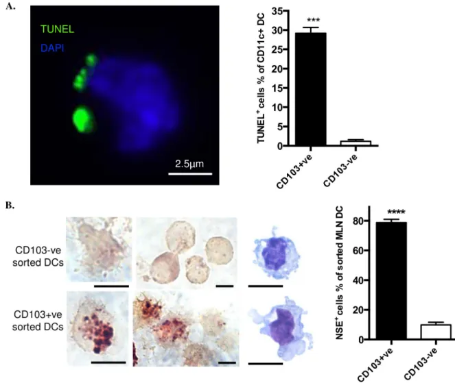
We prepared MLNs from healthy C57BL/6 mice housed in pathogen-limited conditions, selecting CD11c+ve DCs which were sorted for CD103 expression and stained by TUNEL for prior ingestion of cells containing apoptotic, cleaved double-stranded DNA. Inclusions of apo-ptotic material were exhibited 24.7-fold more frequently by CD103+ve DCs, when compared with CD103-ve DCs (Fig 1A). Because rapid degradation of DNA occurs in phagolysosomes,
we also stained for the more stable IEC marker NSE, observing that NSE-positive inclusions were 8-fold more frequent in CD103+ve DCs (Fig 1B). Thus, when compared with CD103-ve MLN DCs, there was evidence that a much greater proportion of CD103+ve MLN DCs had spontaneously ingested apoptotic cells, including IECs, prior to reaching the draining lymph node.
Phagocytosis of apoptotic cells
ex vivo by MLN DCs does not depend on
CD103 expression
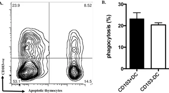
Preferential localisationin vivo of apoptotic cells to CD103+ve DCs isolated from MLNs (Fig 1) could reflect different capacity for phagocytosis of apoptotic cells by mature DC populations defined by CD103 expression. However, when we compared the capacity of isolated CD103 +ve and CD103-ve MLN DCs to ingest apoptotic thymocytesex vivo, no significant difference was observed (Fig 2A and 2B). This indicated that rather than greater phagocytosis of
Fig 1. CD103+ve DCs from MLN contain apoptotic cell remnants. A. FACS-sorted CD103+ve DCs and CD103-ve DCs were prepared
by affinity beads from pooled MLN from three mice and stained with TUNEL (green) and DAPI (blue). One typical image (left) shows TUNEL positive material associated with CD103+ve MLN DC. Chart (right) shows percentage of DCs bearing TUNEL positive material. B. Staining for the marker of intestinal endothelial cells, NSE and Diff-Quick staining (images at right side of left panel) were performed on CD11C+ve DCs isolated from MLNs. Scale bar, 5μm. All data were from four experiments with at least 200 cells counted for each experiment. Data show mean± SEM of four individual experiments.���p<0.001;����p<0.0001; unpaired t test with Welch’s correction.
apoptotic IECsin situ being a passive consequence of mature MLN DC phagocytic capacity co-incidentally associated with the CD103+ve phenotype, phagocytosis of apoptotic cells dur-ing maturation of DC precursors might preferentially drive CD103 expression.
Apoptotic cells drive CD103 expression in myeloid DC precursors
in vitro
To test possible effects of apoptotic cells on myeloid DC maturation we created a model sys-temin vitro, based on myeloid DC precursors isolated by a protocol closely similar to that described by del Rioet al and characterised as having the expected lineage-ve CD11c-ve CD103-ve phenotype [17]. In keeping with monocytic precursors of macrophages [18], such myeloid DC precursors bound but did not ingest either syngeneic apoptotic thymo-cytes or human apoptotic neutrophils (Fig 3A); the latter represent a particularly pure apo-ptotic cell “meal” with low (<2%) levels of secondary necrosis [14,15]. When myeloid DC precursors were co-cultured with apoptotic human neutrophils at a 1:1 ratio, the relative proportion of DCs expressing CD103 at 7 days was increased by 53.2±9.04% (mean±SEM, n = 4) (Fig 3B). When co-cultured at varying DC:Ac (apoptotic cell) ratios, increasing num-bers of apoptotic cells did not recruit more DC precursors into the CD103+ve DC popula-tion at 7 days (Fig 3C) but did increase the intensity of CD103 staining (Fig 3D). When apoptotic cells were added at different times after myeloid DC precursor culture was estab-lished (Fig 3E), recruitment of DCs into the CD103+ve population at 7d required co-culture with apoptotic cells prior to day 3, with maximal effect being observed when apoptotic cells were included from day 0, indicating that DC precursors rather than more mature DCs exhibited upregulation of CD103 when exposed to apoptotic cells. We confirmed (Fig 3F) that apoptotic cells induced DC precursor CD103 expression by qRTPCR analysis of mRNA for CD103 and the associated immunoregulatory marker, theβ8 integrin chain, which is critical for myeloid DCs to activate TGF-β1 and elicit responses such as induction of Tregs [2,3,11,19].Fig 2. Phagocytosis of apoptotic cellsex vivo by MLN DCs does not depend on CD103 expression. A. MLN DCs
co-cultured with fluorescence-labelled apoptotic thymocytes for 2h were stained for CD103 and assessed by flow cytometry; a typical plot is shown; note the similar proportion of DCs ingesting apoptotic cells in both CD103+ve and CD103-ve populations. B. from three such experiments. Data show mean± SEM, no significant difference; unpaired t test with Welch’s correction.
DC precursors co-cultured with apoptotic cells support enhanced
induction of Tregs
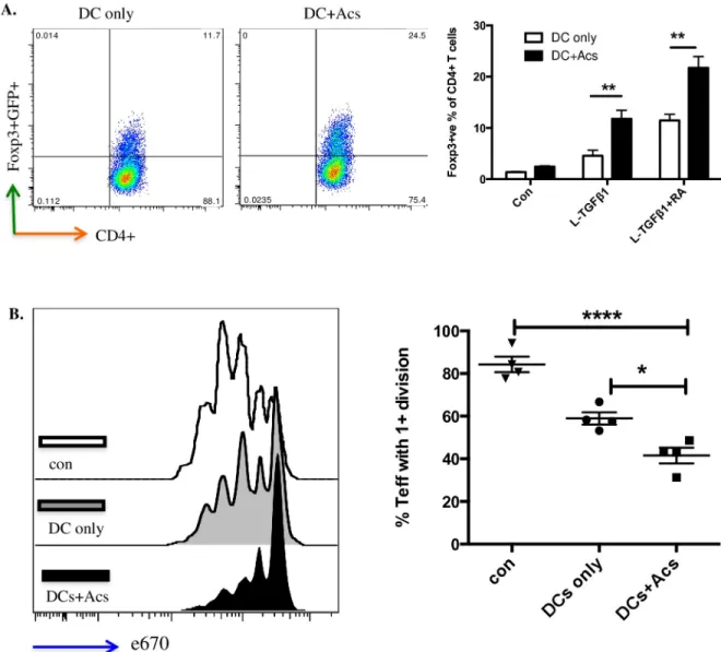
Next, we sought a functional effect of apoptotic cell-directed induction of CD103 expression in DC precursors maturingin vitro. DC precursors cultured for 7 days with (or without, as a con-trol) apoptotic cells were then co-cultured for a further 4 days with murine CD4+ve T cells bearing a FoxP3/GFP transgene marker as described [3,20]. When compared with DCs that had not been exposed to apoptotic cells, induction of FoxP3+ve T cells by either latent TGF-β1 at 2ng/ml or latent TGF-β1 at the same concentration plus retinoic acid at 100nm was signifi-cantly enhanced by DC precursor cultures that had been exposed to apoptotic cells from day 0 (Fig 4A). Furthermore in standard assays of antigen-driven T cell proliferation, suppression of T cell activation was greater with T cell populations induced by DCs exposed to apoptotic cells when compared with DCs that had not (Fig 4B). Thus, apoptotic cell direction of enhanced CD103 expression was indeed correlated with enhanced immunoregulatory function of DCs generatedin vitro.
DC precursor
α
vintegrin orchestrates binding of apoptotic cells with
activation of TGF-β1
Previously we have shown by bone marrow transplantation and other strategies that inαv-tie2
mice [9], in which a tie2-CRE construct deletes floxedαv, the effects on DC function are due
to deletion ofαvin the myeloid line. Having confirmed that myeloid DC precursors fromαv
-tie2 mice lackedαvexpression (Fig 5A), we then undertook an assay of the binding of
apopto-tic cells by myeloid DC precursors (3hrs), observing that this was reduced by about half (Fig 5B), in keeping with many studies of the complex multi-molecular mechanisms underlying myeloid phagocyte recognition of apoptotic cells [8,16,21–24], in which phagocytosis of apo-ptotic cells was reduced by about half where a putative phagocyte receptor for apoapo-ptotic cells, such asαvwas deleted [9]. This indicated that DC precursorαvintegrin is necessary for
maxi-mally efficient binding of apoptotic cells and that other mechanisms, not examined further in this study, contributed to binding. However,αvdeficiency completely abrogated the capacity
of apoptotic cells and/or latent TGF-β1 (administered at day 0) to increase myeloid DC pre-cursor CD103 expression at day 7 (Fig 5C). The dependence of such increased expression upon active TGF-β1 was confirmed by the capacity of blocking antibody to inhibit direction of increased DC precursor CD103 expression by apoptotic cells± latent TGF-β1 irrespective of whetherαvwas expressed (Fig 5C). These observations led us to seek direct evidence thatαv
integrins and latent TGF-β1 might be brought together at points where DC precursors contact apoptotic cells.
We employed three colour fluorescence microscopy to examine non-permeabilised DC precursors at 4˚C after co-culture with apoptotic thymocytes at 37˚C for 3 hours, consistently
Fig 3. Coculture with apoptotic cells promotes CD103 expression by DC precursors. A. Freshly isolated DC precursors stained with CM-orange
were co-cultured for 2hrs with apoptotic mouse thymocytes or apoptotic human neutrophils, both labelled with Cell Tracker Green. DC precursors binding apoptotic cells was measured by flow cytometry. Plots (left) show the gated CM-orange DC precursors with green apoptotic cells. Images (right) show DC precursors binding apoptotic thymocytes; note merged, yellow staining at the binding points. B. Selected bone marrow DC precursors cultured with apoptotic human neutrophils at the ratio of 1:1 (Ac:DC) at day0 in the presence of GM-CSF. Cells were stained with CD11c-APC and CD103-PE after 7days. Flow Cytometry was performed and one plot representative of four is shown (left). The chart (right) shows relative CD103+ve DC percentage from four different experiments. C, D. DC precursors co-cultured with apoptotic neutrophils in different ratios as indicated (Ac:DC) at day0. Relative CD103+ve DC percentage and geometric mean intensity were measured by flow cytometry after 7days. E. DC precursors cultured with apoptotic neutrophils at the ratio of 1:1 (Ac:DC) and apoptotic cells added at days 0, 1, 2 and 3. Relative CD103+ve DC (%) were analysed at day7. F. DC precursors cultured with apoptotic neutrophils at the ratio of 1:1 (Ac:DC) at day0 and relative gene expression was measured by qPCR after 7 days. Data were from three different experiments. All data show mean± SEM.�p<0.05;��p<0.01;���p<0.001;����p<0.0001; unpaired t test with Welch’s
correction (B); one-way ANOVA with Dunnett’s post-hoc test (C, D, E); two-way ANOVA with Tukey’s post-hoc test (F).
finding that 31.23± 3.706 (%) (mean ± SEM, n = 3) precursors bound apoptotic cells. Staining forαvintegrin was often intense at points of contact, withαv-positive DC processes partially
surrounding bound apoptotic cells in cup-like structures (Fig 5D);αvstaining was present at
91.8± 5.7 (%) (mean ± SEM, n = 3) of points of contact. Low intensity staining for latent TGF-β1 was associated with the surface of both DC precursors and apoptotic thymocytes, but high intensity staining co-localised withαv−positive points of contact in 72.2 ± 1.5 (%)
(mean± SEM, n = 3). For obvious reasons we could not seek co-localisation of αvstaining
with latent TGF-β1 staining with DC precursors from αv–tie2 mice, but in the small
Fig 4. CD103+ve DCs induced by apoptotic cells enhance functional Treg development. A. DCs differentiated from bone marrow DC
precursors co-cultured with naïve CD4+ve GFP-ve FoxP3-ve T cells in the presence of 2ng/ml latent TGF- β1 ± 100nm retinoic acid (RA). The percentage of FoxP3+ve T cells was determined at day 5 by flow cytometry (left); chart (right) collates data from three independent experiments. Data show mean± SEM. B. Naïve CD4+ve CD25-ve effector T cells from OTII mouse spleen were labelled with eFluor1 670 (e670) and co-cultured at 1:1 ratio with CD4+ T cells only (con); CD4+ T cells co-cultured with DCs in the presence of 2ng/ml latent TGF-β1 + 100nm RA for 5 days (DCs only); CD4+ T cells co-cultured with DCs exposed to apoptotic neutrophils in the presence of 2ng/ml latent TGF-β1 + 100nm RA (DC+Acs) for 5 days. pOVA pulsed DCs were added in all wells to stimulate effector T cells. Effector T cell proliferation was determined after 72hrs by flow cytometry; plots (left) and chart (right) show one experiment representative of three. Data show mean± SEM of four replicate wells.��p<0.01;���p<0.001; two-way ANOVA with Tukey’s post-hoc test (A); one-way ANOVA with
Bonferroni’s post-hoc test (B).
proportion ofαv-negative points of contact between wild type DC precursors and apoptotic
cells we never observed staining for latent TGF-β1.
Discussion
Myeloid phagocyte populations present in the healthy mammalian gastrointestinal tract are complex and the subject of ongoing investigation in many laboratories. Nevertheless, CD103 +ve myeloid DCs have been characterised in detail in the mouse, with compelling evidence for key roles in keeping the healthy gut free of inflammation, by induction of Tregs and other mechanisms dependent on TGF-β1 activation [2,3,5,19,25]. In this report, we demonstrate that CD103+ve DCs isolated from the MLNs of healthy mice exhibited extensive evidence of prior ingestion of IECs undergoing spontaneous apoptosis whereas, depending on the strain-ing technique used to identify previously strain-ingested apoptotic IECs, CD103-ve MLN DCs were 8–24 fold less likely to do so. Because theex vivo capacity to ingest apoptotic cells was similar for both CD103+ve and CD103-ve MLN DCs, we sought evidence that phagocytosis of apo-ptotic cells might drive CD103-ve DC precursors towards the immunoregulatory phenotype of mature CD103+ve DCs. In anin vitro model system apoptotic cells did indeed direct enhanced expression by co-cultured lineage-ve CD103-ve myeloid DC precursors of the immunoregula-tory CD103+veβ8+ve DC phenotype. These immunoregulatory CD103+ve DC also enhanced induction of functional Tregsin vitro. However, when myeloid DC precursors were isolated from mice deleted forαvintegrin in the myeloid line, DC precursors exhibited reduced
bind-ing of apoptotic cells and complete loss of enhancement of CD103 expression by apoptotic cells and/or latent TGF-β1; a role for αvintegrin-mediated activation of latent TGF-β1 was
confirmed by the capacity of active TGF-β1 to increase CD103 expression in DC precursors fromαv-tie2 mice and wild type mice. Because we observed co-localisation of DC precursorαv
integrin and latent TGF-β1 at points of contact with bound apoptotic cells, we conclude that immature CD103-ve DC precursors can deployαvintegrins to capture apoptotic cells and
co-ordinately activate latent TGF-β1. This results in maturation of CD103-ve DC precursors towards a mature, immunoregulatory CD103+ve DC phenotype. Thus, a hitherto unrecog-nised consequence of myeloid phagocyte binding and uptake of apoptotic cells may be to pro-gramme DCs to promote the tonic suppression of inflammation in the healthy gut.
Evidence of prior ingestion of apoptotic cells by dendritic cells coursing from the gut to mesenteric lymph nodes was first described in the rat by MacPherson’s group [7]. The OX41-ve DC subset in MLNs was found to have preferentially ingested apoptotic IECs and was speculated to have a role in inducing and maintaining self-tolerance [7]. However, despite functional similarities between OX41-ve MLN DCs in rats and CD103+ve MLN DCs in mice, the current study is the first to confirm in healthy unmanipulated mice that CD103+ve MLN
Fig 5. DC precursorαvintegrin orchestrates binding of apoptotic cells, activation of TGF-β1 and enhanced CD103 expression. A. DC precursors from wt andαv-tie2 mice were stained withαv-integrin-PE antibody and flow cytometry performed; plot representative of three
experiments. B. DC precursors (stained with CM-orange) from wild type orαv-Tie2 mice cultured with mouse apoptotic thymocytes (stained
with Cell Tracker Green) at a ratio of 5:1 (Ac:DC). Binding was measured by flow cytometry after three hours. Data were from four mice in two indenpendent experiments. C. CD103 expression by DCs derived from DC precursors cultured in serum free X-vivo medium in presence of GM-CSF. Conditions were DC precursor only (Con); DC precursor cultured with apoptotic cells at 1:2 ratio (Acs); DC precursor cultured with 2ng/ml latent TGF-β1 (L-TGFβ1); DC precursor cultured with apoptotic cells and 2ng/ml latent TGF-β1 (Acs+L-TGFβ1); DC precursor cultured with apoptotic cells and 2ng/ml latent TGF-β1 and 2μg/ml anti-TGF-β (Acs+L-TGFβ1+anti-TGFβ); DC precursor cultured with apoptotic cells and 2μg/ml anti-TGF-β (Acs+anti-TGFβ); DC precursor cultured with 0.2ng/ml active-TGF-β1 (a-TGFβ1). Relative percentage of CD103+ve DCs were measured by flow cytometry after 7days. Data were from three different experiments with three mice per group. D. DC precursors from wild type mice cultured with apoptotic thymocytes at a ratio of 1:5 for three hours. Cells were stained with integrinαv(green),
LAP (latent TGF-β1; red) and DAPI. Images are typical of 3 independent experiments. White arrow points an apoptotic cell binding to a DC precursor. All data show mean± SEM.�p<0.05;��p<0.01;���p<0.001;����p<0.0001; unpaired t test with Welch’s correction (B); two-way
ANOVA with Tukey’s post-hoc test (C).
DCs do indeed exhibit extensive evidence of prior ingestion of apoptotic cells, particularly IECs. Our data confirm the physiological relevance of elegant transgenic studies in which IECs induced to die by apoptosis were tracked as being ingested by CD103+ve DCs exhibiting anti-inflammatory and immunosuppressive gene expression signatures [1].
A key observation in the current studies of MLN DCs was that similar capacity to ingest apoptotic cellsex vivo was observed for CD103-ve and CD103+ve CD11c+ve DCs isolated from MLNs, whereas only the latter exhibited extensive evidence of prior ingestion of apopto-tic cellsin vivo. This raised the possibility that maturation of immature myeloid DCs towards a CD103+veβ8+ve immunoregulatory phenotype might be directed by the effects of contact with apoptotic cells upon immature CD103-ve myeloid DCs. To test this hypothesis directly, it was not necessary to characterise the isolated CD11c+ve MLN DCs further, although in the future it would be intriguing to determine whether apoptotic cell ingestion is preferentially exhibited by migratory (eg integrinβ8+ve [3]) rather than blood-derived resident MLN DCs, or by CD11b+ve rather than CD11b-ve CD103+ve DCs. Furthermore, localisation of DCs bearing apoptotic cells within mesenteric lymph nodes will add information of interest. Never-theless, thein vitro data reported here strongly support the likelihood of contact with apoptotic cells being a major factor, along with TGF-β1 activation, in driving acquisition by immature CD103-ve DCs of the CD103+ve phenotype associated with the capacity to generate function-ally active Tregs. However, it should be noted that these data were obtained in a model system that may not be representative of changes occurring after immature DCs ingest apoptotic epi-thelial cells in the gut wallin vivo prior to migrating to draining mesenteric lymph nodes.
One way of testing the dependence of maturation of gastrointestinal tract myeloid DCs upon binding and ingestion of apoptotic cells would be to characterise mechanisms by which immature DCs interact with apoptotic cells and elaborate active TGF-β1. In the current study we demonstrate, using mice deficient forαvin the myeloid line, thatin vitro myeloid DC
pre-cursor binding of apoptotic cells is partially dependent upon DC prepre-cursorαvintegrin.
Fur-thermore, DCαvintegrin is essential for the capacity of apoptotic cells and/or latent TGF-β1
to enhance CD103 expression by DC precursor populations. Thesein vitro data would predict that mice deficient forαvin the myeloid line would exhibitin vivo (a) reduced capacity of
mye-loid phagocytes to bind and ingest apoptotic cells; (b) diminished numbers of CD103+ve DCs in MLNs; (c) reduced numbers of Tregs in MLNs; and (d) spontaneous inflammation in the gut. All of these characteristics were exhibited byαv-tie2 mice in which the deleterious effects
ofαvdeficiency were shown to be due to effects on myeloid phagocytes [9].
Therefore, we propose that in the gut a hitherto unrecognised consequence of the interac-tion between immature DCs and apoptotic cells arising spontaneously is the beneficial “pro-gramming” of maturing DCs towards the CD103+veβ8+ve phenotype that is known to keep gut inflammation in check by tonic suppressive mechanisms. The capacity of apoptotic cells to suppress inflammation and enhance active TGF-β1 elaboration was initially studied in models of resolving acute inflammation [16,26–28] but the current study emphasises that apoptotic cells may pro-actively prevent inflammation in addition to suppressing active inflammation, in both cases by altering the “programming” of myeloid phagocytes.
Further work is obviously needed to dissect in detail the mechanisms by which binding of apoptotic cells by DC precursors directs subsequent DC maturation. Nevertheless, the current study supports the hypothesis that contact with apoptotic cells allows clustered DC surfaceαv
integrin to orchestrate the formation of multimolecular complexes that can capture and acti-vate latent TGF-β1. Such future work should address which of a number of potential “bridg-ing” molecules allow DC surfaceαvintegrin to bind apoptotic cells. Of those previously
implicated [16,22,23,29], it is intriguing that molecules such as thrombospondin are capable of activating latent TGF-β1 [30]. Furthermore, although enhancement of DC precursor
CD103 expression by apoptotic cells and/or latent TGF-β1 was completely dependent upon DC expression ofαv, the data suggest that other DC surface receptors are probably involved in
apoptotic cell capture, becauseαvdeficiency did not completely eliminate the capacity of DC
precursors to bind apoptotic cells; for example, candidate receptors such asMer are known to co-operate withαv[31]. Indeed, the signalling mechanisms elicited within DC precursors that
deployαvintegrin to bind apoptotic cells await characterisation. Lastly, because careful
microscopy by Blander’s group showed that DCs already expressing CD103 can, in a gut stressed by induced large-scale apoptosis, select and bind apoptotic cells, future work should also seek to demonstrate interactionin vivo between DC precursors and apoptotic cells. For example, means should be sought to determine whether lineage-ve CD11c-ve CD103-ve DC precursors do closely appose to IECs dying by apoptosis in the gut wall, but such studies are beyond the scope of the current work.
To conclude, we report that murine CD103+ve MLN DCs, known to be important in con-stitutive suppression of gut inflammation, do indeed exhibit extensive evidence of prior inges-tion of IECs dying spontaneously by apoptosis. Furthermore, we demonstratein vitro that exposure to apoptotic cells directs the maturation of CD103-ve myeloid DC precursors towards the mature CD103+veβ8+ve myeloid DC phenotype capable of inducing Tregs. The mechanisms responsible involve DC precursorαvintegrin, which orchestrates the binding of
apoptotic cells with the activation of latent TGF-β1 and enhancement of the immunoregula-tory CD103+ve phenotype. These data, taken with earlier observations of spontaneous gut inflammation in animals lackingαvin the myeloid line, suggest that a newly identified
conse-quence for myeloid phagocyte interaction with apoptotic cells is programming of DCs to pre-vent inflammation threatened by micro-environmental stimuli.
Acknowledgments
We thank Stephen Anderton and Richard A. O’Connor for suppling FoxP3 reporter mice and CD45.1 OTII mice. We thank the QMRI Flow Cytometry and Cell Sorting Facility for their excellent technical assistance. Eleanor Bonikowski and Ruth MacInnes provided invaluable secretarial help with the manuscript. We also thank Professor Kev Dhaliwal and Professor Neil Henderson for supporting animal experiments.
Author Contributions
Conceptualization: Ailiang Zhang, Adam Lacy-Hulbert. Data curation: Ailiang Zhang, Helena Paidassi.
Formal analysis: Ailiang Zhang. Funding acquisition: John Savill. Investigation: Ailiang Zhang, John Savill. Resources: John Savill.
Supervision: John Savill.
Writing – original draft: John Savill.
References
1. Cummings RJ, Barbet G, Bongers G, Hartmann BM, Gettler K, Muniz L, et al. Different tissue phago-cytes sample apoptotic cells to direct distinct homeostasis programs. Nature. 2016; 539(7630):565–9. https://doi.org/10.1038/nature20138PMID:27828940
2. Coombes JL, Siddiqui KR, Arancibia-Carcamo CV, Hall J, Sun CM, Belkaid Y, et al. A functionally spe-cialized population of mucosal CD103+ DCs induces Foxp3+ regulatory T cells via a TGF-beta and reti-noic acid-dependent mechanism. J Exp Med. 2007; 204(8):1757–64.https://doi.org/10.1084/jem. 20070590PMID:17620361
3. Paidassi H, Acharya M, Zhang A, Mukhopadhyay S, Kwon M, Chow C, et al. Preferential expression of integrin alphavbeta8 promotes generation of regulatory T cells by mouse CD103+ dendritic cells. Gastroenterology. 2011; 141(5):1813–20.https://doi.org/10.1053/j.gastro.2011.06.076PMID: 21745448
4. Paidassi H, Acharya M, Lacy-Hulbert A. Alpha (v) integrins license regulatory T cells to apoptotic cells and self-associated antigens. Ann N Y Acad Sci. 2010; 1209:68–76. https://doi.org/10.1111/j.1749-6632.2010.05783.xPMID:20958318
5. Coombes JL, Maloy KJ. Control of intestinal homeostasis by regulatory T cells and dendritic cells. Semin Immunol. 2007; 19(2):116–26.https://doi.org/10.1016/j.smim.2007.01.001PMID:17320411
6. Annacker O, Coombes JL, Malmstrom V, Uhlig HH, Bourne T, Johansson-Lindbom B, et al. Essential role for CD103 in the T cell-mediated regulation of experimental colitis. J Exp Med. 2005; 202(8):1051– 61.https://doi.org/10.1084/jem.20040662PMID:16216886
7. Huang FP, Platt N, Wykes M, Major JR, Powell TJ, Jenkins CD, et al. A discrete subpopulation of den-dritic cells transports apoptotic intestinal epithelial cells to T cell areas of mesenteric lymph nodes. J Exp Med. 2000; 191(3):435–44.https://doi.org/10.1084/jem.191.3.435PMID:10662789
8. Rubartelli A, Poggi A, Zocchi MR. The selective engulfment of apoptotic bodies by dendritic cells is mediated by the alpha(v)beta3 integrin and requires intracellular and extracellular calcium. Eur J Immu-nol. 1997; 27(8):1893–900.https://doi.org/10.1002/eji.1830270812PMID:9295024
9. Lacy-Hulbert A, Smith AM, Tissire H, Barry M, Crowley D, Bronson RT, et al. Ulcerative colitis and auto-immunity induced by loss of myeloid alphav integrins. Proc Natl Acad Sci U S A.2007; 104(40):15823– 8.https://doi.org/10.1073/pnas.0707421104PMID:17895374
10. Albert ML, Pearce SF, Francisco LM, Sauter B, Roy P, Silverstein RL, et al. Immature dendritic cells phagocytose apoptotic cells via alphavbeta5 and CD36, and cross-present antigens to cytotoxic T lym-phocytes. J Exp Med. 1998; 188(7):1359–68.https://doi.org/10.1084/jem.188.7.1359PMID:9763615
11. Travis MA, Reizis B, Melton AC, Masteller E, Tang Q, Proctor JM, et al. Loss of integrin alpha(v)beta8 on dendritic cells causes autoimmunity and colitis in mice. Nature. 2007; 449(7160):361–5.https://doi. org/10.1038/nature06110PMID:17694047
12. Lucas M, Stuart LM, Zhang A, Hodivala-Dilke K, Febbraio M, Silverstein R, et al. Requirements for apo-ptotic cell contact in regulation of macrophage responses. J Immunol. 2006; 177(6):4047–54.https:// doi.org/10.4049/jimmunol.177.6.4047PMID:16951368
13. Worthington JJ, Fenton TM, Czajkowska BI, Klementowicz JE, Travis MA. Regulation of TGFbeta in the immune system: an emerging role for integrins and dendritic cells. Immunobiology. 2012; 217 (12):1259–65.https://doi.org/10.1016/j.imbio.2012.06.009PMID:22902140
14. Savill JS, Wyllie AH, Henson JE, Walport MJ, Henson PM, Haslett C. Macrophage phagocytosis of aging neutrophils in inflammation. Programmed cell death in the neutrophil leads to its recognition by macrophages. J Clin Invest. 1989; 83(3):865–75.https://doi.org/10.1172/JCI113970PMID:2921324
15. Stuart LM, Lucas M, Simpson C, Lamb J, Savill J, Lacy-Hulbert A. Inhibitory effects of apoptotic cell ingestion upon endotoxin-driven myeloid dendritic cell maturation. J Immunol. 2002; 168(4):1627–35. https://doi.org/10.4049/jimmunol.168.4.1627PMID:11823490
16. Savill J, Dransfield I, Gregory C, Haslett C. A blast from the past: clearance of apoptotic cells regulates immune responses. Nat Rev Immunol. 2002; 2(12):965–75.https://doi.org/10.1038/nri957PMID: 12461569
17. del Rio ML, Rodriguez-Barbosa JI, Bolter J, Ballmaier M, Dittrich-Breiholz O, Kracht M, et al. CX3CR1+ c-kit+ bone marrow cells give rise to CD103+ and CD103- dendritic cells with distinct functional proper-ties. J Immunol. 2008; 181(9):6178–88.https://doi.org/10.4049/jimmunol.181.9.6178PMID:18941208
18. Mikolajczyk TP, Skrzeczynska-Moncznik JE, Zarebski MA, Marewicz EA, Wisniewska AM, Dzieba M, et al. Interaction of human peripheral blood monocytes with apoptotic polymorphonuclear cells. Immu-nology. 2009; 128(1):103–13.https://doi.org/10.1111/j.1365-2567.2009.03087.xPMID:19689740
19. Johansson-Lindbom B, Svensson M, Pabst O, Palmqvist C, Marquez G, Forster R, et al. Functional specialization of gut CD103+ dendritic cells in the regulation of tissue-selective T cell homing. J Exp Med. 2005; 202(8):1063–73.https://doi.org/10.1084/jem.20051100PMID:16216890
20. Suffner J, Hochweller K, Kuhnle MC, Li X, Kroczek RA, Garbi N, et al. Dendritic cells support homeo-static expansion of Foxp3+ regulatory T cells in Foxp3.LuciDTR mice. J Immunol. 2010; 184(4):1810– 20.https://doi.org/10.4049/jimmunol.0902420PMID:20083650
21. Savill J, Dransfield I, Hogg N, Haslett C. Vitronectin receptor-mediated phagocytosis of cells undergoing apoptosis. Nature. 1990; 343(6254):170–3.https://doi.org/10.1038/343170a0PMID:1688647
22. Savill J, Gregory C, Haslett C. Cell biology. Eat me or die. Science (New York, NY). 2003; 302 (5650):1516–7.
23. Savill J, Hogg N, Ren Y, Haslett C. Thrombospondin cooperates with CD36 and the vitronectin receptor in macrophage recognition of neutrophils undergoing apoptosis. J Clin Invest. 1992; 90(4):1513–22. https://doi.org/10.1172/JCI116019PMID:1383273
24. Park SY, Kim IS. Engulfment signals and the phagocytic machinery for apoptotic cell clearance. Exp Mol Med. 2017; 49(5):e331.https://doi.org/10.1038/emm.2017.52PMID:28496201
25. Jaensson E, Uronen-Hansson H, Pabst O, Eksteen B, Tian J, Coombes JL, et al. Small intestinal CD103+ dendritic cells display unique functional properties that are conserved between mice and humans. J Exp Med. 2008; 205(9):2139–49.https://doi.org/10.1084/jem.20080414PMID:18710932
26. Fadok VA, Bratton DL, Konowal A, Freed PW, Westcott JY, Henson PM. Macrophages that have ingested apoptotic cells in vitro inhibit proinflammatory cytokine production through autocrine/paracrine mechanisms involving TGF-beta, PGE2, and PAF. J Clin Invest. 1998; 101(4):890–8.https://doi.org/10. 1172/JCI1112PMID:9466984
27. Huynh ML, Fadok VA, Henson PM. Phosphatidylserine-dependent ingestion of apoptotic cells pro-motes TGF-beta1 secretion and the resolution of inflammation. J Clin Invest. 2002; 109(1):41–50. https://doi.org/10.1172/JCI11638PMID:11781349
28. Freire-de-Lima CG, Xiao YQ, Gardai SJ, Bratton DL, Schiemann WP, Henson PM. Apoptotic cells, through transforming growth factor-beta, coordinately induce anti-inflammatory and suppress pro-inflammatory eicosanoid and NO synthesis in murine macrophages. The Journal of biological chemis-try. 2006; 281(50):38376–84.https://doi.org/10.1074/jbc.M605146200PMID:17056601
29. Hanayama R, Tanaka M, Miwa K, Shinohara A, Iwamatsu A, Nagata S. Identification of a factor that links apoptotic cells to phagocytes. Nature. 2002; 417(6885):182–7.https://doi.org/10.1038/417182a PMID:12000961
30. Schultz-Cherry S, Murphy-Ullrich JE. Thrombospondin causes activation of latent transforming growth factor-beta secreted by endothelial cells by a novel mechanism. J Cell Biol. 1993; 122(4):923–32. Epub 1993/08/01.https://doi.org/10.1083/jcb.122.4.923PMID:8349738
31. Wu Y, Singh S, Georgescu MM, Birge RB. A role for Mer tyrosine kinase in alphavbeta5 integrin-medi-ated phagocytosis of apoptotic cells. J Cell Sci. 2005; 118(Pt 3):539–53.https://doi.org/10.1242/jcs. 01632PMID:15673687