CONTROL OF CELL BEHAVIOR BY ENGINEERING SUBSTRATA WITH DEFINED SURFACE TOPOGRAPHY AND CHEMISTRY
by
RAHUL SINGHVI
B.Tech. Chemical Engineering
Indian Institute of Technology, Kanpur, India (1987)
M.S. Chemical Engineering Practice Massachusetts Institute of Technology
(1989)
Submitted to the Department of Chemical Engineering in Fulfillment of the Requirement for the Degree of
DOCTOR OF SCIENCE in
CHEMICAL ENGINEERING at the
MASSACHUSETTS INSTITUTE OF TECHNOLOGY February, 1994
© 1994 Massachusetts Institute of Technology . _ All Rights Reserved
Signature of Author
Signature ofAuho - Department of Chemical Engineering January 12, 1994
Certified by _
Professor Daniel I. C. Wang _TlJess Supervisef, DePartment of Chemical Engineering Certified by
by
Certified
?-.--j/ - 'rProfessor - '"- Gregory N. Stephanopoulos· / 9 Thesis Supervisor, Department of Chemical Engineering
Accepted by
Professor Robert Cohen MAssAt r Swwc Chairman, Committee on Graduate Students
Control of Cell Behavior by Engineering Substrata with Defined Surface Topography and Chemistry
BY
RAHUL SINGHVI
Submitted to the Department of Chemical Engineering Massachusetts Institute of Technology
on February 12, 1994
in partial fulfillment for the Degree of Doctor of Science in Chemical Engineering
The role of substratum topography on cell behavior was investigated by preparing glass substrata with defined surface morphologies using photolithography and plasma etching. A parallel grooved surface morphology was chosen as the initial surface texture with groove dimensions in the same order of magnitude as the size of a typical animal cell.
A highly shear sensitive cell line, AtT-20, was used to study the role of substratum morphology on cell adhesion. Using a cell adhesion assay based on centrifugal force, it was shown that confluent cell layers of AtT-20 cells on grooved substrata was more stable than on the smooth controls. The degree of enhancement of cell-substratum adhesion was dependent on dimensions of the groove width (w) and groove depth (d).
Improved cell stability on grooved substrata was found to correlate with the efficiency with which the substratum prevented the formation of an extensive cell-cell network. The ability of a grooved substratum to prevent cell-cell interactions was a function of its capability to confine cells in grooves, or its confinement factor. The confinement factor is a new concept developed in this thesis. Confinement factor was a function of groove dimensions and could be expressed in terms of 'w' and 'd'. Adhesion performance correlated directly with confinement factor of the substratum.
Primary rat hepatocytes were used to study the role of substratum morphology on cell growth and function. Levels of DNA synthesis in hepatocytes cultured on grooved substrata were, in general, lower than that on the smooth controls. DNA synthesis was inversely found to be inversely proportional to the confinement factor of the substratum. In contrast, rate of albumin secretion, a measure of hepatocyte differentiated function, increased with increased confinement factor. These results indicated that the effects of substratum morphology on cell physiology was mediated by the ability of the substratum to confine cells and thereby affect the extent of cell spreading. This is consistent with data from many independent studies that show that the extent of cell spreading modulates cell growth and function.
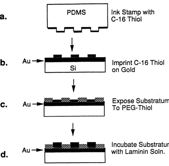
Based on these results, a strategy was devised to better control cell confinement by manipulating surface chemistry of the substratum. Substrata with cell-adhesive islands of precise dimensions were fabricated using self-assembled monolayers of alkanethiols on gold surfaces. These surfaces allowed control over cell confinement in two dimensions as compared to one in grooved substrata. Studies with these
substrata showed that DNA synthesis in hepatocytes could be manipulated simply by changing the size of the underlying adhesive island. Furthermore, substrata could be patterned such that hepatocytes cultured on them exhibited low DNA synthesis and improved differentiated function for extended periods of time. These results show that cell behavior can be controlled by manipulating simple physio-chemical properties of the substratum, even in the presence of saturating amounts of soluble mitogens and extracellular matrix.
Table of Contents
Title Page ...Abstract
...
Table of Contents ...Acknowledgements ....
List of Figures ... List of Tables ... Chapter 1. Introduction ...1.1 Motivation and Problem Statement ... 1.2. Thesis Objectives ... 1.3. Thesis Organization ... Chapter 2. 2.1. Literature Review ... Cell-Surface Interactions ...
2.2. Surface Morphology and Cell Interations ... 2.2.1. Substrata for In Vitro Studies: Early Studies ... 2.2.2. Towards a defined substratum ...
2.2.3. Cell Interaction Studies with Defined Substrata ... 2.2.3.1 Cell Spreading and Migration Studies ... 2.2.3.2 Cell Attachment and Adhesion Studies ... 2.2.3.3 Cell Growth and Function Studies ... 2.2.3.4. Cell Spreading and Cell Behavior ... 2.3. Summary and Potential ...
Chapter 3.
3.1
Materials and Methods ...
Preparation of Substrata with Defined Morphology ... 3.1.1 Mask Design and Preparation ...
3.1.2 Glass Substrata with Grooved Surface Morphology ... 3.1.2.1 Precleaning Wafers for processing ... 3.1.2.2 Aluminum evaporation ... 3.1.2.3 Photolithography ... 3.1.2.4 Plasma Etching ...
3.1.2.5 Cutting and Cleaning Grooved Glass Wafers. 3.1.3 Surface Characterization ...
3.1.3.1 Groove Dimensions ...
3.1.3.2 Surface Elemental Analysis ... 3.2 Cell Culture ... 3.2.1 AtT-20 ... 36 36 36 39 39 39 40 41 43 44 44 46 48 50 1 2 4 8 10 13 14 14 17 18 19 19 19 20 21 26 26 29 31 32 33
3.2.1.1 Cell Culture and Maintenance ... 3.2.1.2 Cell Enumeration ...
3.2.2 Primary Rat Hepatocytes ... 3.2.2.1 Cell Isolation ...
3.2.2.2 Serum-Free Medium and Supplements ... 3.2.2.3 Cell Enumeration ...
3.2.2.4. Fluorescent Cell Viability Assay ... 3.2.2.5 Coating Substrata with Laminin ... 3.2.3 Chinese Hamster Ovary (CHO) ...
3.2.3.1 Cell Culture and Maintenance ... 3.2.3.2 Cell Enumeration ... 3.3 Cell Adhesion Assay ...
3.3.1 Culture Wells for Adhesion Assay ... 3.3.2 Labelling Cells ...
3.3.3 Centrifugation of Attached Cells ... 3.3.4 Quantification of Cell Number ... 3.4 Cell patterning ...
3.4.1 Preparation of Master Patterns ...
3.4.2 Preparation of an Elastomeric Stamp ... 3.4.3 Patterning Gold Substrata ... 3.5 Image Analysis ... 50 51 51 51 51 52 52 54 55 55 56 56 57 57 60 60 62 62 65 67 71
3.6 Cell Culture on microcarriers ... 71
3.6.1 Microcarriers ... 71
3.6.2 Roughening and Characterization of Microcarriers ... 72
3.6.3 Spinner Flask Culture ... 72 3.7 DNA Synthesis Assay ...
3.7.1 3H-thymidine Uptake and Autoradiography ... 3.7.2 BrdU Uptake, Incorporation and Detection ... 3.8 Metabolite and Product Assays ...
3.8.1 Glucose and Lactate ...
3.8.2 Insulin...
3.8.3 Albumin ... 3.8.4 Interferon ...
3.9 Analysis of rhlFNy Heterogeneity ... 3.9.1 SDS-PAGE ...
3.9.2 Western Blotting ... 3.9.3 Densitometer Scan ...
Chapter 4. Surface Characterization of Grooved Substrata ...
73 73 74 75 75 76 78 81 84 84 84 87 88
...
...
...
...
...
Chapter 5. Effects of Substratum Topography on Cell Adhesion ... 5.1 Introduction. ...
5.2 Defined Substrata ... 5.3 Model Cell ... 5.4 Results and Discussion ....
5.4.1 Cell Attachment and Proliferation.
5.4.2 Cell-Substratum Adhesion Strength ... 5.4.2.1 Cell Adhesion Assay ... 5.4.2.2 Adhesion Curves ...
5.4.2.3 Confinement Factor (CF) of Grooved Substrata. 5.4.3 Cell Function ...
5.4.4 AtT-20 Cell Culture on Rough Microcarriers ... 5.5 Conclusions ...
Chapter 6. Effect of Substratum Topography on Cell Growth and Function ... 6.1 Introduction. ...
6.2 Model Cell ... 6.3 Results and Discussion ...
6.3.1 Preliminary DNA Synthesis Studies ... 6.3.2 Emergence of a Mechanism ...
6.3.3 DNA Synthesis Studies with Expanded Set of Substrata 6.3.4 Hepatocyte Function Studies ...
6.4 Conclusions ...
Chapter 7. Engineering Substrata for Controlling Cell Behavior ...
7.1 Introduction...
7.2 Strategies for Two Dimensional Control of Cell Spreading ... 7.2.1 Truncated parallel groove morphology ... 7.2.2 Basket-Weave Morphology ... 7.2.3 Pit Morphology ...
7.2.4 Cell-Adhesive Islands ...
7.3 Engineering Substrata with Cell-Adhesive Islands ... 7.3.1 Macroscopic Cell Patterning ...
7.3.2 Patterning Islands ... 7.3.3 Control of Cell Shape ...
7.3.4 Control of Cell Growth and Function ...
7.4 Conclusions . ...
Chapter 8.
8.1
8.2 8.3
Conclusions and Recommendations ...
Introduction
. ...
Major Accomplishments and Conclusions ... Recommendations for Future Research ...
8.3.1 More Functional assays to judge hepatocyte function ... 8.3.2 Decoupling the role of nuclear and cell spreading ... 8.3.3 Effect of Substratum Morphology on Product Quality ...
8.3.3.1 Glycosylation ... 8.3.3.2 Virus Production ... 94 94 95 95 97 97 97 97 98 102 110 114 117 121 121 123 124 124 125 132 135 138 140 140 140 140 142 142 142 143 143 147 147 150 154 156 156 156 160 160 160 161 161 162
-...
...
...
...
...
8.3.4 Effects on other cell types ... 162
8.3.5 Use and Applications of Patterned Substrata ... 163
8.3.5.1 Basic studies ... 163
8.2.5.2 Applications ... 163
Chapter 9. References ... ... 166
Appendix A. Effects of Substratum Morphology on Growth, and Productivity of rCHO cells. ... 173
Acknowledgements
I would first like to thank my mentor, Professor Daniel . C. Wang, who has had a profound effect over my professional development. Professor Wang has not only shown me how to identify and address the real problem in research, but, by his own example, has taught me what real discipline and hard work is. Professor Wang is a leader and he takes great interest in the development of his students. His consistent effort to help his students, both professionally and personally, is exemplary. He is critical at times and very generous at other occasions, but his motive is always to help the student. He respects his students and never hesitates in assigning responsible jobs to them. He has allowed a great deal of independence in my work. I have
learned a great deal from him and I am fortunate to have been associated with him during the last few years. In addition, it has been a joy to play tennis with him regularly
over the past few years. It has certainly improved my tennis game.
Professor Greg Stephanopoulos has been most supportive of this work. I believe that his early interest and vision in this research was critical in shaping this thesis. His input and brilliant insight into this work has been invaluable and I am indebted to him for this. My thesis committee has been instrumental in keeping me on the right track and making me do the right controls. I am thankful for the constructive criticisms of my thesis committee members, Professor Charles Cooney, Dr. Donald Ingber and Professor Bob Langer. I hope that all those suggestions have led to a much better piece of work. Dr. Ingber's own work has inspired a majority of this research and his continued interest and comments during the course of this work have been invaluable. Dr. Ingber is a devoted researcher and I am thankful to him for instilling in me the passion for science.
A large number of individuals have contributed to the successful completion of this very, interdisciplinary research. Brian Kell , Jeff Bigler and Thanassis Sambanis taught me cell culture. I learned a great deal form Dr. Seujeung Park about experimentation and creativity. Prof. Michael Cima introduced me to the field of photolithography and provided me with the first insights into this work. Octavio Hurtado, Paul Tierney, Rich Perrelli and Tim McClure helped me learn the techniques of photolithography and plasma etching. Vivek Mohindra played a critical role in this research by helping me plasma etch the glass wafers. Sriram helped with numerous material science related problems. Linda Cima and Donald Ingber allowed me to use their fluorescent microscopes. Primary rat hepatocytes were procured from Dr. J. Vacanti's lab at the Children's hospital. Especial thanks are due to Drs. Magali Fontaine, David Mooney and Linda Hansen for isolating the hepatocytes and for helping me with numerous protocols. The contributions from the Whitesides group at Harvard were critical in completing the work with patterned substrata. I would like to thank Professor George Whitesides, Drs Amit Kumar and Gabriel Lopez and Hans Biebuyck for a very productive and enjoyable collaboration. Gino Grampp, Brian Kelley and Craig Zupke were invaluable colleagues at BPEC in terms of discussing research and other miscellaneous issues. Thanks for being a sounding board at innumerable occasions and for being so generous in giving me your time. I enjoyed our times together and I learned a lot from you. Bob Kiss, Jeff Cleland and Per Lindell coached me at
numerous occasions to cope with the real world. Thanks for all your advice. Bill Mullett greatly enhanced my understanding of the biotech business. My UROPs and REU student worked very hard and I want to thank them for all their effort. Thanks to Cenk Sumen, Miguel Valle, Kayo Yamamoto, Cliff Urmaza, Carlonda Russell, Sherri Briesie, Con Esenther, Debbie Bressen and Allen Wong.
Thanks are due to fellow laboratory mates and research group members (both past and present): Jamie Piret, Mark Applegate, John Aunins, Wen Chiou, Gautam Nayar, Dave Stevenson, Eric Scharin, Kai-Chee Loh, Margaret Speed, Marc Shelikoff, Enno Adema, Chris Hwang, Karen Troxel, Jorg Neerman, Gregg Nyberg, Steve Meier, Beth Junker, Ed Osawa, Mike Thein, Brian Kell, Seujeung Park, Thanassis Sambanis, Jeff Bigler, Bruce Woodson, Nick Valkanas, Peter Frier, Sandeep Paliwal, Tim Nadler, Cheng Lee, Keqin Chen, Greg O'Conner, Christine Moore, Chris Dowd, Troy Simpson, Steve Lee, Anna Hagen, Dan lasko, David Chang, Brian Kelley, Craig Zupke, Gino Grampp, John Chung, Grace Colon, Roy Kamimura, J.-F. Hamel, Bob Murray, Catherine Shaw, Joe Vallino, Per Lindell and Liangzhi Xie Thanks for a very enjoyable experience. I apologize if I missed anyone.
Life at MIT was greatly enhanced by social interactions with Sue and Gino Grampp, Lisa and Jack Prior, Dawn Orton, Mark Applegate, Dave Stevenson, Steve Meier, G. K. and Lavanya Raju, Dana Johnson, Brian Laffey and with my first year office buddies Gil Huppert, Gavin Zau, Joy Mendoza and David -Yu-Hsiang (eggplant) Chang. Thanks for your continued friendship and good luck to all of you.
Finally, I would like to thank my dearest friend and lovely wife, Anushri, for her help and support during the past few years. You have been extremely patient during these very trying times. Without your continued love and support, completing this goal would have been a lot harder. I also want to thank my parents and sisters for their love and support during all my years in school. I hope that we get to spend some time together soon.
Financial support for this project was provided by a National Science Foundation Grant CDR-88-03014 to the Biotechnology Process Engineering Center. Additional support was provided by Merck & Co. via a fellowship to the author.
List of Figures
Figure Figure Figure Figure Figure Figure Figure Figure Figure Figure Figure Figure Figure Figure Figure Figure 1.1 1.2 3.1 3.2 3.3 3.4 3.5 3.6 3.7 3.8 3.9 3.10 3.11 3.12 3.13 4.1 Figure 4.2 Figure 5.1 Figure 5.2 Figure 5.3Scanning electron micrograph of a MgO. A1203 ceramic bead ... 15
AtT-20 cells attached on the ceramic bead ... 16
Process for preparing grooved glass substrata ... 37
Choice of surface morphology ... 38
Preparation of grooved glass substrata for cell culture ... 45
Characterization of groove dimensions by SEM and profilometery ...47
Typical scan from Auger spectroscopy ... 49
Cell culture dishes for cell adhesion experiments ... 58
Schematic of a cell adhesion assay using a centrifugal field ... 61
Device for placing lexan wells directly into gamma counter ... 63
Protocol for quantifying cell substratum adhesion strength ... 64
Process for preparing template for the elastomeric stamp ... 66
Preparation of the elastomeric stamp ... 68
Protocol for stamping gold substrata to create cell adhesive islands ...69
Schematic of steps in detecting and quantifying IFNy heterogeneity ...85
Cleaning of plasma etched glass substratum to remove damaged
laye
...
r
90
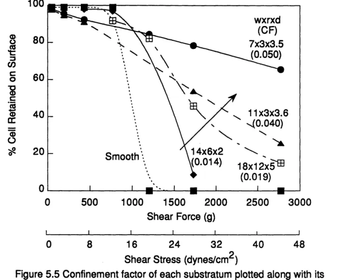
Auger spectroscopy on groove and ridge areas of a grooved substratum ... 92Cell removal from different substrata with increased shear force ... 99
Proposed mechanism for improved stability of AtT-20 cells on parallel grooved substrata ... 01
Figure 5.4
Figure 5.5
Figure 5.6
substrata .103... 103 Schematic representation of cell spreading on grooved substrata with
varying groove dimensions ... 105
Confinement factor of each substratum plotted along with its
corresponding confinement factor ... 108
Measurement of the average slope of each adhesion curve for shear forces greater than 750g ... 109
Figure 5.7 Figure Figure Figure Figure Figure 5.8 5.9 5.10 5.11 5.12 Figure 6.1 Figure 6.2
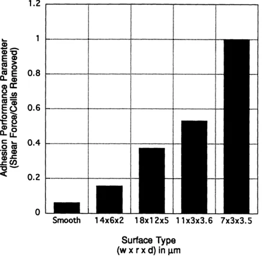
Adhesion performance parameter plotted as a function of surface
type
...
111
Adhesion performance parameter as a function of confinement factor. 112 Function of AtT-20 cells on the various substrata ... 13 SEM of Plastispex microcarriers before and after roughening ... 116 AtT-20 cells cultured on smooth and rough microcarriers ... 118 Lactate accumulation in microcarrier cultures of AtT-20 cells.Comparison between rough and smooth microcarrier cultures ... 1 19 Nuclear labelling index as a function of substratum type (preliminary experim ents) ... 126 Visualization of hepatocyte nuclei on smooth and grooved substrata..127
DNA synthesis correlates with cell and nuclear area ... 129 Nuclear labelling index correlates with nuclear area ... 130 Decline in nuclear labelling index correlates with confinement factor..131 Nuclear labelling index as a function of substratum type for the expanded set of grooved substrata ... 133 Figure Figure Figure Figure 6.3 6.4 6.5 6.6
Nuclear labelling index correlates inversely with confinement factor....134 Figure 6.8 Figure 6.9 Figure 6.10 Figure 7.1 Figure 7.2 Figure 7.3 Figure 7.4 Figure 7.5 Figure 7.6 Figure 7.7
Relative rate of albumin secretion by hepatocytes cultured on various substrata . ...136 Albumin secretion rate by hepatocytes on various grooved substrata is correlated to the substratum's confinement factor ... 137 Opposite effects on hepatocyte nuclear labelling index and albumin
secretion rate due to confinement ... 139
Strategies for greater control over cell confinement by modulating surface morphology and surface chemistry of the substratum ... 141
Alkanethiols chemisorb on gold surfaces to form self-assembled
m onolayers ... 145
Photographs of macroscopically patterned gold substrata with
alkanethiols ... 146
Diagrammatic representation of the method for fabricating the rubber tamp and creating patterned substrata ... 148
High and low magnification views of SEMs showing a patterned region which contains cells adherent to adhesive islands of varying size ... 149
Control of cell shape and size on patterned substrata ... 151 Relationship between adhesive island area, DNA synthesis, and albumin secretion . ... 153
Figure Al Rate of lactate production by CHO cells on different substrata ... 175 Comparison of IFNy production by rCHO cells on different substrata... 176 Macroheterogeneity in IFNy secreted by rCHO cells cultured on different
substrata ... 178
Figure A2 Figure A3 Figure 6.7
List of Tables
Table 2.1 Summary of studies conducted to elucidate the role of surface
morphology on cell physiology ... 24
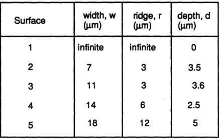
Table 5.1
Table 6.1
Characteristic dimensions of grooved substrata used in adhesion
experim ents ... 96
Characteristic dimensions of grooved substrata used in preliminary and expanded experiments for DNA synthesis ... 122
Table Al Characteristic dimensions of substrata used in CHO cell
Chapter 1. Introduction
1.1 Motivation and Problem Statement
Large-scale cultivation of mammalian cells is extremly important for the production of vaccines and complex recombinant proteins such as erythropoietin, tissue plasminogen activator (tPA) and monoclonal antibodies. As manufacturing of these products becomes increasingly competitive, highly sophisticated and efficient cell cultivation systems need to be developed. A thorough characterization of the cell microenvironment and its effects on cellular physiology is imperative for the successful
implementation of these culture systems because understanding of the
microenvironment leads to better control over cell growth, productivity and quality of the secreted product.
During the development of an animal cell cultivation system that utilized porous
ceramic beads, we were faced with the challenge of characterizing cell
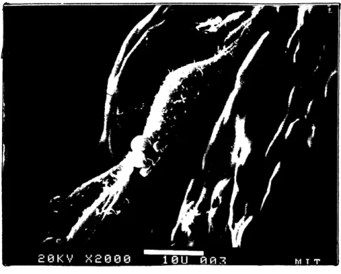
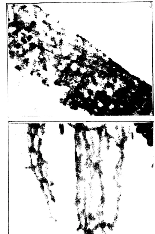
microenvironment in the beads. The beads were made of a magnesium aluminate (MgO. A1203) ceramic and were approximately 500 glm in diameter and 80% porous. The pores ranged between 1-100 gm in diameter; large enough to host animal cells. In this system, the cell microenvironment at any site of the bead consisted not only of the concentration of various nutrients but also the nature of the substratum. Scanning electron micrographs of the beads showed that the ceramic surface available for cell attachment was highly rough and jagged (Figure 1.1). Cells readily attached on the ceramic surface and the material was found to be biocompatible.
During the course of this study, a curious observation was made. Cells at various locations of the same bead spread differently (Figure 1.2) and, more
Figure 1.1. Scanning electron micrograph of a MgO.AI203ceramic bead. The beads were prepared using the citric method and were approximately 5001~m in diameter and 80% porous.
A
B
Figure 1.2. A.) AtT-20 cells attached on a relatively smooth area of a MgO.AI203 ceramic bead. The cells appear to spread extensively. B.) AtT-20 cells on a different site of the same ceramic bead as in A. Cells are not spread and appear highly rounded. Note that the underlying substratum topography is a lot more rough than in A.
importantly, the extent of cell spreading appeared to be modulated by the topography of the underlying substratum. A literature search was conducted to determine if this phenomenon could result in altered cell behavior. The search revealed that an impressive body of literature has emerged in the past 15 years which demonstrates a correlation between the extent of cell spreading (at least in normal cells), and cell growth and differentiated function (see Chapter 2 for review). This data, in conjunction with the hypothesis that substratum morphology modulates extent of cell spreading (or cell shape), suggested that substratum morphology could influence cell growth and function. There was little data in the literature on this particular issue and this further motivated us to investigate, systematically and comprehensively, the role of substratum morphology on cell behavior.
1.2. Thesis Objectives
The overall objective of this research is to systematically elucidate the effects of substratum morphology on cell behavior and to explain these results via biochemical or biophysical mechanisms. To acheive this goal, the specific aims are:
a.) To prepare substrata with defined substratum morphologies b.) To characterize the morphology using suitable surface techniques
c.) To develop suitable assays for measuring cellular response to the substratum morphology (e.g. cell-substratum adhesive strength)
d.) To explain the difference, if any, in cellular response to different surface morphologies
e.) Using the principles learnt in the above studies, to design substrata and achieve better control over cell physiology.
It is hoped that the results from this thesis are useful for biotechnology as well as biomedical applications
1.3. Thesis Organization
This thesis is organized into nine chapters. A comprehensive literature review on the effects of substratum morphology on cells and tissues is presented in Chapter 2. Details of experimental methods and reagents used in the course of this research are presented in Chapter 3 on Materials and Methods. Experimental results are presented and discussed in Chapters 4-7. Chapter 4 deals with the analysis and characterization of the substrata used in this study. In Chapter 5, the results of cell-substratum adhesion studies on various substrata are presented. A mathematical factor based on the dimensions of the surface morphology is introduced that captures the physics of cell adhesion on non-planar substrata and successfully organizes the data. Chapter 6 details experiments elucidating the role of substratum morphology on cell growth and differentiated function. In Chapter 7, an approach is described whereby the principles learned from the previous experiments are applied to engineer substrata with defined surface morphology and chemistry to control cell growth and function. The key conclusions and accomplishements are summarized, and recommendations for future research are provided in Chapter 8. References for the entire thesis are shown in Chapter 9. Finally, data from preliminary experiments with CHO cells is presented in Appendix A.
Chapter 2. Literature Review
2.1. Cell-Surface Interactions
An understanding of cell interactions with artificial surfaces is important for designing implant surfaces and substrata for in vitro animal cell culture. A number of physio-chemical surface properties, including surface composition, surface charge (Maroudas, 1975), surface energy (Maroudas, 1977; Schakenraad et al., 1986) surface oxidation (Ramsey, 1984), solidity (Maroudas, 1979), curvature (Maroudas, 1972), and surface morphology (Zingg et al., 1982; Brunette, 1987), have been shown to affect cell attachment and behavior. Cell attachment to the substratum is almost always mediated by extracellular matrix (ECM) proteins adsorbed on the surface. ECM proteins are present in serum, commonly used in cell culture applications. Surface properties can affect cell attachment by influencing the ability of the substratum to adsorb protein and/or by altering the conformation of the adsorbed protein. When cell attachment is not mediated by ECM proteins, surface properties can affect cell attachment, for example, via electrostatic interactions between the negatively charged cell surface and the charged culture surface (Curtis et al., 1983; 1986a,b). While much attention has focussed on understanding the effect of surface properties such as surface charge and energy on cell attachment (Davies, 1988; Schakenraad et al., 1986), relatively little work has been done to elucidate the role of surface morphology on cell behavior.
2.2. Surface Morphology and Cell Interations
morphology on cell behavior, rough or porous surfaces are routinely used in clinical applications such as orthopedic, dental and cardiovascular prosthesis (Clark et al., 1974; Haddad et al., 1987; Thomas and Cook, 1985; Thomas et al., 1987). In vivo studies conducted with such implants have provided useful insights into how cells and tissues interact with rough substrata. For example, numerous studies have suggested that porous surfaces can enhance tissue integration with implant surfaces to form better tissue-implant seals (Haddad et al., 1987). Roughness has been shown to alter adhesivity of platelets to hydrophobic and hydrophilic surfaces63. Inoue et al. (Inoue et al., 1987) have conducted in vitro experiments on the orientation of fibroblasts with smooth and porous surfaces and predicted that geometrical configuration of implant surface could influence whether a capsule or an oriented fibrous attachment is developed in relation to implant in vivo. Finally, a number of recent studies with porous substrata have indicated that surface morphology could affect cell growth and differentiated function (Blumenthal et al., 1989; Cook et al., 1989). These data suggested that surface morphology could potentially affect cells in a multiple ways and a systematic investigation was warranted.
2.2.1. Substrata for In Vitro Studies: Early Studies
Early attempts to study the effects of surface topography on cells and tissues relied on relatively undefined substrata such as spider webs, plasma clots, scratched polystyrene, fibers, prisms, woven fabrics etc ( Harrison, 1912; Weiss, 1929; Curtis and Varde, 1964; Dunn and Heath, 1976). The earliest known response of cells to substrata with non-smooth morphology was documented by Harrison (Harrison, 1912) who cultured cells on spiders' webs and showed that it influenced the cell's direction
of movement. This phenomenon was confirmed by Loeb and Fleisher (Loeb and Fleisher, 1917) and later by Weiss (Weiss, 1929) by culturing cells on complex networks of fibrin fibers in a plasma clot. This phenomenon of cell orientation in response to the underlying substratum topography was described by Weiss59 as 'contact guidance'.
While qualitative studies such as these began to provide insights into how cells interacted with rough substrata, systematic quantitative studies were needed to unambiguously ascertain the role of surface morphology on cell behavior. Such quantitative studies were hampered primarily due to a lack of uniform surfaces with well defined morphologies.
2.2.2. Towards a defined substratum
First attempts to prepare substrata with relatively uniform surface morphologies were made by Weiss in 1958 (Weiss, 1958). He used a microlathe to cut a spiral on plane glass surface which could be approximated as parallel grooves over short distances. Curtis and Varde (Curtis and Varde, 1964) used silica coated polystyrene replicas of 25 Lm repeat diffraction gratings to study cell orientation. Ridges were sections of circles of 31 m diameter. Rovensky et al. (Rovensky et al., 1971, Rovensky et al., 1974) used grooved polyvinylchloride substrata that were parts of a disc used in sound recording. In these surfaces, grooves were V- shaped while the ridges approximated convex cylindrical shape. Clearly, substrata used by Curtis and Rovensky were complex because they combined properties of both cylinders and grooves. Ohara and Buck (Ohara and Buck, 1979) prepared grooved substrata by mechanically cutting grooves using a diamond stylus with spacings between 5 and 30
gAm in a plastic culture dish.
While these were extremely useful studies, they suffered from numerous problems. All the techniques to prepare the textured substrata lacked flexibility in dimensions. Furthermore, the surfaces were complicated due to heterogeneities introduced during cutting. As a result, studies were limited mostly to contact guidance and the results were, on many occasions, ambiguous.
These problems were solved recently with the use of photolithography, a technique commonly used in microelectronics processing, to prepare substrata with well-defined morphologies. Using this technique, features in the range of micrometer could be etched precisely and uniformly on a variety of substrata such as silicon, plastic and glass. Because a typical animal cell is a few micrometers in diameter (10-20 Im), surface textures generated using photolithography are ideal for studying cell and tissue responses to different surface morphologies. The technique is flexible enough to allow a wide variety of textures and feature sizes. A multiple parallel grooved morphology has been used in most studies perhaps because it is simple and easy to interpret.
Because of the advantages described above, researchers in a number of laboratories have resorted to photolithography techniques to prepare culture substrata with well defined surface morphologies and investigate various aspects of cell behavior both in vitro and in vivo. A variety of different materials, such as titanium coated silicon (Brunette et al, 1983, Brunette, 1986a, 1986b, 1988; Chehroudi et al., 1990, 1992), glass (Singhvi et al., 1992), quartz (Dunn and Brown, 1986; Wood, 1988), silicon dioxide (Meyle et al., 1991) and perspex plastic (Clark et al., 1987, 1990), have been used to prepare grooved surfaces. The range of groove dimensions
were chosen according to the objective of the study and in each application, various protocols based on photolithography and plasma etching were used. Recently, a further refinement of this technique was used (Clark et al.,1991) to study the effects of ultrafine grooves on cell alignment. The ultrafine topography was prepared by using a modified laser holographic method used to define masks for X-ray printing. Table 2.1 summarizes the methods and the range of topographical dimensions used in most of these studies.
Any protocol utilizing photolithography and plasma etching for textured
substrata production must address the issue of chemical homogeneity between the etched and unetched regions. Plasma etching is known to alter surface properties. For example, polystyrene surfaces are known to become hydrophilic upon treatment with plasma (glow discharge). Clark et al. (Clark et al., 1987). who used perspex plastic in their studies, reported heterogeneity in surface energy between the etched and unetched portion. The authors claimed to have solved this problem by re-exposing the entire surface to oxygen plasma (blanket etching) at the end of the process. Wood62 admitted that this was a problem in his work with quartz. He argued that the effect of microheterogeneity between the etched and unetched regions on cell
behavior would be minor compared to that of surface morphology.
Surface homogeneity with respect to surface chemistry and surface energy must, however, be proven. Further, if serum is used in the culture, surfaces should be characterized after treatment with serum to show whether the "all important" protein adsorption to the etched and unetched regions is any different. Sophisticated techniques for surface analysis such as Auger spectroscopy or X-Ray Photoelectron spectroscopy (XPS) are now routinely available. A recent book addresses this issue
Substratum Surface Topggralhv: Cell/Tissue Phenomenon Reference
Material Preparation Characteristic Studied Studied N.
Glass cover Spider webs Fibers Neural tissue Migration Harrison
slips Lymph clots (1912)
Glass cover Stresse Fibers P skin Migration Loeb &
slips lasma clots explants Fisher (1917)
Glass Stressed Fibers Chick embryo Migration Weiss (1929)
plasma clots
Glass Microlathe ParaeT Chick embryo Migration Weiss (1958)
grooves
Si coated Diffraction Parallel Chicken Contact Curtis &
polystyrene grating grooves; heart inhibition, Varde (1964)
replicas w=25gm, fibroblasts Contact
r=31pm guidance,
spreading
Polyvinylchl- Discs for Parallel Chicken Migration, Rovensky et
oride (PVC); sound grooves; s= embryo attachment, al. (1971)
Nickel; PE; recording 25gpm, ridges fibroblasts, Contact
PMMA arc of circles Mouse L guidance
fibroblasts
Glass Fibers, convex, Chicken Contact Dunn &
Prisms concave heart guidance Heath (1976)
curvature fibroblasts
Polystyrene Scratching undefined Fibroblasts, Migration, Rich & Harris
roughness macrophages adhesion (1981)
Plastic; Epon Mechanical V-shaped Chick heart Ohara &
replicas, fish cutting with parallel fibroblasts, guidance Buck (1979)
scales diamond grooves; s= pig kidney
stylus, 5, 30pm; fine epithelial
scratching grooves cells
Glass Diamond V-shaped Chick heart Contact Dunn
stylus parallel fibroblasts guidance, (1982)
grooves; w= spreading
2,4rnm; s = 2,4,8,16jim
Titanium (Ti) Photolith, V-shaped Human Migration, Brunette
coated silicon plasma etch grooves, w= gingival orientation (1983)
(Si) 70, 130 pm; s explants,
=80, 140pm porcine
epithelial
Ti coated Si, Photolith, V- shaped Hman Orientation, Brunette
Epon plasma etch and vertical gingival migration, (1986)
Parallel fibroblasts spreading
grooves; w = 34- 162pm, r
= 24-96pgm; d = 5 - 92nm
i coated Si, Photolith, V-shaped porcine Orientation, Brunette
Epon plasma etch and vertical peridontal migration, (1986)
parallel epithelial spreading
grooves;
d=0.5-60jum,
s = 4.9- 220
Surace Photolith, plasma etch Photolith, plasma etch Photolith, plasma etch Photolith, plasma etch Photolith, plasma etch Laser holography, microelecttro nic fabrication techniques Photolith, plasma etch Photolith, plasma etch on Silicon, Solvent-castinq PS ToDoraDhv: Characteristic Dimensions V-shaped parallel grooves, w = V-shaped grooves, d= 3-22,pm, s= 7-391m Vertical parallel grooves; w= 1-4Wm, r = 1-3.61n, d= 1.21m Vertical parallel grooves; w= r=1, 1.5,2 Im d =1 Am Vertical parallel grooves; s= 4-24pnm, d = 0.2-1.91um Parallel grooves; w = 130 nm, r =130nm, d= 100, 210, 400 nm Vertical parallel grooves, s =10m, d =2lwm Parallel grooves; w= bIm, d = 0.5, 5pm CellTissue Studied Epithelial Epithelial, connective tissue Teleost fin mesenchyme Human gingival and dermal fibroblasts Baby hamster kidney, Chick embryo neurons, MDCK Baby hamster kidney, Chick embryo neurons, MDCK Chick embryo fibroblasts Rat bone cells Phenomenon Studied Attachment Contact guidance, epithelial downgrowth Contact guidance Attachment, shape conformation Topographi-cal guidance Topographi-cal guidance Migration DNA, ECM protein synthesis Reference Brunette (1988) Brunette (1988) Wood (1988) Meyle et al. (1991) Clark et al. (1990) Clark et al. (1991) Schutze et al. (1991) Chemsal (1989)
width (space between grooves), s
Table 2.1. Continued. = pitch or repeat spacing.
Substratum Material 'l' coated i -i coated i Quartz SiO2 Perspex plastic Quartz Glass Polystyrene (PS)
and provides excellent reviews on surface analysis techniques (Ratner, 1988).
Certain protocols, such as the one used by Brunette et al. (Brunette et al., 1983; Brunette, 1986) appear to avoid this problem altogether. In their protocol, grooved silicon surfaces are prepared using photolithography and plasma etching. These are then used as templates to prepare epoxy replicas. Finally, the replicas are coated with a thin film of titanium. Clearly, this results in completely homogenous grooved titanium surfaces.
2.2.3. Cell Interaction Studies with Defined Substrata
Availability of defined surfaces from photolithography has allowed systematic studies on the effects of surface morphology on cell behavioral properties such as cell spreading (Dunn and Brown, 1986), attachment (Brunette, 1988; Singhvi et al., 1992), and growth and differentiated functions (Hong and Brunette, 1987; Chesmel et al.,
1989). Such surfaces have also been used to address several fundamental questions in biology such as mechanisms of contact guidance (Brunette, 1986, 1988) and cell migration (Schutze et al., 1991). A summary of these studies is presented in Table 2.1 and some of the recent work is discussed in the following paragraphs.
2.2.3.1 Cell Spreading and Migration Studies
We mentioned that cells orient themselves in response to the underlying surface morphology. This phenomenon, called contact guidance, has been proposed to be a mechanism for the invasion of tumor cells (Curtis and Varde, 1964). Because cell migration and contact behavior is central in understanding cancer metastasis, contact guidance has attracted great interest since its discovery. Topographical
control of animal cells is also believed to be an important factor in determining cell movement during development (Clark et al., 1990). A number of hypotheses have been proposed to explain contact guidance. For example, Weiss (Weiss, 1945) suggested that a colloidal exudate helped orient the cells along the long axis of a fiber. Rovensky et al.(Rovensky et al., 1971) studied cell migration and orientation on grooved substrata and concluded that cell guidance due to topographical features was a result of differences in the attachment of cells to surfaces with various geometrical configurations. Dunn and Heath (Dunn and Heath, 1976) used fibers of different diameters to propose that cells demonstrated contact guidance because they tend to avoid discontinuities in their movement. Using prism-shaped substrata, they showed that substratum morphology and shape impose restrictions on the formation of linear bundles of microfilaments that are important in cell adhesion and locomotion. Ohara and Buck (Ohara and Buck, 1979) used grooved substrata with features smaller than the cell size and showed that cells were sufficiently rigid to bridge over surface irregularities. They proposed that focal contacts can only form on crests of grooved substrata and such a restriction influences the direction of cell movement.
Only recently, with the routine availability of micromachined grooved substrata, have many of the contact guidance hypotheses been put to test. The quest for elucidating mechanisms of contact guidance has been the prime motivation for a majority of workers to use defined grooved substrata in their studies (Clark et al., 1987, 1990, 1991; Dunn, 1982; Wood, 1988). Brunette (Brunette, 1986a,b) examined the orientation of fibroblasts and epithelial cells on V-shaped grooved substrata and found that varying repeat distance and groove depth had relatively small effect on the degree of orientation; although the feature dimensions used in this study were mostly larger
than the size of a typical cell (Table 2.1) . This and subsequent studies by Brunette and coworkers showed that cell orientation was dependent on groove depth and that cells were able to change shape by conforming to the underlying surface morphology
(Chehroudi et al., 1990, 1992; Hong and Brunette, 1987). Dunn and Brown (Dunn and Brown, 1986) used grooved substrata with varying groove width, ridge (space between grooves) and grooved depth and applied regression analyses to show that for chick heart fibroblast, cell alignment was most dependent on ridge width. Cell alignment was found to be inversely proportional to ridge width. Wood (Wood, 1988) used teleost fin mesenchyme cells, a cell type that is known to be contact guided in vivo, to study contact guidance on microfabricated parallel grooved substrata. Using repeat spacings between 1.8 to 7.4 gtm and a constant groove depth of 1.1 gAm, he showed that the highest cell alignment was observed on the highest repeat spacing substratum. In another study, Clark et al (Clark et al., 1990) used parallel grooved substrata with varying dimensions (4-24 m repeat spacings and 0.2-1.9 Am grooved depth) and three cell types (Baby Hamster Kidney (BHK), Madine-Darby Canine Kidney (MDCK) and chick cerebral neurons), to show that repeat distance had a small effect while groove depth had a more significant effect in determining cell alignment. They also showed that the susceptibility to topography is cell type dependent and that it depends on whether or not cell-cell interaction is allowed. In a subsequent study, Clark et al.(Clark et al., 1991) used parallel grooved substrata with ultrafine features (260nm period and 100-400 nm deep) to "mimic the topography of aligned fibrillar extracellular matrix". They found that the degree of alignment was again dependent on depth and the control of cell behavior by topography at this scale was strongly dependent on cell type and cell-cell interactions. This study appears to contradict the
hypothesis proposed by Ohara and Buck because if the cells were simply bridging the grooves (for subcellular features), there would be no effect of groove depth. In summary, the above studies indicate that the degree of cell alignment on parallel grooved substrata is a strong function of groove depth and a weaker function of repeat spacing. Groove dimensions in these studies were comparable to the size of a typical cell (Table 2.1).
The fundamental cell behavioral property of contact guidance exhibited by grooved substrata has recently been applied to improve performance of dental implants. Chehroudi et al.(Chehroudi et al.,1990,1992) showed that percutaneous dental implants with parallel grooved surfaces resulted in an inhibition of epithelial downgrowth, a severe problem that leads to implant loss. The optimal design of these implant surfaces was possible solely due to the flexibility and precision offered by micromachining.
The mechanism by which substratum morphology affects contact guidance is still not completely clear but the studies with defined substrata have and will continue to provide useful insights into this and other poorly understood responses of cells to topographical cues. For example, in another fundamental study on cell migration, Schutze et al. (Schutze et al., 1991) utilized parallel grooved glass substrata to show that the position of the microtubule-organizing center (MTOC) in directionally migrating fibroblasts was dependent on the nature of the substratum.
2.2.3.2 Cell Attachment and Adhesion Studies
Porous, rough and textured surfaces have been used for several years in medical and dental implants. The underlying assumption in these designs is that the
increased surface areas would improve cell adhesion and tissue ingrowth and result in a mechanically stable device. Although, a number of in vivo studies with orthopedic implants have shown that this is true to a certain extent (Haddad, 1987, Thomas and Cook, 1985, Thomas et al., 1987), very few well defined and systematic in vitro studies
have been conducted to elucidate the effect of surface morphology on cell adhesion. Adhesion of cells to rough substrata was investigated by Rich and Harris in 1981 (Rich and Harris, 1981). They created rough polystyrene surfaces by scratching a smooth surface with a fire-polished end of a glass rod and showed that normal and transformed fibroblast cells preferred the smooth surface and shunned away from the roughened areas. In contrast, macrophages preferred the rough surface, a behavior the authors named rugophilia (rugo, rough; philia, love). One of the concerns in this work has been the possibility of differences in surface chemistry between the smooth and roughened areas of the dish. Because this possibility cannot entirely be ruled out, the observed cell behavior cannot be attributed to differences in surface roughness alone. Nevertheless, the finding that different cell types react differently to surface roughness is of extreme importance in implant design because more than one cell types are expected to interact with the surface.
With the supply of uniform micromachined surfaces, more well-defined experiments to study cell adhesion on rough substrata have been possible. Brunette6
studied cell attachment on titanium grooved substrata (groove width 4 glm, depth 3 Im) and showed that epithelial cells attached with greater efficiency on grooved substrata than on a smooth surface. The increase in the number of attached cells was not found to be due to the increased surface area of the grooved substrata. In another recent study, Meyle et al. (Meyle et al., 1991) used parallel grooved substrata with subcellular
feature dimensions and showed that fibroblasts conformed to the contours of underlying substratum morphology. They concluded that this resulted in mechanical interlocking and reduction in interfacial motion and hypothesized that this could be a strategy to improve tissue attachment to implant surfaces.
2.2.3.3 Cell Growth and Function Studies
Several recent studies with textured, but not well-defined, surfaces have suggested that surface morphology could be playing a much broader role in influencing cell behavior. Cook et al. (Cook et al., 1989) cultured MDCK cells in vitro on microporous membranes and showed that the differentiated state of MDCK cells could be regulated by the microporosity of the surface. Cima et al. (Cima et al., 1991)
speculated that surface texture could affect hepatocyte growth and function.
Blumenthal (Blumenthal, 1989) showed that roughened titanium surfaces enhanced hydroxyapatite formation by osteoblast-like cells in vitro. Data from such studies has suggested systematic investigation of surface morphology effects on cell growth and function.
A literature search revealed two attempts to address this issue. Hong and Brunette (Hong and Brunette, 1987) cultured diploid epithelial cells obtained from porcine periodontal ligaments (PLE) on V-shaped parallel grooved titanium substrata with 60 gm deep grooves and 92 Im repeat spacings. They showed that secretion of proteinases such as neutral protinase and plasminogen activator by these cells on the grooved substrata was significantly higher than on smooth flat substrata. In another study, Chesmel et al. (Chemsel et al., 1989) cultured bone cells isolated from neonatal rat calvaria on micromachined grooved polystyrene culture dishes with 5 m wide
grooves which were either 0.5 gAm or 5 pm deep. The authors found that surface morphology and chemistry had a synergistic effect on bone cell protein synthesis and this could not be predicted from the responses to the individual features alone.
2.2.3.4. Cell Spreading and Cell Behavior
A number of laboratories have reported that the extent of cell spreading or cell shape is a major determinant of cell growth (Folkman and Moscona, 1978; Ingber,
1990; Iwig et al., 1980; Iwig et al., 1981). Cell growth has also been found proportional to nuclear spreading (Ingber et al., 1987; Sims et al., 1992). An impressive body of literature has emerged in the last decade demonstrating that cell shape also controls gene expression of ECM proteins (Ben-Ze'ev, 1987; Li et al., 1987; Ben-Ze'ev, 1991), mRNA stability (Mooney et al., 1992) and differentiated function of cells (Bissell and Barcellos-Hoff, 1987; Glowacki et al., 1983; Lee et al., 1984; Li et al., 1987). Because ample evidence exists to indicate that substratum morphology alters the extent of cell spreading and cell shape (Hong and Brunette, 1987; Rovensky et al., 1971), it has been hypothesized that substratum morphology could affect cell growth and function by modulating cell and nuclear spreading (Brunette, 1988; Singhvi et al., 1992).
Hong and Brunette concluded that differences in proteinase secretion by diploid epithelial cells on grooved substrata was mediated by differences in cell shape. Several methods to modify cell shape, such as mechanical stretching, growth on poly-2-hydroxyethyl methacrylate (PolyHEMA, a less adhesive substratum), and exposure to cholera toxin and 12-o-tetradecanoylphorbol-13-acetate, were used to show that the increase in proteinase secretion was correlated with changes in cell shape. The authors proposed that growth of PLE cells on grooved substrata resulted
in more round cells but did not attempt to investigate how changing feature sizes of the grooved substratum affected cell shape and proteinase secretion. Their conclusion was also based on a rather qualitative assessment of cell shape.
2.3. Summary and Potential
Availability of substrata with uniform and well defined surface morphologies, prepared using techniques such as photolithography and plasma etching, have facilitated a systematic investigation into the role of substratum morphology on cell physiology. These studies have provided new insights into previously known effects, such as contact guidance. For example, it has been shown that interaction of cells with substrata with non-planar surfaces depends on the dimensions of the underlying topography. The response to topography is dependent on cell type and can be different for the same cell depending on whether or not cell-cell interactions are allowed.
Recent studies suggest that substratum morphology may impact cell physiology in many previously unknown ways, such as on cell growth and differentiated function. The effects on cell growth and function appear to be mediated via modulation of cell shape by the underlying surface morphology. Subtle differences in dimensions of the underlying topographical features could translate into significant differences in cell behavior. Clearly, substratum morphology appears to an important surface parameter and it can be exploited to optimize substrata for implants and in vitro cell culture.
While the importance of surface texture is well recognized in the field of implant design, it has not received much attention in culture of animal cells in vitro. The data reviewed in this paper makes two important points that can potentially impact industrial
cell culture. First, for systems that contain textured surfaces, such as porous ceramics, porous collagen, titanium metal plate reactors etc., performance of cell cultures could be affected by the underlying substratum morphology. Depending upon the cell type, the outcome could be adverse or beneficial. Secondly, because substratum morphology affects cell physiology, there is an opportunity to design surfaces of common cell culture substrata with surface textures that can lead to improved culture performance. It should be emphasized that the surface morphology has to be of the same order of magnitude as a cell for the growth, function or adhesion to be affected. We are aware of commercially available roller bottles that have a corrugated surface where the surface morphology is at least two orders of magnitude greater than the cell size. The motivation in this design is to increase surface area. Cell behavior is not expected to be affected because to the cells, the surface appears smooth.
Further opportunities exist in improving implant performance by optimizing surface morphology. Chehroudi et al. has shown how fundamental understanding of cell response to grooved substrata can be used to design better dental implants. Elucidation of cell behavior with respect to cell growth and differentiated function should similarly help design better implants. Design of the surface texture will again be motivated from the understanding that substratum morphology modulates cell shape which, in turn, affects cell growth and function. For example, maintenance of hepatocyte function in vitro has been shown to correlate with extent of cell spreading (Mooney et al, 1992). Hepatocytes dedifferentiate if they are allowed to spread extensively. A surface morphology that can inhibit hepatocyte spreading, yet keeping them attached, should help maintain them in a differentiated state.
example, anchorage of cells (or cell shape) has recently been shown to affect post-translational modifications (Kabat et al., 1985). These results indicate that modulation of cell shape by surface morphology could affect protein processing events, such as proteolysis and glycosylation, in cultured cells.
Chapter 3. Material and Methods
3.1 Preparation of Substrata with Defined Morphology
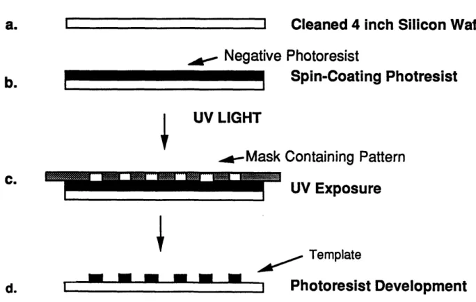
Highly polished glass (Corning 7740) wafers were purchased from Mooney Precision Glass Co. (Huntington, WV) for use in all experiments described in this thesis. Glass wafers were circular with a diameter of either 0.7 inch or 4 inch and thickness of 0.023 inch. Surfaces of these glass substrata were modified with the desired surface morphology using methods of microelectronics processing. The overall flow chart of the process used is shown in Figure 3.1 and the specific details of each step are described below.
3.1.1 Mask Design and Preparation
The process begins with the selection of the desired surface morphology and feature dimensions. A simple parallel grooved morphology was chosen in this study to facilitate data interpretation (Figure 3.2). The size of groove width (w) and spacing between grooves (r) was chosen based on the size of a typical mammalian cell (diameter 10 - 20g1m). Once the surface morphology was selected, a master template of this design, called a mask, was generated.
A computer program called 'kic' (University of California, Berkeley), developed for designing integrated circuits, was used to generate a 2 inch x 2 inch pattern of parallel grooves on a computer (VAXstation II/GPX, DEC, MA). Data from the computer was transferred to a pattern generator (Gyrex model 1005A, USA). The pattern generator transferred the design to a chromium coated quartz plate (5 inch x 5 inch, Precision Photoglass, USA) using a contact printer (Oriel) and a developer
Cleaned 4 inch Glass Wafer
*d.
e.
I 111M-IAluminum
UV LIGHT.-
Mask
Aluminum Evaporation
sitive Photoresist
Spin-Coating Photresist
Containing Pattern
UV Exposure
*Photoresist Development
ILLI t
l-I I I
Aluminum Etching
Etching with CF
4, CHF
3& Ar Plasma
Etched Glass
Photolithography and Plasma Etching Process for Preparing Grooved
b.
C. I I I I f.g-h.
Figure 3.1 ...:·s·:~-:·· :;:55;:;52;·:5.:.:·I·:s·:5·:·! :s·:··:·:·:·s·::t ! I """"""""""""""""""""'' · I:·;··:·S·:·:·i:·;5·f·:·:':':·15 :~::~:::~:::... : : : :·:·::·~··:·::~:·:L·:- I§,:~:::-:::M· --- :&5:" ,I~
~~~~~~~~~...···:.
·
a.
I ITop-view
* Parallel grooved glass substrata
* Groove dimensions chosen based on cell size
* Each surface represented by w x r x d
d
W
Cross-section
Figure 3.2 Choice of Surface Morpholgy
i.
I
I
i-II
mI
I
MI
i,.I
I
wI
I
I
L .(Advance Process Technology, USA). The chromium plate (mask) now contained the desired pattern. All masks were prepared at the Microsystems Technology Laboratories (MTL) at MIT.
3.1.2 Glass Substrata with Grooved Surface Morphology
Masks were used as templates to transfer the desired pattern onto glass substrata using photolithography and plasma etching. Individual steps in this process are described as follows.
3.1.2.1 Precleaning Wafers for processing
Glass wafers were cleaned in a hot piranha solution, which was prepared by mixing 3 volumes of concentrated sulfuric acid (H2SO4) with 1 volume of 30% hydrogen peroxide (J.T. Baker). Piranha cleaning was performed for 10 minutes in a acid-hood (Laminaire) located in the MTL at MIT. The glass wafers were then rinsed profusely with double distilled water in an automatic rinser and then dried with dry N2 in a wafer dryer (Semitool, USA). Glass wafers were dehydrated in a 200C oven for at least 8 hours prior to further processing.
3.1.2.2 Aluminum evaporation
After dehydration, the wafers were coated with a thin layer of aluminum (Figure 3.1b). The aluminum coating was performed in two different systems. For the 4 inch wafers, an electron beam (e-beam) evaporator (Temescal, model VES 2550) was used along with a film deposition controller (FDC-8000) and a power supply (Temescal CV-14) to deposit a 1 m film of aluminum. This procedure was performed
in the Technology Research Laboratories (TRL) of the MTL at MIT.
A vacuum evaporator (Dottie) was used at 5 x 10⁻⁵ torr to deposit a 0.25 gAm film of aluminum on the 0.7 inch glass wafers. This procedure was performed in the Microelectronics Laboratory of the Center for Material Science and Engineering at MIT.
3.1.2.3 Photolithography
Aluminum coated glass wafers were dehydrated at 200'C for at least 8 hours before further processing. Dehydrated wafers were coated with a 1 pgm layer of a positive photoresist (KTI 820/27 centistokes, Union Carbide) using a spin-coater (Solitec, USA). The wafer was centered on the spin-coating chuck and secured by applying vacuum. Photoresist was poured on the wafer and the chuck spun at 5000 RPM for 30 seconds to create a uniform coating of 1m (Figure 3.1c). Non-uniform coating resulted when the wafer was not properly centered. After coating, the wafer was removed from the chuck and placed in a constant temperature oven (Blue M, General Signal, USA) at 90'C for 30 minutes. Next, the wafer was placed in an aligner (Karl Sss, Germany) along with the mask that contained the desired pattern. The wafer was positioned below the mask, in contact with the "chrome side" of the mask. The contact between the mask and the wafer was enhanced by applying vacuum between the interstitial space between the two. The mask was then irradiated with ultra violet (UV) light from top at an intensity of 11 mw/cm2for 2.5 seconds (Fig. 3.1d). The photoresist coated wafer, placed below the mask, was exposed with UV light through the clear regions of the mask that allowed the UV light to pass through. Exposure of UV light rendered the positive photoresist labile to a chemical, called a developer (KTI 934 1:1, Union Carbide; chemical name: tetra methyl ammonium
hydroxide). The wafer was removed from the aligner and exposed to this developer for 40-50 s in a swirling motion. The exposed photoresist was removed by the developer resulting in a patterned photoresist (Figure 3.1 e). The wafer was rinsed in a stream of deionized water for several seconds and dried in a stream of dry nitrogen gas. The aluminum layer below the photoresist was exposed in the areas where the resist was removed. The wafer was inspected under a light microscope (Nikon Optiphot, Japan) to confirm that no defects in the pattern existed. The wafer was also examined in a fluorescent microscope to confirm that all the photoresist was removed from the exposed areas (photoresist appears red and aluminum black under a green fluorescent filter). If the processing was satisfactory, the wafer was baked at 120C for 30 minutes.
Next, the exposed aluminum on the wafer was removed using an aluminum etching solution (Transene Co., Rawley, MA; chemical composition: 80% phosphoric acid, 5% glacial acetic acid, 5% concentrated nitric acid, 10% water) at 50°C. Under these conditions, aluminum was removed at a rate of 0.1 gIm/s. This step was performed very carefully in an acid-hood and the wafer was watched continuously because a slight overexposure of the Al etchant resulted in the removal of the aluminum below the photoresist. Next, the wafer was rinsed profusely with several volumes of deionized water, dried with N2 and inspected in a light microscope to confirm complete removal of aluminum (Figure 3.1f).
3.1.2.4 Plasma Etching
The aluminum etch resulted in the transfer of the desired pattern to the aluminum layer. The final step was to etch the exposed glass that resulted after the
aluminum etch. This could be accomplished by either a wet etch, involving a chemical glass etchant such as hydrofluoric acid (HF) or Buffered Oxide Etchant (BOE, composition: 1:7 40% hydrofluoric acid: ammonium hydroxide), or by a dry etching process, such as plasma etching. Pure HF could not be used because it was too strong and etched the aluminum as well. BOE was successful in etching the glass in a controlled manner but because wet etching is isotropic, grooves deeper than 2 gm could not be etched due to severe undercutting. Therefore, a protocol based on plasma etching protocol was developed.
SiO2 (bulk of Corning 7740 glass) is known to be etched, at a controlled rate, with a CF4 plasma in combination of CHF3 and Ar. Such a plasma is a combination of
a reactive ion etch (via the chemical reaction of F- ions with Si to produce SiF6) and physical etching (via bombardment of Ar ions on the glass surface). An industrial plasma etcher (Applied Precision 5000, Applied Materials, Santa Clara) was used for etching the 4 inch wafers under the following conditions:
Gas Mixture: CF4 = 9 sccm, CHF3 = 30 sccm, Ar = 60 sccm
Power: 550 W
Chamber Pressure: 70 mtorr Magnetic Flux: 70 gauss
The plasma chamber was equipped with a magnetic field to facilitate uniform etching on the entire wafer. Under these conditions, the residual photoresist was etched immediately and glass was etched at a rate of approximately 48 A/s. As intended, the aluminum was resistant to the plasma etch and therefore acted as a good mask. The exposed glass was selectively etched resulting in the formation of a
grooved surface morphology (Figure 3.1f,g). The depth of the grooves could be controlled by changing the time of exposure of the plasma. This was obviously limited by the eventual etching of the aluminum mask which occurred in approximately 10 minutes of exposure to plasma. Undercutting was significantly reduced with plasma etching but was not completely eliminated. Dimensions of the grooves and ridges were significantly different from the original mask design. These were characterized
using scanning electron microscopy on the different substrata (described in detail in a later section). This work was conducted at the Plasma etching laboratory of Prof. Sawin in the Chemical Engineering department at MIT.
Plasma etching on the smaller wafers (0.7 inch diameter) was conducted in a different system because the AP 5000 could not accommodate wafers smaller that 4 inch diameter. The etching was performed in a reactive ion etch (RIE) system (Waf'r Batch 700 series, Plasma-Therm, Inc., USA). The residual photoresist from the wafer, remaining after the Al etching, was removed first by exposure to an oxygen plasma in the RIE system, at a base pressure of 10-5 torr, and an oxygen pressure of 30 mtorr. The exposed glass was etched using a CF4 plasma (70 mTorr CF4pressure) at 10-5 torr base pressure in the same equipment. Etching was much slower compared to the AP 5000 and it proceeded at a rate of 3 A/s. The depth of etching was again controlled by the exposure time to plasma. This work was performed in the microelectronics laboratory in the Center for Material Science and Engineering at MIT.
3.1.2.5 Cutting and Cleaning Grooved Glass Wafers
A computer controlled dicing saw (model DAD-2H/5, Disco Abrasive Systems, Japan) was used to cut the 2 inch x 2 inch patterned area from each 4 inch glass wafer