Christian Jackowski Marcel J. B. Warntjes Johan Berge Walter Bär Anders Persson Received: 5 March 2010 Revised: 28 April 2010 Accepted: 10 June 2010 Published online: 20 July 2010 # European Society of Radiology 2010
Magnetic resonance imaging goes
postmortem: noninvasive detection
and assessment of myocardial infarction
by postmortem MRI
Abstract Objective To investigate the performance of postmortem magnetic resonance imaging (pmMRI) in identification and characterization of lethal myocardial infarction in a non-invasive manner on human corpses. Materials and Methods Before forensic autopsy, 20 human forensic corpses were examined on a 1.5-T system for the presence of myocardial infarction. Short axis, transversal and longitudi-nal long axis images (T1-weighted; T2-weighted; PD-weighted) were acquired in situ. In subsequent autopsy, the section technique was adapted to short axis images. Histological investigations were conducted to confirm autopsy and/or radiological diagnoses.Results Nineteen myocardial lesions were detected and age staged with pmMRI, of which 13 were histologically confirmed (chronic, subacute and
acute). Six lesions interpreted as peracute by pmMRI showed no mac-roscopic or histologicalfinding. Five of the six peracute lesions correlated well to coronary pathology, and one case displayed a severe hypertrophic alteration.ConclusionpmMRI reliably demonstrates chronic, subacute and acute myocardial infarction in situ. In peracute cases pmMRI may display ischemic lesions undetectable at autopsy and routine histology. pmMRI has the potential to substantiate autopsy and to
counteract the loss of reliable infor-mation on causes of death due to the recent disappearance of the clinical autopsy.
Keywords Postmortem imaging . Forensic Radiology . Myocardial infarction
Introduction
Over the last decades clinical autopsy rates have been declining dramatically. Being the main source of today’s postmortem information, this broadly affects the health of the population and the quality of the health care systems, too. As, for example, national mortality statistics directly influence the financial investments intended to support specific research areas in the health care sector, this development impairs the accuracy of research planning and thereby the expected benefit for the health of the population as well. Efforts to counteract this unfavorable trend have predominantly remained unsuccessful [1–5]. However, possible alternative investigation techniques
exist and may have the potential to narrow this informa-tion gap.
In the forensic environment, postmortem cross-sectional imaging is being evaluated and validated to assess different forensic findings in a minimally invasive or non-invasive manner mainly for forensic documentation [6–8]. Within these research efforts, it was also experienced that natural causes of death could be visualized in a non-invasive manner. A variety of natural causes of death were exempla-rily published by predominantly forensic-radiological research groups [9–16]. It should not be surprising for any physician that macro-morphologicalfindings such as major intra-cerebral bleedings, aortic dissection, pericardial tamponades, tumors, etc., are quite easily diagnosed on
C. Jackowski
:
M. J. B. Warntjes:
A. PerssonCenter for Medical Image Science and Visualization, CMIV, University Hospital, University of Linköping,
SE-58185 Linköping, Sweden C. Jackowski
:
J. BergeDepartment of Forensic Medicine, Artillerigatan 12, SE-58133 Linköping, Sweden
C. Jackowski ())
:
W. Bär Institute of Legal Medicine, University of Zürich, Winterthurerstrasse 190/52, CH-8057 Zürich, Switzerland e-mail: [email protected] Tel.: +41-44-6356201 Fax: +41-44-6356850postmortem CT and MRI images. Postmortem imaging is not impaired by patient-related motion artifacts, dose limitations or examination time restrictions. Furthermore, these methods have the potential to provide more detailed information on macroscopic and tissue alterations for the cause of death assessment. In this context, visualization of early ischemic alterations of the myocardium would be most important as cardiac death represents the major portion of the national mortality registers.
The initial cases of myocardial infarction visualized by pmMRI were published from 2003 to 2006 [17–19]. In clinical MR examinations only contrast-enhanced sequences allow sufficient cardiac MR imaging [20]. Postmortem cardiac MR imaging, however, is based on a purely morphological visualization of the investigated structures since only structural tissue alterations influence image contrast. This study focused on cases of myocardial infarction comparing the findings of pmMRI with the pathologicalfindings at autopsy. The aim was to investigate the different stages of myocardial infarction (representing different survival times) in correlation with autopsy and histological findings in a larger study population than that on which the existing literature is based [17–19,21,
22].
Materials and methods Cases
Twenty persons dying under circumstances consistent with a cardiac cause of death were prospectively inves-tigated between May 1, 2007 and March 31, 2008. The study population consisted of 16 men and 4 women [mean (±SD) age at death, 63±10 years]. A pmMRI examination of each body was performed before forensic autopsy. The study was approved by the local ethics committee (Dnr M64-05, 75-05).
Logistics
The corpses were undressed and wrapped twice in artifact-free body bags. As the forensic department is not equipped with MR equipment, the imaging infrastructure of a nearby radiological research institute was used based on a pre-existing collaboration. Transportation was carried out using the institutional mortician vehicle. The MR examinations took place between 7 and 8 o’clock a.m. before regular patient scheduling.
MRI
Each corpse was placed supine in a 1.5-T clinical MR machine (Achieva, Philips Medical Systems, Best, The Netherlands) and examined using a SENSE cardiac or a
SENSE torso coil. Short axis images were acquired using conventional clinically used localizer settings [23] in T1 (TR 650 ms; TE 15 ms), 2× T2 (TR 4,000 ms; TE 70 ms and TR 4,000 ms; TE 100 ms) and proton density (TR 4,000 ms; TE 10 ms) weighting. Slice thickness was 4 mm, resolution 0.7×0.7 mm. Examination time was <20 min. Image interpretation was performed according to Jackowski et al. [19], predominantly based on the signal behavior on T2-weighted images by two investigators in consensus sessions. Additionally, the finding of hypoin-tensity without marginal hyperinhypoin-tensity in T2-weighted images not yet described by Jackowski et al. [19] was interpreted as peracute infarction.
Autopsy
Forensic autopsy was performed by board-certified foren-sic pathologists. The cardiac dissection technique was adapted to match short axis images by slicing the ventricles parallel to the heart base. Extensive photo-graphic documentation was carried out. Histological specimens of the entire circumference of the left ventricle (LV) according to the short axis MR images were investigated. Tissue staining included hematoxylin and eosin (H&E), van Gieson and chromotrop-aniline-blue (CAB). The coronary orifices and the apex of the heart were used as anatomical landmarks for comparison of related findings. Histological grading ranged from acute (early and late) to sub-acute (early and late) and chronic.
Results
In 17 of the 20 study cases, cardiac failure was considered the cause of death. In case 12, severe pneumonia was considered to have promoted cardiac failure. Two study cases (9 and 15) presented with an extra-cardiac cause of death.
Sixteen study cases presented with overall 19 myocar-dial lesions to investigate, and four study cases showed no assessable local ischemic alteration in pmMRI, autopsy or histology. Table 1 displays the 19 lesions separated into three groups. Group 1 consisted of seven pmMRI lesions that correlated well to autopsy and histology. Group 2 included six cases that showed good correlation of pmMRI lesion to histology, but less or no correlation to macroscopic autopsy. There was a good correlation between pmMRI and histology in all group 1 and group 2 cases. Group 3 combined six cases that presented with lesions at pmMRI, but had no myocardial findings at autopsy or histology. All six group 3 cases were radio-logically interpreted as peracute. These six peracute lesions presented with further cardiac autopsy findings that could explain an ischemic situation at the affected myocardial region by either severe coronary event/stenosis
(five cases) or severe myocardial hypertrophy (one case). Acute pulmonary edema and internal congestion indicated acute cardiac failure as the cause of death in all six group 3 cases.
The findings of chronic (Fig. 1), subacute (Fig. 2) and acute infarction (Fig. 3) demonstrated reproducible appearances in pmMRI. Chronic infarction was associ-ated with a broad loss of signal in all applied sequences. In T2 weighting, subacute infarction displayed hyperintense alterations within the affected
myocardium. Acute infarction showed a hypointense center with hyperintense margins because of edema in the outer zone in T2 weighting.
Discussion
In postmortem cardiac MR imaging, not having the possibility to assess late enhancements [24], excellent
Table 1 Findings of study cases grouped according grade of consensus
Case no. Location pmMRI Autopsy Histology Cause of
death
Comments
(a) Lesion group 1: consensus among pmMRI, autopsy and histology
3 Anterior Acute Acute Acute Cardiac Only smallfinding of
5×5 mm, no coronary finding
4 Anteroseptal Acute and subacute Acute and subacute
Acute and subacute
Cardiac Severe LAD stenosis
8a Anterior Chronic Chronic Chronic Cardiac RCA + RCX occluded,
LAD severe stenosis right after left main CA, bypass on RCA occluded, bypass on LAD open
8c Lateral Subacute Subacute Subacute
14 Posteroseptal Chronic Chronic Chronic Cardiac Severe 3 vessel disease with coronary bypass surgery
19 Lateral Subacute Subacute Subacute Cardiac Severe RCX stenosis
20 Anterior Acute Acute Acute Cardiac Severe LAD stenosis
(b) Lesion group 2: consensus between pmMRI and histology 1a Anteroseptal/
anterior
Subacute and chronic Chronic Subacute and chronic
Cardiac Severe 3 vessel desease
1b Lateral Acute and subacute Subacute Acute and subacute
7 Septal Acute - Acute Cardiac Heart weight 613 g
12 Septal Acute - Acute Pulmonary/
cardiac
50% stenosis on LAD and severe
pneumonia 13 Anteroseptal Subacute and chronic Chronic Subacute and
chronic
Cardiac Severe stenosis at proximal LAD
16 Posteroseptal Subacute - Subacute Cardiac Severe RCA stenosis
(c) Lesion group 3: no consensus between pmMRI and autopsy/histology
2 Septal Peracute - - Cardiac Severe stenosis at 2nd
septal branch
6 Anteroseptal Peracute - - Cardiac Severe stenosis at
intermediate branch
8 b) Septal Peracute - - see case no. 8a
10 Anterior/ant. papillary muscle
Peracute - - Cardiac Fresh ruptured soft
plaque in LAD
17 Anteroseptal Peracute - - Cardiac LV hypertrophy due
to severe aortic valve stenosis
18 Anterior Peracute - - Cardiac Severe LAD stenosis
(d) Study cases without myocardialfindings in pmMRI, autopsy and histology
5 - - - - Cardiac Heart weight 690 g
9 - - - - Multiorgan
failure
Pancreatic cancer
11 - - - - Cardiac Heart weight 796 g
15 - - - - Pulmonary Severe pneumonia
Grading of pmMRIfindings and histological findings was assessed according to Jackowski et al. [19]. Autopsy diagnosis was based on the macroscopic appearance at autopsy only. The given cause of death represents thefinal integrative forensic case diagnosis including extra-cardiac autopsy findings. Case no. represents the initial chronological case numbering
image quality can nevertheless be obtained due to the absence of cardiac motion or breathing-related artifacts. The image contrast not based on perfusion differences visualized by contrast agent administration strongly represents morphological alterations within the myocardial tissue. Already in the early 1980s, unenhanced clinical
MR imaging showed promising results for the identi fica-tion of myocardial infarcfica-tion in the living because of the increase of water (edema) within the infarcted muscle [25,
26]. Thereby, acute infarctions appear as hyperintense regions in in-vivo T2-weighted images and dark in native T1-weighted images [27].
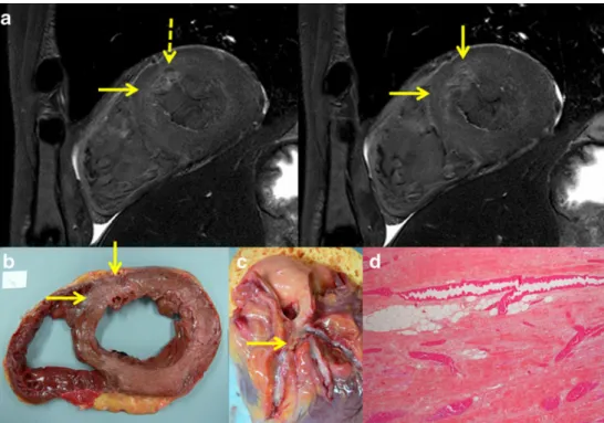
Fig. 1 Chronic myocardial infarction (case 14) in pmMRI. a T2-weighted short axis images (entire FoV and magnification) depict a severe shrinking of the inferior wall with broad decrease of signal (arrows). b Autopsy demonstrates definite collagenous transformation of the infarcted inferior myocardium with scar-caused shrinking. c Histology (H&E) shows cell-free collagen formation as the cause for the significant decrease of signal in MRI as well as fatty degeneration
Fig. 2 Subacute myocardial infarction (case 13) in pmMRI. a T2-weighted short axis images at different levels show a broad hyperintensity (arrows) affecting the anteroseptal wall. Note a tiny spot of signal loss due to a small chronic infarction within the affected anterior wall (left image, dotted arrow). b Autopsy demonstrates discreet shrinking of the anteroseptal wall and minor connective tissue formation. c The coronary artery system presents with a severe stenosis at the LAD. d Histology (H&E) shows loose connective tissue formation and angiogenesis as the reason for the general signal increase in T2
Whereas clinical cardiac MR imaging in recent years has improved different contrast agents and implemented application schedules as well as contrast agent and cardiac motion-adapted sequences to better assess cardiac pathol-ogy [20, 28–31], postmortem cardiac MR imaging fully depends on imaging of unenhanced structural alterations at different stages of myocardial infarction. These stages showed a specific signal behavior in pmMRI that allows an age staging of the infarction. The present study
confirmed the results from our recent study concerning the pmMRI findings in acute, subacute and chronic infarction [19].
Thirteen of the 19 lesions (group 1 and 2) correlated well between pmMRI and histology, whereas 6 of them (group 2) showed poor correlation to macroscopic autopsy. Macroscopic detection of myocardial infarction depends on the extent of discoloration and/or the consistency of the tissue. This makes overlooking tiny
Fig. 3 Acute myocardial infarction (case 3) in pmMRI. a T2-weighted short axis image demonstrates a tiny hypointensity (circle) with indistinctive surrounding edema within the anterior wall. b Autopsy can reveal a small yellowish alteration at the same location (circle). d Histology (H&E)
demonstrates beginning granulocyteous infiltration only in one magnification field as shown
Fig. 4 Peracute myocardial infarction (case 10) in pmMRI. a T2-weighted short axis images at different levels of the anterior papillary muscle present with local hypointensities within the anterior papillary muscle and indistinctively also within the anterior wall. b Autopsy aspect of the specimen shows no visible alteration within the anterior wall and the papillary muscles. Histology also failed in demonstrating ischemic alterations (not shown). c Dissection of the coronary artery system reveals a fresh soft plaque rupture with intimal hemorrhage within the proximal LAD
lesions likely when no distinctive and distended alter-ations are present. Furthermore, an age staging by eye at the autopsy table is extremely uncertain, and histology is needed for validation. Therefore, the group 2 cases show exemplarily that pmMRI provides additional information in cases that are hard to diagnose macroscopically.
In addition to the recent work [19], the present study demonstrated that cases of sudden cardiac death display-ing no myocardial morphological finding at autopsy and histology showed structural alterations in pmMRI (Fig.4). These cases challenge pathologists since generations and all approaches to verify an ischemic situation before sudden cardiac death have not reached widespread application because of less satisfying performance [32–34]. In five of the six group 3 cases, myocardial
pmMRI findings (hypointensities in T2 weighting)
correlated well with coronary pathology at autopsy, whereas according to the literature [35] only half of the peracute cases present a coronary lesion at all. The sixth case revealed massive LV hypertrophy consistent with the possibility of a local relative ischemic incident. All peracute cases in our study group were forensically diagnosed as cardiac deaths in combination with indirect signs of cardiac failure such as internal congestion and acute pulmonary edema. Although statistically not supported because of the still rather small study population, the results of our pmMRI examinations let us assume that pmMRI is a suitable method to detect and display peracute myocardial lesions to be included into the case diagnosis.
In concordance with our previous work [19], acute myocardial infarction lesions were displayed as a zone with a dark ischemic center and a bright edematous margin (T2-weighted images). Since the development of post-infarct myocardial edema takes several minutes, it is not expected in sudden cardiac deaths due to an arrhythmic event resulting in a dark hypointense lesion representing the zone of ischemic myocardium without bright margin as observed in the six peracute cases. In conclusion, pmMRI seems to visualize ischemic myocardium well before the develop-ment of edema occurs. Within this short time period, the onset of a ventricularfibrillation may have caused cardiac failure and sudden death, preventing further vital morpho-logical reactions in the myocardium.
The observation of hypointense areas in T2-weighted images in ischemic myocardial regions is not sufficiently explained yet. A slight decrease of signal also seen in PD-weighted sequences would suggest that the mean water content is lower than in the surrounding myocardium. A lower pH value in combination with local electrolyte changes may also contribute to a relevant T2 relaxation time shortening. However, the anatomical association of “dark myocardium” with a coronary lesion at the coronary artery perfusing the affected myocardium makes a causative correlation to an ischemic situation very likely. Compared to the in-vivo T2 appearance of acute infarc-tions, this is the most striking difference as clinical radiologists know acute infarctions as hyperintense in T2-weighted images. The edema as a vital myocardial
reaction within the infarcted region is present from hours to days after the onset of ischemia. In the majority of cases when acute infarction is imaged postmortem, the patient died in very early stages. In these early time frames after ischemic onset (minutes to hours), the edema starts developing, and this stage is kept after death. Therefore, we see no edema in peracute cases (minutes) yet, but hypointense areas probably as a result of reduced micro-circulation. In case the patient did not die immediately, we see the state of developing edema as a marginal in-growing hyperintensity around the hypointensity in acute cases. Our hypothesis is that most of the in-vivo images are obtained at the stage of more or less fully developed edema and that postmortem images often show earlier stages with beginning edema or no edema.
Autopsy and histology, the “gold standard” to inves-tigate cardiac deaths, are to be challenged by new approaches. However, without a coronaryfinding indicat-ing a local circulation barrier, a peracute lesion causindicat-ing an arrhythmic sudden cardiac death cannot be verified using traditional methods within the myocardium itself. The six cases in group 3 without consensus between pmMRI and autopsy and histology are not “false positive.” It is the gold standard that obviously fails in these cases. Only the view of the coronary artery system helped to interpret the mismatch correctly. We think we have demonstrated that pmMRI may provide further valuable diagnostic information about the myocardium in peracute cardiac deaths; it is capable of visualizing where and how much distension the myocardium has suffered from absolute or relative ischemia.
Application of contrast agents may also be promising in postmortem imaging. Techniques of sufficient perform-ance already exist and enable minimally invasive visual-ization of the coronary artery system [36–39]. In porcine ex vivo experiments, distribution defects of injected gadolinium could be simulated within the porcine myo-cardium, which possibly can be correlated with a disturbed antemortem perfusion as well [36]. A combina-tion of pmMRI and minimally invasive angiography will enhance the diagnostic performance of pm imaging.
Limitations
The study was not conducted in a double-blinded way as radiological findings not detected at autopsy should undergo histological examination as well. Therefore, the forensic pathologist had to be informed about the pmMRI findings in advance to obtain tissue samples from both autopsy and pmMRIfindings.
The study number is rather low, so a confirmation of our results in larger series is needed.
A further limitation may be the lack of a comparably sized control group with a sufficient number of non-cardiac deaths. The four study cases without a local myocardial finding may indicate a consensus for the
absence of myocardial findings, too. However, four cases do not allow for valid conclusions.
No measurements of relative signal intensities within the infarcted myocardium in comparison to skeletal or not affected myocardium are given. From the clinical point of view, this would be expected to lead to better under-standing of the pm appearances of infarcted myocardium. However, the postmortem situation is more complex with respect to relative signal intensities. Both T1 and T2 relaxation are temperature dependent. The decrease of T2 relaxation time at decreasing temperature is less distinc-tive compared to the rather considerable decrease of T1 relaxation time at decreasing temperature. In contrast to the clinical situation where the patient's temperature is a predominantly stable value of about 37°C, we have to deal postmortem with a possible temperature ranging from about 4° to 40°C. Exceptions even lie beyond these values when, e.g., frozen bodies found in glaciers or burned bodies with still elevated core temperatures enter the MR machine. Therefore, any absolute quantified or relative signal intensity on postmortem MR data can only be interpreted with respect to the actual body temperature during data acquisition. As the body core temperature of the study cases was not consistent, it is not reasonable to compare absolute or relative signal intensities between different cases. To address this problem, a separate study is needed and already in preparation that will perform an absolute quantification of T1, T2 and proton density for every body tissue (grey matter, white matter, myocardium, liver tissue, etc.) at defined temperatures ranging from 4° to 40°C to compile a data collection that can be used on one hand for optimal sequence design and on the other hand for diagnostic purposes to characterize pathological alterations.
Conclusion
With pmMRI ischemic lesions can be detected, assessed and age staged. Indeed, in peracute lesions, as in sudden
cardiac death when the lesion may not be visible with conventional autopsy techniques, it may provide addi-tional information for the pathologist.
The present study demonstrates that pmMRI imaging provides an alternative and/or complementary postmortem examination technique to autopsy. Additionally, it should be considered for cases not undergoing a traditional autopsy. Depending on the country, up to more than 90% of the deceased do not undergo comprehensive postmortem examination. The negative consequences for medical education, quality assurance in medicine, public health and mortality statistics are substantial, as discussed by numerous authors [1–3, 5, 40]. Postmortem imaging has matured to a valuable postmortem examination method to acquire patho-anatomic details in a spatial resolution not achievable by routine autopsy. An increas-ing number of forensic institutes have started to install CT and MRI systems to use the imaging techniques for the purpose of quality improvement. However, it is the natural death that is significantly under-autopsied today, and it seems obvious to us that clinicians should become aware of the potential and the recent progress of pmCT and especially pmMRI as a non- or minimally invasive method for the investigation of the deceased patient. The aim is not to replace clinical autopsies, but to complement or offer an alternative when autopsy is not agreed to by the next of kin and to reestablish a reliable base of data of cause of death in our society.
Acknowledgements The authors would like to thank the team of forensic autopsy technicians and forensic examiners at the Depart-ment of Forensic Medicine, Linköping, Sweden, for the reliable support at autopsy and logistic aspects of the study. We furthermore express our gratitude to the team of radiological technicians at the Center for Medical Image Science and Visualization, Linköping, Sweden, who performed the data acquisition.
Funding Supported by a grant from the Swiss National Science Foundation (PBBE33-115060), a grant from the Swedish Knowledge Foundation (KK-stiftelsen 2007/0170) and a grant from the Swedish National Board of Forensic Medicine (RMV) (all to Dr. Jackowski).
References
1. Shojania KG, Burton EC (2008) The vanishing nonforensic autopsy. N Engl J Med 358:873–875
2. Shojania KG, Burton EC, McDonald KM, Goldman L (2005) Overestimation of clinical diagnostic performance caused by low necropsy rates. Qual Saf Health Care 14:408–413
3. Loughrey MB, McCluggage WG, Toner PG (2000) The declining autopsy rate and clinicians' attitudes. Ulster Med J 69:83–89
4. Sinard JH (2001) Factors affecting autopsy rates, autopsy request rates, and autopsyfindings at a large academic medical center. Exp Mol Pathol 70:333–343
5. Ward HE, Clarke BE, Zimmerman PV, Cleary MI (2002) The decline in hospital autopsy rates in 2001. Med J Aust 176:91
6. Dirnhofer R, Jackowski C, Vock P, Potter K, Thali MJ (2006) VIRTOPSY: minimally invasive, imaging-guided virtual autopsy. Radiographics 26:1305–1333
7. Levy AD, Harcke HT, Getz JM, Mallak CT, Caruso JL, Pearse L, Frazier AA, Galvin JR (2007) Virtual autopsy: two- and three-dimensional
multidetector CTfindings in drowning with autopsy comparison. Radiology 243:862–868
8. Levy AD, Abbott RM, Mallak CT, Getz JM, Harcke HT, Champion HR, Pearse LA (2006) Virtual autopsy: preliminary experience in high-velocity gunshot wound victims. Radiology 240:522–528 9. Yen K, Lovblad KO, Scheurer E,
Ozdoba C, Thali MJ, Aghayev E, Jackowski C, Anon J, Frickey N, Zwygart K, Weis J, Dirnhofer R (2007) Post-mortem forensic neuroimaging: correlation of MSCT and MRIfindings with autopsy results. Forensic Sci Int 173:21–35
10. Aghayev E, Yen K, Sonnenschein M, Ozdoba C, Thali M, Jackowski C, Dirnhofer R (2004) Virtopsy post-mortem multi-slice computed tomography (MSCT) and magnetic resonance imaging (MRI)
demonstrating descending tonsillar herniation: comparison to clinical studies. Neuroradiology 46:559–564 11. Shiotani S, Watanabe K, Kohno M,
Ohashi N, Yamazaki K, Nakayama H (2004) Postmortem computed tomographic (PMCT)findings of pericardial effusion due to acute aortic dissection. Radiat Med 22:405–407 12. Arai A, Shiotani S, Yamazaki K, Nagata
C, Kikuchi K, Suzuki M, Atake S, Kohno M, Ohashi N (2006) Postmortem computed tomographic (PMCT) and postmortem magnetic resonance imaging (PMMRI) demonstration of fatal massive retroperitoneal
hemorrhage caused by abdominal aortic aneurysm (AAA) rupture. Radiat Med 24:147–149
13. Ikeda G, Yamamoto R, Suzuki M, Ishikawa H, Kikuchi K, Shiotani S (2007) Postmortem computed tomography and magnetic resonance imaging in a case of terminal-stage small cell lung cancer: an experience of autopsy imaging in tumor-related death. Radiat Med 25:84–87
14. Shiotani S, Kohno M, Ohashi N, Yamazaki K, Nakayama H, Watanabe K, Oyake Y, Itai Y (2004) Non-traumatic postmortem computed tomographic (PMCT)findings of the lung. Forensic Sci Int 139:39–48 15. Jackowski C, Dirnhofer S, Thali M,
Aghayev E, Dirnhofer R, Sonnenschein M (2005) Postmortem diagnostics using MSCT and MRI of a lethal
streptococcus group A infection at infancy: a case report. Forensic Sci Int 151:157–163
16. Jackowski C, Thali MJ, Buck U, Aghayev E, Sonnenschein M, Yen K, Dirnhofer R, Vock P (2006)
Noninvasive estimation of organ weights by postmortem magnetic resonance imaging and multislice computed tomography. Invest Radiol 41:572–578
17. Thali MJ, Yen K, Schweitzer W, Vock P, Boesch C, Ozdoba C, Schroth G, Ith M, Sonnenschein M, Doernhoefer T, Scheurer E, Plattner T, Dirnhofer R (2003) Virtopsy, a new imaging horizon in forensic pathology: virtual autopsy by postmortem multislice computed tomography (MSCT) and magnetic resonance imaging (MRI)—a feasibility study. J Forensic Sci 48:386–403 18. Jackowski C, Schweitzer W, Thali M,
Yen K, Aghayev E, Sonnenschein M, Vock P, Dirnhofer R (2005) Virtopsy: postmortem imaging of the human heart in situ using MSCT and MRI. Forensic Sci Int 149:11–23
19. Jackowski C, Christe A, Sonnenschein M, Aghayev E, Thali MJ (2006) Postmortem unenhanced magnetic resonance imaging of myocardial infarction in correlation to histological infarction age characterization. Eur Heart J 27:2459–2467
20. Kim RJ, Wu E, Rafael A, Chen EL, Parker MA, Simonetti O, Klocke FJ, Bonow RO, Judd RM (2000) The use of contrast-enhanced magnetic resonance imaging to identify reversible
myocardial dysfunction. N Engl J Med 343:1445–1453
21. Jachau K, Heinrichs T, Kuchheuser W, Krause D, Wittig H, Schoening R, Beck N, Beuing O, Doehring W, Jackowski C (2004) Computed tomography and magnetic resonance imaging compared to pathoanatomicfindings in isolated human autopsy hearts. Rechtsmedizin 14:109–116
22. Shiotani S, Yamazaki K, Kikuchi K, Nagata C, Morimoto T, Noguchi Y, Suzuki M, Atake S, Kohno M, Ohashi N (2005) Postmortem magnetic resonance imaging (PMMRI)
demonstration of reversible injury phase myocardium in a case of sudden death from acute coronary plaque change. Radiat Med 23:563–565
23. Axel L (1992) Efficient method for selecting cardiac magnetic resonance image locations. Invest Radiol 27:91–93 24. Hunold P, Schlosser T, Vogt FM,
Eggebrecht H, Schmermund A, Bruder O, Schuler WO, Barkhausen J (2005) Myocardial late enhancement in contrast-enhanced cardiac MRI: distinction between infarction scar and non-infarction-related disease. AJR Am J Roentgenol 184:1420–1426
25. Herfkens RJ, Sievers R, Kaufman L, Sheldon PE, Ortendahl DA, Lipton MJ, Crooks LE, Higgins CB (1983) Nuclear magnetic resonance imaging of the infarcted muscle: a rat model. Radiology 147:761–764
26. Higgins CB, Herfkens R, Lipton MJ, Sievers R, Sheldon P, Kaufman L, Crooks LE (1983) Nuclear magnetic resonance imaging of acute myocardial infarction in dogs: alterations in magnetic relaxation times. Am J Cardiol 52:184–188
27. West AM, Kramer CM (2010) Cardiovascular magnetic resonance imaging of myocardial infarction, viability, and cardiomyopathies. Curr Probl Cardiol 35:176–220
28. Krombach GA, Wendland MF, Higgins CB, Saeed M (2002) MR imaging of spatial extent of microvascular injury in reperfused ischemically injured rat myocardium: value of blood pool ultrasmall superparamagnetic particles of iron oxide. Radiology 225:479–486 29. Kim RJ, Fieno DS, Parrish TB, Harris
K, Chen EL, Simonetti O, Bundy J, Finn JP, Klocke FJ, Judd RM (1999) Relationship of MRI delayed contrast enhancement to irreversible injury, infarct age, and contractile function. Circulation 100:1992–2002
30. Steuer J, Bjerner T, Duvernoy O, Jideus L, Johansson L, Ahlstrom H, Stahle E, Lindahl B (2004) Visualisation and quantification of peri-operative myocardial infarction after coronary artery bypass surgery with contrast-enhanced magnetic resonance imaging. Eur Heart J 25:1293–1299
31. Fieno DS, Kim RJ, Chen EL, Lomasney JW, Klocke FJ, Judd RM (2000) Contrast-enhanced magnetic resonance imaging of myocardium at risk: distinction between reversible and irreversible injury throughout infarct healing. J Am Coll Cardiol 36:1985– 1991
32. Varga M, Zsonda L (1988) A simple method for postmortem detection of acute myocardial infarction. Forensic Sci Int 37:259–263
33. Hansen SH, Rossen K (1999) Evaluation of cardiac troponin I immunoreaction in autopsy hearts: a possible marker of early myocardial infarction. Forensic Sci Int 99:189–196 34. Ribeiro-Silva A, SM CC, Rossi MA
(2002) Is immunohistochemistry a useful tool in the postmortem recognition of myocardial hypoxia in human tissue with no morphological evidence of necrosis? Am J Forensic Med Pathol 23:72–77
35. Farb A, Tang AL, Burke AP, Sessums L, Liang Y, Virmani R (1995) Sudden coronary death. Frequency of active coronary lesions, inactive coronary lesions, and myocardial infarction. Circulation 92:1701–1709
36. Jackowski C, Sonnenschein M, Thali MJ, Aghayev E, von Allmen G, Yen K, Dirnhofer R, Vock P (2005) Virtopsy: postmortem minimally invasive angiography using cross section techniques–implementation and preliminary results. J Forensic Sci 50:1175–1186
37. Jackowski C, Bolliger S, Aghayev E, Christe A, Kilchoer T, Aebi B, Perinat T, Dirnhofer R, Thali MJ (2006) Reduction of postmortem angiography-induced tissue edema by using polyethylene glycol as a contrast agent dissolver. J Forensic Sci 51:1134–1137 38. Jackowski C, Persson A, Thali MJ
(2008) Whole body postmortem angiography with a high viscosity contrast agent solution using poly ethylene glycol as contrast agent dissolver. J Forensic Sci 53:465–468
39. Ross S, Spendlove D, Bolliger S, Christe A, Oesterhelweg L, Grabherr S, Thali MJ, Gygax E (2008) Postmortem whole-body CT angiography: evaluation of two contrast media solutions. AJR Am J Roentgenol 190:1380–1389 40. Shojania KG, Burton EC (2004) The
persistent value of the autopsy. Am Fam Physician 69:2540–2542