Prof. Fabrice Clément. Directeur, Université de Neuchâtel Prof. Alan J. Pegna, co-Directeur, University of Queensland
______________________________________________________________
An electroencephalographic approach to the processing of
ambiguous stimuli under social influence: the role of
perception, attention, emotion and metacognition
THÈSE
Présentée à l’Université de Neuchâtel
Pour obtenir le grade de Docteure en sciences cognitives
par
JULIE ZANESCO
de
Neuchâtel, Suisse
Doctorat en sciences cognitives
Thèse de
Julie ZANESCO
Intitulée: An electroencephalographic approach to the processing of ambiguous
stimuli under social influence: the role of perception, attention, emotion and
metacognition
(*) La Faculté des lettres et sciences humaines, sur préavis d’une commission formée par les professeurs : Fabrice Clément, directeur, Université de Neuchâtel ; Alan J. Pegna, co-directeur, University of Queensland ; Roland Maurer, Université de Genève ; Joseph Krummenacher, Université de Freiburg
Autorise l’impression de la présente thèse, sans prétendre par là émettre d’opinion sur les propositions qui y sont énoncées.
NEUCHÂTEL, le 20 décembre 2019
Le doyen :
Pierre-Alain Mariaux
N.B. La thèse doit porter la déclaration précédente (*) et remplir les conditions énumérées dans les « recommandations aux étudiants qui présentent une thèse »
ACKNOWLEDMENTS
During these four years of PhD, I have been extremely lucky to be surrounded by incredible people who had a direct or indirect impact on the elaboration of this thesis.
First, I would like to thank my academic directors, Professor Fabrice Clément and Professor Alan Pegna. Thank you, Fabrice, for having given me the opportunity to work on a highly interesting subject which is metacognition and social influence, and having taught me the importance of philosophical questions and their complementary contribution to the field of cognitive neurosciences. Thank you, as well, Fabrice, for having trusted me throughout this thesis. I would like to thank my mentor, Professor Alan Pegna, for teaching me everything I know in the field of neuroscience and neuropsychology, for having taught me to think creatively as a scientist, for having supported me in absolutely all aspects of this thesis. Thank you, Alan, for always being there for me, not only professionally but also psychologically, and this since the first time we met in 2009. Thank you, Alan, for being who you are and for accepting me the way I am.
I deeply thank my colleague, co-author and very good friend, Dr Eda Tipura, without whom I wouldn’t have been able to do this thesis. Thank you, Eda, for having taught me how to analyse the data, for helping me in creating this novel paradigm, for your valuable advice and for being next to me all the way, technically and emotionally.
Next, I would like to thank Professor Krummenacher and Professor Maurer for being part of the jury and for their investment in this work.
Thank you, Dr Andrés Posada, for helping me in the programming of the experimental task and for your technical support during the EEG recordings.
My biggest thank you goes to my beloved and wonderful husband, for his emotional support, for his patience, for his trust, for his insightful comments and suggestions. Thank you, Sandro, for always being interested in what I do. Thank you, Sandro, for sharing your life with me. Thank you to my mother and to my sister for listening to me, day after day, for your trust and support. I thank you for loving me.
Finally, I would like to thank my very good friends, Alexandre Caldara, Isabel Tissières, Marzia del Zotto, Hélène Manesse and Adriana Meichtry for their friendship, fidelity and trust.
TABLE OF CONTENTS
Abstract
Résumé en français
1. GENERAL INTRODUCTION ... 1
2. THEORETICAL PART ... 4
2.1. EEG and ERP SPECIFICITIES ... 4
2.1.1. PHYSIOLOGICAL BASIS OF ELECTROENCEPHALOGRAPHY ... 6
2.1.2. EARLY ERP COMPONENTS ... 9
2.1.3. LATE ERP COMPONENTS ... 11
2.2. VISUAL PERCEPTION AND ATTENTION ... 13
2.2.1. BRAIN STRUCTURES INVOLVED IN VISUAL PERCEPTION ... 13
2.2.2. BRAIN STRUCTURES INVOLVED IN ATTENTION ... 15
2.2.3. ERP DATA FROM VISUAL PERCEPTION AND EARLY ATTENTION ... 17
2.2.4. EMOTIONAL FACE PERCEPTION ... 19
2.2.4.1. BRAIN STRUCTURES INVOLVED IN FACE PERCEPTION ... 19
2.2.4.2. ERP DATA FROM FACE PERCEPTION ... 21
2.3. METACOGNITION AND UNCERTAINTY ... 24
2.3.1. BRAIN STRUCTURES INVOLVED IN METACOGNITION ... 25
2.3.2. ERP DATA FROM METACOGNITION ... 26
2.4. SOCIAL INFLUENCE AND SOCIAL CONFLICT ... 28
2.4.1. BRAIN STRUCTURES INVOLVED IN SOCIAL CONFLICT... 28
2.4.2. ERP DATA FROM SOCIAL CONFLICT ... 30
2.5.1. COGNITIVE MECHANISMS INVOLVED IN SOCIAL ANXIETY ... 32
2.5.2. UNCERTAINTY AND AMBIGUITY IN SOCIAL ANXIETY ... 34
3.1. STUDY 1: SEEING IS BELIEVING: ERALY PERCEPTUAL BRAIN PROCESSES ARE MODIFIED BY SOCIAL FEEDBACK1 ... 36
3.1.1. INTRODUCTION ... 37
3.1.2. MATERIALS AND METHODS ... 40
3.1.2.1. PARTICIPANTS ... 40
3.1.2.2. STIMULI AND EXPERIMENTAL PROCEDURE ... 40
3.1.2.3. EEG ACQUISITION ... 43
3.1.2.4. EEG PROCESSING... 44
3.1.2.5. BEHAVIOURAL ANALYSIS ... 44
3.1.2.6. ELECTROPHYSIOLOGICAL RECORDINGS AND ANALYSIS ... 45
3.1.3. RESULTS ... 46 3.1.3.1. BEHAVIOURAL RESULTS ... 46 3.1.3.2. ELECTROPHYSIOLOGICAL RESULTS ... 48 3.1.4. DISCUSSION ... 54 3.1.5. CONCLUSION ... 57 3.1.6. SUPPLEMENTARY MATERIAL ... 57
3.2. STUDY 2: PATTERNS OF ELECTRICAL BRAIN ACTIVATION IN RESPONSE TO SOCIALLY-DISPUTED PERCEPTUAL JUDGMENTS2 ... 63
3.2.1. INTRODUCTION ... 64
3.2.2. MATERIALS AND METHODS ... 65
3.2.2.2. STIMULI AND EXPERIMENTAL PROCEDURE ... 65 3.2.2.3. EEG ACQUISITION ... 65 3.2.2.4. EEG PROCESSING... 66 3.2.3. RESULTS ... 67 3.2.3.1. BEHAVIOURAL RESULTS ... 67 3.2.3.2. ELECTROPHYSIOLOGICAL RESULTS ... 68 3.2.4. DISCUSSION ... 71 3.2.5. CONCLUSION ... 73
3.3. STUDY 3: AM I REALLY SEEING WHAT’S AROUND ME ? AN ERP STUDY ON SOCIAL ANXIETY UNDER SPEECH INDUCTION, UNCERTIANTY AND SOCIAL FEEDBACK3 ... 74
3.3.1. INTRODUCTION ... 76
3.3.2. MATERIALS AND METHODS ... 80
3.3.2.1. PARTICIPANTS ... 80
3.3.2.2. STIMULI AND EXPERIMENTAL PROCEDURE ... 81
3.3.2.3. EEG ACQUISITION ... 82
3.2.2.4. EEG PROCESSING... 82
3.3.2.5. BEHAVIOURAL ANALYSIS ... 82
3.3.2.6. ELECTROPHYSIOLOGICAL RECORDINGS AND ANALYSIS ... 83
3.3.3. RESULTS ... 85
3.3.3.1. BEHAVIOURAL RESULTS ... 85
3.3.3.2. ELECTROPHYSIOLOGICAL RESULTS ... 87
3.3.5. CONCLUSION ... 96
4. GENERAL CONCLUSION ... 98
5. REFERENCES... 102
6. ANNEXES ... 121
6.1. Liebowitz questionnaire (French version)... 121
ABSTRACT
Humans, as social beings, are susceptible to social influence in their judgements, beliefs and behaviours. Over the last 70 years, a great wealth of data from experimental social psychology has demonstrated that social knowledge affects the way humans perceive and interpret their environment, giving rise to cognitive, perceptual, attentional and motivational biases. Already in the 1950’s, Solomon Ash (1951), in a series of perceptual judgement experiments, demonstrated that humans could alter their responses when these differed from a majority. Moreover, the degree of uncertainty triggered by stimuli in the environment has been shown to increase human’s vulnerability to social opinion. More recently, neuroscience began to explore automatic unconscious and controlled conscious brain responses to social stimuli, mainly face expressions. To date, however, the temporal unfolding of attentional and perceptual processes under uncertainty and social conflict remains unclear, particularly regarding the perception of ambiguous stimuli other than faces in a social context. Thus, the aim of the three studies in this thesis, was to investigate whether early perception and higher order cognitive processes were altered when healthy and socially anxious subjects were presented with ambiguous stimuli and when their responses were either endorsed or disputed. For this, a novel experimental paradigm was created allowing us to measure event-related potentials (ERPs) in response to visual stimuli, before and after social feedback indicated by a face displaying a happy (agreement) or disgusted expression (disagreement). Participants were asked to judge the colour of a square (the probe) that was either clearly blue or green (distinct probes) or were highly ambiguous bluish-green colour (ambiguous probes). They were also asked to indicate their level of confidence in those judgments, after which they received social feedback either endorsing or disputing the participants’ responses. Participants were then presented the stimulus again and asked to reconsider their decision and subjective confidence level. Behavioural results showed that confidence levels decreased whereas the number of revisions increased, both with task difficulty
and with conflicting social feedback across healthy subjects. Moreover, this pattern was enhanced across socially anxious individuals. Event-related-potential data revealed differences beginning at already 100 ms after ambiguous stimuli presentations compared to distinct stimuli, as well as enhanced early amplitudes following disputed feedback. These findings are compatible with heightened sensory facilitation of visual information, demonstrating that uncertainty and social pressure modify early perceptual brain processes. The same pattern was reduced across socially anxious individuals suggesting a reduction in early attentional processes to external ambiguous stimuli due to excessive self-focusing and anticipation of the social situation. Additionally, later ERP components, starting at around 300 ms, were decreased for distinct stimuli compared to ambiguous probes in line with higher subjects’ signal detection accuracy and metacognitive experiences.
Overall, findings indicate that unconscious perception to stimuli in social environments are modified when subjects are faced with uncertainty and social pressure and that these perceptual processes are diminished in the socially anxious population, whereas, self-awareness metacognitive processes begin at a later stage when subjects are certain about the physical attributes of the stimuli.
Key words:
Event-related potentials, early visual perception, attentional mechanisms, uncertainty, social feedback, metacognition, social anxiety disorderRésumé en français
Les êtres humains, en tant qu'êtres sociaux, sont susceptibles d'influence sociale dans leurs jugements, leurs croyances et leurs comportements. Au cours des 70 dernières années, une grande quantité de données issues de la psychologie sociale expérimentale a démontré que les connaissances sociales affectent la façon dont les humains perçoivent et interprètent leur environnement, ce qui entraîne des biais cognitifs, perceptuels, attentionnels et motivationnels. Déjà dans les années 50, Solomon Ash (1951), dans une série d’expériences de jugement perceptuel, démontrait que les humains pouvaient modifier leurs réponses lorsque celles-ci différaient de la majorité. De plus, il a été démontré que le degré d’incertitude provoqué par des stimuli environnementaux augmentait la vulnérabilité de l’homme à l’opinion sociale. Plus récemment, les neurosciences ont commencé à explorer les réponses cérébrales inconscientes et contrôlées aux stimuli sociaux, principalement les expressions de visages. À ce jour, toutefois, le déroulement temporel des processus d’attention et de perception dans des situations d’incertitude et de conflits sociaux reste flou, en particulier en ce qui concerne la perception de stimuli ambigus, autres que les visages, dans un contexte social. L'objectif des trois études de cette thèse était donc de déterminer si la perception précoce et les processus cognitifs d'ordre supérieur étaient altérés lorsque des sujets tout-venants ainsi que des sujets présentant une anxiété sociale, étaient face à des stimuli ambigus et que leurs réponses étaient soit approuvées, soit contestées. Pour cela, un nouveau paradigme expérimental a été créé, nous permettant de mesurer les potentiels évoqués (ERP) en réponse à des stimuli visuels, avant et après le retour social (expression heureuse/accord ou de dégoût/désaccord). Les participants ont été invités à juger la couleur d'un carré (la sonde) qui était soit clairement bleu ou vert (sondes distinctes), soit de couleur bleue-verte très ambiguë (sondes ambiguës). On leur a également demandé d'indiquer leur degré de confiance à l'égard de leurs jugements, après avoir reçu le feed-back social
approuvant ou contestant leurs réponses. Le même stimulus est ensuite présenté à nouveau et les participants sont invités à reconsidérer leur décision et leur niveau de confiance subjective. Les résultats comportementaux chez les sujets sains, ont montré que les niveaux de confiance diminuaient alors que le nombre de révisions augmentait, à la fois en raison de la difficulté de la tâche et du feedback social. Cette tendance était renforcée chez les personnes socialement anxieuses. Les données de potentiels évoqués ont révélé des différences commençant déjà à 100ms après la présentation des stimuli ambigus. Ces résultats démontrent une modification précoce des processus visuels par une facilitation sensorielle accrue de l'information lorsque les sujets se trouvent en situation d’incertitude et sous pression sociale. Cette même configuration était réduite chez les sujets anxieux sociaux, ce qui suggère une réduction des processus attentionnels précoces aux stimuli ambigus externes en raison d'une focalisation excessive sur soi et d'une anticipation de la situation sociale. De plus, les composantes ERP tardives, commençant à environ 300 ms, étaient diminuées pour les stimuli distincts par rapport aux ambiguës, conformément à une plus grande détection de précision du signal de précision et à des expériences métacognitives.
Dans l’ensemble, les résultats indiquent que la perception inconsciente des stimuli dans l’environnement social est modifiée lorsque les sujets sont confrontés à l’incertitude et à la pression sociale. Ces processus perceptuels sont atténués dans la population socialement anxieuse, alors que les processus métacognitifs commencent plus tard, lorsque les attributs physiques des stimuli renvoient vers un sentiment de certitude
Mots-clés : potentiels évoqués, perception visuelle précoce, mécanismes attentionnels
incertitude, feedback social, métacognition, trouble de l'anxiété sociale.1
1. GENERAL INTRODUCTION
The nature of human vision has been extensively investigated in cognitive psychology and more recently in the field of neurosciences. Cognitive models such as Marr’s model of visual perception (Marr, 1982) or Humphreys & Riddoch’s model (Humphreys & Riddoch, 1987a)
showed that recognition of stimuli in our environment depends on a hierarchical processing of visual information involving sensory, perceptual and cognitive processing. The human brain, first analyses the visual elementary properties of stimuli, then forms a percept of the stimulus and finally, compare this percept to its memory representations in order to recognise the stimulus. Neuroscientific work, using structural and temporal methods, has focalised on these different stages of processing in the human brain. Typical questions that were raised have been for example, how low-level properties of a stimulus influence behavioural and neuronal outcomes. Findings showed that attentional mechanisms had a direct effect on how we perceive information in our environment. Since our attentional capacities are limited, salient or threatening stimuli capture our attention in an automatic bottom-up manner (e.g. LeDoux, 2000), whereas, controlled top-down attentional processes enable to direct our attention towards what we consider relevant at a certain time and location, thus involving more effortful orienting processes (e.g. Hopfinger & West, 2006). An important question raised in neurosciences, is how does top-down endogenous attention interact with bottom-up exogenous attention on visual processing. Results have shown that endogenous attention is enhanced in early processing when a stimulus discrimination is necessary to perform a task (e.g. Luck, Heinze, Mangun & Hillyard, 1990). As social beings, humans are often obliged to perform under social pressure. The field of social psychology and social neuroscience, has been largely devoted to the neural and behavioural responses to environmental information under social pressure. Thus, the social context in which we live may affect, not only people’s thoughts, but also the way we perceive physical stimuli.
2
Additionally, it has been demonstrated that when facing uncertain information, the influence of social context is increased (Cialdini & Goldstein, 2004), and that this effect is modulated by the ability of humans to reconsider their decisions. The latter suggests the involvement of metacognitive processes, that is, the processes by which we monitor and control our cognitive processes (Frith, 2012).
The general aim of this thesis is to investigate the electrophysiological correlates associated with the processing of ambiguous uncertain stimuli under social pressure. Thus, the early unconscious perception and the later metacognitive experiences have been explored under positive and negative social feedback within the healthy population as well as across socially anxious individuals.
The work in this thesis has been divided into three parts: a theoretical part, an experimental part and a general conclusion. In the theoretical part, we first give an overview of the electroencephalographic methodology and we review the main findings in neurosciences across the visual, attentional, metacognitive and social fields. At the end of this theoretical part, we report the effects of uncertainty and social pressure within socially anxious subjects, giving rise to attentional biases.
The second section of this thesis, the experimental part, includes the three studies that were conducted to investigate the temporal dynamics of early perception and metacognition under uncertainty and social feedback. The first study focalises on the modulation of early visual ERP (event-related potentials) when healthy subjects are under uncertainty and social influence. The second study looks at later components in response to this subjective feeling of uncertainty and social feedback in order to understand how these aspects influence metacognitive processes. Finally, the third study, using the same novel paradigm, explores early visual and attentional biases in socially anxious individuals compared to healthy subjects. In this last study, a stressor (speech induction) is added prior to the experimental task in order to enhance the effect of
3
uncertainty and social feedback and see the different consequences in these two groups of subjects.
Finally, a general conclusion is presented with the integration of the main findings of the experimental part, as well as an overview of the theoretical implications of this thesis.
4
2. THEORETICAL PART
2.1. EEG and ERP SPECIFICITIES
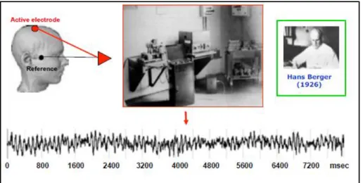
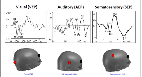
Although EEG (electroencephalography) is an old technique to measure brain neural activity discovered in the 1930s, the use of ERPs (event-related-potentials) has progressed over the last century. In 1929, Hans Berger, was the first to put two electrodes on the surface of the scalp. He looked at the difference between these two electrodes and found that there was a fluctuation of electrical activity across time (ms). He demonstrated that the human brain’s electrical neuronal activity could be measured by placing an electrode on the surface of the scalp, and through the amplification of the signal, he could plot the changes in voltage (microvolts) over time (Berger, 1929) (Fig. 1). This electrical activity measured on the scalp is called an electroencephalogram. However, the EEG is a rough measure of neural brain activity and cannot be used in its raw form to measure specific neural processes (Fig. 2). In other words, the raw data obtained is made up of several different neural sources of activity
(Luck, 2014). Thus, in order to isolate the different cognitive processes embedded within the EEG, the extraction of neural responses associated with sensory, motor and cognitive activity, is required. This is done through averaging techniques that enable to look at the potentials related to a particular event, called event-related-potentials (ERPs). The resulting averaged ERP waveforms consist of a sequence of positive and negative voltage deflections which are called peaks or components, and are labelled P1, N1, P2, N2, P3, N400…indicating positive (P) or negative (N) going peaks. The number next to the P or N indicates the position within the waveform or the latency of the peak in ms (Fig. 3).
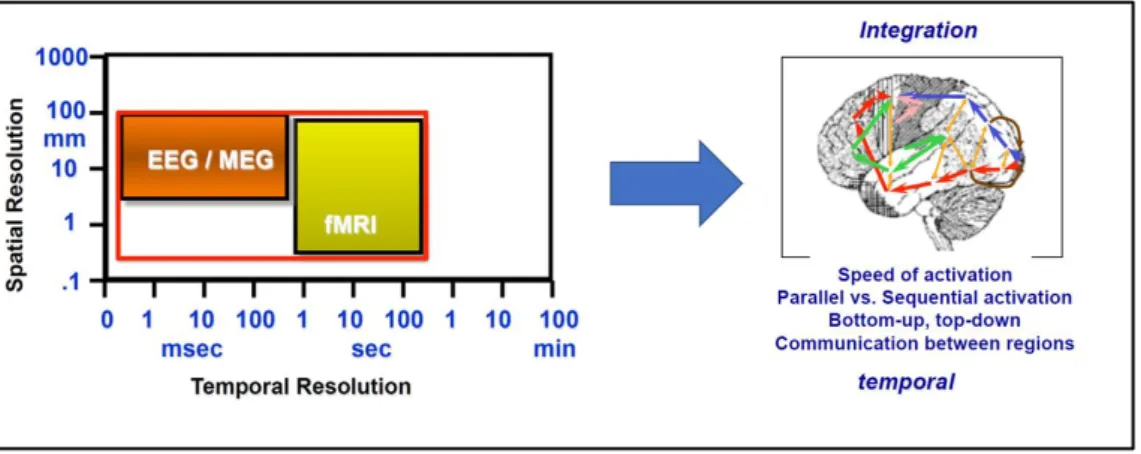
EEG has a very precise temporal resolution (1ms) compared to hemodynamic measures (hundreds of ms), which have a strong spatial resolution in the mm range, but the latter cannot match the former in terms of timing (Fig. 4). Compared to behavioural measures, ERPs provide an online measure of processing when behaviour responses are impossible. This is
5
called covert measurement of processing. Thus, ERPs can be used to create models of distribution of activity over cortical surface.
Figure 1. The discovery of the electroencephalogram (EEG) by Hans Berger in 1929.
Figure 2. Example of EEG raw data
6
Figure 4. Temporal resolution in EEG compared to fMRI (functional magnetic resonance imaging).
2.1.1. PHYSIOLOGICAL BASIS OF ELECTROENCEPHALOGRAPHY
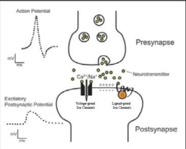
Neuronal activity is mainly characterised by electrical discharges. EEG measures fluctuations in electrical potential on the scalp’s surface resulting from post-synaptic activity when the neurotransmitter binds to the receptor, thus changing the flow of ions across the cell membrane. Neurons in the brain have a resting potential which is characterised by negative potential inside and positive potential outside the cell (Fig. 5). When neurons are activated, actions potentials are coming in through the neuron, neurotransmitter is released and goes to the post-synaptic neuron (Fig. 6). This process will change the channels and make ions going in and out of the extra neuronal space. However, we do not have direct access to the electrical activity, as the distance between the electrode and the neuron is too large (Fig. 7). We have access to the voltage related to the electrical activity of synchronous activity of parallel organised pyramidal cells that together generate a sufficiently strong dipolar field. The electrical activity spreads through the brain and passes the skull to the scalp, where it can be picked up by electrodes. Scalp ERPs are not produced by action potentials. When postsynaptic potentials occur, they create an electrical dipole, thus ERPs can be measured when the dipoles of thousands of similarly oriented neurons sum together forming a vector sum. Because of this spatial arrangement, we can detect the activity of synchronously firing neurons. The
7
voltage recorded on the surface of the scalp will be positive on one side of the dipole and negative on the other side with a line zero voltage separating the positive and negative sides
(Luck, 2014).
As discussed, it is erroneous to attribute any change in potential to the tissue beneath the electrode. Nevertheless, the entire neuronal population contributes to produce changes in the electrical field measured at the surface of the skull, leading to temporally precise measurements of brain activity, while spatial resolution remains quite poor (e.g. Michel, Murray, Lantz, Gonzalez, Spinelli & Peralta, 2004).
Figure 5. The resting potential of neurons
8
Figure 7. Large distance between electrode and neuron: activation of a few neurons is not observable on the surface. Large areas need to be active to be seen on the surface.
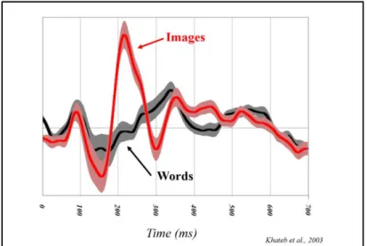
In sum, EEG is recorded from multiple electrodes distributed across the scalp with a conductive gel between each electrode and the skin to make a stable electrical connection. The electrical potential (voltage) is recorded from each electrode resulting in a separate waveform for each electrode site. This waveform is a mixture of actual brain activity, biological electrical potential produced outside the brain (skin, eyes, muscles) and induced activity from external devices that is picked up by the head. Thus, in order to get what is related to the task, we need to extract the evoked response linked to an event/stimulus (averaging technique). The event-related-potentials will enable to isolate the temporal activation of cortical processes associated with cognitive events (Fig. 8).
9
Figure 8. Example of ERPs. Comparison between two conditions (words vs. images)
2.1.2. EARLY ERP COMPONENTS
A distinction is made between early exogenous components and late endogenous components. Early exogenous components vary according to the physical features of a stimulus and are observed early after the presentation of the stimulus. These responses mainly reflect sensory information (vision, audition and touch) (Fig. 9). These exogenous sensory components, although triggered by the presence of a stimulus, may be modulated by top-down processes. The main visual, auditory and somatosensory ERPs are the P1, N1, N170 and P2/N2 components. The visual P1 and N1 are considered the earliest electrical markers of visual processing and are influenced both, by the low-level features of the stimuli (Johannes, Münte, Heinze & Mangun, 1995) and by attentional processes (Luck et al., 1990). The P1 is an occipital component peaking at around 100 ms after stimulus presentation. It is generated in the extrastriate visual area (V1) and reflects early sensory processing for stimuli presented in a location where attention is focused whereas, the N1, peaking between 100 ms and 200 ms, represents the orienting of attention to a task-relevant stimulus (Luck et al., 1990). However, there are several visual N1 subcomponents contributing to the same deflection. The anterior
10
N1 peaks earlier than the two posterior ones (parietal and lateral occipital), but both are influenced by spatial attention (e.g. Hillyard & Anllo-Vento, 1998) as well as task discrimination (e.g. Vogel & Luck, 2000).
The N170 component, peaking between 150 ms and 200 ms after stimulus onset, is a face-sensitive component assumed to be generated in occipito-temporal cortex and posterior fusiform gyrus (e.g. Bötzel, Schulze & Stodieck, 1995). It reflects sensory perceptual stages of face processing for subsequent face recognition (Eimer, 2000). Section 2.2.3 (ERP data from visual perception and early attention) will discuss the main visual components in further details. Section 2.2.4.2 (ERP data from face perception) discusses the main components in face recognition.
The P2 component, peaking between 200 ms and 300 ms, is at the boundary between exogenous and endogenous processes (Luck, 2014). This component is usually larger for target features and enhanced when targets are rare (Luck & Hillyard, 1994). This component indexes attention oriented towards certain features of objects (Hermann & Knight, 2001). Finally, the N2, peaking between 200 ms and 300 ms, is usually observed for deviant stimuli and auditory mismatches at anterior sites (for a review see: Folstein & Van Petten, 2008).
11
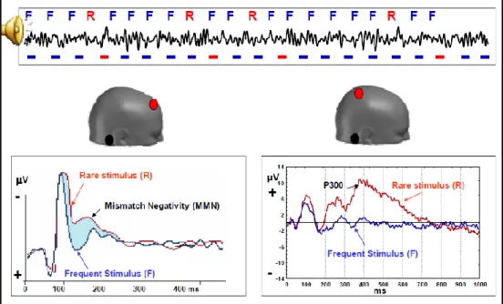
2.1.3. LATE ERP COMPONENTS
Late endogenous components are observed at later stages of conscious processing and can or cannot take place for a particular stimulus depending on the degree of attention allocation of the subject. These responses reflect cognitive treatment (Fig. 10), and are entirely dependent on the task. The main endogenous components are the P3, N400 and the late posterior positivity (LPP). Concerning the P3, there are several components that have been identified in the time range between 300 ms and 450 ms (for a review, see: Polich 2004; 2012). Mainly, the P3, a positive-going deflection on midline sites (Donchi & Coles, 1988) has been associated with stimulus predictability (Picton, 1992), allocation of additional attentional resources and task difficulty (e.g. Isreal, Chesney, Wickens & Donchin, 1980), stimulus categorisation and post categorisation processes (Kutas, McCarthy & Donchin, 1977; Polich & Bondurant, 1997) and more recently, with expectancy towards outcome (Zhou, Yu & Zhou, 2010), feedback valence (Hajcak, Holroyd, Moser & Simons, 2005) and variations in perceptual certainty (Selimbeyoglu, Keskin-Ergen &Demiralp, 2012), as well as metacognitive awareness (Murphy, Robertson, Harty & O’Connell, 2015; Desender, Van Opstal, Hughes & Van den Bussche, 2016) and post-decision accumulation of sensory evidence that leads to a judgement (Murphy et al., 2015; Kelly & O’Connell, 2013; Twomey,
Murphy, Kelly & O’Connell, 2015). Thus, P3 is highly dependent on the experimental manipulation.
Concerning the N400, this component has been associated to situations of semantic violations of meaning (Kutas & Hillyard, 1980). However, recent research in social neurosciences have found an N400 for context violations suggesting that this semantic-related component extends beyond language (e.g. Amaruso, Gelormini, Aboitiz, Gonzalez, Manes, Cardona & Ibanez, 2013).
12
The LPP, a midline ERP starting at around 300 ms after stimulus onset and lasting up to 600 ms, has mainly been linked to cognitive strategies of emotional regulation (e.g. Hajcak, MacNamara & Olvet, 2010). It reflects interactions of bottom-up attentional allocation and top-down sustained elaboration (e.g. Weinberg, Ferri & Hajcak, 2013).
Figure 10. Example of cognitive evoked potentials: the brain can differentiate between what is regular (F; frequent stimulus), what is rare (R) and what is deviant (mismatch negativity).
13
2.2. VISUAL PERCEPTION AND ATTENTION
2.2.1. BRAIN STRUCTURES INVOLVED IN VISUAL PERCEPTION
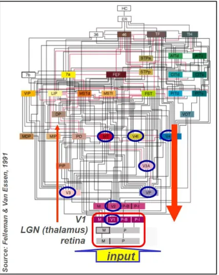
Occipital structures are the beginning of visual processing but visual mechanisms extend beyond the striate cortex. Visual regions are hierarchically organised (Hubel & Wiesel, 1962)
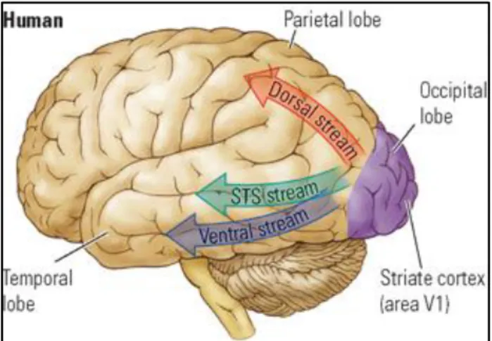
with parallel and interconnecting pathways at each level to account for a progressive increase in complexity (Felleman & Van Essen, 1991) (Fig. 11). The information spreads from the primary visual area V1 (striate cortex) to higher order visual areas. Thus, V1 is the first processing level in the distributed hierarchy. It receives the largest input from the lateral geniculate nucleus of the thalamus and projects to all other occipital areas. V2 (pre-striate cortex) is the second level and it is a mirror image map of V1. Striate and extra-striate areas in humans (V1 and V2) are sensitive to shapes and spatial orientations. After V2, parallel pathways go to the parietal cortex, inferior temporal cortex and superior temporal sulcus. Object recognition as well as colour recognition takes place in the ventral pathway (temporal lobe) whereas visual action takes place in the dorsal stream (parietal lobe) (Mishkin, Ungerleider & Macko, 1983) (Fig. 12). Accordingly, from V1 and V2 areas, the information either goes to V4, where cells respond to colour, or to V3, which is sensitive to the shape of objects in motion, or to V5, which hosts neurons sensitive to segments that move (Kolb & Wishaw, 2009). As we go forward towards anterior regions, neurons respond to more and more complex refined stimuli.
Neuropsychological research has shown that selective lesions in the hierarchy produce specific deficits in visual processing. For example, patients with damage to V4, will see the world in shades of grey and will not be able to recall colours neither to imagine them
(Meadows, 1974; Sacks & Wasserman, 1987), whereas lesions to V5 will produce an inability to perceive objects in movement (Schenk, Ellison, Rice & Milner, 2005).
14
However, visual processing does not end in secondary areas (V3, V4, V5). Several visual regions in temporal and parietal lobes respond to complex stimuli. For instance, within the temporal lobe, the fusiform face area is responsible for face recognition, the extrastriate body area for body analysis, the superior temporal sulcus for biological motion, the lateral occipital area for object analysis and the parahippocampal place area for the analysis of landmarks
(Kolb & Wishaw, 2009). Lesions to specific regions in temporal cortex will result in specific agnosia, this is the incapacity to analyse or recognise objects or familiar faces without any impairment of visual primary system. Nevertheless, I will not go into further details on these neuropsychological visual deficits as it is not relevant to the purpose of this thesis.
Figure 11. Hierarchy of visual regions showing a densely interconnected network hierarchically organised with feedforward and feedback connections. This hierarchy shows 32 visual cortical areas as well as subcortical visual stages (retinal ganglion cell layer and the lateral genicular nucleus) plus
15
several non-visual areas such as the somatosensory cortex (7b), perirhinal area (36), and the hippocampal complex. These areas are connected by 187 linkages, most of which have been demonstrated to be reciprocal pathways.
Figure 12. Visual pathways in the human brain. The parietal cortex (dorsal stream) controls the voluntary orienting of attention towards a location of interest. The ventral stream is involved with object and visual identification and recognition. The STS (superior temporal sulcus) separates the superior temporal gyrus from the middle temporal gyrus and it involved with the identification human biological motion and specific social inputs suggesting its role in social perception.
2.2.2. BRAIN STRUCTURES INVOLVED IN ATTENTION
Bottom-up attention or exogenous attention is captured by the stimuli features, as opposed to top-down attention (endogenous attention), in which visual attention will be oriented on the basis of our expectations, knowledge and objectives. Thus, there is a functional segregation of cortical pathways for bottom-up and top-down attention (Corbetta, Kincade, Ollinger, McAvoy & Shulman, 2000) (Fig. 13).
The ventral pathway responds to unexpected or deviant events. Regions within the ventral stream inhibit parietal regions, whereas, the dorsal pathway identifies the localisation (goal-oriented) and gets activated when attention is reoriented in space, as well as when a specific
16
localisation is selected. Thus, fronto-parietal regions are activated in focalised attention
(Corbetta et al., 2000).
Visual search paradigms have been used to test attention competition in laboratory settings
(e.g. Theeuwes, 1994). In these tasks, subjects are required to search for a target among distractors. When targets are salient in terms of shape and colour, bottom-up activations are generated based on the differences in stimulus features (singleton detection). When targets are similar to distractors, the searching is done in a serial manner involving top-down processes
(Bacon & Egeth, 1994). Thus, the parietal cortex is activated for attention to location and the occipito-temporal cortex is activated for attention to features such as colour and shape
(Treisman & Gelade, 1980). Moreover, several frontal regions, mainly the anterior cingulate, are involved when attentional effort is required (Kolb & Wishaw, 2009). Consequently, sensory processing is affected by early exogenous and late endogenous mechanisms of attention that interact according to task manipulations. Exogenous attention is oriented more rapidly whereas endogenous attention requires more cognitive resources involving a larger attentional network including frontal eye fields, intraparietal sulcus, superior temporal gyrus and anterior cingulate (Hopfinger & West, 2006).
17
Figure 13. Cortical pathways involved in bottom-up and top-down- attention: The dorsal system is bilateral and composed of the intraparietal sulcus (IPS) and the junction of the precentral and superior frontal sulcus (frontal eye field, FEF) in each hemisphere. It is involved in voluntary (top-down) attention. The ventral system is right-lateralised and composed of the right temporal parietal junction (TPJ) and the right ventral frontal cortex (VFC). This system is involved in bottom-up capture attention.
2.2.3. ERP DATA FROM VISUAL PERCEPTION AND EARLY ATTENTION
As mentioned in the previous section, regions activated during attention suggest separable endogenous and exogenous attentional systems implying different stages of processing that electroencephalography can clarify. Thus, we would expect to observe distinct stages of processing preceding conscious attention. Nevertheless, EEG studies have shown that both mechanisms interact (e.g. Desimone & Duncan, 1995; Hopfinger & West, 2006) as endogenous mechanisms modulate activity in neurons coding the spatial location (Bisley & Goldberg, 2003) suggesting a unitary focus of attention with top-down influences on
18
exogenous orientation (Folk, Remington & Johnston, 1992). For example, Hopfinger & Mangun (1998) showed that exogenous attention enhances the visual early P1 component and this same stage of processing can also be enhanced by endogenous attention when converged at the same location. These results suggest that visual ERPs are strongly modulated by attention-related activity. Consequently, top-down neural mechanisms such as directed attention may increase or decrease brain activity related to visual processing affecting the amplitudes of the visual P1 component, whereas, the N1 amplitudes are enhanced only by endogenous attention (Hopfinger & West, 2006). These findings are of great relevance for the upcoming studies of the present thesis and suggest a functional dissociation of P1 and N1 components. Indeed, Luck et al. (1990) showed that the P1 indexed a facilitation of sensory information for stimuli presented to a location where attention was focused, whereas, the N1 represented the orienting of attention to task- relevant stimuli.
Following the anterior N1 wave, two visual/attentional components are observed, the P2 and the N2. The P2 is a positive-going centro-parietal potential, peaking at around 200ms after stimulus onset, and represents some aspects of higher-order perceptual processes modulated by attention (Luck & Hillyard, 1994). This component is larger for targets and is enhanced for infrequent targets (Luck, 2004).Finally, the anterior N2 sensitive to the anterior cingulate, has been associated to conflicting responses. For instance, within a social context, the N2 is enhanced for negative or deviant feedback (e.g. Carretié, Hinojosa, Martin-Loeches, Mercado & Tapia 2004; Daffner, Scinto, Calvo, Faust, Mesulam, West & Holcomb, 2000).
19
2.2.4. EMOTIONAL FACE PERCEPTION
Face perception can be considered as the most developed visual perceptual skill in humans suggesting a special status for face processing (Ishai, 2008). Faces provide additional social information based on the perception of changeable aspects of the face such as the expression and eye gaze (Haxby, Hoffman & Gobbini, 2000). The face perception system encompasses invariant aspects of a face that allow to recognise the identity, and changeable aspects which facilitate social communication (gaze, expression and lip movement). Thus, the changeable aspects of faces underlie the perception of information that facilitates social communication. These two processes proceed in a relatively independent manner and constitute the core system. Thus, the core system is responsible for the visual analysis of faces whereas another system, the extended system, processes the meaning of information in a face such as the emotions (Haxby et al., 2000; Haxby, Hoffman & Gobbini, 2002).Bruce & Young (1986), proposed two parallel routes underlying face expressions and identity processing. These routes work independently allowing the processing of expression without the processing of identity
(Breen, Caine & Colheart, 2000). Neuropsychological data supports this model by showing in some prosopagnosic patients an ability to recognise expressions (Damasio, Damasio & Van Hoesen, 1982), whereas other patients show intact identity recognition but impaired processing of facial expressions. For example, damage to the insula impairs recognition of disgust (Calder, Keane, Manes, Antoun & Young, 2000).
2.2.4.1. BRAIN STRUCTURES INVOLVED IN FACE PERCEPTION
The extraction of face information relies on the activation and interaction of several brain structures. The changing aspects in face perception generate activity in the superior temporal sulcus (STS), whereas the perception of identity activates the lateral fusiform gyrus whose
20
activation is usually bilateral and consistently on the right (Ishai, 2008; Hoffman & Haxby, 2000; Haxby et al., 2000) (see Fig. 14).
For the purpose of the studies completed in this thesis, I will focus on the brain regions activated during facial expressions. The expression on another’s face provides information about the emotional state of that person and can trigger this emotion in oneself (Haxby et al., 2002). Hemodynamic responses show activation of the inferior occipital gyrus, fusiform gyrus, STS and inferior frontal gyrus when seeing facial expressions compared to neutral ones. Moreover, the right STS is more activated when perceiving averted gaze faces compared to direct gaze faces, suggesting distinct neural systems for gaze direction and expressions (Engell & Haxby, 2007). A significant enhanced activity in the fusiform extrastriate areas is associated with the four basic emotions (fear, disgust, happiness and sadness), and these responses are increased for fear and disgust compared to happiness and sadness. The perception of fear also evokes a response in the amygdala as well as in regions involved in social and cognitive responses, such as the STS, cingulate and parietal regions (Vuilleumier & Pourtois, 2007). The perception of disgust in the face of another activates the anterior insula but not the amygdala, and strong disgust also activates regions associated with the limbic cortico-striatal-thalamic circuit (Phillips, Young, Senior, Brammer, Andrews, Calder, Bullmore, Perret, Rowland, Williams, Gray & David, 1997). Another neural system involved in the perception of happy emotions is the orbitofrontal cortex associated with reward and social reinforcement (Haxby et al., 2002).
21
Figure 14. Model of the distributed human neural system for face perception (Haxby et al 2000). Interaction of two systems: the core system enabling visual analysis of faces with activity in superior temporal sulcus for the encoding of changeable aspects and activity in lateral fusiform gyrus for the encoding of identity; the extended system allowing the extraction of complementary features.
2.2.4.2. ERP DATA FROM FACE PERCEPTION
From an electroencephalographic point of view, a wealth of ERP studies of face perception has consistently shown an evoked potential at around 170 ms at posterior sites called the N170. This component is observed after the presentation of human upright faces as well as for inverted faces but not for non-face stimuli such as cars, butterflies or animal faces. Inverted faces show a delay in the N170 response relative to upright faces. This response to faces is larger over right hemisphere leads. The fact that the N170 is present for both, upright and inverted faces suggests that this response is not associated with face recognition, as it is not modulated by familiarity neither by emotional expressions, but only by the structural encoding of visual stimuli that enable to categorise a stimulus as a face (Bentin, Allison, Puce, Perez & McCarthy, 1996). Additionally, when presenting isolated face components, the N170 is also enhanced corroborating the interpretation of this ERP as an early detection of structural features characterising human faces. Moreover, the N170 response is larger for isolated eyes
22
relative to the whole face indicating that the neural mechanisms generating N170 responses are specific to eyes.
When presenting famous and non-famous faces, the N170 is unaffected. Face recognition occurs later, at around 250 ms (N250) over lateral occipito-temporal sites and is only observed for known faces compared to familiar ones (Gosling & Eimer, 2011). Thus, the N170 precedes face recognition and the N250 underlies neural generators in the face specific regions of the fusiform gyrus (Schweinberger, Pickering, Jentzsch, Burton & Kaufmann, 2002) suggesting an activation of stored long-term representations of famous faces. Additionally, famous faces elicit a sustained positivity around 600 ms. This longer latency positivity for famous faces is not linked to familiarity but to explicit face identification (Gosling & Eimer, 2011).
Concerning emotional expressions, ERP results are contradictory. Some authors have shown an enhanced frontocentral P200 for fearful expressions and an enhanced P3 for disgusted face expressions, but no N170 modulations (Ashley, Vuilleumier & Swick, 2004). These findings suggest that structural encoding and expression processing are independent processes (Eimer & Holmes, 2003). In contrast, other research (Batty & Taylor, 2003) have explored the timing of processing of basic emotions in implicit emotional tasks and observed significant effects starting at around 100 ms (P1) as well as differences in amplitude and latency at around 140 ms, indicating that the N170 is sensitive to emotions suggesting an early automatic encoding of emotional faces. The authors interpret these results as a facilitated identification due to the activation of a subcortical pathway when presented with relevant emotions. Moreover, they found later N170 latencies for negative emotions compared to neutral and positive ones which are explained by additional information sent from the subcortical pathway to ventral regions
(Batty & Taylor, 2003; Garvert, Friston, Dolan & Garrido, 2014). It seems, though, that the experimental task as well as the instructions influence the processing of emotional faces. For example, Ratner & Amodio (2013) showed larger N170 amplitudes to ingroup faces compared
23
to outgroup faces, suggesting top-down effects on early perception when subjects are required to focus attention on the social significance of the stimuli compared to passive viewing. This interpretation is corroborated by Holmes et al. (Holmes, Vuilleumier & Eimer (2003) as they found an enhanced N170 for attended faces whereas emotional faces outside the attentional focus did not trigger any emotional expression effects at these early stages of processing. We can conclude that affective valence and arousal work independently (Olofsson, Nordin, Sequeira & Polich, 2008).
24
2.3. METACOGNITION AND UNCERTAINTY
Metacognition refers to the processes by which we monitor and control our cognitive processes (Frith, 2012). It represents the top of the hierarchy of control over cognitive processes. In our everyday life, we are constantly pushed to make decisions involving two or more options. It seems thus important to be able to quickly evaluate the adequacy of a response and monitor our behaviour accordingly. Evaluation and monitoring are two aspects underlying the concept of metacognition (Proust, 2014), defined as cognition about cognition
(Shea, Boldt, Bang, Yeung, Heyes & Frith, 2014), which ranges on a continuum from total uncertainty to complete certainty. At these extremes lies pathological behaviour. Excessive doubt is related to mental disorders such as obsessive-compulsive disorder (OCD), while overconfidence, might be the manifestation of psychosis (Ron, Oren & Dar, 2016). In healthy individuals, such extremes are rarely reached but these reflective processes are nevertheless subject to inter-individual differences (Song, Kanai, Fleming, Weil, Schwarzkopf & Rees, 2011; Rouault, McWilliams, Allen & Fleming, 2018) and are sensitive to the degree of ambiguity of the stimulus sensory information (Selimbeyoglu et al., 2012; Yeung & Summerfield, 2012).
Recently, related metacognitive processes have been explored in signal detection and reaction time tasks (Fleming, Dolan & Frith, 2012) and focalised on error detection resulting in increased reaction times and behavioural measures such as judgment revisions. However, error detection may occur without explicit awareness. Thus, two forms of metacognition exist: an implicit form characterised by rapidity and automatism without awareness and an explicit form characterised by self-monitoring and awareness (Frith, 2012). The latter corresponds to reportable knowledge about our behaviour in decision making and represents the aspect of metacognition we are interested here, as it creates the experience of agency (Moretto, Walsh & Haggard, 2011), enhances social interactions by communicating our thoughts to others and
25
enables the engagement in mentalising, a crucial aspect of theory of mind (Wellman, Cross & Watson, 2001). For example, deficits in metacognitive ability have been observed in schizophrenia. These patients have difficulties in detecting whether they have made an error and present poor neurocognitive function resulting in a lack of social cognitive ability
(Lysaker, Leonhardt, Pijnenborg, van Donkersgoed, de Jong & Dimaggio, 2014), as well as overconfidence and delusional symptoms. By opposition, patients suffering from obsessive compulsive disorder manifest acute doubt as a result of deficient conviction and feeling of knowing (Ron et al., 2016; Szechtman & Woody, 2004). Thus, judgements of uncertainty in humans involve metacognition (Fleming et al., 2012). In healthy adults, perceptual metacognitive efficiency declines with age which supports the link between metacognition and executive function (Palmer, David & Fleming, 2014).
2.3.1. BRAIN STRUCTURES INVOLVED IN METACOGNITION
Aspects of metacognition such as monitoring and control of cognitive processes are closely linked to working memory and executive control (Shimamura, 2000). These processes take place in prefrontal cortex. Indeed, activation in prefrontal medial cortex is associated with subsequent conforming behavioural adjustments (Shestakova, Rieskamp, Tugin, Ossadtchi, Krutitskaya and Klucharev, 2012). Neuropsychological data corroborates these findings as prefrontal lesions disrupt metacognitive judgements about perception (Shimamura, 2000; Del Cul, Dehaene, Reyes, Bravo & Slachevsky, 2009). However, recent research has shown that retrospective and prospective judgments are associated with distinct neural substrates
(Fleming & Dolan, 2012). Retrospective monitoring refers to the ability to evaluate the adequacy of a response and is associated with activity in anterior and dorsolateral prefrontal cortex, whereas prospective judgements, that is, the ability to evaluate the capacity to perform
26
a future cognitive task, is linked to activity in medial prefrontal cortex (Proust, 2013; Fleming & Dolan, 2012). Methods for investigating the neural components of retrospective and prospective metacognition differ (Fleming et al., 2012; Fleming & Dolan, 2012). The former is measured with subjective confidence ratings about the decision whereas the latter is measured with subjective judgements about the ability to learn (see Arbuckle & Cuddy, 1969)
and feelings of knowing (see Hart, 1965), occurring after task completion. Prospective metacognition involves memory processes and future imagery, thus, this aspect of metacognition also implies increased connectivity with medial temporal lobe (Schnyers, Nicholls & Verfaellie, 2005).
The contribution of prefrontal cortex to metacognition is associated with task uncertainty as a consequence of self-generated information processes and attention to internal representations
(Yoshida & Ishii, 2006). Accordingly, the role of uncertainty to optimise decision making is crucial to metacognitive experiences (Fiser, Berkes, Orbán & Lengyel, 2010).
2.3.2. ERP DATA FROM METACOGNITION
Electrophysiologically, most studies have focalised exclusively on the error-related negativity (ERN), a negative ongoing wave appearing after the subjects’ response and peaking at around 100 ms (for a review see: Larson, Clayson & Clawson, 2014). This ERP component originates in the anterior cingulate cortex (e.g. Holroyd & Coles, 2002). The ERN appears in speeded reaction time tasks in which errors are mostly caused by impulsive responding because of the limited response time (Selimbeyoglu et al., 2012). Thus, this ERP is associated with conflict detection but not with regulative control processes such as the allocation of attentional resources allowing dynamic behavioural adjustments. For that, other components, locked to the stimulus and associated with the processing of different stages of attention, such as the P2 and the P3 (Herman & Knight, 2001) need to be examined. As mentioned in the previous
27
section, evidence from neuropsychology shows that patients with damage to the anterior medial prefrontal cortex have impaired self-reflection, suggesting that this process relies strongly on anterior areas (Shimamura, 2000; Fleming et al., 2012). These studies showed that high executive control in terms of attentional resources lead to increased self-reflective processes.
Few studies have explored the temporal dynamics of metacognition indexed by an additional allocation of attentional resources internally oriented, that is, controlled attention leading to increased self-reflective processes. A recent study (Desender et al., 2016) investigated activity from metacognitive processes and showed that recategorising an ambiguous stimulus followed by a disputing social feedback required additional attention that is necessary for improved control over a response. The authors found an increased P3 amplitude and proposed that this component in situations of ambiguity and social conflict could be the neural correlate of metacognitive awareness as explicit awareness goes beyond one’s ability to discriminate between one’s correct and incorrect responses. Indeed, the P3 component has been shown to be increased under high demanding tasks and decreased under less demanding tasks (Kok, 2001; Olofsson & Polich, 2007). Studies investigating the relationship between the P3 and the participants’ level of uncertainty/ certainty induced by task difficulty found that P3 builds up as sensory evidence increases (Kelly & O’Connell, 2013; Murphy et al., 2015; Twomey et al.,
2015). Consequently, we can conclude that increased perceptual difficulty necessitates greater attentional engagement leading to an increased P3 component. In contrast, less demanding tasks would induce immediately a higher feeling of certainty preventing P3 amplitude to increase (Zanesco, Tipura, Clément & Pegna, 2019).
28
2.4. SOCIAL INFLUENCE AND SOCIAL CONFLICT
The attempt to understand and explain how thoughts and behaviours of individuals are influenced by the presence of others started with the study of social influence about 70 years ago. Social life is characterised by conflict and controversy in which individuals try to change the thoughts and behaviours of others. The normative social aspects have regulated the behaviour in human interactions and yielded social pressure creating conformity at a public level, yet not always at a private level. Thus, in the recent years, researchers have tried to explore social influence processes that are indirect and nonconscious (for a review see: Cialdini & Goldstein, 2004). This work has been done in laboratory using neuroscientific methods, such as fMRI and electroencephalography.
2.4.1. BRAIN STRUCTURES INVOLVED IN SOCIAL CONFLICT
Over the course of the last decade, a large number of studies in social neurosciences have examined the neurocognitive correlates of social influence (for reviews see: Izuma, 2013). These studies have used functional magnetic resonance imaging (fMRI) and have focused on the brain networks implicated in social conformity- the act of changing one’s behaviour to match the responses of others (Cialdini and Goldstein, 2004) - as well as in the subsequent changes in behaviour. Disagreement with group opinion is associated with activity in posterior medial frontal cortex (pMFC), specifically, the rostral cingulate and the anterior insula as well as deactivation of ventral striatum (Klucharev, Hytönen, Rijpkema, Smidts and Fernández, 2009; Berns, Capra, Moore and Noussair, 2009). Individuals with a stronger tendency towards conformity show higher activation of pMFC and insula (Berns et al., 2009). Agreement with group opinion induces activity in ventral frontostriatal circuity implicated in the anticipation of reward (Wu & Zhi, 2016; Galvan, Hare, Davidson, Spicer, Glover and Casey, 2005), particularly the nucleus accumbens (Knutson and Cooper,2005). This activity in the striatum
29
is selectively enhanced when participants conform to the ingroup as compared to the outgroup
(Stallen, Smidts and Stanfey, 2013).
It is thought that pMFC activation reflects a prediction-error signal implicated in reinforcement learning (Klucharev et al., 2009; Shestakova et al., 2012; Kim, Liss, Rao, Singer and Compton, 2012; Campbell-Meiklejohn, Bach, Roepstorff, Dolan and Frith, 2010), which subjects try to decrease by adjusting their subsequent behaviour (Klucharev et al., 2009; Mars, Coles, Grol, Holroyd, Nieuwenhuis, Hulstijn and Toni, 2005; Holroyd and Coles, 2002; Ridderinkhof, Ullsperger, Crone and Nieuwenhuis, 2004).
An important point should be emphasised which is that, these studies have focused on the regulatory processes required when individuals are presented with incongruent social feedback. Such adaptive cognitive control suggests the implication of higher cortical areas associated with executive functions. However, to investigate whether a group opinion modulates not only self-reported preference but also its neural representation (Izuma, 2013), one needs to distinguish between private acceptance (i.e. genuine change in one’s attitude to match the group) and public compliance (i.e. expressive form of conformity) (Cialdini and Goldstein, 2004). Changes in activation in the striatum and ventromedial prefrontal cortex (VMPFC) have been associated with changes in self-preference, reflecting private acceptance
(Zaki, Schirmer and Mitchell, 2011)
.
These regions track subjects’ preferences for variousstimuli (Klucharev et al., 2009; Campbell-Meiklejohn et al., 2010; Zaki et al., 2011; Izuma, Matsumoto, Murayama, Samejima, Sadato and Matsumoto, 2010). They seem to encode the subjective value associated with the processing of reward, thus constituting a valuation system
(Bartra, McGuire and Kable, 2013).
One way by which neurosciences research may contribute to elucidate whether conformity occurs at an explicit level or whether it influences individuals’ actual perception is by
30
investigating the effect of group opinion on the presentation of visual stimuli. Few studies have examined the impact of conformity at low-level perceptual and attentional processes as opposed to a decision taken at an executive level (Trautmann-Lengsfeld and Herrmann, 2013; Berns, Chappelow, Zink, Pagnoni, Martin-Skurski and Richards, 2005; Stapel and Koomen, 1997). Asch (1951) already raised the possibility that social pressure could alter perception
(Asch, 1951). Berns et al. (2005), using fMRI, provided the first evidence for alterations in perceptual processes (i.e. in occipito-parietal networks) when subjects were confronted with incorrect peer feedback regarding the degree of rotation of an abstract figure. However, the limited temporal resolution of hemodynamic measures does not allow to determine if this posterior activation is associated with early or late processes. Yet the temporal characteristics would facilitate the understanding of this activity by indicating whether this activity arises rapidly, or long after higher cortical areas.
2.4.2. ERP DATA FROM SOCIAL CONFLICT
In line with this idea of conflict monitoring and error processing, recent electroencephalography (EEG) studies (Shestakova et al., 2012; Chen, Wu, Tong, Guan and Zhou, 2012) showed that the activity in pMFC, sensitive to prediction error, generates a negative deflection, called feedback-related negativity (FRN) or medial frontal negativity (MFN), which peaks between 200 ms and 400 ms at frontocentral sites after the presentation of the social cue (Shestakova et al., 2012; Chen et al., 2012; Long, Jiang and Zhou, 2012). The FRN appears after negative feedback and is similar to the response-locked error-related negativity (ERN) which typically appears in speeded reaction time tasks following response errors (Walsh and Anderson, 2012). Dipole source models indicate that the topography of the
31
ERN and FRN components is consistent with activity in the anterior cingulate (Dehaene, Posner and Tucker, 1994; Gruendler, Ullsperger and Huster, 2011).
Substantial research has addressed how face processing can be biased by social factors. Results show that P1 and the face - sensitive N170 ERP components are maximum when the combination of co-emitted social cues clearly represent a threat for the participant (i.e. anger associated with direct gaze) suggesting a sensory system specifically optimised for biologically relevant stimuli (El Zein, Gamond, Conty and Grèzes, 2015). Similarly, group membership cues may change the way we see a face (Ratner & Amodio, 2013). Studies assessing the influence of social group membership (i.e. ingroup/outgroup) on early stages of face processing show a larger N170 to ingroup faces than to outgroup faces (Ratner & Amodio, 2013; Ibáñez, Gleichgerrcht, Hurtado, González, Haye and Manes, 2010). The differential and early effects of social cues and group membership on early ERP components suggest that relevant social information is analysed rapidly and effectively by the brain.
However, only two ERP studies have investigated the influence of social context on early perceptual processes for non-face stimuli (Trautmann-Lengsfeld & Herrmann, 2013; Zanesco, Tipura, Posadas, Clément & Pegna, 2018). The findings showed that social influence can have a top-down effect on these early perceptual processes. In Trautmann-Lengsfeld & Herrmann’s (2013) study, participants showed a larger P1 amplitude when they conformed to the correct group opinion compared to situations where they conformed to the incorrect one. The authors did not find any significant differences in P1 amplitude when participants adapted to the incorrect opinion compared to conditions where they did not. The difference became evident at 150 ms, with the analysis of the N1 component, which reflects the orientation of attention towards task relevant stimuli (Luck et al., 1990). In our study (Zanesco et al., 2018), ERPs were measured to ambiguous and distinct stimuli before and after social feedback. Results showed an increase in P1 amplitude for ambiguous stimuli following a disputed feedback with
32
no effect on distinct stimuli, corroborating the fact that social influence is most effective under situations of uncertainty induced by task difficulty (Cialdini & Goldstein, 2004).
2.5. SOCIAL ANXIETY DISORDER
According to the categorical approach from the DSM-V (APA, 2013), social anxiety disorder is characterised by a constant and intense fear in social situation when the person is observed by others, a fear that others will notice their anxiety signs and an avoidance of social situations. These aspects persist more than six months and affect their daily functioning.
The DSM criteria represents social anxious responses to social situations but do not take into account the causes of the disorder. In order to understand the underlying mechanisms of social anxiety, several cognitive models have been proposed based on behavioural and neural responses during the performance of experimental tasks.
2.5.1. COGNITIVE MECHANISMS INVOLVED IN SOCIAL ANXIETY
Attentional bias towards threat is a key phenomenon observed in all experimental tasks and across all anxiety disorders (Bar-Haim, Lamy, Pergamin, Bakermans-Kranenburg & van Ijzendoorn, 2007; MacLeod, Mathews & Tata, 1986; Mogg & Bradley, 1998; Williams, Mathews & MacLeod, 1996). An attentional bias towards threat refers to a strong selective and automatic attentional allocation to threatening social stimuli such as human face emotions. Different components of attentional bias can be observed in experimental tasks such as facilitated and faster detection of threatening social stimuli, difficulty to disengage attention from threatening social stimuli and a tendency to avoid the threatening stimuli by allocating attention towards an opposite location (for a review see: Cisler & Koster, 2010). Most ERP
33
studies in social anxious disorder (SAD) suggest that this early attentional bias is reflected by an enhanced P1 to socially threatening stimuli (e.g. Peschard, Philippot, Joassin & Rossignol, 2013; Rossignol, Philippot, Bissot, Rigoulot & Campanella., 2012; Rossignol, Campanella, Bissot, Philippot, 2013; Mueller, Hofmann, Santesso, Meuret, Bitran & Pizzagalli., 2009; Morel, George, Foucher, Chammat & Dubal, 2014). These studies have focalised on the response to emotional faces but haven’t taken into account the anticipatory processing, which seems to play a role in SAD (Clark & Wells, 1995; Deiters, Stevens, Hermann & Gerlach, 2013; Sluis, Boschen, Neumann & Murphy, 2017).
The neurocognitive mechanisms underlying these attentional biases components include a rapid, automatic and unconscious fear pathway mediated by the amygdala (Ohman, 2005; LeDoux, 2000) which enhances selective attention towards threatening social stimuli. At a higher order strategic level, the attentional control theory (ACT; Eysenck, Derakshan, Santos & Calvo, 2007), postulates three executive components that seem to mediate the ability to disengage from threatening stimuli (Berggren & Derakshan, 2013). The first, inhibition, defined as the ability to suppress task-irrelevant information, whose impairment can be observed by an increased antisaccade latency in socially anxious individuals leading to a reduction in top-down control in inhibiting reflexive processes (Derakshan & Eysenck, 2009). Additionally, socially anxious individuals show an increased attentional capture by irrelevant stimuli (Poy, Eixarch & Avila, 2004) and an increased interference for task-irrelevant distractors (Pacheco-Unguetti, Acosta, Callejas & Lupiañez, 2010). The second executive aspect is shifting, this is, a reduced capacity to shift attention in a flexible manner between relevant task demands. In this sense, socially anxious individuals are slower in task requiring to switch (e.g. Wisconsin Card Sorting Task; Goodwin & Sher, 1992), thus showing a lower performance than non-anxious individuals. Finally, impaired updating is also observed in socially anxious subjects, which is the inability to update and monitor representation in