Human 100-kDa homologous DNA-pairing protein is the splicing factor PSF and promotes DNA strand invasion
9
0
0
Texte intégral
(2) Nucleic Acids Research, 2000, Vol. 28, No. 16 3023. of hPOMp75/TLS are still unclear, its ability to promote HP including D-loop formation suggests a role in some aspects of recombination/repair. This hypothesis is further supported by recent findings that Tls–/– mice exhibit severe defects in chromosome pairing during meiosis, increased sensitivity to ionising radiation and high levels of chromosomal instability (35,36). Finally, both hPOMp75/TLS and hPOMp100 activities are associated with cell proliferation (33). In the present report, we describe the purification and identification of hPOMp100 as the human splicing factor PSF (37). It binds both single-stranded (ss) and double-stranded (ds) DNA and facilitates the renaturation of complementary ssDNA molecules. Importantly, hPOMp100/PSF promotes the formation of D-loops in superhelical duplex DNA. hPOMp100/PSF serves as an efficient substrate for protein kinase C (PKC) in vitro. PKC phosphorylation of hPOMp100/PSF stimulates its DNA binding and D-loop formation activity suggesting a possible regulatory mechanism. MATERIALS AND METHODS Reagents Nitrocellulose membranes Hybond C-super (0.45 µm) were purchased from Amersham Pharmacia Biotech (Dübendorf, Switzerland). ATP, ADP, ATPγS, dNTPs, the complete protease inhibitors mixture, proteinase K, DNA restriction and modification enzymes and E.coli RecA protein were purchased from Roche Molecular Biochemicals (Rotkreuz, Switzerland), SeeBlue pre-stained protein standards from Novex (San Diego, CA), Ni-NTA Superflow resin from Qiagen AG (Basel, Switzerland) and BSA from New England Biolabs (Beverly, MA). DNA substrates M13mp18 RFI DNA, M13mp19 RFI DNA and plasmid pUC19 were from Amersham Pharmacia Biotech. and 24mer M13/pUC sequencing primer from New England Biolabs. The plasmids pUC-Sγ and psv2neo have been described previously (28,34). Duplex DNA was labelled with 32P by filling in the 5′ protruding ends using the Klenow fragment of polymerase I. Synthetic oligonucleotides 19mer 5′-GTAAAACGACGGCCAGTGA-3′; 24mer M13/pUC sequencing primer 5′-CGCCAGGGTTTTCCCAGTCACGAC-3′; two complementary 49mer oligonucleotides 5′-TCACACTCCCACCTTCCCCAGCTCCCCCACAGCTGCTAGGACTGGTCCC-3′ and 5′-GGGACCAGTCCTAGCAGCTGTGGGGGAGCTGGGGAAGGTGGGAGTGTGA-3′; 60mer 5′-AGTGAATTCGAGCTCGGTACCCGGGGATCCTCTAGAGTCGACCTGCAGGCATGCAAGCTT-3′ were 5′-end-labelled with [γ-32P]ATP (Amersham Pharmacia Biotech) and T4 polynucleotide kinase. The annealing of two complementary 49mer Sγ-oligonucleotides was performed in 10 mM Tris–HCl (pH 7.5), 1 mM EDTA, 0.2 M NaCl, 10 mM MgCl2. The oligonucleotide mixture was heated for 10 min at 95°C and further incubated for 60 min at 56°C. All DNA concentrations are expressed in moles of nucleotides. Purification of hPOMp100 and peptide microsequencing All purification procedures were performed at 4°C. Nuclear extract (Fraction I) from 5–10 × 1010 HeLa cells was dialysed extensively against TED buffer (20 mM Tris–HCl, pH 7.5,. 0.1 mM EDTA, 1 mM DTT, 10% glycerol) with 50 mM NaCl and the complete protease inhibitor cocktail as described (34). The precipitate formed during dialysis was collected by centrifugation at 100 000 g for 2 h and the pellet was extracted with TEDU buffer (TED buffer + 6 M urea) + 100 mM NaCl (Fraction II). Fraction II was loaded onto a HiTrap Q column (2 × 5 ml; Amersham Pharmacia Biotech) equilibrated with TEDU + 100 mM NaCl. The flow-through (Fraction III) was loaded directly onto a HiTrap SP column (5 ml; Amersham Pharmacia Biotech), which was developed with a 60 ml gradient of 100–600 mM NaCl in TEDU buffer. The fractions containing the peak of hPOMp100 activity eluted at ∼300 mM NaCl were pooled (Fraction IV), 2-fold diluted with TEDU buffer to lower the ionic strength and loaded onto a HiTrap heparin column (1 ml; Amersham Pharmacia Biotech). The hPOMp100 activity was eluted with a 12 ml gradient of 200– 600 mM NaCl in TED buffer. The active fractions eluted at about 350 mM NaCl were pooled (Fraction V) and applied directly onto a Bio-Scale CHT-I hydroxyalpatite column (2 ml; BioRad) equilibrated in TED buffer + 350 mM NaCl. Elution was performed with a 25 ml linear gradient of 50–500 mM sodium phosphate in TED. hPOMp100 eluted from the column at ∼300 mM sodium phosphate, the pool of which (Fraction VI) was concentrated in a Centricon-30 microconcentrator (Amicon, Wallisellen, Switzerland) and stored in small aliquots at –70°C. Protein concentrations were determined using the Bio-Rad protein assay kit with BSA as a standard. For microsequencing, purified hPOMp100 (Fraction VI) was resolved on a SDS–10% PAGE and stained with Amido Black. The 100-kDa protein was cut from the gel and was subjected for digestion with endopeptidase and internal peptide sequencing. The obtained peptide sequences were compared with the EBI and SwissProt data banks using the computer programs FastA and Blast. Purification of recombinant PSF His6-tagged recombinant PSF (rPSF) was expressed in E.coli as described (37). rPSF was purified under denaturing conditions in the presence of 6 M urea using a Ni–NTA resin (Qiagen) followed by heparin affinity chromatography under native conditions. POM assay The POM assay was performed essentially as described previously (32). Briefly, the assay measures the ability of nuclear proteins separated by SDS–PAGE and transferred onto a nitrocellulose membrane to pair labelled linear dsDNA with homologous ssDNA immobilised on the membrane. Autoradiography revealed homologous pairing activity as a radioactive band, corresponding to the migration of the proteins displaying this activity. Electrophoretic mobility shift assay (EMSA) EMSAs were performed in 10 µl reaction mixtures containing 64 nM (6000 c.p.m.) probe DNA in 20 mM HEPES (pH 7.5), 1 mM DTT, 100 µg/ml BSA and protein as indicated. 5′-32Plabelled oligonucleotides were used as DNA probe as specified in the figure legends. In competition experiments, competitor DNAs were added 5 min before the probe. After incubation for 20 min at room temperature, protein–DNA complexes were resolved by electrophoresis at 4°C in a non-denaturing 6%.
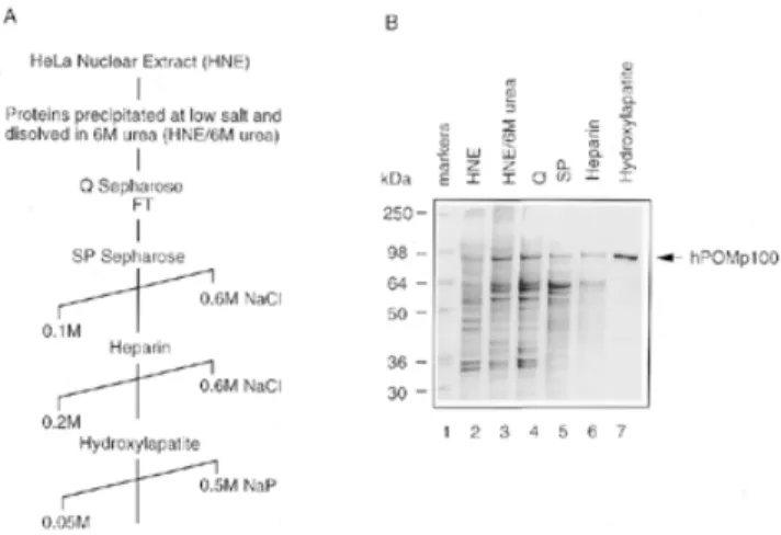
(3) 3024 Nucleic Acids Research, 2000, Vol. 28, No. 16 polyacrylamide gel in 0.5× TBE. 32P-labelled DNA was detected by autoradiography and quantified using a Molecular Dynamics PhosphorImager. DNA renaturation assays DNA renaturation was monitored by the S1 nuclease protection assay (28,38). Reaction mixtures (40 µl) contained 20 mM Tris–HCl (pH 7.5), 10 mM MgCl2, 1 mM DTT, 1 mM ATP, 2.1 µM heat-denatured HindIII-linearised [3H]pSV2neo DNA (2.5 × 10⁵ c.p.m./nmol) and various amounts of protein. After incubation at 37°C for 30 min, reactions were stopped by the addition of 4 µl 10% SDS. The mixtures were then diluted into 550 µl of S1 digestion buffer containing 150 mM NaCl, 50 mM sodium acetate (pH 4.6), 1 mM ZnCl2; then 1 µl of heat-denatured calf thymus DNA (5 mg/ml) and 70 U S1 nuclease were added. Digestions were performed at 37°C for 30 min and the reactions were terminated by the addition of 4 µl calf thymus DNA (5 mg/ml) and 600 µl of 10% trichloroacetic acid. Acid-precipitable radioactivity was measured by liquid scintillation counting. Complementary DNA strands used in the second DNA renaturation assay were obtained by heat denaturation of a 3′32P-labelled 422-bp DNA fragment, produced by AvaI digestion of M13mp18 RFI DNA (28). Reaction mixtures (10 µl) contained 20 mM Tris–HCl (pH 7.5), 10 mM MgCl2, 1 mM DTT, 1 mM ATP, 0.13 µM heat-denatured 32P-labelled 422-bp DNA fragment and various amounts of protein. After incubation at 37°C for 30 min, reactions were stopped by the addition of 0.1 vol 10% SDS and 100 mM EDTA and analysed by SDS–7.5% PAGE. 32P-labelled DNA was detected by autoradiography and quantified using a Molecular Dynamics PhosphorImager. D-loop assay In standard reactions, various amounts of protein were preincubated at room temperature for 5 min with 0.4–0.5 µM 32Plabelled 49mer in a reaction mixture (10 µl) containing 20 mM Tris–HCl (pH 7.5), 1 mM DTT, 5 mM MgCl2, 1 mM ATP or ATPγS and 100 µg/ml BSA. After preincubation, superhelical pUC-Sγ DNA was added at 30–60 µM and reactions were incubated at 37°C for 30 min, unless otherwise stated. At various times, reactions were stopped and deproteinised at 37°C for 15 min in 0.5% SDS and 2 mg/ml proteinase K. The products were analysed on 0.8% agarose gels in TAE buffer containing 5 mM MgCl2 run at 2 V/cm for 16 h at 4°C, and visualised by ethidium bromide staining. 32P-labelled DNA was detected by autoradiography and quantified using a Molecular Dynamics PhosphorImager. In vitro phosphorylation of PSF by PKC Purified rPSF was phosphorylated by PKC in the presence of Ca2+, phosphatidylserine and diacylglycerol using an in vitro PKC assay kit (Upstate Biotechnology Inc., Lake Placid, NY) and according to the manufacturer’s protocol. Controls for these reactions included all components except PKC. Phosphorylation reaction mixtures were incubated at room temperature for 15 min.. Figure 1. Purification of hPOMp100 from HeLa cell nuclei. (A) A purification scheme. (B) Coomassie-stained SDS–10% polyacrylamide gel with peak fractions from each step in (A). Lane 1, SeeBlue prestained molecular mass standards; lane 2, HNE (Fraction I); lane 3, nuclear proteins precipitated at low salt and resuspended with 6 M urea (Fraction II); lanes 4–7, fractions eluted from HiTrap Q (Fraction III), HiTrap SP (Fraction IV), HiTrap heparin (Fraction V) and Bio-Scale hydroxylapatite (Fraction VI), respectively; lane 7 contains 0.5 µg of purified hPOMp100 (Fraction VI).. RESULTS Isolation and identification of hPOMp100 as the human polypyrimidine tract-binding protein-associated splicing factor (PSF) We have previously described a 100-kDa protein, designated hPOMp100, as one of the major DNA pairing proteins in different mammalian cell lines (32). Using the POM assay as a functional test, we purified hPOMp100 from HeLa nuclear extracts (HNE) to near homogeneity (Fig. 1). In all purification steps, the hPOMp100 activity co-purified with a 100-kDa protein. Purified hPOMp100 was devoid of ssDNA or dsDNA exo- and endonuclease, topoisomerase I and II and ATPase, either in the presence or absence of DNA (data not shown). Moreover, the purified protein did not contain any hRAD51 protein or hPOMp75/TLS as revealed by immunoblotting of the preparation with anti-RAD51 antibodies (Oncogene, Cambridge, MA) or with anti-TLS monoclonal antibody (39), respectively (data not shown). Purified hPOMp100 (Fraction VI) was resolved by SDS– PAGE and submitted for microsequencing. Seven internal peptide sequences were obtained (Fig. 2A) and found to be identical to that of the human PSF (37). To ensure that isolated hPOMp100 was PSF, His6–rPSF was expressed in E.coli, purified using Ni–NTA and HiTrap heparin affinity chromatography and assayed by the POM assay. Both proteins, hPOMp100 purified from HeLa nuclei and bacterially expressed rPSF, exhibited similar electrophoretic mobility and DNA-pairing activity (Fig. 2B and C). Purified rPSF was used in the studies below and is designated throughout as hPOMp100/PSF. hPOMp100/PSF binds to ssDNA and dsDNA HP activity of hPOMp100/PSF implies the ability to interact with DNA; however, its binding to DNA has never been.
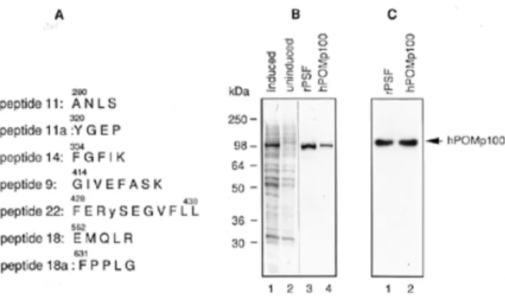
(4) Nucleic Acids Research, 2000, Vol. 28, No. 16 3025. Figure 2. Identification of hPOMp100 as the human splicing factor PSF. (A) Peptide sequences of hPOMp100. Fifty pmol of the gel-purified hPOMp100 was microsequenced and found to be the hPSF. Amino acid residues in one-letter code are numbered according to the published hPSF sequence (37). Cys431 in the reported hPSF sequence was miscalled as a Tyr (indicated in lower case) in peptide 22. It is rather unlikely that the original hPSF DNA sequencing was in error. (B) Purification of recombinant PSF (rPSF) E.coli cellular proteins after and before induction with IPTG (lanes 1 and 2, respectively); rPSF purified from E.coli and hPOMp100 purified from HeLa nuclei (Fraction VI) (lanes 3 and 4, respectively). Samples were analysed by SDS–PAGE and Coomassie staining. (C) rPSF displays the POM activity. Bacterially expressed and purified rPSF and hPOMp100 purified from HeLa nuclei (Fraction VI) were analysed by the POM assay (lanes 1 and 2, respectively). Positions of SeeBlue prestained markers and hPOMp100/PSF are indicated.. studied. Therefore, we have used EMSA to investigate its DNA-binding properties. Upon incubation of increasing amounts of hPOMp100/PSF with the 32P-labelled 49mer oligonucleotide, two protein–DNA complexes that exhibited retarded mobility were detected (Fig. 3A). At concentrations up to 10 nM hPOMp100/PSF, there was a single predominant band with retarded mobility. As the concentration of the protein increased, a second, more slowly migrating band appeared, suggesting that more than one protein molecule may. bind to one oligonucleotide molecule under these conditions. At higher concentration of hPOMp100/PSF (>10 nM) large protein–DNA aggregates which did not migrate into the gel were also observed (lanes 5–7). To compare hPOMp100/PSF affinity for ssDNA and dsDNA, incubation of the protein with the labelled 49-bp duplex was also performed. Binding to this substrate was less efficient than to ss 49mer and a more slowly migrating protein–DNA complex was also noted at higher (40 nM) protein concentrations (Figure 3A, lanes 8–13). In competition experiments, the binding reactions with the labelled 49mer oligonucleotide were carried out in the presence of increasing amounts of unlabelled competitor φX174 dsDNA or ssDNA. In agreement with results shown in Figure 3A, ssDNA competed more efficiently with the labelled oligonucleotide for hPOMp100/PSF binding than did dsDNA. A 50-fold molar excess of ssDNA completely abolished binding while at least a 100-fold molar excess of duplex competitor was needed to achieve a similar effect (Fig. 3B, compare lanes 5 and 6 with 8 and 9). The addition of up to 5 mM Mg2+ or 1mM ATP to the reaction had little effect on ssDNA binding (data not shown). Dependence of DNA binding on length of ssDNA was also examined using 19, 24, 49 and 60mer synthetic oligonucleotides (Fig. 3C). The efficiency of binding increased with an increase of DNA length; the 19mer oligonucleotide was very poorly bound by hPOMp100/PSF, and only one protein–DNA complex was detected. Thus far, no sequence specificity was found for hPOMp100/PSF. hPOMp100/PSF promotes DNA renaturation The simplest HP reaction is DNA renaturation, the conversion of two complementary ssDNA molecules into a duplex form. To test if hPOMp100/PSF can promote this reaction, we used the S1 nuclease digestion and agarose gel electrophoresis assays. In the S1 nuclease digestion assay, heat-denatured,. Figure 3. DNA-binding properties of hPOMp100/PSF. (A) Varying concentrations of hPOMp100/PSF were incubated with either 64 nM 32P-labelled 49mer oligonucleotide (lanes 1–7) or 64 nM 32P-labelled 49-bp ds-oligonucleotide (lanes 8–13). Concentrations of hPOMp100 were 0.1, 0.2, 1, 2, 10 and 20 nM (lanes 2–7) and 2, 5, 10, 20 and 40 nM (lanes 9–13). (B) 2 (lane 2) or 10 nM (lanes 3–9) hPOMp100/PSF were incubated with 64 nM 32P-labelled 49mer oligonucleotide in the absence (lanes 2 and 3) or in the presence of 25-, 50- or 100-fold molar excess of unlabelled ds- (lanes 4–6) or in the presence of 10-, 25- or 50-fold molar excess of unlabelled ssφX174 DNA (lanes 7–9). (C) 10 (lanes 2, 6, 10 and 14), 20 (lanes 3, 7, 11 and 15) or 40 nM (lanes 4, 8, 12 and 16) hPOMp100/PSF were incubated with 60 nM 32P-labelled 19 (lanes 1–4), 24 (lanes 5–8), 49 (lanes 9–12) or 60mer (lanes 13–16) oligonucleotides. The left part of the gel (lanes 1–8) was exposed 3 times longer than the right part (lanes 9–16) in order to detect a comparable amount of protein–DNA complexes. Protein–DNA complexes were analysed by native 6% PAGE. no, no protein was added..
(5) 3026 Nucleic Acids Research, 2000, Vol. 28, No. 16. 422 nt DNA strands in a concentration dependent manner with an efficiency comparable to that of RecA. After ∼20 min of incubation at 37°C, the reaction reached a plateau corresponding to 62% of ssDNA being converted into duplex form (Fig. 4C). Under our conditions, optimal activity was observed at one hPOMp100/PSF molecule per 50–60 nt ssDNA. As in the S1 nuclease digestion assay, the reaction required Mg2+ but not ATP (Fig. 4B, lanes 10 and 11, and data not shown). hPOMp100/PSF mediates D-loop formation. Figure 4. DNA-renaturation activity of hPOMp100/PSF. (A) The S1 nuclease protection assay. The indicated amounts of hPOMp100/PSF were incubated with 1 µM heat-denatured HindIII-linearised pSV2neo [3H]DNA. The activity is expressed as percent of total DNA that is resistant to degradation with S1 nuclease. (B) The effect of hPOMp100/PSF concentration on DNA renaturation determined by the gel assay. The indicated amounts of RecA (lanes 2 and 3) or hPOMp100/PSF (lanes 5–9) were incubated with 130 nM heat-denatured 32P-labelled 422-bp DNA fragment. 2 nM hPOMp100/PSF was added in the reactions of lanes 10 and 11 and ATP or MgCl2 were omitted from the reactions in lanes 10 and 11, respectively. (C) Kinetics of DNA renaturation promoted by hPOMp100/PSF. 130 nM heat-denatured 32P-labelled 422-bp DNA fragment and 2 nM hPOMp100/PSF were incubated at 37°C for the indicated times. The products were analysed by SDS–PAGE. native, undenatured dsDNA was loaded; –hPOMp100, no protein was added, the reaction was incubated at 37°C for 30 min.. HindIII-linearised [3H]pSV2neo DNA was incubated with hPOMp100/PSF at 37°C for 30 min and the formation of dsDNA was monitored by determining the proportion of the DNA that was converted to an S1 nuclease resistant form. The addition of increasing amounts of hPOMp100/PSF to the reactions resulted in increased formation of dsDNA; up to 73% of the DNA became S1 nuclease resistant at roughly one protein molecule per 50 nt of ssDNA substrate after 15 min incubation at 37°C (Fig. 4A). DNA-reannealing activity of hPOMp100/PSF was confirmed by a second independent assay which utilised shorter (422 nt) ssDNA substrates and visualised formed products by gel electrophoresis followed by autoradiography. As shown in Figure 4B, hPOMp100/PSF promoted renaturation of. The results presented above demonstrated that hPOMp100/ PSF can efficiently promote renaturation of ssDNAs. To test whether the protein can mediate a more specific recombination reaction, the invasion of a duplex DNA by a ssDNA (D-loop formation), hPOMp100 was incubated with the 32P-labelled 49mer oligonucleotide and the homologous superhelical dsDNA. D-loops were detected as the comigration of radioactivity with duplex DNA. As shown in Figure 5, hPOMp100/ PSF promoted D-loop formation between homologous ssDNA and dsDNA (lane 2–6), but not between heterologous DNA substrates (lane 9). Since at elevated temperatures D-loop formation can proceed spontaneously without participation of any proteins (40), all our D-loop assays included control reactions which were incubated for 30 min at 37°C. Under our reaction conditions, the yield of D-loops formed spontaneously never exceeded 0.6% (lane 1). As a positive control, we examined the D-loop formation from the same substrates catalysed by RecA protein (lanes 17 and 18). Two major DNA product bands were observed; the more slowly migrating DNA species that comigrated with relaxed dsDNA might represent less efficient assimilation of ssDNA into relaxed dsDNA present in the superhelical DNA preparation. The yield of D-loops formed by hPOMp100/PSF depended on the protein concentration and duration of incubation (Fig. 5). It increased up to 20% at 10 nM of POMp100/PSF after 40 min of incubation at 37°C. Under our conditions, optimal activity was observed at one protein molecule per 20–25 nt ssDNA. hPOMp100/PSF was also able to catalyse the formation of D-loops with 24mer oligonucleotide and homologous superhelical pUC19 or M13mp19 DNAs, implying that the reaction is not specific for a particular DNA sequence context (data not shown). In contrast to the RecA-like-promoted reaction (14,15,41,42), the addition or omission of ATP or ATPγS to the hPOMp100/ PSF-promoted reaction had no effects on D-loop formation (Fig. 5, compare lane 5 with 7 and 8). The reaction required Mg2+; optimal activity occurred at 1–2 mM Mg2+ (Fig. 6, lanes 1–8 and lower panel). Mg2+ can be replaced by Mn2+ but not Ca2+ (lanes 13–16). When the protein was loaded onto ssDNA at 1 mM Mg2+, followed by shift up to 10 mM Mg2+ (the conditions optimal for the RecA-promoted reaction), the efficiency of D-loop formation was not changed (data not shown). D-loop formation was optimal at 0–25 mM NaCl and decreased at higher salt concentrations (Fig. 6, lanes 9–12 and lower panel). We did not detect any significant differences whether hPOMp100/PSF was preincubated with ssDNA followed by the addition of dsDNA (the conditions required for the hRAD51-promoted HP; 43), or the protein was added to a mixture of ss- and dsDNA (data not shown). We also studied the effect of varying dsDNA concentration as a function of hPOMp100/PSF concentration on the formation of D-loops (Fig. 7). At a constant protein/ssDNA.
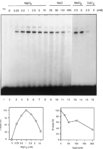
(6) Nucleic Acids Research, 2000, Vol. 28, No. 16 3027. Figure 5. The formation of D-loops promoted by hPOMp100/PSF. Schematic diagram of DNA substrates and the expected product of the reaction is shown above the gel. Stoichiometric requirements for hPOMp100/PSF (lanes 1–6). Reactions contained 32P-labelled 49mer oligonucleotide, pUCSγ superhelical dsDNA and 1.25, 2.5, 5, 10 or 20 nM hPOMp100/PSF (lanes 2–6) were incubated at 37°C for 30 min. ATP was omitted from the reaction of lane 7 and 1 mM ATPγS was added instead of ATP in the reaction of lane 8. Heterologous M13mp18 dsDNA was added instead of pUCSγ dsDNA in the reaction of lane 9. Time course of D-loop formation (lanes 10–16). DNAs and hPOMp100/PSF (10 nM) were incubated for the indicated times. 10 nM hPOMp100/PSF was added in the reactions of lanes 7–16. RecA protein (90 or 300 nM) was added and incubated at 37°C for 30 min in the reactions of lanes 17 and 18, respectively. Reaction products were analysed by agarose gel electrophoresis as described in ‘Materials and Methods’. no, no protein was added, the reaction was incubated at 37°C for 30 min. The D-loop products were quantified and are shown below the gel.. ratio which varied from one protein molecule per 160 nt to one protein molecule per 40 nt, increasing the concentration of dsDNA from 10 to 60 mM resulted in an increased amount of D-loops formed. When the linear dsDNA was substituted for the superhelical dsDNA, no D-loops were detected (lane 17), consistent with the known stabilisation of D-loops by supercoiling (42).. Figure 6. Cofactor dependence of the hPOMp100/PSF-mediated D-loop formation. 32P-labelled 49mer oligonucleotide, pUCSγ superhelical dsDNA and 8 nM hPOMp100/PSF were incubated in reaction buffer adjusted to the indicated MgCl2 (lanes 2–8), NaCl (lanes 9–12), MnCl2 (lanes 13 and 14) or CaCl2 (lanes 15 and 16) concentrations. no, no protein was added. Reactions were incubated at 37°C for 30 min and products were analysed as described in ‘Materials and Methods’. The D-loop products were quantified and are shown below the gel.. As the positively charged RNA/DNA-binding proteins such as splicing factors SF2/ASF and U2AF65 and histone H1 are able to promote DNA renaturation in vitro (44–46), we examined if these proteins can mediate D-loop formation. Although all three control proteins mediated DNA renaturation more efficiently than hPOMp100/PSF, unlike the latter, none of these proteins could promote D-loop formation under various reaction conditions (data not shown). Phosphorylation of hPOMp100/PSF by PKC stimulates its DNA binding and D-loop formation activity Because PKC in the presence of Ca2+ and phospholipids phosphorylates PSF in vitro resulting in inhibition of its RNA binding (47), we examined whether PKC phosphorylation of hPOMp100/PSF also affects its DNA binding and D-loop formation activity. When purified protein was phosphorylated by PKC its binding to ssDNA was significantly increased compared to the unphosphorylated protein (Fig. 8A) whereas,.
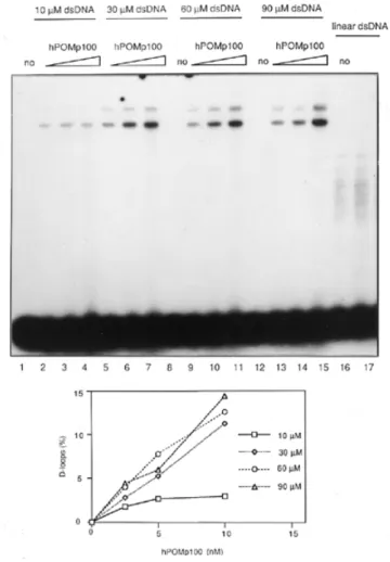
(7) 3028 Nucleic Acids Research, 2000, Vol. 28, No. 16. Figure 8. The PKC phosphorylation of hPOMp100/PSF stimulates its DNA binding and D-loop formation activity. Purified protein was phosphorylated in vitro by PKC as described in ‘Materials and Methods’. (A) ssDNA binding. 50 nM 32P-labelled 49mer oligonucleotide was incubated with 2.5, 5 or 10 nM hPOMp100/PSF before (lanes 2–4) or after (lanes 5–7) phosphorylation by PKC. Protein–DNA complexes were analysed by a native 6% PAGE. (B) Dloop formation. The 32P-labelled 49mer oligonucleotide and superhelical pUCSγ dsDNA were incubated with 5, 10 or 20 nM hPOMp100/PSF before (lanes 2–4) or after (lanes 5–7) phosphorylation by PKC. no, no protein was added. Reactions were incubated at 37°C for 30 min and products were analysed as described in ‘Materials and Methods’.. Figure 7. Dependence of the hPOMp100/PSF-mediated D-loop formation on dsDNA concentration. 32P-labelled 49mer oligonucleotide and the indicated amounts of pUCSγ superhelical dsDNA were incubated with 2.5 (lanes 2, 5, 9 and 13), 5 (lanes 3, 6, 10 and 14) or 10 (lanes 4, 7, 11, 15 and 17) nM hPOMp100/PSF. 60 µM Asp700-linearised pUCSγ dsDNA was used instead of pUCSγ superhelical dsDNA in the reactions of lanes 16 and 17. no, no protein was added. Reactions were incubated at 37°C for 30 min and products were analysed as described in ‘Materials and Methods’. The D-loop products were quantified and are shown below the gel.. in agreement with results reported previously (47), binding to poly(U) was decreased (data not shown). The formation of a more slowly migrating DNA–protein complex was enhanced in particular. Consistently, PKC phosphorylation of hPOMp100/PSF stimulated its D-loop formation activity (Fig. 8B). Quantitation of the reaction products using a PhosphorImager revealed a 2–3-fold increase in D-loop formation by the phosphorylated protein compared to unmodified protein. DISCUSSION We have previously described two HP activities in nuclear extracts from different mammalian cells (32). One of these proteins, hPOMp100, has been purified from HeLa nuclei to near homogeneity. Seven internal peptides of purified hPOMp100 revealed identity to the human polypyrimidine tract-binding protein-associated splicing factor, PSF, required. early in spliceosome formation in vitro (37,48). The identification of hPOMp100 as PSF was further confirmed by the demonstration of the ability of bacterially expressed rPSF to promote HP under the POM conditions. PSF contains two RNA recognition motifs and a proline- and glutamine-rich N terminus, which are present not only in many RNA-binding proteins but also in many transcription factors (37). However, its ability to bind DNA has never been addressed. hPOMp100/PSF binds both ssDNA and dsDNA in a non-sequence specific manner with a slightly higher affinity for ssDNA. Unlike RecA protein or its yeast and human homologues, ATP does not affect its DNA binding. The efficiency of hPOMp100/PSF binding to ssDNA increases with an increase in DNA length from 19 to 60mer. Two major protein–DNA complexes observed with longer (49 and 60mer) oligonucleotides suggest that more than one protein molecule is bound to the oligonucleotides. In contrast to RecA protein (49) and similar to hRAD51 protein (43), hPOMp100/PSF does not bind to ssDNA with high cooperativity. Proteins involved in homologous recombination like E.coli RecA, RecT and RecO, Ustilago maydis Rec2 and yeast and human RAD52 are able to catalyse the renaturation of two complementary DNA strands (38,50–54). Using two independent assays with two different DNA substrates, we have demonstrated that hPOMp100/PSF has a similar activity. The hPOMp100/PSF-promoted renaturation requires Mg2+ but is ATP-independent. In contrast to DNA renaturation, a rather simple reaction that can occur non-enzymatically and can be facilitated by unrelated proteins, D-loop formation is a much more specific recombination reaction (2). According to the current models.
(8) Nucleic Acids Research, 2000, Vol. 28, No. 16 3029. for homologous recombination, D-loop is the first heteroduplex DNA intermediate generated between two recombining DNA molecules. Moreover, as discussed by Kowalczykowski and Eggleston (2), this in vitro reaction is less prone to artifacts than strand exchange. hPOMp100/PSF is able to catalyse the formation of D-loops in superhelical duplex DNA. The formation of D-loops by hPOMp100/PSF is slow and does not exhibit the pseudoreversibility that is a feature of the RecA-mediated reaction (42,55). Similar characteristics have been reported for D-loop formation by RecO, RecT, scRad51 and hDMC1 proteins (56–59). It is worth noting that although the efficiency of D-loop formation by hPOMp100/PSF is lower than that reported for RecA, it is comparable with those reported for scRad51 and hDMC1 (58,59). Like the formation of D-loops by RecO protein (56) and HP and strand transfer by hRAD51 (43), the hPOMp100/ PSF-mediated reaction is optimal at low Mg2+ concentrations. This could be explained in part by the inability of these proteins to remove secondary structures from ssDNA. The most significant difference in HP mediated by both POM proteins, hPOMp100/PSF and hPOMp75/TLS, from that by RecA-like proteins is the absence of requirement for a high energy cofactor. The E.coli recombination proteins RecT and RecO display a similar feature (50,51,57,58). Recently discovered SMC proteins from both yeast and mammals are also able to catalyse ATP-independent DNA reannealing (28,29) whereas the reaction promoted by bacterial SMC is highly stimulated by ATP (30). According to current models, DNArenaturation activity of pro- and eukaryotic SMC proteins might be essential for their roles in recombination/repair as well as in chromosome condensation and chromatid cohesion (6). Another important difference which suggests that the mechanism of D-loop formation by hPOMp100/PSF is different from that of the RecA-like catalysed reaction, is the demonstration that the amount of the protein required for optimal activity is determined by the concentration of dsDNA. In contrast to the RecA-like promoted reaction which is mediated by a nucleoprotein filament (1,2,12,13,60), D-loop formation by hPOMp100/PSF might involve binding to both ssDNA and dsDNA, unwinding of the latter followed by reannealing of the ssDNA to complementary sequences. A similar mechanism was proposed for the RecO-catalysed reaction (56). In agreement with this, the order of addition of DNA substrates and protein has no effect on the efficiency of the hPOMp100/PSFpromoted D-loop formation, whereas for efficient HP catalysed by RecA and especially by hRAD51, it is essential to preincubate the proteins with ssDNA, to form nucleoprotein filament, prior to the addition of dsDNA. Finally, D-loop formation activity is a specific feature of both POM proteins. Indeed, despite splicing factors SF2/ASF and U2AF65 as well as histone H1 displaying somewhat similar biochemical characteristics (highly basic charge, RNA/DNA binding and DNA renaturation; 44–46), they are unable to catalyse the formation of D-loops. This also implies that D-loop formation by hPOMp100/PSF is not a simple consequence of its annealing activity. The biological importance of D-loop formation is emphasised by emerging evidence that this reaction could also be implicated in re-establishing stalled replication forks (8–10).. In this context, our results demonstrating that both POMp100/ PSF and POMp75/TLS activities are associated with cell proliferation (33) further suggest that their in vitro HP activities may be physiologically relevant. Evidence for an additional intriguing similarity between hPOMp100/PSF and hPOMp75/TLS linking both proteins to tumourigenesis has recently emerged. In papillary renal cell carcinoma, chromosomal translocation results in the fusion of PSF to the TFE3 transcription factor gene (61). Given the structurally similar fusion of TLS to the CHOP gene in lyposarcomas (62,63) and our recent finding that the oncoprotein TLS–CHOP cannot promote DNA pairing (34), it will be of interest to study HP activity of the fusion protein PSF–TFE3 to define cellular functions of hPOMp100/PSF. PSF was first discovered as a splicing factor associated with the polypyrimidine tract-binding protein and essential for the second catalytic step of pre-mRNA splicing in vitro (37,48). The same protein appears to be purified as a 100-kDa DNAbinding subunit of a p100/p52 heterodimer of unknown function (64). More recently, PSF has been demonstrated to interact with a variety of cellular targets including the human pro-oncoproteins EWS and hPOMp75/TLS and calmodulin (47), the RNA/DNA-binding nuclear protein p54nrb/NonO (the homologue of PSF) and DNA topoisomerase I (65). We also observed stable PSF/p54nrb heterodimers in HeLa nuclear extracts whereas association with hPOMp75/TLS was less stable (A.T.Akhmedov, unpublished results). Interestingly, the PSF/p54nrb dimer not only copurifies with DNA topoisomerase I but also stimulates in vitro activity of the enzyme (65), which itself is involved in various aspects of DNA dynamics. These interactions, together with the relative abundance of hPOMp100/PSF, suggest that it may get recruited by protein– protein interactions to various RNA and/or DNA metabolic pathways. Emerging evidence indicates that radiation-induced phosphorylation of hRAD51 recombinase may regulate RAD51dependent recombination/repair, although the current data are controversial (66,67). In this context, our finding demonstrating that PKC phosphorylation of hPOMp100/PSF stimulates its binding to DNA and D-loop formation but inhibits its binding to RNA implies an additional or alternative regulatory mechanism to direct this multifunctional protein to DNA metabolism. Significantly, a similar regulatory switch between RNA and DNA binding has been recently proposed for one of the hPOMp100/PSF interacting partners, p54nrb (68). Given the essential role of hPOMp100/PSF in pre-mRNA splicing and the absence of its homologue in yeast, it is difficult to provide genetic evidence for auxiliary roles of this multi-faceted protein in DNA metabolism. Significantly, the recombinational potential of another POM protein, hPOMp75/ TLS, which was also first described as an RNA-binding protein, was supported by the phenotypes of TLS-deficient mice (35,36). In this regard, detailed biochemical characterisation of hPOMp100/PSF is of particular importance. Although based on demonstrated biochemical characteristics it is tempting to speculate on a role for hPOMp100/PSF in some aspects of DNA recombination/repair, these novel features might also be important for its role in spliceosome formation during pre-mRNA splicing..
(9) 3030 Nucleic Acids Research, 2000, Vol. 28, No. 16. ACKNOWLEDGEMENTS We thank Pascale Bertrand for the contribution to the initial stages of this project; Jacques d’Alayer for peptide sequencing; James Patton for pET-PSF-expressing vector; David Ron for anti-TLS monoclonal antibody; Adrian R. Krainer for SF2/ ASF and Jerard Hurwitz for U2AF65; Heidi Baechtold for excellent technical assistance; and Rolf Jessberger and Ekaterina Revenkova for critical reading of the manuscript and for useful discussions. This work was supported in part by the Association pour la Recherche contre le Cancer (to B.S.L.). The Basel Institute for Immunology was founded and is supported by F. Hoffmann-La Roche Ltd., Basel, Switzerland. REFERENCES 1. Roca,A.I. and Cox,M.M. (1997) Prog. Nucleic Acid. Res. Mol. Biol., 56, 129–223. 2. Kowalczykowski,S.C. and Eggleston,A.K. (1994) Annu. Rev. Biochem., 63, 991–1043. 3. Shinohara,A. and Ogawa,T. (1995) Trends Biochem. Sci., 20, 387–391. 4. Kleckner,N. (1996) Proc. Natl Acad. Sci. USA, 93, 8167–8174. 5. Roeder,G.S. (1997) Genes Dev., 11, 2600–2621. 6. Jessberger,R., Frei,C. and Gasser,S.M. (1998) Curr. Opin. Genet. Dev., 8, 254–259. 7. Rossignol,J.-L. and Faugeron,G. (1994) Experientia, 50, 307–317. 8. Formosa,T. and Alberts,B.M. (1986) Cell, 47, 793–806. 9. Kogoma,T. (1996) Cell, 85, 625–627. 10. Haber,J.E. (1999) Trends Biochem. Sci., 24, 271–275. 11. Steitz,J.A. (1992) Science, 257, 888–889. 12. Radding,C.M. (1991) J. Biol. Chem., 266, 5355–5358. 13. Baumann,P. and West,S.C. (1998) Trends Biochem. Sci., 23, 247–251. 14. Sung,P. (1994) Science, 265, 1241–1243. 15. Baumann,P., Benson,F.E. and West,S.C. (1996) Cell, 87, 757–766. 16. Thacker,J. (1999) Trends Genet., 15, 166–168. 17. Lim,D.-S. and Hasty,P. (1996) Mol. Cell. Biol., 16, 7133–7143. 18. Tsuzuki,T., Fujii,Y., Sakumi,K., Tominga,Y., Nakao,K., Sekiguchi,M., Matsushiro,A., Yoshimura,Y. and Morita,T. (1996) Proc. Natl Acad. Sci. USA, 93, 6236–6240. 19. Sonoda,E., Sasaki,M.S., Buerstedde,J.-M., Bezzubova,O., Shinohara,A., Ogawa,H., Takata,M., Yamaguchi-Iwai,Y. and Takeda,S. (1998) EMBO J., 17, 598–608. 20. Sturzbecher,H.-W., Donzelmann,B., Henning,W., Knippschild,U. and Buchhop,S. 1996. EMBO J., 15, 1992–2002. 21. Scully,R., Chen,J., Plug,A., Xiao,Y., Weaver,D., Feunteun,J., Ashley,T. and Livingston,D.M. (1997) Cell, 88, 265–275. 22. Moynahan,M.E., Chiu,J.W., Koller,B.H. and Jasin,M. (1999) Mol. Cell, 4, 511–518. 23. Mizuta,R., LaSalle,J.M., Cheng,H.L., Shinohara,A., Ogawa,H., Copeland,N., Jenkins,N.A., Lalande,M. and Alt,F.W. (1997) Proc. Natl Acad. Sci. USA, 94, 6927–6932. 24. Sharan,S.K., Morimatsu,M., Albrecht,U., Lim,D.-S., Regel,E., Dinh,C., Sands,A., Eichele,G., Hasty,P. and Bradley,A. (1997) Nature, 386, 804– 810. 25. Maldonado,E., Shiekhattar,R., Sheldon,M., Cho,H., Drapkin,R., Rickert,P., Lees,E., Anderson,C.W., Linn,S. and Reinberg,D. (1996) Nature, 381, 86–89. 26. Shen,Z., Pardington-Purtymun,P.E., Comeaux,J.C., Moyzis,R.K. and Chen,D.J. (1996) Genomics, 36, 271–279. 27. Kovalenko,O.V., Plug,A.W., Haaf,T., Gonda,D.K., Ashley,T., Ward,D.C., Radding,C.M. and Golub,E.I. (1996) Proc. Natl Acad. Sci. USA, 93, 2958–2963. 28. Jessberger,R., Riwar,B., Baechtold,H. and Akhmedov,A.T. (1996) EMBO J., 15, 4061–4068. 29. Sutani,T. and Yanagida,M. (1997) Nature, 388, 798–801. 30. Hirano,M. and Hirano,T. (1998) EMBO J., 17, 7139–7148. 31. Bertrand,P., Corteggiani,E., Dutreix,M., Coppey,J. and Lopez,B.S. (1993) Nucleic Acids Res., 21, 3653–3657.. 32. Akhmedov,A.T., Bertrand,P., Corteggiani,E. and Lopez,B.S. (1995) Proc. Natl Acad. Sci. USA, 92, 1729–1733. 33. Bertrand,P., Akhmedov,A.T., Delacote,F., Durrbach,A. and Lopez,B.S. (1999) Oncogene, 18, 4515–4521. 34. Baechtold,H., Kuroda,M., Sok,J., Ron,D., Lopez,B.S. and Akhmedov,A.T. (1999) J. Biol. Chem., 274, 34337–34342. 35. Kuroda,M., Sok,J., Webb,L., Baechtold,H., Urano,F., Yin,Y., Chung,P., Akhmedov,A., de Rooij,D.G., Ashley,T. and Ron,D. (2000) EMBO J., 19, 453–462. 36. Hicks,G.G., Sigh,N., Nashabi,A., Mai,S., Bozek,G., Klewes,L., Arapovic,D., White,E.K., Koury,M.J., Oltz,E.M., Van Kaer,L. and Ruley,H.E. (2000) Nature Genet., 24, 175–179. 37. Patton,J.G., Porro,E.B., Galceran,J., Tempst,P. and Nadal-Ginard,B. (1993) Genes Dev., 7, 393–406. 38. Weinstock,G.M., McEntee,K. and Lehman,I.R. (1979) Proc. Natl Acad. Sci. USA, 76, 126–130. 39. Zinszner,H., Sok,J., Immanuel,D., Yin,Y. and Ron,D. (1997) J. Cell Sci., 110, 1741–1750. 40. Beattie,K.L., Wiegand,R.C. and Radding,C.M. (1977) J. Mol. Biol., 116, 783–803. 41. McEntee,K., Weinstock,G.M. and Lehman,I.R. (1979) Proc. Natl Acad. Sci. USA, 76, 2615–2619. 42. Shibata,T., DasGupta,C., Cunningham,R.P. and Radding, C M. (1979) Proc. Natl Acad. Sci. USA, 76, 1638–1642. 43. Baumann,P. and West,S.C. (1997) EMBO J., 16, 5198–5206. 44. Krainer,A.R., Conway,G.C. and Kozak,D. (1990) Genes Dev., 4, 1158– 1171. 45. Lee,C.-G., Zamore,P.D., Green,M.R. and Hurwitz,J. (1993) J. Biol. Chem., 268, 13472–13478. 46. Cox,M.M. and Lehman,I.R. (1981) Nucleic Acids Res., 9, 389–400. 47. Deloulme,J.C., Prichard,L., Delattre,O. and Storm,D.R. (1997) J. Biol. Chem., 272, 27369–27377. 48. Gozani,O., Patton,J.G. and Reed,R. (1994) EMBO J., 13, 3356–3367. 49. Menetski,J.P. and Kowalzcykowski,S.C. (1985) J. Mol. Biol., 181, 281– 295. 50. Hall,S.D., Kane,M.F. and Kolodner,R.D. (1993) J. Bacteriol., 175, 277– 287. 51. Luisi-DeLuca,C. and Kolodner,R. (1994) J. Mol. Biol., 236, 124–138. 52. Kmiec,E.B., Cole,A. and Holloman,W.K. (1994) Mol. Cell. Biol., 14, 7163–7172. 53. Mortensen,U.H., Bendixen,C., Sunjevaric,I. and Rothstein,R. (1996) Proc. Natl Acad. Sci. USA, 93, 10729–10734. 54. Reddy,G., Golub,E.I. and Radding,C.M. (1997) Mutat. Res., 377, 53–59. 55. Shibata,T., Ohtani,T., Iwabuchi,M. and Ando,T. (1982) J. Biol. Chem., 257, 13981–13986. 56. Luisi-DeLuca,C. (1995) J. Bacteriol., 177, 566–572. 57. Noirot,P. and Kolodner,R.D. (1998) J. Biol. Chem., 273, 12274–12280. 58. Tracy,R.B., Baumohl,J.K. and Kowalczykowski,S.C. (1997) Genes Dev., 11, 3423–3431. 59. Li,Z., Golub,E.I., Gupta,R. and Radding,C.M. (1997) Proc. Natl Acad. Sci. USA, 94, 11221–11226. 60. Stasiak,A. and Egelman,E.H. (1994) Experientia, 50, 192–203. 61. Clark,J., Lu,Y.-J., Sidhar,S.K., Parker,C., Gill,S., Smedley,D., Hamoudi,R., Linehan,W.M., Shipley,J. and Cooper,C.S. (1997) Oncogene, 15, 2233–2239. 62. Crozat,A.Y., Aman,P., Mandal,N. and Ron,D. (1993) Nature, 363, 640– 644. 63. Rabbitts,T.H., Forster,A., Larson,R. and Nathan,P. (1993) Nature Genet., 4, 175–180. 64. Zhang,W.-W., Zhang,L.-X., Busch,R.K., Farres,J. and Busch,H. (1993) Biochem. J., 290, 267–272. 65. Straub,T., Grue,P., Uhse,A., Lisby,M., Knudsen,B.R., Tange,T.O., Westargaard,O. and Boege,F. (1998) J. Biol. Chem., 273, 26261–26264. 66. Yuan,Z.M., Huang,Y., Ishiko,T., Nakada,S., Utsugisawa,T., Kharbanda,S., Wang,R., Sung,P., Shinohara,A., Weichselbaum,R. and Kufe,D. (1998) J. Biol. Chem., 273, 3799–3802. 67. Chen,G., Yuan,S.-S.F., Liu,W., Xu,Y., Trujillo,K., Song,B., Cong,F., Goff,S.P., Wu,Y., Arlinghaus,R., Baltimore,D., Gasser,P.J., Park,M.S., Sung,P. and Lee,E.Y.-H.P. (1999) J. Biol. Chem., 274, 12748–12752. 68. Basu,A., Dong,B., Krainer,A.R. and Howe C.C. (1997) Mol. Cell Biol., 17, 677–686..
(10)
Figure

+2
Documents relatifs