HAL Id: hal-03014910
https://hal.archives-ouvertes.fr/hal-03014910
Submitted on 7 Dec 2020HAL is a multi-disciplinary open access archive for the deposit and dissemination of sci-entific research documents, whether they are pub-lished or not. The documents may come from teaching and research institutions in France or abroad, or from public or private research centers.
L’archive ouverte pluridisciplinaire HAL, est destinée au dépôt et à la diffusion de documents scientifiques de niveau recherche, publiés ou non, émanant des établissements d’enseignement et de recherche français ou étrangers, des laboratoires publics ou privés.
A Fast Visual Recognition Memory System in Humans
Identified Using Intracerebral ERP
Elodie Despouy, Jonathan Curot, Martin Deudon, Ludovic Gardy, Marie
Denuelle, Jean-Christophe Sol, Jean-Albert Lotterie, Luc Valton, Emmanuel
Barbeau
To cite this version:
Elodie Despouy, Jonathan Curot, Martin Deudon, Ludovic Gardy, Marie Denuelle, et al.. A Fast Visual Recognition Memory System in Humans Identified Using Intracerebral ERP. Cerebral Cortex, Oxford University Press (OUP), 2020, 30 (5), pp.2961-2971. �10.1093/cercor/bhz287�. �hal-03014910�
PRE
-
PRINT PUBLICATION
A Fast Visual Recognition Memory System in Humans
Identified Using Intracerebral ERP
Elodie Despouy1,2,3*, Jonathan Curot1,2,4, Martin Deudon1,2, Ludovic Gardy1,2, Marie
Denuelle4, Jean-Christophe Sol5,6, Jean-Albert Lotterie5,7, Luc Valton1,2,4, Emmanuel
J. Barbeau1,2
1- Centre de Recherche Cerveau et Cognition, Université de Toulouse, Université Paul Sabatier Toulouse, 31052, France.
2- Centre National de la Recherche Scientifique, CerCo, UMR 5549, Toulouse, 31052, France. 3- Dixi medical, Chaudefontaine, 25640, France.
4- Explorations neurophysiologiques, Hôpital Purpan, Université de Toulouse, Toulouse, 31059, France.
5- INSERM, U1214, TONIC, Toulouse Mind and Brain Institute, Toulouse, 31024, France 6- Neurochirurgie, Hôpital Purpan, Université de Toulouse, Toulouse, 31059, France
7- Radiochirurgie stéréotaxique, Hôpital Purpan, Université de Toulouse, Toulouse, 31059, France
*Corresponding Author: Elodie Despouy; CerCo CNRS UMR 5549, CHU Purpan Pavillon Baudot, BP 25202, 31052 Toulouse Cedex; +33(0)562746120; [email protected]
ABSTRACT
One key item of information retrieved when surveying our visual world is whether or not objects are familiar. However, there is no consensus on the respective roles of medial temporal lobe structures, particularly the perirhinal cortex and hippocampus. We considered whether the perirhinal cortex could support a fast recognition
memory system independently from the hippocampus. We recorded the intracerebral EEG activity of epileptic patients while they were performing a fast visual recognition memory task, constraining them to use their quickest strategy. We performed ERP and classification analyses. The perirhinal cortex was, by far, the earliest region involved in recognition memory. This activity occurred before the first behavioral responses and was found to be related to reaction times, unlike the hippocampus. Single-trial analyses showed that decoding power was equivalent in the perirhinal cortex and hippocampus but occurred much earlier in the perirhinal cortex. A critical finding was that recognition memory-related activity occurred in different frontal and parietal regions, including the supplementary motor area, before the hippocampus. These results, based on ERP analyses, suggest that the human brain is equipped with a fast recognition memory system which may bypass the hippocampus and in which the perirhinal cortex plays a critical role.
KEYWORDS
recognition memory, iEEG, hippocampus, perirhinal cortex.
ABBREVIATIONS
ERP = Event Related Potential; iEEG = intracerebral EEG; MTL = Medial Temporal Lobes; minRTs = minimum Reaction Times; MVPA = Multivariate Pattern Analysis;
INTRODUCTION
We invariably survey our visual world for subsumed information. One key item of information retrieved during this process is whether or not objects are familiar. The system backing this ability has a massive and detailed storage capacity (Brady et al. 2008), can support very long-term memory (even for objects initially seen for only a few seconds (Larzabal et al. 2018)), and is also quite rapid as behavioral responses start to occur around 350-400 ms following presentation of the stimulus (Besson et al. 2012). This shows that the ability to recognize familiar objects is a powerful natural cognitive system.
How the brain actually processes recognition memory is unclear. Two regions of the medial temporal lobes, namely the perirhinal cortex and the hippocampus, are critically involved in this ability, but their respective roles are contested. Lesions limited to the perirhinal cortex are sufficient to produce a severe loss in visual recognition memory tasks and this causes far more damage than that witnessed in any other single structure of the medial temporal lobes (Meunier et al. 1993; Winters et al. 2004; Bowles et al. 2007). In addition, the hippocampus is not needed to perform visual recognition memory tasks in patients with isolated lesions of this structure (Aggleton and Shaw 1996; Vargha-Khadem 1997; Mayes et al. 2002; Aggleton et al. 2005; Barbeau et al. 2011; Patai et al. 2015; Duzel et al. 2001). Contrasting results have, however, been reported in other apparently similar patients (Manns et al. 2003). To explain some of these conflicting results, some authors have suggested that the perirhinal cortex supports familiarity while the hippocampus is critical for recollection. However, intracerebral EEG (iEEG) recorded in the
hippocampus of epileptic patients can be related to familiarity (Merkow et al. 2015). Likewise, familiarity neurons have been recorded in the hippocampus of human
patients (Rutishauser et al. 2006), which contradicts the idea of a simple functional dichotomy between these structures.
This study adopts a completely novel approach to this debate. Given that the activity of the hippocampus is delayed by several tens of milliseconds compared to that of the perirhinal cortex (Barbeau et al. 2008; Mormann et al. 2008; Staresina et al. 2012), we assumed that the perirhinal cortex could support a fast recognition memory system, bypassing the hippocampus. To identify this fast system, we used a specific recognition memory task requiring subjects to use their quickest strategy by applying a response deadline (Besson et al. 2012, 2015, 2017). Two concurring hypotheses could be tested in this way. Firstly, the perirhinal cortex should show recognition memory activity before the quickest behavioral responses, while the hippocampus should show activity occurring too late to be related to these same behavioral responses. Secondly, the hypothesis of a fast recognition memory circuit bypassing the hippocampus also logically presupposes early activity related to
recognition memory processes in brain regions other than the medial temporal lobes, to account for processes such as decision, confidence judgements and motor
activity. We thus expected to observe early activity, i.e., earlier than that in the hippocampus, in the frontal (Meunier et al. 1997; Swick and Knight 1999; Bastin et al. 2006) and parietal lobes (Gonzalez et al. 2015; Rutishauser et al. 2018).
MATERIALS AND METHODS
Patients
Fourteen patients with drug-refractory epilepsy were included in this study (6 women, median age: 34.5 years old, range: [21; 63], 8 right-handers and 2
behavioral performance (9 women, median age: 31 years old, range: [23; 66], all right-handers). Epileptic patients were admitted to the Epilepsy Monitoring Unit in Toulouse to identify their epileptic zone for possible subsequent resection. As non-invasive assessment failed to precisely indicate the spatial organization of the epileptogenic zone, these patients underwent depth electrode implantation using stereoelectroencephalography (SEEG). The placement of all electrodes was determined exclusively following clinical criteria. Each patient underwent specific implantation individually tailored to the seizure onset zone (i.e., patients did not systematically have electrodes implanted in the perirhinal cortex and hippocampus). The depth electrodes had a diameter of 0.8 mm and contained from 8 to 18
platinum/iridium contacts, 2 mm long (Microdeep depth electrode, DIXI medical, France). Eight to 13 depth electrodes were implanted stereotaxically in each patient (Supplementary Table 1).
Prior to implantation, all patients underwent a high-resolution 3T T1- weighted volumetric MRI. After electrode implantation, they underwent a high-resolution CT-scan. The pre-operative MRI and post-operative CT images were fused and normalized to the MNI brain atlas to pinpoint each contact exactly.
This study was approved by the local University Hospital Ethics Committee (CER No. 47-0913). Informed consent forms were signed for the implantation and use of EEG data for research purposes.
As we are aware that the speeded recognition memory task described below is highly demanding, we didn’t propose it to patients whom we knew had cognitive difficulties (n=5). Some other patients were included but dropped out due to the difficulty of the task and, ultimately, weren’t included in the final sample of patients
(n=3). The performance of the group of patients ultimately included in the current study therefore does not reflect that of epileptic patients in general in such tasks.
Recordings
Intracerebral EEG activity was recorded using two synchronized 64-channel acquisition systems (SystemPlus Evolution, SD LTM 64 EXPRESS, Micromed, France) at a sampling rate of 256 Hz for patients 1 and 2 and either at 1024 or 2048 Hz for the others (respectively, anti-aliasing filter: 115.8 Hz, 463.3 Hz and 926.7 Hz; high pass-filter: 0.15 Hz). No patients had seizures in the 6 hours leading up to the recordings.
Experimental task
We used a visual recognition memory task, namely the Speed and Accuracy Boosting procedure (SAB), which required subjects to use their quickest strategy as assessed in previous studies (Besson et al. 2012; Barragan-Jason et al. 2013). Each block began with an encoding phase during which 30 trial-specific stimuli (targets) were presented individually for at least 3s (self-paced) in the center of a grey screen. Participants were explicitly instructed to remember all stimuli. We compared the duration of picture presentation during the encoding phase and found a mean ± SD of 1.39 ± 1.29 s for epileptic patients and 1.52 ± 1.24 s for healthy subjects (not significantly different). The encoding phase was followed by a distracting phase during which participants viewed a cartoon video with sound on for 3 minutes. The recognition memory phase followed, during which participants had to recognize the stimuli presented during the encoding phase, interspersed with 30 new stimuli (distractors) that they had never seen before. During this recognition phase, a short
response deadline and an audio-feedback required subjects to answer as quickly and accurately as possible. Based on previous studies (Besson et al. 2012), we used a response deadline between 500 and 800 ms depending on the patient’s cognitive ability. This did not change the minimal reaction time (Supplementary Table 1). Responses were based on a go/no-go design. If a go response was made before the response deadline, a positive audio-feedback was played if the stimulus was a target (hit), and a negative one was played if it was a distractor (false alarm) (Fig. 1). If a no-go response was made, a positive audio-feedback was played if the stimulus was a distractor (correct rejection), and a negative one was played if the stimulus was a target (miss). Participants answered by raising their finger from an infrared response pad as quickly as possible. The SAB is demanding and required one or two short training sessions, which were not included in subsequent analyses. Patients underwent 7 to 10 SAB blocks.
Figure 1 - Example of stimuli used during the encoding phase and illustration of the
SAB procedure with a response deadline at 600 ms. The stimuli were presented in color.
Behavioral Performance Analyses
In order to evaluate the subject’s performance, d prime (discrimination ability) was computed for each participant (Rousselet et al. 2003). To estimate the minimal processing time required to recognize targets, the minimal behavioral reaction time (minRT) was computed by determining the latency at which correct go-responses (hits) started to significantly outnumber incorrect go-responses (false alarms) (Besson et al. 2012). For each participant, we used 30 ms time bins and a Fisher’s exact test (p < 0.05), followed by at least two significant consecutive bins.
ERP Processing
We used bipolar montage between adjacent macrocontacts to ensure that the signal recorded had a local origin (Lachaux et al. 2003). After downsampling to 256 Hz, ERPs were computed off-line using the MATLAB toolbox EEGlab v12.0.2.6b (Delorme and Makeig 2004) between 0 and 800 ms with a pre-stimulus baseline of 200 ms. Preliminary visual inspection of the EEG as well as manual artefact rejection procedures were used to reject the interictal activity period (an average of 14% of all trials (range: [8%-22%]) were excluded across participants). This procedure
decreases the risk of including ERPs modified by the patients’ epilepsy. As ERP recordings in the perirhinal cortex and hippocampus have already been studied and published completely independently from the current study, by us (i.e., in other patient groups or clinical settings (Barbeau et al. 2008, 2017)) or other groups (Trautner et al. 2004), we were also able to compare these and verify that they were similar or showed similar differential latencies.
To precisely track the time course of visual recognition memory while limiting possible confounding issues due to multiple comparisons, we performed a paired two-tailed permutation test based on the tmax statistic (Maris and Oostenveld 2007). This analysis was used to compare ERPs for hits versus correct rejections.
Multivariate Pattern Analysis (MVPA)
MVPA was conducted on single-trial ERPs (Cauchoix et al. 2014). A linear classifier was trained to discriminate between hits and correct rejections from single-trial individual time bins (4 ms) of the unfiltered iEEG signal across electrode sub-sets. Accurate measurements were derived by averaging the performance of the
classifier over multiple random data splits. This type of decoding analysis characterizes temporal changes in the category signal. Each input (electrode potential) was normalized across trials in advance using normalization between 0 and 1. A linear support vector machine (SVM) classifier from liblinear (Fan et al. 2008) was used as a classifier. The total number of hits and correct rejections was balanced in the database using random under sampling.
To avoid overfitting, we used a nested cross validation (Pereira et al. 2009; Granholm et al. 2012). We split our database into 3 inner folds and 3 outers folds. The 3 inner folds were used to tune the C parameter of the model. Then the
accuracy of the best model was evaluated on the remaining outer fold. This process was repeated until all outer folds were used as the test set. With such procedure, the algorithm is tested on the entire database.
This procedure was repeated 10 times for contact sub-sets of interest. A single measure of accuracy was obtained by averaging the classification
performance over all repetitions. Chance levels were measured by performing the same analysis on permuted labels. This allowed the latency of recognition
information to be estimated via a paired two-tailed permutation test based on the tmax statistic (Maris and Oostenveld 2007), using a familiarity α-level of 0.05.
MVPA was computed on two adjacent contacts showing the highest amplitude in the perirhinal cortex and in the hippocampus (i.e., the same contacts that were used for the ERPs analyses, MNI coordinates available in Supplementary Table 2).
ERP Image
We explored trial-to-trial differences in event-related EEG epochs using ERP-image plots (Delorme et al. 2015). The brain activity for each trial was coded as a
row of values in an ERP-image matrix. Then, row vectors for the single trials were stacked and smoothed vertically (moving vector smoothing = 30 for the perirhinal cortex and the hippocampus and 10 for the supplementary motor area), forming an ERP-image matrix of dimensions in terms of the number of epoch latencies x
number of trials (Delorme et al. 2015). In order to relate cerebral activity with patient response, single trials were sorted by increasing reaction time latencies. We
performed an analysis across patients by merging the different ERP images after amplitude normalization using Z-scoring and polarity reversal if needed (the main component of interest was usually negative in the perirhinal cortex and positive in the hippocampus in most patients as in previous studies (Trautner et al. 2004; Barbeau et al. 2008); however, the position of the electrodes regarding the dipole can vary from patient to patient). These amplitudes normalized and polarity corrected ERPs were thus used for correlation analyses. As different patterns of correlations could be observed (see ERP image, Figure 4), two metrics were developed. In the perirhinal cortex, no correlation with corrected ERP onset was observed while a correlation with the length of the ERP was suggested. We thus computed the total time below 0 µV after the onset of the evoked response. We then averaged these durations per bin of 50 trials for all trials pooled across subjects for correlations with RTs (i.e., we created super trials to reduce the noise that may occur in a single trial (Hebart et al. 2018)). In the supplementary motor area, a correlation with the corrected ERPs onset could be observed. We therefore used the onset of the evoked response as our metric and then averaged these onset latencies per bin of 20 trials as there were less trials overall, again pooled across subjects. Onset latencies of evoked
responses were determined using the median rule method (Letham and Raij 2011). Other numbers of trials were tested for both areas without changing the results. A
Pearson correlation coefficient was computed to assess the linear relationship
between the evoked response duration and the reaction times in the perirhinal cortex or between the evoked response onset and the reaction time in the supplementary motor area. These same analyses were performed and are reported for the
hippocampus for the sake of comparison, although no such correlation was observed. All correlations are reported in Supplementary Figure 1.
RESULTS
Behavioral Performance
A recognition memory task requiring subjects to use their quickest strategy was given to fourteen patients with drug-refractory epilepsy. To assess whether their performance was similar to that of healthy subjects, their ability to discriminate between targets and distractors (d’) and their minimum reaction times (minRTs, i.e., the moment when the number of hits significantly outnumber the number of false alarms), were compared with those of fourteen healthy matched subjects. No difference was observed (d’: median = 1.46 [0.92; 2.54] for epileptic patients and 1.46 [0.82; 2.84] for healthy subjects, Mann-Whitney test, U=93, p = 0.83; minRTs: median = 450 ms [390;510] for epileptic patients and 420 ms [390; 480] for healthy subjects, Mann-Whitney test, U=135, p = 0.08, Supplementary Fig. 2).
Dissociation Between the Perirhinal Cortex and the Hippocampus
We recorded typical and focal ERPs in the perirhinal cortex and the hippocampus of the patients (Trautner et al. 2004; Barbeau et al. 2008) (see example in Fig. 2a). Differential activity between hits and correct rejections was recorded in these two structures. It occurred systematically earlier in the perirhinal
cortex than in the hippocampus (median delay between the two structures = 273 ms, range = [137; 418] for 6 patients with electrodes, simultaneously in the two structures (2 with bilateral implantations), Wilcoxon test, W=0, p = 0.007, Fig. 2b).
We also used a multivariate pattern analysis (MVPA) (Kamitani and Tong 2005; Cauchoix et al. 2014) to investigate the information carried in each single trial to distinguish activity between hits and correct rejections. The MVPA was computed between hits and correct rejections on 2 adjacent contacts located in the perirhinal cortex (for 10 electrodes in 8 patients) and in the hippocampus (for 13 electrodes in 9 patients). Importantly, the decoding power recorded at the peak of maximum accuracy was similar between the perirhinal cortex and the hippocampus (median accuracy = 62%, Mann-Whitney test, U=88, p = 0.48) but occurred earlier in the perirhinal cortex compared to the hippocampus (median delay between both
structures = 277 ms, Mann-Whitney test, U=0, p < 0.001) (Fig. 2c). Furthermore, the median decoding onset occurred 272 ms before the minRTs in the perirhinal cortex (range = [197; 417]), versus only 19 ms in the hippocampus (range = [-120; 68], Mann-Whitney test, U=150, p < 0.001; Fig. 2d). Critically, the decoding onset for MVPA was similar when applied only to the activity generated by the perirhinal cortex or the hippocampus or when applied simultaneously to activity from both the perirhinal cortex and the hippocampus (Mann-Whitney test, U=25, p = 0.50). The same finding was recorded when this analysis was carried out at the peak of
maximum accuracy (U=35, p = 0.79). This implies that the interaction between these structures does not facilitate clearer differentiation of the activity related to hits and correct rejections, i.e., their contribution appears to be independent.
Figure 2 – Electrophysiological Activity Recorded in the Perirhinal Cortex and in the
Hippocampus. (a) Example of focal ERPs recorded in the perirhinal cortex (contact 3) and in the hippocampus (contacts 2 and 3) of patient 14 (p14, monopolar
montage). (b) Left: example of ERPs for hits and correct rejections (CR) recorded in the perirhinal cortex and in the hippocampus of patient 5 using bipolar montage (significant differences estimated from a permutation paired t-test using the tmax method are shown in orange). Right, onset of the differential activity recorded in the hippocampus (HPC) minus the onset recorded in the perirhinal cortex (PRC) for 6 patients with electrodes located in both structures (results sorted in increasing order of difference). The differential activity between hits and CR occurs systematically earlier (at least 140 ms) in the perirhinal cortex compared to the hippocampus, in both hemispheres. (c) Example of multivariate pattern analysis (MVPA) computed between hits and CR in the perirhinal cortex and in the hippocampus of patient 5. The distribution of reaction times is depicted by the dashed red line and the minimum reaction time (minRT) by the vertical black line. The decoding accuracy estimated from permuted labels was used to assess chance level. Paired two-tailed
permutations tests were used to compute the latency of the first significant decoded bin (1 bin = 30 ms; horizontal purple line for the perirhinal cortex, blue line for the hippocampus). (d) Notched box plots of MVPA metrics obtained across the perirhinal cortex and the hippocampus.
Spatio-Temporal Dynamics of Visual Recognition Memory Throughout the Whole Brain
1450 contacts were recorded across the 14 patients and many areas of the brain displayed differential activity between hits and correct rejections, suggesting
their involvement in recognition memory (Fig. 3a). In order to perform statistical comparisons, we selected a sub-set of brain regions according to the following criteria (Barbeau et al. 2008): (1) ERPs exhibiting similar waveforms and latencies in the same region; (2) being generated focally, as demonstrated by polarity reversal between adjacent contacts and/or steep voltage gradient and high voltage
fluctuations between adjacent contacts; (3) presenting differential activity between hits and correct rejections; (4) reaching a minimum of 3 patients per region across our population of 14 patients to ensure consistency, with the exception of the
supplementary motor area (SMA) due to the scarcity of electrode implantation in this structure. If several contacts in the same region matched these criteria, the contacts showing the earliest differential activity between hits and correct rejections were selected (this occurred for 7 electrode pairs and the mean difference between adjacent contacts was 13 ms (range: [8-20]). Contacts from both hemispheres were merged to increase power but are detailed in Supplementary Table 2.
A total of 74 bipolar contacts meeting these criteria were selected,
corresponding to 10 different brain structures (Fig. 3b; MNI coordinates available in Supplementary Table 2 and details for each bipolar contact in Supplementary Fig. 3). Looking at the onset of the differential activity between hits and correct rejections in each structure, we found that the perirhinal cortex was the first structure in the brain where a differential activity occurred. It also lasted the longest. It should be noted that electrodes were also regularly implanted in the posterior and middle ventral visual pathway but did not show differential activity, or only of late onset and
inconsistently. Differential activity then occurred simultaneously in the supplementary motor area, the second fastest region after the perirhinal cortex, as well as in a number of frontal and parietal areas. The last structures in which differential activity
was observed were other regions of the medial temporal lobes, such as the amygdala, hippocampus and temporal pole, as well as the anterior cingulum (Fig. 3c).
Figure 3 – Spatio-temporal dynamics of visual recognition memory throughout the
whole brain. (a) A large number of contacts (in red) recorded across the 14 patients displayed differential activity between hits and correct rejections. (b) 74 bipolar
contacts were selected, corresponding to 10 different brain structures following criteria (see text). For each structure, the median of the minimal differential activity (in bold), the range and the number of bipolar contacts (n) are represented. Each dot represents the bipolar contact of a different electrode. Further information is provided in Fig. S2. (c) Duration of the differential activities between hits and correct rejections across the 10 brain structures in all patients. SMA: supplementary motor area.
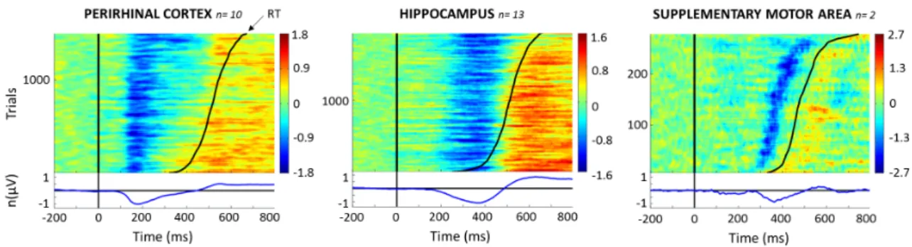
ERP Images
ERP images are plots of event-related single-trial activities ordered in this study in terms of increasing reaction times across patients (Fig. 4). In the present case, it reveals potential relationships between ERP components and reaction times. Different patterns could be observed for the perirhinal cortex, hippocampus and the supplementary motor area. The first negative potential of the ERP was time-locked to stimulus presentation in the perirhinal cortex, suggesting a partly exogenous response in this region, consistent with the idea that the perirhinal cortex processes sensory information and is strongly related to the visual ventral pathway. In this region, the duration of the first negative response correlated with reaction times. In the supplementary motor area in contrast, it was the onset of the negative response that appeared to be related to reaction times. In the hippocampus in contrast, no obvious relationship with reaction times could be observed. Complementary analyses are provided in Supplementary Figure 1.
Figure 4 – ERP images. Upper panel: single-trial ERP collapsed across subjects
and ordered by reaction times (black continuous curve). They were computed for each structure across patients after amplitude normalization, polarity adjustment and vertical smoothing across trials (see Methods). Y axis: number of single trials
included in the ERP image collapsed across subjects. n= number of subjects
included in the analysis. Voltage variations of each trial are represented using a color scale in microvolts. Bottom panel: the blue line represents the normalized evoked response averaged across trials. Note that this figure represents trials for go responses, not differential activity between hits and correct rejection.
DISCUSSION
In this study, we could identify a fast visual recognition memory system. The core component of this system is the perirhinal cortex. Moreover, the hippocampus is unlikely to play a role in this fast system.
Across the whole brain, the perirhinal cortex was the inceptive region displaying differential activity between hits and correction rejections in a specific recognition memory task. Such result is in accordance with many previous studies which have consistently shown an early differential activity of the perirhinal cortex or subhippocampal structures using intracerebral recordings by different groups and
varying kinds of stimuli (Fernandez et al. 1999; Trautner et al. 2004; Barbeau et al. 2008) or MEG (Gonsalves et al. 2005). The most notable and novel finding was that this differential activity reliably occurred much earlier (about 100 ms) than in any other brain region as assessed here through extensive recordings in the visual ventral stream as well as in the frontal and parietal lobes. This activity preceded behavioral responses and was found to be related to reaction times across patients. The next fastest region was the supplementary motor area (SMA), whose activity correlated to reaction times. This region is involved in the motor planning and execution of the response (Cona and Semenza 2017). These two regions thus defined a fast recognition memory system, which we could identify using a recognition memory task that required subjects to use their quickest strategy.
In contrast, the differential activity recorded in the hippocampus occurred remarkably late, about 270 ms after that of the perirhinal cortex, which strongly supports the idea that the perirhinal cortex is involved in a fast system while the hippocampus is involved in a different, much slower system. Furthermore, it appears that, in many instances, the differential activity in the hippocampus started too late to account for the fastest reaction times because the median difference between single-trial decoding onset and the minimum reaction time was about 20 ms. This means that, in some patients, the fastest behavioral responses occurred earlier than the onset of the differential activity in the hippocampus. The delay between the onset of the differential activity in the perirhinal cortex and the SMA was about 100 ms (Fig. 3c). As there is no reason, as far as we know, to suggest that the hippocampus is connected to the SMA by a faster route, this, in fact, indicates that much of the activity of the hippocampus occurs too late to be involved in most of the fastest behavioral responses. Importantly, decoding analyses indicated that the level of
decoding was similar in both the perirhinal cortex and the hippocampus and that decoding involving both regions simultaneously did not increase decoding accuracy. This strongly suggests that the perirhinal cortex and the hippocampus make an independent contribution to recognition memory, albeit at different time frames. Overall, it is highly unlikely that the hippocampus is involved in the fastest behavioral responses, as assessed in this study.
We initially predicted that the activity of the hippocampus occurred too late to account for the fastest behavioral responses. Hence, we should be able to identify differential activities in the frontal lobe areas occurring after the perirhinal cortex but before the hippocampus. The frontal lobes have been shown to be involved in
recognition memory in various studies in humans and non-human primates (Meunier et al. 1997; Swick and Knight 1999; Bastin et al. 2006). We also expected to observe early activity in motor and sensory-motor areas in relation to behavioral responses, which in this study involved replying with movements of the fingers. This was indeed what we found. A number of areas in the frontal region, including the SMA, as well as the parietal lobes, in accordance with previous studies (~200-300 ms in Gonzalez et al. 2015), displayed differential activity occurring earlier than that in the
hippocampus. In fact, these frontal and parietal lobe areas also showed earlier differential activity than other medial temporal lobe areas, such as the amygdala and temporal pole, highlighting, within MTL structures, the specific and pivotal role of the perirhinal cortex in visual recognition memory. The participation of these frontal and parietal regions is also consistent with the concept of a fast recognition memory system activated before a second system related to hippocampal activity. In other words, it is consistent with a circuit of brain areas involved in fast recognition memory.
It may seem surprising that the delay between the perirhinal cortex and the hippocampus is so protracted. A simpler model could be that the perirhinal cortex initially processes familiarity, which would then be rapidly relayed to the
hippocampus via the entorhinal cortex. The speeded recognition memory task we used may, in fact, have helped to show that these two structures belong to two completely different systems. This may not have been apparent in earlier
electrophysiological studies in which no speed constraint was used (Trautner et al. 2004; Barbeau et al. 2008). Overall, our findings reveal a two-stage model in which the perirhinal cortex is initially involved in the early detection of familiar objects followed by activation of other frontal and parietal cortical areas. This finding is consistent with the notion that the perirhinal cortex is the highest area in the hierarchy of the visual ventral stream whilst also being related to memory
(Ranganath and Ritchey 2012; Tamura et al. 2017). Essentially, this fast recognition memory system could have a strong ecological value, as this is the one potentially used to react at a glance. In our view, this perirhinal signal is automatic (i.e. the automatic result of ventral visual stream processes) and relatively cost-efficient compared to the computation necessary to retrieve more elaborate memories. In this sense, it may also differ from other signals required to solve more complex
recognition memory tasks (e.g. Remember/Know or Process Dissociation Procedures).
Following this initial stage, the hippocampus and other MTL areas are activated, probably making a qualitatively different contribution to recognition memory as postulated in neurocomputational models (Norman and O’Reilly 2003), and possibly with greater confidence (Rutishauser et al. 2015). These two stages may correspond to two fundamentally different cognitive entities: the fast system
being related to an interaction with the world (i.e., processing of immediate relevant stimuli present in our environment), while the slower system would be related to the retrieval of other information from memory. The switch from attention paid to the external world to attention paid to the internal world may require major reorganization of the brain (Brincat and Miller 2016; Barbeau et al. 2017) and prove
time-consuming, hence the delay between the perirhinal cortex and hippocampus.
Most objects in this study were familiar in the sense that similar objects of the same basic level and same name had already been seen (by reference to the visual hierarchy of object processing). Our view, however, is that what subjects do when they have to recognize whether or not they’ve seen an object during the recognition stage is to recognize them at the exemplar (or individual item) level. This is perhaps most clear when we run the same type of recognition memory task using abstract pictures, many of which are similar. In this case, the minimum reaction times remain the same (Besson et al. 2012).
A question might be related to context integration taking place during the encoding stage. In our protocol, as in many others, the use of context during recognition is not particularly useful, as subjects remain in the same context than during the encoding stage (same room, same people in the room, external events minimized as much as possible, etc.). Time context is also reduced to a minimum as a mere sense of recency (rather than referring to a specific moment in time) is
enough to recognize a stimulus as familiar in our experiment. Reference to context during memory tasks is often thought to depend on the hippocampus. The fact that the hippocampus doesn’t seem to be necessary in our task seems to be in
agreement with these interpretations. Overall, we think we assess in this study a very basic process underlying recognition memory.
Analyses in this study were limited to evoked responses and MVPA however, and the absence of an ERP signal in the hippocampus doesn’t completely prove that this structure was not involved in fast recognition memory. Analyses of time
frequencies (Mormann et al. 2005; Gonzalez et al. 2015; Colgin 2016) or single neuron activity might indeed have revealed an earlier involvement of the
hippocampus, although evidence is currently lacking to support such a hypothesis. Activity in the gamma range could reveal differences between conditions that were not seen using averaged evoked responses, particularly if such activity is induced, as opposed to evoked (Tallon-Baudry and Bertrand, 1999). Likewise, the association between the large and relatively slow components analyzed in this study and
neuronal activity is not straightforward (Buzsáki et al, 2012). It has also been
suggested that the brain could process novelty even faster than shown in the current study (Bunzeck et al. 2009). This magnetoencephalography study, however, did not allow analyzing clearly from which region, the frontal or temporal lobes, such rapid signal originated and whether this signal was related to conscious or unconscious forms or recognition. An early signal (250-300 ms) related to stimulus repetition has nonetheless been suggested to originate from the hippocampus (Nahum et al. 2011; Raynal et al. 2019), but this has been attributed to an encoding stage prior to
consolidation rather than recognition per se. Complex, and early, interactions
between the hippocampus and perirhinal cortex could also play a role in recognition processes (Staresina et al. 2012). Therefore, overall, the current study supports the idea that rapid recognition memory does not require the hippocampus. However, future studies, possibly using novel experimental conditions (Bunzeck et al. 2012) or other EEG analyses, may reveal a more refined picture of the involvement of the hippocampus in the processes underlying fast recognition.
As we used a go/no-go task, some of the ERPs that we analyzed may have reflected motor preparation and movement, rather than familiarity and decision processes. It was the case, for example, in the SMA, where we found a strong correlation between the onset of single EEG components and reaction times. It is unlikely to be the case in the perirhinal cortex and hippocampus in contrast, as we never found any within subject correlations in these structures (although we usually had more than 100 trials per subject, suggesting it unlikely it is a problem of power). Dedicated control tasks may help to disentangle this issue in future studies.
In this study, we intended to avoid reference to predominant
familiarity/recollection dichotomy. We make no claim as to the cognitive processes used by our subjects in our fast recognition memory task, although there are indications that tasks relying on response deadlines mainly rely on familiarity
(Sauvage et al. 2010; Ranganath and Ritchey 2012) and that familiarity consistently occurs rapidly and before recollection (Hintzman and Curran 1994; Brown and Xiang 1998; Brown and Aggleton 2001). Indeed, as we have seen, familiarity signals or neurons can sometimes be identified in the hippocampus (Rutishauser et al. 2006; Merkow et al. 2015), suggesting that familiarity may depend on different MTL structures. It has been suggested that familiarity may in fact be a poorly defined concept (Hintzman, 2011). In this context, there have also been calls to abandon this, perhaps too simple, perirhinal/familiarity and hippocampus/recollection
dichotomy (Wixted and Squire 2011). Ultimately, our approach, which departs from conventional theory, allowed us to identify a fast recognition memory system that may be more akin to neurophysiological brain systems.
ACKNOWLEDGEMENTS
This work was supported by the European Research Council, under the European Union's Seventh Framework Programme (FP/2007-2013) / ERC Grant Agreement N. 323711 (M4 project).
CONFLICT OF INTERESTS
ED is funded through a CIFRE Industrial Research Agreement (No. 2015/1135) signed between Dixi Medical and the Centre de recherche Cerveau et cognition (CerCo). The remaining authors have no competing interests.
REFERENCES
Aggleton JP, Shaw C. 1996. Amnesia and recognition memory: A re-analysis of psychometric data. Neuropsychologia. 34:51–62.
Aggleton JP, Vann SD, Denby C, Dix S, Mayes AR, Roberts N, Yonelinas AP. 2005. Sparing of the familiarity component of recognition memory in a patient with hippocampal pathology. Neuropsychologia. 43:1810–1823.
Barbeau EJ, Chauvel P, Moulin CJA, Regis J, Liégeois-Chauvel C. 2017. Hippocampus duality: Memory and novelty detection are subserved by distinct mechanisms. Hippocampus. 27:405–416.
Barbeau EJ, Pariente J, Felician O, Puel M. 2011. Visual recognition memory: A double anatomo-functional dissociation. Hippocampus. 929–934.
Barbeau EJ, Taylor MJ, Regis J, Marquis P, Chauvel P, Liegeois-Chauvel C. 2008. Spatio temporal dynamics of face recognition. Cereb Cortex. 18:997–1009.
Barragan-Jason G, Besson G, Ceccaldi M, Barbeau EJ. 2013. Fast and Famous: Looking for the Fastest Speed at Which a Face Can be Recognized. Frontiers Psychol. 4.
Bastin C, Van der Linden M, Lekeu F, Andrés P, Salmon E. 2006. Variability in the impairment of recognition memory in patients with frontal lobe lesions. Cortex. 42:983–994.
Besson G, Barragan-Jason G, Thorpe SJ, Fabre-Thorpe M, Puma S, Ceccaldi M, Barbeau EJ. 2017. From face processing to face recognition: Comparing three different processing levels. Cognition. 158:33–43.
Besson G, Ceccaldi M, Didic M, Barbeau EJ. 2012. The speed of visual recognition memory. Vis cogn. 20:1131–1152.
Besson G, Ceccaldi M, Tramoni E, Felician O, Didic M, Barbeau EJ. 2015. Fast, but not slow, familiarity is preserved in patients with amnestic mild cognitive impairment. Cortex. 65:36–49.
Bowles B, Crupi C, Mirsattari SM, Pigott SE, Parrent AG, Pruessner JC, Yonelinas AP, Köhler S. 2007. Impaired familiarity with preserved recollection after anterior temporal-lobe resection that spares the hippocampus. Proc Natl Acad Sci. 104:16382–16387.
Brady TF, Konkle T, Alvarez GA, Oliva A. 2008. Visual long-term memory has a massive storage capacity for object details. PNAS. 105:14325–14329.
Brincat SL, Miller EK. 2016. Prefrontal Cortex Networks Shift from External to Internal Modes during Learning. J Neurosci. 36:9739–9754.
Brown MW, Aggleton JP. 2001. Recognition memory: what are the roles of the perirhinal cortex and hippocampus? Nat Rev Neurosci. 2:51–61.
Brown MW, Xiang J-Z. 1998. Recognition memory: neuronal substrates of the judgement of prior occurrence. Prog Neurobiol. 55:149–189.
Bunzeck N, Doeller CF, Dolan RJ, Duzel E. 2012. Contextual interaction between novelty and reward processing within the mesolimbic system. Hum Brain Mapp. 33:1309– 1324.
Bunzeck N, Doeller CF, Fuentemilla L, Dolan RJ, Duzel E. 2009. Reward Motivation
Accelerates the Onset of Neural Novelty Signals in Humans to 85 Milliseconds. Curr Biol. 19:1294–1300.
Buzsáki G, Anastassiou CA, Koch C. 2012. The origin of extracellular fields and currents--EEG, ECoG, LFP and spikes. Nat Rev Neurosci. 13(6):407-20.
Cauchoix M, Barragan-Jason G, Serre T, Barbeau EJ. 2014. The Neural Dynamics of Face Detection in the Wild Revealed by MVPA. J Neurosci. 34:846–854.
Colgin LL. 2016. Rhythms of the hippocampal network. Nat Rev Neurosci. 17:239–249. Cona G, Semenza C. 2017. Supplementary motor area as key structure for domain-general
sequence processing: A unified account. Neurosci Biobehav Rev. 72:28–42. Delorme A, Makeig S. 2004. EEGLAB: an open source toolbox for analysis of single-trial
Delorme A, Miyakoshi M, Jung T-P, Makeig S. 2015. Grand average ERP-image plotting and statistics: A method for comparing variability in event-related single-trial EEG
activities across subjects and conditions. J Neurosci Methods. 250:3–6. Duzel E, Vargha-Khadem F, Heinze HJ, Mishkin M. 2001. Brain activity evidence for
recognition without recollection after early hippocampal damage. Proc Natl Acad Sci. 98:8101–8106.
Fan R-E, Chang K-W, Hsieh C-J, Wang X-R, Lin C-J. 2008. LIBLINEAR: A Library for Large Linear Classification. J Mach Learn Res. 9:1871–1874.
Fernández G, Effern A, Grunwald T, Pezer N, Lehnertz K, Dümpelmann M, Van Roost D, Elger CE. 1999. Real-Time Tracking of Memory Formation in the Human Rhinal Cortex and Hippocampus. Science. 285:1582–1585.
Gonzalez A, Hutchinson JB, Uncapher MR, Chen J, LaRocque KF, Foster BL, Rangarajan V, Parvizi J, Wagner AD. 2015. Electrocorticography reveals the temporal dynamics of posterior parietal cortical activity during recognition memory decisions. Proc Natl Acad Sci. 112:11066–11071.
Gonsalves BD, Kahn I, Curran T, Norman KA, Wagner AD. 2005. Memory strength and repetition suppression: multimodal imaging of medial temporal cortical contributions to recognition. Neuron, 47:751–761.
Granholm V, Noble WS, Käll L. 2012. A cross-validation scheme for machine learning algorithms in shotgun proteomics. BMC bioinformatics. 13:S3.
Hebart MN, Bankson BB, Harel A, Baker CI, Cichy RM. 2018. The representational dynamics of task and object processing in humans. eLife. 7:e32816.
Hintzman DL. 2011. Research Strategy in the Study of Memory: Fads, Fallacies, and the Search for the "Coordinates of Truth". Perspect Psychol Sci. 6(3):253-71.
Hintzman DL, Curran T. 1994. Retrieval dynamics of recognition and frequency judgments: evidence for separate processes of familiarity and recall. J Mem Lang. 33:1–18. Kamitani Y, Tong F. 2005. Decoding the visual and subjective contents of the human brain.
Lachaux JP, Rudrauf D, Kahane P. 2003. Intracranial EEG and human brain mapping. J Physiol Paris. 97:613–628.
Larzabal C, Tramoni E, Muratot S, Thorpe SJ, Barbeau EJ. 2018. Extremely long-term memory and familiarity after 12 years. Cognition. 170:254–262.
Letham B, Raij T. 2011. Statistically robust measurement of evoked response onset latencies. J Neurosci Methods. 194:374–379.
Manns JR, Hopkins RO, Reed JM, Kitchener EG, Squire LR. 2003. Recognition memory and the human hippocampus. Neuron. 37:171–180.
Maris E, Oostenveld R. 2007. Nonparametric statistical testing of EEG- and MEG-data. J Neurosci Methods. 164:177–190.
Mayes AR, Holdstock JS, Isaac CL, Hunkin NM, Roberts N. 2002. Relative sparing of item recognition memory in a patient with adult-onset damage limited to the hippocampus. Hippocampus. 12:325–340.
Merkow MB, Burke JF, Kahana MJ. 2015. The human hippocampus contributes to both the recollection and familiarity components of recognition memory. Proc Natl Acad Sci. 112:14378–14383.
Meunier M, Bachevalier J, Mishkin M. 1997. Effects of orbital frontal and anterior cingulate lesions on object and spatial memory in rhesus monkeys. Neuropsychologia. 35:999–1015.
Meunier M, Bachevalier J, Mishkin M, Murray EA. 1993. Effects on visual recognition of combined and separate ablations of the entorhinal and perirhinal cortex in rhesus monkeys. J Neurosci. 13:5418–5432.
Mormann F, Fell J, Axmacher N, Weber B, Lehnertz K, Elger CE, Fernández G. 2005. Phase/amplitude reset and theta-gamma interaction in the human medial temporal lobe during a continuous word recognition memory task. Hippocampus. 15:890–900. Mormann F, Kornblith S, Quiroga RQ, Kraskov A, Cerf M, Fried I, Koch C. 2008. Latency
Nahum L, Gabriel D, Spinelli L, Momjian S, Seeck M, Michel CM, Schnider A. 2011. Rapid consolidation and the human hippocampus: Intracranial recordings confirm surface EEG. Hippocampus. 21: 689-693.
Norman KA, O’Reilly RC. 2003. Modeling hippocampal and neocortical contributions to recognition memory: A complementary-learning-systems approach. Psychol Rev. 110:611–646.
Patai EZ, Gadian DG, Cooper JM, Dzieciol AM, Mishkin M, Vargha-Khadem F. 2015. Extent of hippocampal atrophy predicts degree of deficit in recall. Proc Natl Acad Sci. 112:12830–12833.
Pereira F, Mitchell T, Botvinick M. 2009. Machine learning classifiers and fMRI: A tutorial overview. NeuroImage. 45:S199–S209.
Ranganath C, Ritchey M. 2012. Two cortical systems for memory-guided behaviour. Nat Rev Neurosci. 13:713–726.
Raynal E, Schnider A, Manuel AL. 2019. Early signal from the hippocampus for memory encoding. Hippocampus.1– 7.
Rousselet GA, Macé MJ-M, Fabre-Thorpe M. 2003. Is it an animal? Is it a human face? Fast processing in upright and inverted natural scenes. J Vis. 3:5–5.
Rutishauser U, Aflalo T, Rosario ER, Pouratian N, Andersen RA. 2018. Single-neuron
representation of memory strength and recognition confidence in left human posterior parietal cortex. Neuron. 97:209-220.e3.
Rutishauser U, Mamelak AN, Schuman EM. 2006. Single-trial learning of novel stimuli by individual neurons of the human hippocampus-amygdala complex. Neuron. 49:805– 813.
Rutishauser U, Ye S, Koroma M, Tudusciuc O, Ross IB, Chung JM, Mamelak AN. 2015. Representation of retrieval confidence by single neurons in the human medial temporal lobe. Nat Neurosci. 18:1041–1050.
Staresina BP, Fell J, Do Lam ATA, Axmacher N, Henson RN. 2012. Memory signals are temporally dissociated in and across human hippocampus and perirhinal cortex. Nat Neurosci. 15:1167–1173.
Swick D, Knight RT. 1999. Contributions of prefrontal cortex to recognition memory: electrophysiological and behavioral evidence. Neuropsychology. 13:155.
Tallon-Baudry C, Bertrand O. 1999. Oscillatory gamma activity in humans and its role in object representation. Trends Cogn Sci. 3(4):151-162.
Tamura K, Takeda M, Setsuie R, Tsubota T, Hirabayashi T, Miyamoto K, Miyashita Y. 2017. Conversion of object identity to object-general semantic value in the primate temporal cortex. Science. 357:687–692.
Trautner P, Dietl T, Staedtgen M, Mecklinger A, Grunwald T, Elger CE, Kurthen M. 2004. Recognition of famous faces in the medial temporal lobe: An invasive ERP study. Neurology. 63:1203–1208.
Vargha-Khadem F. 1997. Differential effects of early hippocampal pathology on episodic and semantic memory. Science. 277:376–380.
Winters BD, Forwood SE, Cowell RA, Saksida LM, Bussey TJ. 2004. Double dissociation between the effects of peri-postrhinal cortex and hippocampal lesions on tests of object recognition and spatial memory: heterogeneity of function within the temporal lobe. J Neurosci. 24:5901–5908.
Wixted JT, Squire LR. 2011. The medial temporal lobe and the attributes of memory. Trends Cogn Sci. 15:210–217.