HAL Id: hal-02043232
https://hal.archives-ouvertes.fr/hal-02043232
Submitted on 20 Feb 2019
HAL is a multi-disciplinary open access
archive for the deposit and dissemination of
sci-entific research documents, whether they are
pub-lished or not. The documents may come from
teaching and research institutions in France or
abroad, or from public or private research centers.
L’archive ouverte pluridisciplinaire HAL, est
destinée au dépôt et à la diffusion de documents
scientifiques de niveau recherche, publiés ou non,
émanant des établissements d’enseignement et de
recherche français ou étrangers, des laboratoires
publics ou privés.
chronic myeloid leukemia: the IMMUNOSTIM study
Delphine Rea, Guylaine Henry, Zena Khaznadar, Gabriel Etienne, François
Guilhot, Franck Nicolini, Joëlle Guilhot, Philippe Rousselot, Françoise
Huguet, Laurence Legros, et al.
To cite this version:
Delphine Rea, Guylaine Henry, Zena Khaznadar, Gabriel Etienne, François Guilhot, et al.. Natural
killer-cell counts are associated with molecular relapse-free survival after imatinib discontinuation in
chronic myeloid leukemia: the IMMUNOSTIM study. Haematologica, Ferrata Storti Foundation,
2017, 102 (8), pp.1368-1377. �10.3324/haematol.2017.165001�. �hal-02043232�
Received: January 21, 2017. Accepted: May 8, 2017. Pre-published: May 18, 2017.
©2017 Ferrata Storti Foundation
Material published in Haematologica is covered by copyright. All rights are reserved to the Ferrata Storti Foundation. Use of published material is allowed under the following terms and conditions:
https://creativecommons.org/licenses/by-nc/4.0/legalcode. Copies of published material are allowed for personal or inter-nal use. Sharing published material for non-commercial pur-poses is subject to the following conditions:
https://creativecommons.org/licenses/by-nc/4.0/legalcode, sect. 3. Reproducing and sharing published material for com-mercial purposes is not allowed without permission in writing from the publisher.
Correspondence:
delphine.rea@aphp.fr or nicolas.dulphy@univ-paris-diderot.fr Ferrata Storti Foundation EUROPEAN HEMATOLOGY ASSOCIATIONHaematologica 2017
Volume 102(8):1368-1377
doi:10.3324/haematol.2017.165001Check the online version for the most updated information on this article, online supplements, and information on authorship & disclosures: www.haematologica.org/content/102/8/1368
D
espite persistence of leukemic stem cells, patients with chronic
myeloid leukemia who achieve and maintain deep molecular
responses may successfully stop the tyrosine kinase inhibitor
ima-tinib. However, questions remain unanswered regarding the biological
basis of molecular relapse after imatinib cessation. In IMMUNOSTIM,
we monitored 51 patients from the French Stop IMatinib trial for
periph-eral blood T cells and natural killer cells. Molecular relapse-free survival at
24 months was 45.1% (95% CI: 31.44%-58.75%). At the time of imatinib
discontinuation, non-relapsing patients had significantly higher numbers
of natural killer cells of the cytotoxic CD56
dimsubset than had relapsing
patients, while CD56
brightnatural killer cells, T cells and their subsets did
not differ significantly. Furthermore, the CD56
dimnatural killer-cell count
was an independent prognostic factor of molecular-relapse free survival in
a multivariate analysis. However, expression of natural killer-cell
activat-ing receptors, BCR-ABL1
+leukemia cell line K562-specific degranulation
and cytokine-induced interferon-gamma secretion were decreased in
non-relapsing and non-relapsing patients as compared with healthy individuals.
After imatinib cessation, the natural killer-cell count increased
significant-ly and stayed higher in non-relapsing patients than in relapsing patients,
while receptor expression and functional properties remained unchanged.
Altogether, our results suggest that natural killer cells may play a role in
controlling leukemia-initiating cells at the origin of relapse after imatinib
cessation, provided that these cells are numerous enough to compensate
for their functional defects. Further research will decipher mechanisms
underlying functional differences between natural killer cells from
patients and healthy individuals and evaluate the potential interest of
immunostimulatory approaches in tyrosine kinase inhibitor
discontinua-tion strategies. (ClinicalTrial.gov Identifier NCT00478985)
Natural killer-cell counts are associated with
molecular relapse-free survival after imatinib
discontinuation in chronic myeloid leukemia:
the IMMUNOSTIM study
Delphine Rea,1,2,3*Guylaine Henry,4Zena Khaznadar,1,5Gabriel Etienne,3,6
François Guilhot,3,7Franck Nicolini,3,8Joelle Guilhot,3,7Philippe Rousselot,3,9
Françoise Huguet,3,10Laurence Legros,3,11Martine Gardembas,3,12Viviane
Dubruille,3,13Agnès Guerci-Bresler,3,14Aude Charbonnier,3,15Frédéric Maloisel,16
Jean-Christophe Ianotto,17Bruno Villemagne,18François-Xavier Mahon,3,6
Hélène Moins-Teisserenc,1,4,5Nicolas Dulphy1,4,5*and Antoine Toubert1,4,5
1INSERM UMRS-1160, Paris; 2Service d’Hématologie Adulte, Hôpital Saint-Louis, Paris; 3France Intergroupe des Leucémies Myéloïdes Chroniques (Fi-LMC), Institut Bergonié,
Bordeaux; 4Laboratoire d’Immunologie et Histocompatibilité, Hôpital Saint-Louis, Paris; 5Institut Universitaire d'Hématologie, Université Paris Diderot-Paris 7; 6Service
d'Oncologie Médicale, Institut Bergonié, Bordeaux; 7INSERM CIC 1402, CHU de Poitiers; 8Service d'Hématologie Clinique, CHU Lyon Sud, Pierre Bénite; 9Service d’Hématologie
Oncologie et INSERM UMR-1173, Centre Hospitalier de Versailles, Le Chesnay; 10Service
d'Hématologie, IUCT Oncopole, Toulouse; 11Service d'Hématologie Clinique, Hôpital de
l'Archet, CHU de Nice; 12Service des Maladies du Sang, CHRU Angers; 13Service
d'Hématologie Clinique, Hôpital Hôtel Dieu, Nantes; 14Service d’Hématologie, CHU
Brabois, Vandoeuvre les Nancy; 15Service d’Onco-Hématologie, Institut Paoli Calmettes,
Marseille; 16Groupe Oncologie-Maladies du Sang, Clinique Sainte Anne, Strasbourg; 17Service Hématologie Clinique, Hôpital Morvan, CHRU de Brest and 18Service Médecine
Onco-hématologie, CH de la Roche sur Yon, France
*DR and ND contributed equally to this work.
Introduction
Chronic myeloid leukemia (CML) is a myeloprolifera-tive neoplasia caused by the fusion of the BCR and ABL1 genes, as the result of the acquired reciprocal t(9;22)(q34;q11) translocation. In the early 2000s, imatinib, the first ATP-competitive inhibitor of the BCR-ABL1 oncoprotein, revolutionized the management of CML, providing most patients a dramatic progression-free sur-vival benefit.1Since then, newer generations of tyrosine
kinase inhibitors (TKI) have been developed in order to overcome some of the drawbacks of imatinib, but ima-tinib remains one of the key initial therapies for newly diagnosed patients.2
When imatinib treatment is addressed appropriately, life expectancy of adult patients diagnosed with chronic-phase CML (CP-CML) is close to that of the general pop-ulation.3.4 However, the current recommendation is to
administer treatment lifelong because of the inability of imatinib and other TKI to eliminate quiescent leukemic stem cells.5-8This recommendation represents a substantial
challenge with respect to long-term safety, quality of life and economic burden. Therefore in the past few years, clinical trials have investigated the feasibility of discontin-uing imatinib treatment in patients with sustained deep molecular responses. In the pioneering STIM trial, patients on imatinib therapy for a minimum of 3 years in whom
BCR-ABL1 transcripts were undetectable for at least 2
years had a probability of maintaining deep molecular responses without any treatment of about 40%, challeng-ing the statement that TKI may never be stopped.9These
findings were rapidly corroborated by the independent TWISTER trial.10 However, a definitive cure remains
uncertain in patients who do not relapse. Indeed, serial assessments with reverse transcriptase quantitative poly-merase chain reaction (RT-qPCR) showed that peripheral blood BCR-ABL1 transcripts could be detected in patients who successfully stopped imatinib, albeit in low amounts.9The use of genomic DNA-based PCR as a
mon-itoring tool revealed that patients continued to harbor the
BCR-ABL1 gene after discontinuation of imatinib, even
when the corresponding transcripts were undetectable.11
In patients who had been off TKI therapy for several years, BCR-ABL1 transcripts could be amplified in CD34+
cell-derived colony-forming cells and long-term culture-initiating cells despite undetectable residual disease in the peripheral blood.8Altogether, these results indicate that a
reservoir of primitive leukemic cells persists in most if not all TKI-treated patients regardless of outcome after treat-ment discontinuation.
There is great clinical interest in trying to identify patients who are more likely to succeed in discontinuing imatinib in order to minimize potential risks of a leukemic rebound and to avoid undesirable drug-withdrawal symp-toms.12So far, the search for clinical variables predictive of
outcome has been challenging but factors such as the Sokal score, duration of therapy, depth of molecular response and duration of deep molecular response have provided some insights into the probability of successful imatinib discontinuation in several studies.9,13,14 However,
biological factors directing the fate of residual leukemic cells once TKI pressure is released are unclear. Given the susceptibility of CML to adaptive and innate immune cel-lular attack, an efficient autologous anti-CML response might help to control the leukemic load beyond cessation
of TKI treatment.15,16We designed and conducted an
ancil-lary biological study within the STIM trial, named IMMUNOSTIM, with the goal of analyzing peripheral blood T cells and natural killer (NK) cells and investigated whether immune parameters were associated with molec-ular relapse-free survival.
Methods
Patients
IMMUNOSTIM is a sub-study of the STIM trial approved by French health authorities (NCT00478985).9Written informed
con-sent was given in agreement with the Declaration of Helsinki. Imatinib was stopped after ≥3 years of therapy and ≥2 years of undetectable BCR-ABL1 transcripts. Stringent monitoring by RT-qPCR was performed after imatinib discontinuation to detect a molecular relapse.9 The assay sensitivity was ≥4.5 log.
Consecutively detectable peripheral blood BCR-ABL1 transcripts showing a ≥1 log increase or loss of a major molecular response [BCR-ABL1/ABL1 internationally standardized (IS) ratio ≤0.1%] defined molecular relapse and triggered imatinib resumption. In IMMUNOSTIM, heparinized blood was collected at baseline, bi-monthly for 6 months then every 6 months until 24 months unless imatinib was resumed. Healthy donors were recruited through the Paris Saint-Louis Blood Donation Center and gave informed con-sent. Experiments were performed in a centralized fashion, allow-ing ≤48 h from blood collection to processallow-ing.
Immunophenotyping
Patients’ whole blood cell counts were determined using a Sysmex XS 1000i analyzer. T cells and NK cells were quantified by dual-platform flow cytometry using monoclonal antibodies recog-nizing CD3, CD4, CD127, CD25, CD8, CD45RA, CCR7, CD27, CD56, CD16 and NKG2D (Online Supplementary Methods). Peripheral blood mononuclear cells (PBMC) were purified with Ficoll density-gradient centrifugation and cryopreserved in liquid nitrogen. NK-cell receptor expression was studied by flow cytom-etry using thawed PBMC stained with monoclonal antibodies rec-ognizing CD56, CD16, CD3, DNAM-1, KIR2D, NKp46, NKp30, NKG2A, CD94 and CD57 (Online Supplementary Methods).
Natural killer-cell functional assays
To assess degranulation, PBMC were thawed, maintained in 10% fetal calf serum-RPMI-1640 overnight at 37°C and incubated with or without the HLA class I-deficient BCR-ABL1+leukemia
cell line K562 at a 1:1 effector to target ratio for 18 h at 37°C with a CD107a monoclonal antibody. Thereafter, PBMC were stained with monoclonal antibodies recognizing CD137, CD56, CD3 and CD16. CD137+and CD107a+ NK cells were detected by flow
cytometry (Online Supplementary Methods). To study interferon (IFN)-γ production, thawed PBMC were activated overnight in medium supplemented with interleukin (IL)-12 and IL-18 (10 ng/mL each). The following day, brefeldin A was added for 4 h. IFN-γ–producing CD3-CD56brightCD16-/low NK cells were detected
by flow cytometry as previously described (Online Supplementary
Methods).17
Statistical analyses
A Mann-Whitney U-test, a Kruskal-Wallis test and a Wilcoxon matched-pairs signed ranked test were used to compare, respec-tively, quantitative variables from two independent groups, more than two groups with a Dunn test for multiple comparisons, and quantitative variables following imatinib discontinuation. Molecular relapse-free survival was estimated with the
Kaplan-Meier method.9Clinical variables and NK cells were assessed as
potential prognostic factors for molecular relapse-free survival. Quantitative factors were categorized into two groups with cut-offs set at the median. All variables were assessed by univariate analysis using the Kaplan-Meier method and the two-tailed log-rank test. A backward stepwise multivariate Cox proportional analysis was performed to determine the influence of variables potentially associated with outcome in univariate analysis (P≤0.10). In the final model, P values <0.05 were considered statis-tically significant. Quantitative variables were categorized into two groups with cut-offs set at the median. Hazard ratios and 95% confidence intervals (CI) were estimated from the Cox regression analysis.
Results
Patients’ characteristics
Fifty-one of the 100 patients enrolled in the STIM trial who agreed to participate to IMMUNOSTIM and from whom blood samples were received and processed within 48 h of collection were included; their baseline character-istics are detailed in Table 1. Thirty-three patients (64.7%) were females and the Sokal score at diagnosis of CP-CML was low in 28 (54.9%). Single-agent imatinib was given as first-line TKI therapy in all patients, either soon after the diagnosis of CML (n=24) or after resistance or intolerance to IFN-α (n=27). None of the patients had a history of allo-geneic stem cell transplantation. The median duration of imatinib treatment was 57 months (range; 36-94). The median age at imatinib discontinuation was 63 years (range; 39-81) and the median follow-up after imatinib dis-continuation was 63 months (range; 47-72). After treat-ment cessation, 28 patients (54.9%) experienced a proto-col-defined molecular relapse and all restarted imatinib treatment. The median BCR-ABL1/ABL1 IS ratio was 0.015% (range; 0.0004-0.201) at first detection of molecu-lar relapse, 0.056% (range; 0.006-1.54) at confirmation of molecular relapse and 0.165% (range; 0.024-4.49) at tinib resumption (Figure 1A). The median time from ima-tinib discontinuation to first detection of molecular relapse was 2 months (range, 1-20) and all molecular relapses but
one occurred before 6 months. The median time until ima-tinib resumption was 4 months (range, 3-29). At 24 months, molecular relapse-free survival was 45.1% (95% CI: 31.44%-58.75%) (Figure 1B). These findings were con-sistent with those of the entire STIM study population.18
The median follow-up for non-relapsing patients was 63 months (range, 48-72).
Leukocyte, T-cell and natural kill-cell counts
at imatinib discontinuation
Leukocyte and lymphocyte counts at the time of ima-tinib discontinuation did not differ significantly between non-relapsing and relapsing patients (Online Supplementary
Table S1). When CD3+ T cells, CD4+ and CD8+ T cells,
naïve, central memory, effector memory CD4+and CD8+
subsets and CD4+CD25+CD127low/-regulatory T cells were
analyzed, no significant differences were observed between non-relapsing and relapsing patients (Online
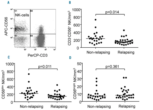
Supplementary Table S1). In contrast, although CD3-CD56+
NK cells were within the range established in our labora-tory using samples from healthy donors (data not shown), non-relapsing patients had significantly higher frequencies
Table 1. Baseline characteristics of the patients (n=51).
Parameters Results (n=51)*
Median age (range) 63 years (39-81) Female gender n, (%) 33 (64.7) Sokal score at diagnosis n, (%)
Low 28 (54.9)
Intermediate 18 (35.3)
High 4 (7.8)
Unknown 1 (2)
IFN-α intolerance or resistance prior to imatinib n, (%) 27 (52.9) Median duration of imatinib treatment (range) 57 months (36-94) Daily dose of imatinib n, (%)
400 mg 44 (86.3)
<400 mg 4 (7.8)
Unknown 3 (5.9)
Median follow-up (range) 63 months (47-72)
*70 patients were enrolled in IMMUNOSTIM but only 51 were included. The 19 other patients were excluded because of missing baseline samples (n=4) or arrival of sam-ples more than 48 h after blood was drawn (n=15).
Figure 1. Evolution of BCR-ABL1 transcripts in relapsing patients and molecu-lar relapse-free survival.(A) Scatter dot plots represent BCR-ABL1 IS % for each individual relapsing patient at first detection of relapse, relapse confirmation and at imatinib reintroduction; median values (horizontal bars) are also shown. (B) Kaplan-Meier estimate of molecular relapse-free survival defined as the time interval between imatinib discontinuation and first occurrence of molecular relapse or death, whichever came first. Data were censored at last molecular assessment for patients who were alive and had not relapsed.
A
and counts of CD3-CD56+ NK cells than had relapsing
patients, with a median count of 233/mm3(range, 70-727)
in the former and 145/mm3 (range, 67-450) in the latter
(P=0.014) (Figure 2A,B). This finding was not explained by differences in age or imatinib doses between the two groups (data not shown). In addition, we did not find any association between CD3-CD56+NK-cell counts and
dura-tion of imatinib therapy (data not shown). CD3-CD56+NK
cells were thus subtyped based upon cell-surface density of the adhesion molecule CD56 and the FcRγIII receptor CD16. Counts of the cytotoxic CD3-CD56dimCD16+
popu-lation (hereafter named CD56dim) were significantly greater
in non-relapsing patients [median: 216/mm3 (range,
55-723)] than in relapsing patients [median: 139/mm3(range,
58-438)] (P=0.011) and accounted for higher total NK-cell numbers in the former (Figure 2C). Indeed, counts of the cytokine-secreting CD3-CD56brightCD16-/low fraction
(here-after named CD56bright) did not differ between the two
groups of patients (Figure 2D).
Baseline prognostic factors for molecular relapse
In the STIM trial, the Sokal risk group and duration of imatinib treatment were associated with outcome follow-ing imatinib discontinuation.9,18Here, clinical andbiologi-cal variables and NK-cell counts at baseline were analyzed as potential prognostic factors for molecular relapse-free survival using univariate analysis. Patients with a low
Sokal score had a significantly higher molecular relapse-free survival rate than patients with an intermediate or high Sokal score (P=0.017) (Figure 3A). There was a trend toward a longer duration of imatinib treatment and higher estimated molecular relapse-free survival rate (P=0.078) (Figure 3B). Notably, patients with CD56dimNK-cell counts
higher than the median (>162/mm3) at baseline had a
sig-nificantly higher molecular relapse-free survival rate than those with a lower CD56dim NK-cell count (P=0.0008)
(Figure 3C). No significant association with age, sex, or prior IFN-α therapy was found (Figure 3D-F). After multi-variable analysis, CD56dimNK-cell count was identified as
an independent prognostic factor for molecular-relapse-free survival (Table 2). These findings led us to analyze NK cells further in the setting of immunoprofiling and func-tional experiments.
Natural killer-cell receptors and maturation marker
at imatinib discontinuation
NK-cell receptors play a key role in recognizing targets and transducing activating or inhibitory signals upon bind-ing to their ligands, thereby controllbind-ing cell function.19We
thus examined the expression of a large panel of NK-cell receptors and that of the carbohydrate antigen CD57, a marker linked to NK-cell terminal differentiation.20,21 No
statistically significant differences were found on NK cells between non-relapsing and relapsing patients with respect
Figure 2. Natural killer cells at imatinib discontinuation in non-relapsing and relapsing patients. (A) Flow cytometry dot plot from a representative patient showing CD3-CD56+NK cells in the upper left quadrant after lymphocyte gating using the side and forward scatter display. (B) Scatter dot plots represent CD3-CD56+NK-cell
counts for each individual non-relapsing and relapsing patient; median values (horizontal bars) are also shown. (C) Scatter dot plots represent CD3-CD56dimNK-cell
counts for each individual non-relapsing and relapsing patient; median values (horizontal bars) are also shown. (D) Scatter dot plots represent CD3-CD56brightNK-cell
counts for each individual non-relapsing and relapsing patient; median values (horizontal bars) are also shown. P values (by the Mann-Whitney U-test) are indicated for each panel.
A B
to expression of the activating receptors CD16, NKG2D, NKp46, NKp30 and DNAM-1, the maturation marker CD57, the activating and inhibitory KIR2D isoforms and the C-type lectin inhibitory receptor NKG2A/CD94 (data
not shown). However, when we compared NK-cell receptor
expression profile of patients with that of healthy individ-uals, significant alterations were found. The proportions
of NK cells expressing the activating receptors NKp46 and DNAM-1 were significantly lower in the CD56dim and
CD56bright subsets of non-relapsing and relapsing patients
(Figure 4). In addition, the CD56brightsubset of
non-relaps-ing and relapsnon-relaps-ing patients showed significantly increased KIR2D and CD57 and decreased NKG2A expression as compared to the expression in healthy donors (Figure 4).
Figure 3. Molecular relapse-free survival after discontinuation of imatinib according to clinico-biological factors. (A) Sokal risk group, (B) imatinib treatment dura-tion, (C) CD56dimNK-cell counts at baseline, (D) age, (E) sex, and (F) prior exposure to IFN-α. For each survival plot, a corresponding log-rank P value is shown.
B A D C F E
Natural killer-cell function at imatinib discontinuation
The capacity of NK cells to degranulate cytolytic vesi-cles was measured through the delocalization of the lyso-somal-associated membrane protein-1 CD107a onto the cell surface after stimulation with the NK-sensitiveBCR-ABL1+ cell line K562. This assay is considered a general
indicator of NK activity and particularly of tumor cell lysis.22 Raw quantification of CD107a in the presence of
K562 indicated a comparable degranulation in non-relaps-ing and relapsnon-relaps-ing patients and in healthy donors (Figure 5A). However, the propensity of NK cells for surface CD107a expression in the absence of K562 revealed signif-icantly higher levels of spontaneous degranulation in both groups of patients (Figure 5A). Consequently, K562-specif-ic degranulation of NK cells from non-relapsing and relaps-ing patients was significantly weaker than that of NK cells from healthy donors (Figure 5B). This finding was corrob-orated by a lower induction of the tumor necrosis factor receptor CD137 (also known as 4-1BB) upon K562 encounter in patients, a marker upregulated following NK-cell activation (Figure 5C). We also investigated the ability of the CD56brightNK-cell subset to secrete IFN-γ after
stim-ulation by IL-12 and IL-18. We found that IFN-γ produc-tion was comparable in non-relapsing and relapsing patients and significantly reduced compared to that in healthy donors (Figure 5D).
Natural killer cells after imatinib discontinuation
Several studies support the notion that TKI exert off-tar-get effects on NK cells and other cells of the immune sys-tem.23-26We thus wondered whether cell counts, receptorexpression and functional features assessed at baseline could have been influenced at least in part by imatinib. To address this point, experiments were repeated after cessa-tion of imatinib. Because most relapsing patients resumed imatinib within a short time frame (64.3% within 3 months and 85.7% within 6 months), follow-up samples obtained within 6 months after imatinib discontinuation were used, prior to imatinib reintroduction. After imatinib was stopped, median leukocyte and lymphocyte counts increased in non-relapsing and relapsing patients from 4810/mm3(range, 2250-7860) to 5950/mm3(range,
3760-10440) (P<0.0001) and 1310/mm3 (range, 720-2610) to
1520/mm3 (range, 670-3060), respectively (P<0.0001). A
rise in NK cells also occurred in both groups of patients, from a median value of 179/mm3(range, 67-727) at
base-line to 205/mm3(range, 74-736) after imatinib
discontinu-ation (P=0.0011). This rise was observed within both the CD56dimsubset [median count 164/mm3(range, 55-723) at
baseline and 195/mm3(range, 62-723) after imatinib
dis-continuation (P=0.0032)] and the CD56brightfraction
[medi-an count 7/mm3 (range, 2-31) at baseline and 10/mm3
(range, 4-37) after imatinib discontinuation (P=0.0008)]. Of note, CD3+T cells also increased from a median value
of 889/mm3 (range, 315-1949) at baseline to 990/mm3
(range, 265-1817) after imatinib discontinuation (P=0.0445). Importantly, the comparison between groups after imatinib discontinuation showed that NK cells of the CD56dim subset remained significantly higher in
non-relapsing patients, with a median value of 249/mm3
(range, 85-723), than in relapsing patients, who had a median value of 148/mm3 (range, 62-442) (P=0.0179)
(Table 3). Immunophenotypic analyses revealed no signif-icant modification in the expression of NKp46, NKp30, DNAM-1 and KIR2D after imatinib discontinuation in
either group of patients (Online Supplementary Figure
S1A-D). A significant reduction in NKG2A+ NK cells was
observed in non-relapsing patients but not in relapsing patients (Online Supplementary Figure S1E). The proportion of CD57+NK cells was significantly decreased in relapsing
patients (Online Supplementary Figure S1F). NK-cell degran-ulation and activation and IFN-γ production did not show any significant enhancement (Online Supplementary Figure
S1G-I).
Discussion
The current principles of CML management rely on the use of life-saving TKI through the induction of an optimal response to prevent progression to blast crisis, followed by maintenance of the optimal response by means of lifelong TKI treatment.7However, recent demonstrations that
ima-tinib can be successfully stopped in a substantial propor-tion of patients with deep and durable molecular respons-es is currently modifying this view and avoiding lifelong dependency on TKI treatment may become the ultimate goal.27-29While serial molecular monitoring is essential to
detect a relapse rapidly after an attempt to discontinue TKI, in order to trigger medical intervention and avoid any negative impact on the patient’s outcome, our ability to predict relapses accurately remains limited and determi-nants of relapse are unknown.30
Here, we demonstrated that patients free from molecu-lar relapse after imatinib discontinuation within the STIM trial had higher numbers of circulating NK cells than had patients who relapsed. In addition to CML-related vari-ables, such as the Sokal score and imatinib treatment dura-tion, a greater load of CD56dimNK cells at the time of
ima-tinib discontinuation was associated with a greater chance of treatment cessation being successful in univariate analysis. Although IFN-α has the ability to activate NK cells, no association was found between past exposure to IFN-α and molecular relapse-free survival, in agreement with what was previously found in the single-agent ima-tinib discontinuation studies STIM and TWISTER.9,10
Multivariable analysis enabled us to show that the amount of CD56dim NK cells was the only independent
prognostic factor for molecular relapse-free survival in this population of patients. Two recent and independent stud-ies are in line with our results. Indeed in the DADI dasa-tinib discontinuation trial and in the EUROSKI imadasa-tinib cessation immunological sub-study, greatest peripheral NK-cell burden at the time TKI therapy was stopped was associated with a higher probability of treatment-free remission.31,32Given the importance of NK cells in immune
Table 2. Potential prognostic factors of molecular relapse-free survival: multivariable Cox proportional analysis.
Molecular relapse-free survival over time (n=51)
Variable Hazard ratio (95% CI) P value
Sokal score* (n=50) 0.474 (0.219-1.024) 0.0576 Imatinib duration (n=51) 0.573 (0.256-1.286) 0.177 CD56dimNK-cell counts (n=51) 0.292 (0.122-0.699) 0.0057
*The Sokal score was categorized as low versus intermediate and high. Quantitative variables were categorized into two groups with cut-offs set at the median. P<0.05 was considered statistically significant.
surveillance against malignancies, these results and ours led us to hypothesize that NK cells and especially the dif-ferentiated and cytotoxic CD56dim subset20 might
con-tribute to prevent overt CML relapse originating from residual leukemic cells, in a direct or indirect fashion, after imatinib discontinuation. The involvement of NK cells in immune surveillance against CML is also supported by
other lines of evidence. BCR-ABL1+CD34+cells from CML
patients express NK activating receptor ligands.33-37In
addi-tion, BCR-ABL1+CD34+cells from CML patients are
sen-sitive to NK-cell-mediated lysis in vitro. Indeed, Sconocchia and colleagues34found that IL-2-stimulated NK cells from
allogeneic HLA-matched siblings inhibited BCR-ABL1+
CD34+-derived colony-forming unit –
granulocyte-Figure 4. Expression of natural killer cell receptors in CD56dimand CD56bright
natu-ral killer cell subsets. Scatter dot plots for healthy donors (black dots, n=44), non-relapsing patients (filled gray dots, n=19) and relapsing patients (empty black dots, n=27) with median values (horizontal bars) are shown. Overall P value (by the Kruskall-Wallis test) is indi-cated for each panel.
macrophage growth in colony formation assays. Yong and colleagues38 reported that primitive quiescent BCR-ABL1+
CD34+stem cells were sensitive to lysis by NK cells from
HLA-identical siblings, although to a lesser extent than cycling CD34+ leukemic cells. Cervantes and colleagues39
observed that IL-2-activated NK cells were capable of sup-pressing autologous BCR-ABL1+ CD34+ cell-derived
colony-forming cells and long-term culture-initiating cells. A further indication of a protective role of NK cells against CML is that early recovery of NK cells was able to predict a positive clinical outcome for patients undergoing T-cell-depleted allogeneic transplantation from HLA-identical
siblings.40 Moreover during treatment with the
NK-cell-stimulating cytokine, IFN-α, an association between the achievement of a complete cytogenetic response and NK-cell cytolytic activity was described.41,42Finally, cross-talk
between NK cells and other immune cells, such as dendrit-ic cells, may potentiate anti-CML adaptive immune responses.43,44
Despite striking quantitative differences, we found that the immunophenotypic and functional features of NK cells from non-relapsing and relapsing patients at the time of imatinib discontinuation were comparable. In addition, NK cells from both groups of patients differed phenotypi-cally and functionally from those of healthy individuals. Expression of the activating receptors NKp46 and DNAM-1 was decreased. Overall degranulation upon encounter of K562 cells was preserved, as found in the immunological EUROSKI sub-study but contrary to EUROSKI, we were able to show lower activation marker induction and high-er basal CD107a mobilization in patients, suggesting poor-er K562-specific degranulation. These diffpoor-erences between the immunological EUROSKI sub-study and our study remain unexplained. A high level of spontaneous degran-ulation was described in other pathological settings, such as in human immunodeficiency virus infection, and was linked to chronic inflammation but its significance in well-controlled CML remains unclear.45 Unfortunately, it was
not possible to assess degranulation toward autologous leukemic cells because it is not routine practice to collect and store CD34+BCR-ABL1+cells at CML diagnosis, it is
challenging to find and isolate very rare residual leukemic stem cells at imatinib discontinuation and this was not planned in our study. Finally, we found that IFN-γ produc-tion in response to cytokine stimulaproduc-tion in the CD56bright
subset was lower in patients irrespective of their relapse status. Although NK cells are not derived from the leukemic clone in CP-CML, deficient NK-cell function has been described by several groups.25,46,47In newly diagnosed
CP-CML prior to any treatment, NK-cell proliferation in response to the K562 leukemic cell line and IL-2 is reduced, as is degranulation triggered by K562 alone.25
Figure 5. Natural killer-cell function. (A) NK-cell degranulation assay against K562 or medium control. (B) Target-specific degranulation estimated by the CD107a ratio. (C) NK-cell activation estimated by CD137 expression in the pres-ence of K562 or medium control. (D) Production of IFN-γ upon stimulation with IL-12 and IL-18. Scatter dot plots for healthy donors (black dots, n=43), non-relapsing patients (filled gray dots, n=15) and non-relapsing patients (empty black dots, n=25) at baseline with median values (horizontal bars) are shown. Overall
P value (by the Kruskall-Wallis test) is indicated for each panel.
A
B
C
D
Table 3. Leukocytes, lymphocytes and natural killer-cell subsets after imatinib discontinuation.
Non-relapsing Relapsing P value*
(n=23) (n=26) Leukocytes /mm3 Median 6360 5465 Range 3760-10440 3920-7100 Lymphocytes /mm3 Median 1580 1375 Range 1140-3060 670-2500 CD3-CD56+NK cells /mm3 Median 260 155 Range 99-736 74-452 CD56dimNK cells /mm3 Median 249 148 Range 85-723 62-442 CD56brightNK cells /mm3 Median 11 9 Range 5-37 4-27
*The Mann-Whitney U test was used to compare variables from non-relapsing and relapsing patients with a level of significance of 0.05. Median values and range (min-max) are shown.
0.072 0.0134
0.0205 0.0179 0.4455
These alterations are partially restored by imatinib, there-by suggesting that the disease itself has a deleterious impact on NK cells which persists beyond the imatinib-induced remission.25
In our study, imatinib discontinuation was accompanied by a rapid and significant increase in leukocytes and lym-phocytes, as also observed by Park and colleagues.48 In
addition, a significant increase in NK cells occurred. This finding is concordant with the inhibitory impact of ima-tinib on NK-cell expansion in vitro, although such an impact was described at higher concentrations than the trough plasma concentrations usually expected in patients treated with imatinib at a dose of 400 mg daily.25It is also
consistent with the fact that patients in deep molecular response who have successfully stopped imatinib have higher NK-cell counts than those with a comparable level of response but who do not stop the drug.49However, it is
important to note that in our study, non-relapsing patients maintained higher counts of NK cells than relapsing patients after imatinib cessation and that the imatinib dose prior to discontinuation was similar in non-relapsing and relapsing patients. Although imatinib plasma levels were not measured prior to discontinuation, this argues against a different drug exposure as the sole explanation for a lower NK-cell burden in relapsing patients. Other fac-tors involved in NK-cell peripheral expansion, such as stro-mal cell factors or the immunoregulatory cytokine IL-15, need to be explored in future research studies.50
In our experimental setting, discontinuation of imatinib did not result in the disappearance of NK-cell differences with healthy individuals, ruling out a major deleterious effect of the drug on NK-cell function. Expression of the activating receptors NKp46 and DNAM-1 remained unchanged. A significant decrease in NKG2A+NK cells in
non-relapsing patients was observed but there was no gain in cytolytic and CD56bright-IFN-γ secretion capacities. Our
study was not designed to decipher the underlying mech-anisms of NK-cell defects in CML but several hypotheses can be made, including a negative impact of residual leukemic stem cells or their microenvironment on immune cell function or a weaker activation of the NK-cell repertoire due to HLA class I/KIR variation as compared to that in healthy individuals.36 In the immunological
EUROSKI sub-study, individual KIR genes and haplotypes were not associated with imatinib discontinuation out-come but, unfortunately, comparisons with the general population were not performed.32
To conclude, we provide evidence that NK cells are associated with outcome after imatinib discontinuation in CP-CML patients in deep molecular response. Beyond functional aspects, larger amounts of cytotoxic CD56dim
NK cells delivered to the leukemic reservoir in non-relaps-ing patients may play an important role in controllnon-relaps-ing residual CML-initiating cells and their progeny soon after cessation of imatinib treatment while a reduced CD56dim
compartment, and thus a lower effector to target cell ratio in relapsing patients, may leave less chance to counterat-tack in an efficient manner. Of course, our results do not preclude the role of additional biological aspects in dictat-ing patients’ outcome, such as the involvement of other immunological factors or functional heterogeneity of the residual BCR-ABL1+ hematopoietic stem-cell reservoir.
Thus integration of NK-cell counts into the TKI discontin-uation decision-making process is not reasonable at this stage. In addition, NK-cell alterations in patients suggest that the use of agents that stimulate NK-cell expansion or function, such as the IL-15 cytokine, or antibodies modu-lating NK-cell receptors may increase the chance of suc-cessfully TKI discontinuation, an aspect that may be addressed in future studies.
Acknowledgments
The authors would like to thank the patients and the personnel from clinical centers involved in this study and Ms. Nicole Biscay, Valérie Hubert, Eliane Melgire and Véronique Delasse from the Cell Therapy Unit, Hôpital Saint-Louis, Paris, France for expert logistic and technical assistance.
Funding
This work was supported by research grants from the Association pour la Recherche sur le Cancer (ZK, #DOC20100600956), Association LMC France (#R13102HH), Assistance Publique-Hôpitaux de Paris (Translational Research Grant in Biology 2010 #RTB10002) and the French Ministry of Health/Institut National du Cancer (Programme Hospitalier de Recherche 2006).
References
1. Druker BJ, Guilhot F, O'Brien SG, et al; IRIS Investigators. Five-year follow-up of patients receiving imatinib for chronic myeloid leukemia. N Engl J Med. 2006;355(23):2408-2417.
2. Kalmanti L, Saussele S, Lauseker M, et al. Safety and efficacy of imatinib in CML over a period of 10 years: data from the random-ized CML-study IV. Leukemia. 2015;29(5): 1123-1132.
3. Gambacorti-Passerini C, Antolini L, Mahon FX, et al. Multicenter independent assess-ment of outcomes in chronic myeloid leukemia patients treated with imatinib. J Natl Cancer Inst. 2011;103(7):553-561. 4. Sasaki K, Strom SS, O'Brien S, et al. Relative
survival in patients with chronic-phase chronic myeloid leukaemia in the
tyrosine-kinase inhibitor era: analysis of patient data from six prospective clinical trials. Lancet Haematol. 2015;2(5):e186-e193.
5. Corbin AS, Agarwal A, Loriaux M, Cortes J, Deininger MW, Druker BJ. Human chronic myeloid leukemia stem cells are insensitive to imatinib despite inhibition of BCR-ABL activity. J Clin Invest. 2011;121(1):396-409. 6. Hamilton A, Helgason GV, Schemionek M,
et al. Chronic myeloid leukemia stem cells are not dependent on Bcr-Abl kinase activity for their survival. Blood. 2012;119(6):1501-1510.
7. Baccarani M, Deininger MW, Rosti G, et al. European LeukemiaNet recommendations for the management of chronic myeloid leukemia: 2013. Blood. 2013;122(6):872-884. 8. Chomel JC, Bonnet ML, Sorel N, et al. Leukemic stem cell persistence in chronic myeloid leukemia patients in deep molecu-lar response induced by tyrosine kinase
inhibitors and the impact of therapy discon-tinuation. Oncotarget. 2016;7(23):35293-35301.
9. Mahon FX, Réa D, Guilhot J, et al; Intergroupe Français des Leucémies Myéloïdes Chroniques. Discontinuation of imatinib in patients with chronic myeloid leukaemia who have maintained complete molecular remission for at least 2 years: the prospective, multicentre Stop Imatinib (STIM) trial. Lancet Oncol. 2010;11(11): 1029-1035.
10. Ross DM, Branford S, Seymour JF, et al. Safety and efficacy of imatinib cessation for CML patients with stable undetectable min-imal residual disease: results from the TWISTER study. Blood. 2013;122(4):515-522.
11. Ross DM, Branford S, Seymour JF, et al. Patients with chronic myeloid leukemia who maintain a complete molecular
response after stopping imatinib treatment have evidence of persistent leukemia by DNA PCR. Leukemia. 2010;24(10):1719-1724.
12. Richter J, Söderlund S, Lübking A, et al. Musculoskeletal pain in patients with chron-ic myeloid leukemia after discontinuation of imatinib: a tyrosine kinase inhibitor with-drawal syndrome. J Clin Oncol. 2014;32 (25):2821-2823.
13. Mori S, Vagge E, le Coutre P, et al. Age and dPCR can predict relapse in CML patients who discontinued imatinib: the ISAV study. Am J Hematol. 2015;90(10):910-914. 14. Lee SE, Choi SY, Song HY, et al. Imatinib
withdrawal syndrome and longer duration of imatinib have a close association with a lower molecular relapse after treatment dis-continuation: the KID study. Haematologica. 2016;101(6):717-723.
15. Rohon P. Biological therapy and the immune system in patients with chronic myeloid leukemia. Int J Hematol. 2012;96(1):1-9. 16. Barrett AJ, Ito S. The role of stem cell
trans-plantation for chronic myelogenous leukemia in the 21st century. Blood. 2015;125(21):3230-3235.
17. Khaznadar Z, Henry G, Setterblad N, et al. Acute myeloid leukemia impairs natural killer cells through the formation of a defi-cient cytotoxic immunological synapse. Eur J Immunol. 2014;44(10):3068-3080. 18. Etienne G, Guilhot J, Rea D, et al, on behalf
of the Intergroupe Français des Leucémies Myéloïdes Chroniques (Fi-LMC). Long-term follow-up of the French Stop Imatinib Study (STIM1) in chronic myeloid leukemia patients. J Clin Oncol. 2017;35(3):298-305. 19. Carrillo-Bustamante P, Ke mir C, de Boer RJ.
The evolution of natural killer cell receptors. Immunogenetics. 2016;68(1):3-18. 20. Björkström NK, Riese P, Heuts F, et al.
Expression patterns of NKG2A, KIR, and CD57 define a process of CD56dim NK-cell differentiation uncoupled from NK-cell edu-cation. Blood. 2010;116(19):3853-3864. 21. Nielsen CM, White MJ, Goodier MR, Riley
EM. Functional significance of CD57 expres-sion on human NK cells and relevance to dis-ease. Front Immunol. 2013;4(422):1-8. 22. Alter G, Malenfant JM, Altfeld M. CD107a
as a functional marker for the identification of natural killer cell activity. J Immunol Methods. 2004;294(1-2):15-22.
23. Cebo C, Da Rocha S, Wittnebel S, et al. The decreased susceptibility of Bcr/Abl targets to NK cell-mediated lysis in response to ima-tinib mesylate involves modulation of NKG2D ligands, GM1 expression, and synapse formation. J Immunol. 2006;176(2):864-872.
24. Salih J, Hilpert J, Placke T, et al. The BCR/ABL-inhibitors imatinib, nilotinib and dasatinib differentially affect NK cell reactiv-ity. Int J Cancer. 2010;127(9):2119-2128.
25. Chen CI, Koschmieder S, Kerstiens L, et al. NK cells are dysfunctional in human chronic myelogenous leukemia before and on ima-tinib treatment and in BCR-ABL-positive mice. Leukemia. 2012;26(3):465-474. 26. Zitvogel L, Rusakiewicz S, Routy B, Ayyoub
M, Kroemer G. Immunological off-target effects of imatinib. Nat Rev Clin Oncol. 2016;13(7):431-446.
27. Rea D, Rousselot P, Guilhot J, Guilhot F, Mahon FX. Curing chronic myeloid leukemia. Curr Hematol Malig Rep. 2012;7 (2):103-108.
28. Hughes TP, Ross DM. Moving treatment-free remission into mainstream clinical prac-tice in CML. Blood. 2016;128(1):17-23. 29. Saußele S, Richter J, Hochhaus A, Mahon
FX. The concept of treatment-free remission in chronic myeloid leukemia. Leukemia. 2016;30(8): 1638-1647.
30. Deininger MW. Molecular monitoring in CML and the prospects for treatment-free remissions. Hematology Am Soc Hematol Educ Program. 2015;(2015):257-263. 31. Imagawa J, Tanaka H, Okada M, et al; DADI
Trial Group. Discontinuation of dasatinib in patients with chronic myeloid leukaemia who have maintained deep molecular response for longer than 1 year (DADI trial): a multicentre phase 2 trial. Lancet Haematol. 2015;2(12):e528-e535.
32. Ilander M, Olsson-Strömberg U, Schlums H, et al. Increased proportion of mature NK cells is associated with successful imatinib discontinuation in chronic myeloid leukemia. Leukemia. 2017;31(5):1108-1116. 33. Salih HR, Antropius H, Gieseke F, et al.
Functional expression and release of ligands for the activating immunoreceptor NKG2D in leukemia. Blood. 2003;102(4):1389-1396. 34. Sconocchia G, Lau M, Provenzano M, et al.
The antileukemia effect of HLA-matched NK and NK-T cells in chronic myelogenous leukemia involves NKG2D-target-cell inter-actions. Blood. 2005;106(10):3666-3672. 35. Boissel N, Rea D, Tieng V, et al. BCR/ABL
oncogene directly controls MHC class I chain-related molecule A expression in chronic myelogenous leukemia. J Immunol. 2006;176(8):5108-5116.
36. Verheyden S, Demanet C. NK cell receptors and their ligands in leukemia. Leukemia. 2008;22(2):249-257.
37. Hilpert J, Grosse-Hovest L, Grünebach F, et al. Comprehensive analysis of NKG2D ligand expression and release in leukemia: implica-tions for NKG2D-mediated NK cell respons-es. J Immunol. 2012;189(3):1360-1371. 38. Yong AS, Keyvanfar K, Hensel N, et al.
Primitive quiescent CD34+ cells in chronic myeloid leukemia are targeted by in vitro expanded natural killer cells, which are func-tionally enhanced by bortezomib. Blood. 2009;113(4):875-882.
39. Cervantes F, Pierson BA, McGlave PB,
Verfaillie CM, Miller JS. Autologous activat-ed natural killer cells suppress primitive chronic myelogenous leukemia progenitors in long-term culture. Blood. 1996;87(6): 2476-2485.
40. Savani BN, Rezvani K, Mielke S, et al. Factors associated with early molecular remission after T cell-depleted allogeneic stem cell transplantation for chronic myel-ogenous leukemia. Blood. 2006;107(4):1688-1695.
41. Pawelec G, Da Silva P, Max H, et al. Relative roles of natural killer- and T cell-mediated anti-leukemia effects in chronic myeloge-nous leukemia patients treated with interfer-on-alpha. Leuk Lymphoma. 1995;18(5-6):471-478.
42. Meseri A, Delwail V, Brizard A, et al. Endogenous lymphokine activated killer cell activity and cytogenetic response in chronic myelogenous leukaemia treated with alpha-interferon. Br J Haematol. 1993;83(2):218-222.
43. Degli-Esposti MA, Smyth MJ. Close encoun-ters of different kinds: dendritic cells and NK cells take centre stage. Nat Rev Immunol. 2005;5(2):112-124.
44. Walzer T, Dalod M, Robbins SH, Zitvogel L, Vivier E. Natural-killer cells and dendritic cells: "l'union fait la force". Blood. 2005;106(7):2252-2258.
45. Lichtfuss GF, Cheng WJ, Farsakoglu Y, et al. Virologically suppressed HIV patients show activation of NK cells and persistent innate immune activation. J Immunol. 2012;189(3):1491-1499.
46. Takahashi N, Miura I, Saitoh K, Miura AB. Lineage involvement of stem cells bearing the philadelphia chromosome in chronic myeloid leukemia in the chronic phase as shown by a combination of fluorescence-activated cell sorting and fluorescence in situ hybridization. Blood. 1998;92(12):4758-4763.
47. Mellqvist UH, Hansson M, Brune M, Dahlgren C, Hermodsson S, Hellstrand K. Natural killer cell dysfunction and apoptosis induced by chronic myelogenous leukemia cells: role of reactive oxygen species and reg-ulation by histamine. Blood. 2000;96(5): 1961-1968.
48. Park JS, Lee SE, Jeong SH, et al. Change of health-related profiles after Imatinib cessa-tion in chronic phase chronic myeloid leukemia patients. Leuk Lymphoma. 2016;57(2):341-347.
49. Ohyashiki K, Katagiri S, Tauchi T, et al. Increased natural killer cells and decreased CD3(+)CD8(+)CD62L(+) T cells in CML patients who sustained complete molecular remission after discontinuation of imatinib. Br J Haematol. 2012;157(2):254-256. 50. Fehniger TA, Caligiuri MA. Interleukin 15:
biology and relevance to human disease. Blood. 2001;97(1):14-32.
Natural killer-cell counts are associated with molecular relapse-free
survival after imatinib discontinuation in chronic myeloid leukemia:
the IMMUNOSTIM study
Delphine Rea,1,2,3*Guylaine Henry,4Zena Khaznadar,1,5Gabriel Etienne,3,6François Guilhot,3,7Franck Nicolini,3,8Joelle
Guilhot,3,7Philippe Rousselot,3,9Françoise Huguet,3,10Laurence Legros,3,11Martine Gardembas,3,12Viviane Dubruille,3,13
Agnès Guerci-Bresler,3,14Aude Charbonnier,3,15Frédéric Maloisel,16Jean-Christophe Ianotto,17Bruno Villemagne,18
François-Xavier Mahon,3,6Hélène Moins-Teisserenc,1,4,5Nicolas Dulphy1,4,5*and Antoine Toubert1,4,5
1INSERM UMRS-1160, Paris; 2Service d’Hématologie Adulte, Hôpital Saint-Louis, Paris; 3France Intergroupe des Leucémies Myéloïdes Chroniques (Fi-LMC), Institut Bergonié, Bordeaux; 4Laboratoire d’Immunologie et Histocompatibilité, Hôpital Saint-Louis, Paris; 5Institut Universitaire d'Hématologie, Université Paris Diderot-Paris 7; 6Service d'Oncologie Médicale, Institut Bergonié, Bordeaux; 7INSERM CIC 1402, CHU de Poitiers; 8Service d'Hématologie Clinique, CHU Lyon Sud, Pierre Bénite; 9Service d’Hématologie Oncologie et INSERM UMR-1173, Centre Hospitalier de Versailles, Le Chesnay; 10Service d'Hématologie, IUCT Oncopole, Toulouse; 11Service d'Hématologie Clinique, Hôpital de l'Archet, CHU de Nice; 12Service des Maladies du Sang, CHRU Angers; 13Service d'Hématologie Clinique, Hôpital Hôtel Dieu, Nantes; 14Service d’Hématologie, CHU Brabois, Vandoeuvre les Nancy; 15Service d’Onco-Hématologie, Institut Paoli Cal-mettes, Marseille; 16Groupe Oncologie-Maladies du Sang, Clinique Sainte Anne, Strasbourg; 17Service Hématologie Clinique, Hôpital Morvan, CHRU de Brest and 18Service Médecine Onco-hématologie, CH de la Roche sur Yon, France
*DR and ND contributed equally to this work.
©2017 Ferrata Storti Foundation. This is an open-access paper. doi:10.3324/haematol.2017.165001 Received: January 21, 2017.
Accepted: May 8, 2017. Pre-published: May 18, 2017.
Online Supplement
Supplementary Methods
Immunophenotyping
To analyze T-cells, NK-cells and their subsets by flow cytometry, a 4-color staining of
fresh blood was performed followed by red blood cell lysis. Leucocytes were acquired with a
BD Biosciences a FACSCalibur™ and data were analyzed with the BD CellQuest software.
PerCP-CD3 (clone SK7), PerCP-CD4 (clone SK3), PE-CD4 (clone SK3), PerCP-CD8 (clone
SK1), PE-CD8 (clone SK1), CD45RA (clone HI100), PE-CD27 (clone M-T271),
APC-CD56 (clone B159), FITC-CD25 (clone 2A3) and FITC-CD16 (clone 3G8) were from BD
Biosciences. FITC-CCR7 (clone 150503) was from R&D Systems). PE-CD127 (clone R34),
and PE-NKG2D (clone ON72) were from Beckman Coulter. To analyze NK-receptor
expression by flow cytometry on thawed PBMCs, an 8-color staining with mAb was
performed, leucocytes were acquired with a BD Biosciences FACSCanto™ II and data were
analyzed with the DIVA v6.1.3 software. FITC-DNAM-1 (clone DX11), APC-H7-CD3
(clone SK7) and PercP-Cy5.5-CD56 (clone B159) were from BD Biosciences. PE-KIR2D
(clone NKVFS1), PE-NKp30 (clone AF29-4D12), APC-NKp46 (clone 9E2) and
Vioblue-CD57 (clone TB03) were from Miltenyi Biotec. PE-Cy7-NKG2A (clone Z199) and
APC-CD94 (clone HP-3B1) were from Beckman Coulter. BV510-CD16 (clone 3G8) was from
Ozyme.
NK-cell functional assays
To analyze CD107a and CD137 expression by flow cytometry on CD3
-CD56
+NK-cells in a degranulation assay, leucocytes stained with mAbs were acquired with a BD
Biosciences FACSCanto™ II and data were analyzed with the DIVA v6.1.3 software.
APC-CD137 (clone 4B4-1), PE-Cy7-CD56 (clone B159) and APC-H7-CD3 (clone SK7) were from
BD Biosciences and BV510-CD16 (clone 3G8) was from Ozyme). For IFN-
detection by
flow cytometry in CD56
brightNK-cells after cytokine stimulation, IFN-
intracellular staining
was performed using the AlexaFluor488-labelled B27 clone (BD Biosciences). Leucocytes
were acquired with a BD Biosciences FACSCanto™ II and data were analyzed with the
DIVA v6.1.3 software.
Supplementary Table S1. Leucocytes, lymphocytes and T-cell subsets at baseline
Parameters All (n=51) Non-relapsing (n=23) Relapsing (n=28) p value* Leucocytes/mm3 4820 (2550-7860) 5020 (2550-7470) 4800 (3400-7860) 0.557 Lymphocytes/mm3 1310 (720-2610) 1400 (810-2610) 1300 (720-1970) 0.185 CD3+ T-cells/mm3 CD3+CD4+/mm3 CD3+CD8+/mm3 Ratio CD4/CD8 897 (315-1949) 534 (228-1248) 279 (72-998) 2.02 (0.5-14.2) 896 (543-1949) 534 (352-1248) 295 (77-998) 1.93 (0.5-5.94) 898 (315-1576) 538 (228-1021) 276 (72-562) 2.09 (0.79-14.2) 0.172 0.297 0.092 0.489 CD3+CD4+ subsets Naïve (CD4+CD45RA+CCR7+CD27+) % CD4+ /mm3Central memory (CD4+CD45RA-CCR7+)
% CD4+
/mm3
Effector memory (CD4+CD45RA-CCR7-)
% CD4+ /mm3 Regulatory (CD4+CD25+CD127low/-) % CD4+ /mm3 35.1 (8.7-75.9) 200 (32-774) 45.1 (19.8-74.7) 259 (80-570) 15.7 (2.4-53) 91 (25-273) 7 (1.2-13.1) 37 (4-94) 35.5 (13.1-57.7) 189 (82-286) 46.7 (25.1-74.7) 289 (120-570) 13.6 (9.3-29.3) 97 (41-273) 7.1 (1.2-12.1) 40 (4-94) 34.7 (8.7-75.9) 213 (32-774) 44.3 (19.8-64.6) 223 (80-394) 18.9 (2.4-53) 85 (25-232) 6.4 (3.2-13.1) 36 (12-53) 0.949 0.949 0.339 0.091 0.171 0.974 0.300 0.136 CD3+ CD8+ subsets Naïve (CD8+CD45RA+CCR7+CD27+) % CD8+ /mm3
Central memory (CD8+CD45RA-CCR7+)
% CD8+
/mm3
Effector memory (CD8+CD45RA-CCR7-)
% CD8+
/mm3
Effector memory (CD8+CD45RA+CCR7-)
% CD8+ /mm3 26.2 (6.5-71.9) 75 (9-239) 12 (1-38.6) 30 (2-113) 24.2 (1.6-58.3) 61 (4-345) 33.3 (6.8-84.6) 108 (9-416) 23.7 (6.5-62.7) 75 (28-239) 13.4 (1-23.7) 38 (2-110) 20.7 (1.6-58.3) 64 (4-345) 38.8 (2-110) 111 (20-416) 28 (7.5-71.9) 72 (9-236) 11.1 (2-38.6) 29 (6-113) 27.9 (3.2-55.7) 56 (9-237) 33 (6.8-58.7) 85 (9-271) 0.841 0.342 0.920 0.367 0.325 0.972 0.494 0.211 *
The Mann-Whitney U test was used to compare variables from non-relapsing and relapsing patients, with a level of significance of 0.05. Median values (min-max) are shown.
Supplementary Figure S1
Figure S1. NK-cell receptor and function after imatinib discontinuation. (A-F) NK-cell
receptor expression, (G) degranulation capacities, (H) activation marker expression and (I)
IFN-
production at baseline and after imatinib discontinuation in non-relapsing (n=6) and
relapsing patients (n=6). Scatter dot plots with median are shown. P values (by Wilcoxon
matched-pairs signed ranked test) are shown for each set of data.
Figure S1
Baseline After Baseline After 0 20 40 60 80 100 % N k p 4 6 + N K -c e lls Non-relapsing Relapsing p=0.1250 p>0.9999 A
Baseline After Baseline After 0 20 40 60 80 100 Non-relapsing Relapsing p=0.8438 p=0.0938 D
Baseline After Baseline After 0 20 40 60 Non-relapsing Relapsing p=0.1875 p=0.0625
Baseline After Baseline After 0 20 40 60 80 100 Non-relapsing Relapsing p=0.6250 p>0.9999 B
Baseline After Baseline After 0 20 40 60 80 100 Non-relapsing Relapsing p=0.0313 p=0.6875 F
Baseline After Baseline After 0 10 20 30 40 Non-relapsing Relapsing p=0.1250 p=0.1250
Baseline After Baseline After 0 20 40 60 80 100 Non-relapsing Relapsing p>0.9999 p=0.8125 C
Baseline After Baseline After 0 20 40 60 80 100 Non-relapsing Relapsing p=0.0313 p=0.4375 E
Baseline After Baseline After 0 20 40 60 Non-relapsing Relapsing p=0.6875 p=0.3125 % N k p 3 0 + N K -c e lls % D N A M -1 + N K -c e lls % K IR 2 D + N K -c e lls % N K G 2 A + N K -c e lls % C D 5 7 + N K -c e lls % C D 1 0 7 a + N K -c e lls % C D 1 3 7 + N K -c e lls % I F N -+ N K -c e lls