HAL Id: hal-03096390
https://hal.archives-ouvertes.fr/hal-03096390
Submitted on 22 Mar 2021
HAL is a multi-disciplinary open access
archive for the deposit and dissemination of
sci-entific research documents, whether they are
pub-lished or not. The documents may come from
teaching and research institutions in France or
abroad, or from public or private research centers.
L’archive ouverte pluridisciplinaire HAL, est
destinée au dépôt et à la diffusion de documents
scientifiques de niveau recherche, publiés ou non,
émanant des établissements d’enseignement et de
recherche français ou étrangers, des laboratoires
publics ou privés.
Deciphering sulfoglycolipids of Mycobacterium
tuberculosis
Emilie Layre, Diane Cala-de Paepe, Gerald Larrouy-Maumus, Julien
Vaubourgeix, Sathish Mundayoor, Buko Lindner, Germain Puzo, Martine
Gilleron
To cite this version:
Emilie Layre, Diane Cala-de Paepe, Gerald Larrouy-Maumus, Julien Vaubourgeix, Sathish
Mun-dayoor, et al.. Deciphering sulfoglycolipids of Mycobacterium tuberculosis. Journal of Lipid
Re-search, American Society for Biochemistry and Molecular Biology, 2011, 52 (6), pp.1098-1110.
�10.1194/jlr.M013482�. �hal-03096390�
1098 Journal of Lipid Research Volume 52, 2011
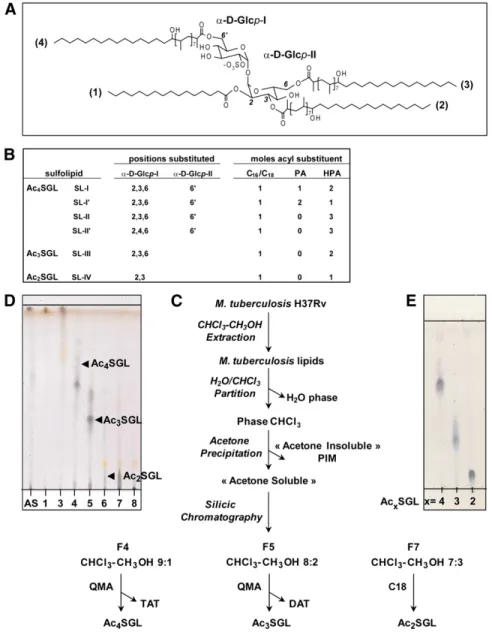
Copyright © 2011 by the American Society for Biochemistry and Molecular Biology, Inc. Sulfoglycolipids (SGLs) were originally discovered in 1959 by Middlebrook et al. ( 1 ) in human (H37Rv) and bovine (Vallée) virulent strains of Mycobacterium tuberculo-sis . Almost 2 decades later, Goren et al. ( 2 ) showed that this family of lipids was specifi c to M. tuberculosis and pro-posed that their amount correlated with the virulence of the M. tuberculosis strain in the guinea pig model of infection. Sulfolipids consist of multi-acylated forms of trehalose sul-fate that differ by the number, structure, and location of acyl moieties ( 3 ). The major sulfolipid, SL-I, was character-ized as 2,3,6,6 ′ -tetraacyl- a - a ’-trehalose-2 ′ -sulfate acylated by two hydroxyphthioceranates (HPA), one phthioceranate (PA), and one palmitate (C 16 ) or stearate (C 18 ) ( Fig. 1A ). HPA are complex dextrorotatory fatty acids specifi c to the Mycobacterium genus and contain one hydroxyl group and several methyl groups arranged in a 2,4,6 pattern. PA are HPA analogs devoid of the hydroxyl group ( 3–5 ). Three other minor tetra-acylated forms of sulfolipids, differing from SL-I by the fatty acyl composition, namely, SL-I ′ , SL-II, and SL-II ′ , and one tri-acylated form, SL-III, have been described previously ( Fig. 1B ) ( 6 ). Later on, a compound called SL-IV and tentatively assigned to a 2,3-diacyltrehalose-2 ′ -sulfate was isolated from clinical isolates of M. tuberculo-sis ( 7 ) and proposed to correspond to the more polar Abstract For 4 decades, in vivo and in vitro studies have
suggested that sulfoglycolipids (SGLs) play a role in the virulence or pathogenesis of the tubercle bacilli. However, the SGL structure and biosynthesis pathway remain only partially elucidated. Using the modern tools of structural analysis, including MALDI-time-of-fl ight MS, MS/MS, and two-dimensional NMR, we reevaluated the structure of the different SGL acyl (di-, tri-, and tetra-acylated) forms of the reference strain Mycobacterium tuberculosis H37Rv, as well as those produced by the mmpL8 knockout strains previously described to intracellularly accumulate di-acylated SGL. We report here the identifi cation of new acyl forms: di-acylated SGL esterifi ed by simple fatty acids only, as well as mono-acylated SGL bearing a hydroxyphthioceranoic acid, which were characterized in the wild-type strain. In a clinical strain, a complete family of mono-acylated SGLs was characterized in high abundance for the fi rst time. For the mmpL8 mutant, SGLs were found to be esterifi ed i ) by an oxophthiocera-noic acid, never observed so far, and ii ) at nonconventional positions in the case of the unexpected tri-acylated forms. Our results further confi rm the requirement of MmpL8 for the complete assembly of the tetra-acylated forms of SGL and also provide, by the discovery of new intermedi-ates, insights in terms of the possible SGL biosynthetic pathways. —Layre, E., D. Cala-De Paepe, G. Larrouy-Maumus, J. Vaubourgeix, S. Mundayoor, B. Lindner, G. Puzo, and M. Gilleron. Deciphering sulfoglycolipids of Mycobacterium
tuberculosis . J. Lipid Res . 2011. 52: 1098–1110.
Supplementary key words acylation • biosynthesis • glycolipids • lip-ids • mycobacteria • structure elucidation • sulfoliplip-ids
Support for the Institut de Pharmacologie et de Biologie Structurale (IPBS) re-search group was provided by the European Union (Cluster for a tuberculosis vaccine, QLK-CT-1999-01093). The IPBS NMR equipment was fi nanced by French research ministry, CNRS, Université Paul Sabatier, the Région Midi-Pyrénées, and the European structural funds.
Manuscript received 7 December 2010 and in revised form 28 February 2011. Published, JLR Papers in Press, April 11, 2011
DOI 10.1194/jlr.M013482
Deciphering sulfoglycolipids of Mycobacterium
tuberculosis
Emilie Layre , 1,2 , * ,† Diane Cala-De Paepe , 1 , * ,† Gérald Larrouy-Maumus , * ,† Julien Vaubourgeix , 3 , * ,† Sathish Mundayoor , § Buko Lindner , ** Germain Puzo , * ,† and Martine Gilleron 4 , * ,†
* CNRS; IPBS ( Institut de Pharmacologie et de Biologie Structurale ); 205 route de Narbonne, F-31077, Toulouse, France ; UPS, † Université de Toulouse, UPS; IPBS; F-31077 Toulouse, France ; Rajiv Gandhi Centre for Biotechnology , § Trivandrum, Kerala, India ; and Division of Biophysics,** Research Center Borstel, Center for Medicine and Biosciences , Borstel, Germany
Abbreviations: DAT, 2,3-di-acyl trehalose; F, fractions; Glc p , glu-copyranosyl unit; HPA, hydroxyphthioceranoic acid; OPA, oxophthio-ceranoic acyl; PA, phthiooxophthio-ceranoic acid; PAC n , phthioceranoic acid with
n carbon atoms; PIM, phosphatidyl- myo -inositol mannosides; QMA,
quaternary methyl ammonium; RP-HPLC, reverse-phase HPLC; SGL, sulfoglycolipid; SL, sulfolipid; TAT, 2,3,6-tri-acyl trehalose; TOF, time-of-fl ight; WT, wild type.
1 E. Layre and D. Cala-De Paepe contributed equally to this work.
2
Present address of E. Layre: Harvard Medical School Brigham and Women’s Hospital, 1 Jimmy Fund Way, Smith 514, Boston, MA 02115.
3
Present address of J. Vaubourgeix: Department of Microbiology and Immunology, Weill Cornell Medical College, New York, NY 10065.
4
To whom correspondence should be addressed. e-mail: [email protected]
The online version of this article (available at http://www.jlr.org ) contains supplementary data in the form of two tables and fi ve fi gures.
molecules accumulating in the knockout mutant strain and characterized them as di-acylated sulfolipids. Finally, we identifi ed di-acylated sulfolipids, renamed SGLs (e.g., Ac 2 SGL [in Ac X SGL, X refers to the total number of acyl groups, whatever the nature of the fatty acids, which could be either palmitic, stearic, hydroxyphthioceranoic, phthi-oceranoic, or oxophthioceranoic acids]), as new lipidic antigens presented by CD1b to T cells ( 14 ). The structure of Ac 2 SGL was for the fi rst time fully established by a com-bination of MS and NMR analyses ( 14 ).
In the present study, we reevaluated the structure of SGLs from the reference strain M. tuberculosis H37Rv, as well as those produced by the mmpL8 knockout strains that have been described to intracellularly accumulate uncharacterized sulfatide observed by Goren et al. ( 8 ) in
the M. tuberculosis H37Rv strain. However, this compound was subsequently found to lack a sulfate ester group ( 9, 10 ), requiring its classifi cation as a sulfatide to be revised ( 11 ). Nevertheless, the presence of di-acylated sulfolipids in the cell envelope of M. tuberculosis was defi nitively proven by three recent studies. Using Fourier transform ion cyclotron resonance MS to identify sulfated metabo-lites by virtue of their metabolic labeling with a stable sulfur isotope, Mougous et al. ( 12 ) showed evidence of several new sulfated molecules in M. tuberculosis , among them di-acylated sulfolipids. Studying the role of the MmpL8-mediated lipid transport in sulfolipid biogene-sis, Domenech et al. ( 13 ) characterized polar sulfated
Fig. 1. Purifi cation of Ac 2 -, Ac 3 -, Ac 4 SGL of M. tuberculosis H37Rv. A: Structure of the major M. tuberculosis
H37Rv Ac 4 SGL is shown. Position (1) is always acylated by a palmitic or a stearic acid, while positions (2), (3),
and (4) are acylated mainly by HPA but also by PA. Ac 2 SGL are acylated in positions (1) and (2) , and Ac 3 SGL
are acylated in positions (1), (2), and (3). B: Structure of SL-I to SL-IV. C: Purifi cation schemes are shown for the different acyl forms of SGL. D: TLC analysis of the different fractions issued from the silicic column eluted by CHCl 3 (1, 2), CHCl 3 /CH 3 OH 9/1 (3, 4), CHCl 3 /CH 3 OH 8/2 (5, 6), CHCl 3 /CH 3 OH 7/3 (7, 8), and CH 3 OH
(9). AS, acetone-soluble fraction. F4 to F7 contained SGL. E: TLC analysis of the different acyl forms of SGL after purifi cation. All the TLC were developed with CHCl 3 /CH 3 OH, 85:15, v/v, and sprayed with anthrone.
2 ml of methanol-water (9:1, v/v) (F7.1 to F7.3), methanol (F7.4 to 7.6), and chloroform-methanol (9:1, v/v) (F7.7 to F7.9). One mil-ligram of Ac 2 SGL was obtained by combining F7.4 with F7.6.
Purifi cation was checked fi rst by TLC on aluminum-backed silica gel plates (Alugram Sil G; Macherey-Nagel, Duren, Ger-many), using a migration solvent system of chloroform-methanol 9:1 (v/v). Orcinol or anthrone was used to detect carbohydrate-containing lipids. Purifi cation was also checked by MALDI-TOF-MS analysis in both positive- and negative-ion modes.
Analysis of SGL from the clinical M. tuberculosis CAS isolate RGTB264
A chloroform-methanol extract was prepared as previously de-scribed, further dissolved in a minimum volume of chloroform and allowed to precipitate by the addition of acetone overnight at 4°C. The precipitate was centrifuged (3,000 g at 4°C for 15 min) to generate both an soluble phase and an acetone-insoluble phase. The acetone-soluble phase was concentrated and purifi ed with a Sep-pak ® Light silica cartridge and eluted six times with 1 ml of chloroform (F1 to F6) and then in chloroform containing 10% (F7 to F72), 20% (F13 to F18), and 30% (F19 to F24) methanol. F 9–10, F13–14, F16–18, and F20–22 were pooled and contained Ac 4 SGL to Ac 1 SGL, respectively.
Evaluation of SGL presence in strains other than M. tuberculosis H37Rv
A chloroform-methanol extract was prepared from each of the following strains, Mycobacterium bovis bacillus Calmette-Guerin Pasteur, M. tuberculosis H37Ra, Mycobacterium chelonae , Mycobacterium fortuitum , Mycobacterium gastri , Mycobacterium kansasii , Mycobacte-rium marinum , MycobacteMycobacte-rium smegmatis mc 2 155, and Mycobacterium xenopi , and concentrated as described previously for M. tuberculo-sis H37Rv cells. For each extract, an acetone-soluble phase was prepared as previously explained and analyzed by MALDI-MS for SGL content.
Reverse-phase HPLC
HPLC was performed using an Atlantis reverse-phase (C 18 )
column (5 µm, 4.6 mm × 250 mm) (Waters Corp.), and lipids were eluted for a period of 1 h at a fl ow rate of 1 ml/min, using an elution solvent gradient in the range of 0%–100% solvent B in solvent A, with solvent A consisting of 2% water in methanol and solvent B consisting of 30% dichloromethane in methanol. A 250 µg solution of Ac 2 SGL was solubilized in
dichloromethane-meth-anol (4:1, v/v) and injected. Sixty 1 ml fractions were collected, and their contents were analyzed by MALDI-TOF-MS in negative-ion mode.
Reduction of Ac 2 SGL from the M. tuberculosis H37Rv and MmpL8 mutant strains
A 20 µg aliquot of Ac 2 SGL was reduced at room temperature
for 1 h in NH 4 OH-EtOH (1:1, v/v) containing 10 mg/ml NaBD 4 .
The reaction was quenched by the addition of 2 droplets of acetic acid. After evaporation of the solvent, the reduced Ac 2 SGL was
solubilized with chloroform and washed three times with water. The dry residue was dissolved in 20 µl of chloroform-methanol (8:2, v/v) prior to MALDI-TOF-MS analysis.
MALDI-TOF-MS analysis
Analysis by MALDI-TOF-MS was carried out with a 4700 model proteomics analyzer (Voyager DE-STR unit with TOF-TOF optics; Applied Biosystems, Framingham, MA) using the refl ectron mode. Ionization was achieved by irradiation with an Nd:YAG laser (355 nm) operating at pulses of 500 picoseconds with a frequency of 200 Hz. Glycolipids were analyzed in the
Ac 2 SGL ( 13, 15 ), by using MALDI-time-of-fl ight (TOF)-MS and MS/MS and two-dimensional NMR methods. The pat-tern of SGLs present in a clinical strain was compared with that in M. tuberculosis H37Rv, and the question of the pres-ence of SGLs in nontuberculous mycobacteria was also ad-dressed. Results are discussed in the context of possible SGL biosynthetic pathways.
MATERIALS AND METHODS Bacterial strain and culture conditions
M. tuberculosis H37Rv (American Type Culture Collection no. 27294), clinical M. tuberculosis (Chemical Abstract Service [CAS] isolate RGTB264) ( 16 ), D mmpL8 jcm108 M. tuberculosis Erdman strain ( 15 ), and mmpL8 :: hyg M. tuberculosis H37Rv Pasteur strain ( 13 ) were grown at 37°C on Sauton’s medium as surface pellicles. Cells were harvested after 4 weeks, separated from the culture medium, and killed by incubation in a chloroform-methanol (2:1, v/v) solution for 2 days at room temperature.
Purifi cation of SGL from M. tuberculosis H37Rv
Bacterial cells (25 g) were suspended in a metha-nol (1:1, v/v) solution and fi ltered four times. The chloroform-methanol extract, which constituted the whole lipid extract (16.2 g), was concentrated and further partitioned between wa-ter and chloroform. Both phases were evaporated (wawa-ter phase, 12g, and chloroform phase, 4.3 g). The chloroform phase was then dissolved in a minimum volume of chloroform and allowed to precipitate by the addition of acetone overnight at 4°C. The precipitate was centrifuged (3,000 g at 4°C for 15 min) to gener-ate both an “acetone-soluble” phase (2 g) and an “acetone-insol-uble” phase (1.7 g). Part of the acetone-soluble phase (1 g) was fractionated on a silica column (22 × 2 cm) irrigated succes-sively with 70 ml of chloroform (fractions [F] 1 and 2) and chlo-roform containing 10% (F3 and F4), 20% (F5 and F6), and 30% (F7 and F8) methanol. F5 (20 mg) and F4 (90 mg) contained Ac 3 - and Ac 4 SGL, respectively; F7 (50 mg) contained Ac 2 SGL;
and F6 (75 mg) contained a mixture of Ac 2 SGL and Ac 3 SGL.
F4 was further purifi ed by anion exchange quaternary methyl ammonium (QMA) chromatography, using a Sep-pak ® Light car-tridge (Waters Corp., Milford, CT) by eluting 3 times with 2 ml of chloroform (F4.1 to F4.3), 3 times with 2 ml of chloroform-meth-anol (8:2, v/v) (F4.4 to F4.6), 2 times with 2 ml of methchloroform-meth-anol (F4.7 and F4.8), and fi nally, 10 times with 2 ml of chloroform-metha-nol (1:1, v/v) containing 0.2 M ammonium acetate (F4.9 to F4.18). Then, 24 mg of Ac 4 SGL was purifi ed by pooling F4.11
with F4.17, while 2,3,6-tri-acyl trehalose (TAT) was recovered from F4.4 and F4.5.
F5 was further purifi ed by anion exchange QMA chromatogra-phy with a Sep-pak ® cartridge by eluting with 1 ml of chloroform (F5.1), two times with 2 ml of chloroform-methanol (8:2, v/v) (F5.2 and F5.3), two times with 2 ml of methanol (F5.4 and F5.5), two times with 2 ml of chloroform-methanol (8:2, v/v) containing 0.1 M ammonium acetate (F5.6 and F5.7), two times with 2 ml of chloroform-methanol (1:1, v/v) containing 0.1 M ammonium ac-etate (F5.8 and F5.9), four times with 2 ml of chloroform-metha-nol (1:1, v/v) containing 0.2 M ammonium acetate (F5.10 to F5.13), and three times with 2 ml of chloroform-methanol (1:1, v/v) containing 0.5 M ammonium acetate (F5.14 to F5.16). Less than 1 mg of Ac 3 SGL was recovered from F5.12 to F5.15, whereas
2,3-di-acyl trehalose (DAT) was found in fractions F5.2 to F5.4. F7 was further purifi ed by reverse phase chromatography using a Sep-pak ® Light C 18 cartridge (Waters Corp.) eluted three times with
The neutral compounds were eliminated by QMA anion exchange chromatography and their structures were eluci-dated by COSY and HOHAHA NMR analysis (data not shown). The neutral product in F4, whose positive mass spec-trum was dominated by two pseudo molecular ions, [M+Na] + at m/z 1,344 and 1,372, was assigned as a 2,3,6-triacyl treha-lose (TAT). The neutral compound in F5 was characterized by the major pseudo molecular ion [M+Na] + at m/z 981, corresponding to 2,3-di-acyl trehalose (DAT) ( 18 ). F7 did not contain neutral lipids but did contain a dye that was sepa-rated from Ac 2 SGL by reverse-phase chromatography ( 14 ).
From 8 g of M. tuberculosis H37Rv cells (3 liters of cul-ture), approximately 25 mg of Ac 4 SGL, which is the most abundant of the SGL acyl forms, less than 1 mg of Ac 3 SGL, was obtained, and from 1 to 3 mg of Ac 2 SGL was usually recovered, using this purifi cation strategy ( Fig. 1E ). MALDI-TOF-MS reveals the heterogeneity of SGL acyl forms
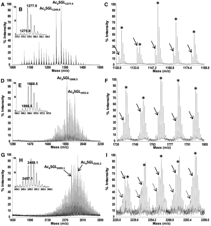
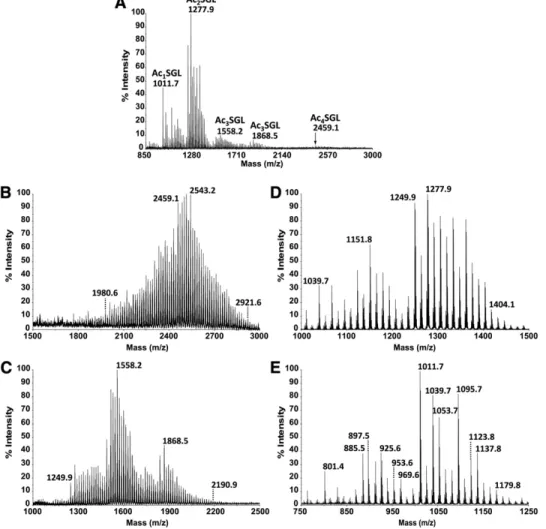
Negative-ion mode MALDI-TOF-MS of purifi ed SGLs ( Fig. 1E ) proved to be the method of choice for charac-terizing the heterogeneity in acylation of the different SGL species. Indeed, for Ac 2 SGL, the mass spectrum ( Fig. 2A ) exhibited a set of peaks ranging from m/z 1,067.7 to 1,446.1, with two major molecular ions, [M-H] 2 at m/z 1,249.9 and m/z 1,277.9. The relative intensities of these ions varied from one culture batch to another, depend-ing on the culture duration (see supplementary Fig. IA, B). The mass range indicated that HPA contained 25 to 54 carbon atoms ( Table 1 ). However, despite this major series of ions, peaks of much weaker intensities were also observed. For instance, the major molecular ion at m/z 1,277.9 dominated another one at m/z 1,275.9 (ratio, 15:1) ( Fig. 2B ), which was tentatively assigned as Ac 2 SGL containing one phthioceranoic acid with 43 carbon atoms (PAC 43 ) and one palmitic acid ( Table 1 ). The relative in-tensity of this series of peaks increases in the m/z range of 1,120–1,240 ( Fig. 2C and Table 1 ), in agreement with those found in the study by Goren ( 6 ), in which PAC 34 and PAC 37 are the major PA released from SGLs. To con-fi rm this assignment, an exact mass measurement was per-formed by using ESI-FT-MS analysis, where the resolution obtained was above 130,000, and the mass accuracy was in the range of 1.5 ppm. The molecular ion at m/z 1,277.9 showed only one molecular species at m/z 1,277.922, agreeing with the calculated molecular mass of 1,277.926 Da for an Ac 2 SGL containing one HPAC 40 or one HPAC 42 (see below). Weak intensity of the molecular ion at m/z 1,275.9 prevented an accurate mass measurement; how-ever, a fraction enriched for this species obtained by HPLC fractionation of Ac 2 SGL (see below) allowed measurement of an accurate mass at m/z 1,275.945, in agreement with the presence of PAC 43 within the molecule (calculated molecular mass of 1,275.947 Da) ( Table 1 ). Nevertheless, molecular species bearing PA represent less than 15% of Ac 2 SGL, with the majority of them containing HPA.
The negative-ion mode MALDI mass spectrum of Ac 3 SGL ( Fig. 2D ) showed a set of peaks with a major ion at m/z 1,868.5. The mass range varied according to the culture batch, illustrated by two examples shown in supplementary
negative- or positive-ion mode, as specifi ed in the text. Spectra from 2,500 to 5,000 laser pulses were summed to obtain the fi -nal spectrum. The 2-(4-hydroxyphenylazo)benzoic acid (HABA) or 2,5-dihydroxybenzoic acid (DHB) matrix (Sigma) was used at a concentration of ⵑ 10 mg/ml in ethanol-water (1:1, v/v). In a typical experiment, 4 µl of glycolipid (5–10 µg) in chloroform-methanol (8:2, v/v) and 4 µl of the matrix solution were mixed with a micropipette, and 0.3 µl of the mixture was then depos-ited on the target. The measurements were internally calibrated at two points with mycobacterial phospholipids myo -inositol mannosides [PIM]).
ESI-FT-MS analysis
SGLs were analyzed by high-resolution Fourier-transform ion cyclotron resonance MS using a hybrid apex-Qe FT-MS system (Bruker Daltonics, Bremen, Germany) equipped with a 7 Tesla actively shielded superconducting magnet and an Apollo dual-ESI/MALDI ion source. Samples ( ⵑ 10 ng • µl 2 1 ) were dissolved in a 50:50:0.001 (v/v/v) mixture of 2-propanol, water, and trieth-ylamine (pH ⵑ 8.5) and sprayed at a fl ow rate of 2 µl • min 2 1 . The capillary entrance voltage was set to 3.8 kV, and the drying tem-perature was set to 150°C. Data acquisition and analysis were per-formed using Apex Control version 3.0 and data analysis version 3.4 software (Bruker Daltonics, Bremen), respectively. The mass spectra were acquired in the negative-ion mode with 1 M data points in the mass range between 1,020 and 1,360 atomic mass units. Mass scale calibration was performed externally with a mix-ture of lipids with known strucmix-tures. The mass resolution ob-tained was above 130,000, and the mass accuracy was in the range of 1.5 ppm.
NMR analysis
NMR spectra were recorded with an Avance DMX500 spec-trometer (Bruker GmbH, Karlsruhe, Germany) equipped with an Origin 200 SGI, using Xwinnmr version 2.6 software. Native SGLs were dissolved in CDCl 3 -CD 3 OD (4:1, v/v) and analyzed in
200 × 5 mm 535 PP NMR tubes at 295 K. Proton chemical shifts are expressed in ppm downfi eld from the signal of chloroform ( d H /TMS 7.27 and d C /TMS 77.7). All details concerning the use
of 1 H- 1 H COrrelation SpectroscopY (COSY), 1 H- 1 H Homonu-clear Hartman Hahn (HOHAHA), and 1 H- 13 C Heteronuclear Multiple Quantum Coherence (HMQC) sequences and experi-mental procedures were as reported previously ( 17 ).
RESULTS
A purifi cation method for the different SGL acyl forms of M. tuberculosis H37Rv
The fi rst objective of this study was to design a protocol to prepare fractions containing pure and unique SGL acyl forms, namely the tetra-, tri-, and di-acylated SGL forms (Ac 4 -, Ac 3 -, and Ac 2 SGL, respectively) from M. tuberculosis H37Rv lipid extract. The purifi cation was monitored by negative-ion MALDI-TOF-MS. Following acetone precipi-tation, the three acyl forms were detected in the acetone-soluble fraction ( Fig. 1C ) ( 14 ). This fraction was further fractionated on a silicic acid column irrigated by chloro-form containing increasing amounts of methanol. Ac 4 SGL were recovered from F4, Ac 3 SGL from F5 and F6, and Ac 2 SGL from F6 and F7. However, F4 to F6 contained ad-ditional compounds, as revealed by TLC analysis ( Fig. 1D ) and positive-ion mode MALDI-TOF-MS (not shown), which highlighted the presence of neutral compounds.
2,248.9, 2,459.1, and 2,543.2 corresponded to Ac 4 SGL bearing three HPA and one C 16 /C 18 . In this case, two mi-nor series of ions at 2 and 4 u less were recovered ( Fig. 2I , as indicated by solid and dashed arrows, respectively). They were tentatively assigned to the molecular species containing two HPA, one PA, and one C 16 /C 18 and to one HPA, two PA, and one C 16 /C 18 , respectively.
MS/MS uncovers an additional level of molecular complexity
A MS/MS-based approach was developed to characterize fatty acids of each acyl form of SGL. Ac 2 SGL species at m/z Fig. IC and D, with ions ranging from m/z 1,630.3 to 2,120.8
or from m/z 1,404.0 to 2,375.1. From the m/z values, we deduced that the major series of ions corresponded to Ac 3 SGL bearing two HPA and one C 16 /C 18 . Again, how-ever, a minor series of ions at 2 u less ( Fig. 2F , arrow) was observed and tentatively assigned to molecular species containing one PA instead of an HPA, although ESI-FT-MS analysis was not performed in this case.
As for Ac 3 SGL, the mass range of ions recorded for Ac 4 SGL varied according to culture batches, for example, from m/z 1,936.6 to 2,851.6 or from m/z 1,756.4 to 2,725.4 (see supplementary Fig. IE, F). The main ions at m/z
Fig. 2. MALDI MS spectra in negative-ion mode of M. tuberculosis H37Rv Ac 2 SGL (A–C), Ac 3 SGL (D–F), and Ac 4 SGL (G–I). The different
views correspond to different expanded areas. In panels B, E, and H, the isotopic profi le of one major ion is presented. In panels A, D, and G, the arrows indicate the species targeted for MS/MS analysis shown in Fig. 3 and supplementary Fig. II. In panels C, F, and I, the species containing HPA are indicated by asterisks, the species containing one PA are indicated by arrows, and the species containing two PA are indicated by dashed arrows.
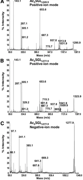
described above, generated fragment ions arising from the loss of the fatty acid in the 3-position only (not shown). For instance, the precursor ions [M-H] 2 at m/z 1,277.9 showed the loss of HPAC 40 or HPAC 42 with fragment ions recorded at m/z 669.3 and 641.3, respectively ( Fig. 3C ). However, loss of C 16 or C 18 was not observed for these pre-cursor ions or for those at m/z 1,249.9, confi rming that the short fatty acids are located on the 2-position, whereas HPA are located on the 3-position of the Glc p unit.
When applied to Ac 3 SGL and Ac 4 SGL, this method re-vealed the level of molecular complexity of each m/z spe-cies. Concerning Ac 3 SGL, we specifi cally focused on the species detected in negative-ion mode at m/z 1,868.5 (Ac 3 SGL 1868.5 ) and m/z 1,952.6 (Ac 3 SGL 1952.6 ) ( Fig. 2D ). As expected, positive MS/MS analysis of the sodiated precur-sor ions [M+2Na] + of Ac 3 SGL 1868.5 at m/z 1,914.5 showed [RCOONa + Na] + fragment ions corresponding to both HPA and short fatty acids (C 16 and C 18 ). The major HPA ions (see supplementary Fig. IIA) were not only at m/z 653.6 (HPAC 40 ), as ions of lower intensity at m/z 569.5 (HPAC 34 ), 611.5 (HPAC 37 ), 667.6 (HPAC 41 ), 681.6 (HPAC 42 ), 695.6 (HPAC 43 ), 737.7 (HPAC 46 ) were also present. Therefore, Ac 3 SGL 1868.5 corresponds not only to SGL acylated by one C 18 and two HPAC 40 or one C 16 /one HPAC 40 /one HPAC 42 but also to SGL acylated by one C 16 / two HPAC 41 , one C 18 /one HPAC 34 /one HPAC 46 , and one C 18 /one HPAC 37 /one HPAC 43 . For Ac 3 SGL 1952.6 , HPAC 40 was again the major HPA species detected alongside ions with lower intensities, indicating HPAC 41 , HPAC 42 , HPAC 43 , HPAC 45 , and HPAC 46 (see supplementary Fig. IIB) in addi-tion to the short fatty acids C 16 and C 18 . Ac 3 SGL 1952.6 was, thus, also a combination of at least fi ve different forms 1,249.9 (Ac 2 SGL 1249.9 ) were therefore unambiguously
as-signed to contain a unique composition of one C 16 /one HPAC 40 , whereas the species at m/z 1,277.9 (Ac 2 SGL 1277.9 ) corresponded to both a major form with one C 18 /one HPAC 40 and a minor form with one C 16 /one HPAC 42 . In-deed, the sodiated precursor ion [M+2Na] + of Ac 2 SGL 1249.9 at m/z 1,295.9 in positive mode showed fragment ions at m/z 653.6 and m/z 301.2, corresponding to sodiated cat-ions [RCOONa + Na] + of HPAC 40 and C 16 , respectively, and at m/z 687.3 arising from the loss of HPAC 40 ( Fig. 3A ). Pre-cursor ions of Ac 2 SGL 1277.9 at m/z 1,323.9 gave HPAC 40 and C 16 fragment ions and additional ions at m/z 681.6 and m/z 329.2 arising from HPAC 42 and C 18 , respectively, with HPAC 40 and C 18 fragment ions being the most abundant ( Fig. 3B ). Fragment ions at m/z 687.3 and 715.3 corre-sponding to the loss of HPAC 42 or HPAC 40 , respectively, were also observed ( Fig. 3B ). MS/MS spectra showed ad-ditional informative fragment ions corresponding to glu-cose or trehalose units bearing HPA. Among them were ions attributed to (anhydro)-di-acylated-sodiated-glucose structures at m/z 1,031.9 (anhydro form at m/z 1,013.9) for Ac 2 SGL 1249.9 ( Fig. 3A ) or at m/z 1,059.9 (anhydro form at m/z 1,041.9) for Ac 2 SGL 1277.9 ( Fig. 3B ). These ions, arising from the loss of (anhydro)-sulfo-glucose, indicated that both fatty acyl appendages were located on the same glu-copyranosyl (Glc p ) unit, i.e., the unit that does not bear the sulfate group. Interestingly, the relative distribution of the different fatty acids on positions 2 and 3 of the Glc p could be deduced from negative-ion MALDI-MS/MS. In-deed, using synthetic Ac 2 SGL molecules with defi ned fatty acids on each position, we observed that MS/MS in the negative-ion mode, in contrast to the positive-ion mode
TABLE 1. m/z values of the different Ac 2 SGL acyl forms containing one C 16 or C 18 at the 2-position of the
acylated glucose and one HPA, one PA, or one OPA at the 3-position
one HPAC 46 ; one HPAC 34 /one HPAC 42 /one HPAC 44 ; one HPAC 34 /two HPAC 43 ; two HPAC 37 /one HPAC 46 ; one HPAC 37 /one HPAC 40 /one HPAC 43 ; one HPAC 37 /one HPAC 41 /one HPAC 42 ; or three HPAC 40 .
New SGL molecular species revealed by HPLC-based fractionation of Ac 2 SGL acyl forms
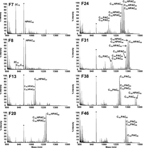
The data described above clearly show that even the sim-plest SGLs, i.e., Ac 2 SGL, is actually a complex mixture of molecular species. We therefore explored the possibility of resolving this complexity by (C 18 ) reverse-phase HPLC (RP-HPLC) monitored by off-line negative MALDI-TOF-MS analysis ( Fig. 4 ). An increasing gradient of dichloromethane in methanol was used as mobile phase to successively elute the different acyl forms, from the most polar to the most apolar molecular species. The fi rst SGL eluted were ob-served in F7 and F8 and corresponded to unexpected spe-cies at m/z 897.5 and 925.6 and were assigned by MS/MS experiments to SGL esterifi ed by short fatty acids (two C 16 and one C 16 /one C 18 , respectively), as well as a compound at m/z 1,011.6 assigned by positive-ion mode MS/MS ex-periments (not shown) to a previously unknown Ac 1 SGL esterifi ed by a single HPAC 40 . Indeed, in the MS/MS spec-trum, only fragment ions corresponding to HPAC 40 at m/z 653.6 and no fragment ions corresponding to C 16 or C 18 at m/z 301.2 or 329.2 were detected. Moreover, in negative-ion mode, we observed fragment negative-ions arising from the loss of HPAC 40 , proving that HPAC 40 is located on the 3-position of the glucose. Next , in fractions F9 to F40, a series of com-pounds characterized by MALDI-TOF-MS with peaks at m/z from 1,039.7 to 1474.1 were assigned by MS/MS experi-ments to Ac 2 SGL containing one C 16 /C 18 and one HPAC 25 to HPAC 55 ( Table 1 ). We could not separate Ac 2 SGL popu-lations containing HPA from those containing PA. Indeed, intermediate fractions, such as F24, F31, or F38 contained Ac 2 SGL with long HPAs together with Ac 2 SGL with shorter PAs. Ac 2 SGLs with PAC 31 to PAC 48 were observed from F22 to F49.
SGL of strains other than the M. tuberculosis H37Rv laboratory strain
SGLs were investigated in strains other than M. tuberculosis H37Rv, either from the tuberculosis complex, such as Mycobacterium bovis bacillus Calmette-Guérin or M. tuberculosis H37Ra, or not, such as Mycobacterium chelonae , Mycobacterium fortuitum , Mycobacterium gastri , Mycobacterium kansasii , Mycobacterium marinum , Mycobacterium smegmatis , and Myco-bacterium xenopi . For each extract, the acetone-soluble phase was prepared and analyzed by negative-ion mode MALDI-TOF-MS for SGL content. None of these strains was found to produce SGLs. The absence of SGLs in M. tuberculosis H37Ra has already been correlated with a point mutation in the PhoP regulator ( 19, 20 ). These data are in agree-ment with those of previous studies showing that SGLs are specifi c glycolipids from M. tuberculosis species ( 21, 22 ).
Because M. tuberculosis H37Rv is a laboratory strain, one could argue that the acyl profi les could be merely a result of the passage of this strain for over 70 years. Then, the SGL acyl profi le of a clinical isolate belonging to the Central containing either one C 16 /one HPAC 40 /one HPAC 46 ; one
C 16 /one HPAC 41 /one HPAC 45 ; one C 16 /2 HPAC 43 ; one C 18 /1 HPAC 41 /one HPAC 43 ; or one C 18 /two HPAC 42 .
As for Ac 4 SGL, MS/MS analysis targeted the major species detected in negative-ion mode at m/z 2,459.1 (Ac 4 SGL 2459.1 ) and m/z 2,543.2 (Ac 4 SGL 2543.2 ) ( Fig. 2G ). As we expected, we observed the same fragment ion signa-ture in the MS/MS spectra (see supplementary Fig. IIC, D) as those observed for Ac 3 SGL. In fact, Ac 4 SGL 2459.1 and Ac 4 SGL 2543.2 corresponded to Ac 3 SGL 1868.5 and Ac 3 SGL 1952.6 acylated by an additional HPAC 40 , respectively. Indeed, the major HPA ions were detected at m/z 653.6 (HPAC 40 ), with ions of lower intensity corresponding to HPAC 34 , HPAC 37 , and HPAC 41 to HPAC 47 , resulting in many possi-ble combinations for each acyl form. For example, consid-ering Ac 4 SGL 2459.1 (see supplementary Fig. IIC) and only the forms containing C 18 , we can propose seven different acyl forms containing either one HPAC 34 /one HPAC 40 /
Fig. 3. MALDI-TOF MS/MS spectra of M. tuberculosis H37Rv
Ac 2 SGL are shown. A: Positive-ion mode MS/MS spectrum is
shown of precursor ions [M+2Na] + at m/z 1,295.9 of Ac 2 SGL 1249.9 . B:
Positive-ion mode MS/MS spectrum is shown of precursor ions [M+2Na] + at m/z 1,323.9 of Ac 2 SGL 1277.9 . C: Negative-ion mode
m/z 1,039.7 to 1404.1, with peaks ranging between m/z 1,249.9 and 1,404.1 predominating, as previously observed for M. tuberculosis H37Rv Ac 2 SGL (see supplementary Fig. IB). The new result concerns the purifi cation of an entire family of mono-acylated SGL observed in the mass spec-trum from m/z 801.4 to m/z 1,179.8 ( Fig. 5E ). This was con-fi rmed by MS/MS analysis, which revealed that these SGLs are acylated by one HPAC 25 to HPAC 52 . Moreover, in this fraction, species at m/z 897.5, 925.6, and 953.6 assigned by MS/MS experiments to SGLs esterifi ed by short fatty acids (two C 16 , one C 16 /1C 18 , and two C 18 , respectively) were also observed ( Fig. 5E ).
Unexpected SGLs in M. tuberculosis mmpL8 knockout strains
M. tuberculosis mmpL8 knockout mutants (from H37Rv and Erdman strains) have been reported to show both a dramatic defect in Ac 4 SGL synthesis and an intracellular accumulation of Ac 2 SGL ( 13, 15 ). We purifi ed Ac 2 SGL Asian India clade ( 16 ) was also studied. The complete SGL
profi le was fi rst analyzed by negative-ion mode MALDI-TOF-MS of the acetone-soluble phase ( Fig. 5A ). Surpris-ingly, the previously characterized M. tuberculosis H37Rv Ac 1 SGL species at m/z 1,011.7 was very abundant, and the ions corresponding to Ac 4 SGL were very weak compared with those of the MALDI-TOF-MS assay results of the M. tuberculosis H37Rv acetone-soluble phase ( 14 ). Then, the different acyl forms were purifi ed and analyzed by MALDI-TOF-MS (Fig. 5B–E ). In all cases, the major SGL species remained essentially the same as the ones described in M. tuberculosis H37Rv. The MS profi le of Ac 4 SGL ( Fig. 5B ) was very similar to that of M. tuberculosis H37Rv (see supple-mentary Fig. IE). As for Ac 3 SGL, ions ranging from m/z 1,249.9 to 2,190.9 were observed ( Fig. 5C ), clearly indicat-ing two families of Ac 3 SGL: one family with two C 16 /C 18 and one HPA and one family with one C 16 /C 18 and two HPA, as confi rmed by MS/MS analysis. The Ac 2 SGL mass spectrum ( Fig. 5D ) exhibited a set of peaks ranging from
Fig. 4. RP-HPLC purifi cation of Ac 2 SGL, monitored by off-line negative-ion mode MALDI-TOF-MS is shown. MALDI-MS mass spectra are
shown of specifi c 1 ml fractions collected after injection of 50 µg of Ac 2 SGL on C 18 Atlantis column. The fatty acid composition of Ac 2 SGL
group (i.e., oxophthioceranoic acyl [OPA], with a calcu-lated molecular mass of 1,275.910 Da) rather than the presence of a PA on the molecule (calculated molecu-lar mass of 1,275.947 Da). To confi rm this assumption, m1Ac 2 SGL and m2Ac 2 SGL were submitted to reduction in the presence of sodium borodeuteride (NaBD 4 ). Based on the premise that only a ketonic group and not an alcoholic group would be sensitive to the reduction, we expected this treatment to displace the molecular species contain-ing one OPA of 3 u but not those containcontain-ing a PA or an HPA. As expected, no shift was observed with WT M. tuber-culosis Ac 2 SGL (see supplementary Fig. IIIA,B), whereas the species at m/z 1,275.9 in m1Ac 2 SGL was displaced to m/z 1,278.9 (see supplementary Fig. IIIC,D). The intensity of the remaining signal at m/z 1,275.9 after reduction (see supplementary Fig. IIID) reveals the proportion of Ac 2 SGL containing one PA, as observed for the WT strain ( Fig. 2B ). The fatty acid composition of the Ac 2 SGL molecular species at m/z 1,275.9 was confi rmed by positive MS/MS analysis (not shown). OPAC 40 and OPAC 42 were detected at m/z 651.6 and 679.6, respectively, in addition to the presence of C 16 and C 18 , indicating that the Ac 2 SGL species is esteri-fi ed mainly by one OPAC 40 /one C 18 and less frequently by one OPAC 42 /one C 16 ( Table 1 ).
from both mutant strains (called m1Ac 2 SGL for the mu-tant generated by Domenech et al. [ 13 ] and m2Ac 2 SGL for the mutant generated by Converse et al. [ 15] ). Both of these strains gave a similar negative-ion mode MALDI-TOF-MS profi le ( Fig. 6A,B ). However, their profi les dif-fered from that of the M. tuberculosis H37Rv wild-type (WT) strain Ac 2 SGL ( Fig. 6C ) by the presence of a second series of peaks with the same intensities but with a difference of 2 u less ( Fig. 6D, E, and F ). This difference could be due to the presence of either i ) a double bond on HPA; ii ) a PA instead of an HPA, as a fatty acid devoid of hydroxyl group (meaning PA instead of HPA, 2 16) and of one CH 2 unit longer (+14) results in a fi nal difference of 2 2; or iii ) a ketonic instead of an alcoholic function on the fatty acid. The fi rst hypothesis was ruled out by 1 H NMR analysis (not shown), which did not reveal any signal at approximately 6 ppm, indicative of a double bond. To investigate the two last hypotheses, we fi rst performed an exact mass measure-ment by ESI-FT-MS of m1Ac 2 SGL, which gave single mo-lecular species at m/z 1,275.909 and 1,277.921. As expected, the latter measurement was in good agreement with the calculated molecular mass of 1,277.926 Da for an Ac 2 SGL containing an HPA, whereas the former ion corresponded better to the presence of a ketonic function on the acyl
Fig. 5. SGLs of a clinical M. tuberculosis CAS isolate. MALDI-TOF-MS spectra in negative-ion mode of the “Acetone-Soluble” fraction (A), and of purifi ed Ac 4 SGL (B), Ac 3 SGL (C), Ac 2 SGL (D) and Ac 1 SGL (E).
Fig. ID). Indeed, positive MS/MS analysis of WT Ac 3 SGL 1516.2 , observed in the MALDI mass spectrum presented in sup-plementary Fig. ID, revealed an Ac 3 SGL esterifi ed by two C 16 /one HPAC 42 or by one C 16 /one C 18 /one HPAC 40 . Moreover, interestingly, the m1Ac 3 SGL structure differed from that of the WT Ac 3 SGL, as deduced from
1
H- 1 H COSY, 1
H- 1 H HOHAHA, and 1 H- 13 C HMQC NMR analyses (see supplementary Fig. V). Indeed , the presence of an acyl group could be easily deduced from 1 H chemical shifts, as the germinal acyloxy proton is deshielded compared with the proton of a nonacylated position. The carbon chemi-cal shifts are almost unaffected by the acylation ( 23 ). Con-sidering WT Ac 2 SGL, as previously published ( 14 ), the downfi eld chemical shifts of Glc p -II H2 and H3 at 4.74 and 5.24 ppm, respectively, proved that fatty acyl appendages are located on C2 and C3 of Glc p -II (see supplementary Fig. VA and supplementary Table I). For WT Ac 3 SGL, H6 of Glc p -II is deshielded at 4.26 ppm, compared with d 3.49/3.82 for Ac 2 SGL (see supplementary Fig. VB), in ac-cordance with the presence of a fatty acid on C6 of Glc p -II, as previously described ( 8 ). For WT Ac 4 SGL, H6 of Glc p -I is deshielded at 4.15 ppm (see supplementary Fig. VC), com-pared with d 3.59/3.55 for Ac 3 SGL, in accordance with the presence of another fatty acid on C6 of Glc p -I. In the case of m1Ac 3 SGL,
1
H chemical shifts proved that they are acy-lated on C2 and C3 of the nonsulfated a -D-Glc p -II and on C6 ′ of the sulfated a -D-Glc p -I (see supplementary Fig. VD and supplementary Table I). This acylation pattern was fur-ther confi rmed by positive-ion mode MS/MS analysis of m1Ac 3 SGL 1528.2 , which showed fragment ions at m/z 543.2, assigned as a palmitoyl-sulfo-Glc p unit, instead of the ions at m/z 305.0, corresponding to the sulfo-Glc p usually de-tected for the WT Ac 3 SGL (not shown).
DISCUSSION
In order to gain better insight into the structure/func-tion relastructure/func-tionships underlying the CD1 presentastructure/func-tion of SGL to T cells ( 14 ), we made great attempts to improve both their purifi cation and structural characterization.
The fi rst objective of this study was to prepare fractions containing pure and unique SGL acyl forms, namely Ac 2 SGL, Ac 3 SGL, and Ac 4 SGL. To achieve this goal, two steps were crucial: i ) to get rid of the phospholipids, par-ticularly mycobacterial PIM, from the lipidic extract, thus making the acetone-precipitation an inevitable step; and ii ) to eliminate neutral lipid contaminants such as TAT and DAT from SGL fractions, using anion exchange chromatography. Goren et al. previously characterized fi ve SGL families, four of which were tetra-acylated (SL-I, SL-I ′ , SL-II, and SL-II ′ ), and one was tri-acylated (SL-III) (3 – 6). Those au-thors used an elegant strategy based on chemical degrada-tion/modifi cation and subsequent MS and infrared spectroscopy analyses that allowed i ) identifi cation of tre-halose-2-sulfate, ii ) determination of the acylated positions on the trehalose core, iii ) identifi cation of the acyl sub-stituents (C 16 /C 18 , HPA, and PA), and iv ) the determina-tion that the 2-posidetermina-tion of SL-I is acylated by one C 16 or C 18 and that the 3-position is occupied almost exclusively by Only trace levels of Ac 3 SGL and Ac 4 SGL could be
purifi ed from the M. tuberculosis H37Rv mmpL8 mutant (m1Ac 3 SGL and m1Ac 4 SGL). Whereas 1 to 1.5 mg of Ac 2 SGL was purifi ed from 1 liter of M. tuberculosis H37Rv mutant culture, only 200 to 300 µg of Ac 3 SGL and 20 to 50 µg of Ac 4 SGL were obtained, respectively. Surprisingly, m1Ac 3 SGL and m1Ac 4 SGL analyses also gave unexpected negative-ion mode MALDI mass spectra. Indeed, their molecular masses were lower than that of the SGL present in the WT strain (see supplementary Fig. IV). However, because m1Ac 4 SGL were present at trace levels, their analysis by MS/MS and their purifi cation for NMR studies could not be completed. Concerning m1Ac 3 SGL, the major species were observed at m/z 1,514.2 and 1,528.2 and were shown esterifi ed by one OPA and two short fatty acids (C 16 /C 18 ). Indeed, as demonstrated by positive MS/MS analysis, they are mainly esterifi ed by one C 16 /one C 18 /one OPAC 40 or by two C 16 / one OPAC 42 and one C 16 /one C 18 /one OPAC 40 , respec-tively (not shown). It is noteworthy that an Ac 3 SGL popula-tion bearing two simple fatty acids can be detected in some batches of WT M. tuberculosis H37Rv (see supple mentary
Fig. 6. Comparative MALDI-MS analyses of Ac 2 SGL purifi ed
from both D mmpL8 mutants of M. tuberculosis and M. tuberculosis H37Rv are shown. Negative-ion mode MALDI-TOF-MS spectra of Ac 2 SGL of mmpL8 :: hyg M. tuberculosis H37Rv Pasteur strain (A) and
D mmpL8 jcm108 M. tuberculosis Erdman strain (B) and M.
tuberculo-sis H37Rv (C) are shown. Expanded area ( m/z 1,272–1.,284) is
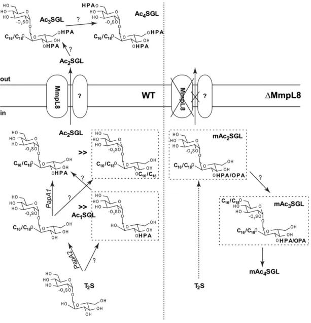
M. tuberculosis H37Rv SGL species, i.e., Ac 2 SGL esterifi ed by simple fatty acids only, namely palmitoyl-SGL, di-stearoyl-SGL, mono-palmitoyl-mono-di-stearoyl-SGL, and Ac 1 SGL bearing a single HPA. This last species was even more abundant in the clinical strain, and a complete fam-ily of Ac 1 SGL acylated by one HPA of variable length was unambiguously characterized. The identifi cation of this mono-acylated species was unexpected, as the fi rst acyla-tion step is considered to be a palmitoylaacyla-tion/stearoyla- palmitoylation/stearoyla-tion on the 2-posipalmitoylation/stearoyla-tion of the sulfated trehalose ( Fig. 1A ) and because PapA1, the acyl-transferase that adds the HPA on C2-palmitoyl/stearoyl-sulfated trehalose, has been shown to harbor no activity toward sulfated trehalose ( 25 ). More clues about the biosynthetic pathway of SGLs arose from an in-depth analysis of SGLs produced by D mmpL8 mutants. Our present knowledge indicates that sulfation of trehalose, catalyzed by the sulfotransferase Stf0, is the fi rst step in SGL synthesis ( 26 ). Palmitate or stearate acyl chains derive from fatty-acid synthase-I (FAS-I), whereas HA and HPA are synthesized by polyketide synthase 2 (Pks2) ( 27 ) working in conjunction with the FadD23 fatty-acyl-CoA ligase ( 28, 29 ). Then, two acyl-transferases, PapA1 on one hand and PapA2 associated with Pks2 on the other hand, would act sequentially ( 25, 30 ), where PapA2 con-verts sulfated trehalose into 2-palmitoyl/stearoyl-sulfated trehalose, and PapA1 further elaborates Ac 2 SGL by adding a methyl-branched fatty acid on the 3-position of treha-lose. The MmpL8 protein, predicted to be a lipid trans-porter, is described as playing a role in mediating the transport of the Ac 2 SGL precursor forms from the cytosol into the cell envelope ( 13, 15 ). Indeed, characterization of the D mmpL8 mutants showed that synthesis of mature Ac 4 SGL was halted and that Ac 2 SGL concomitantly accu-mulated within the cell. This strongly suggests that trans-port and biogenesis of SGLs are coupled and that the fi nal step in SGL biosynthesis might be the extracellular acyla-tion of the C6 posiacyla-tion of both a -D-Glc p units ( 13 ). Here, we show that the D mmpL8 mutant was still able to generate Ac 3 SGL but with a structure different from that of the WT strain. Indeed, the third added fatty acid was not only C 16 / C 18 , rather than HPA/HA, but was in a different position: C6 ′ of the sulfated a -D-Glc p instead of the C6 of the 2,3-di-acylated a -D-Glc p unit. This result proves that Ac 2 SGL must be transported (or translocated) across the cell mem-brane by MmpL8 in order to be correctly transformed into the fi nal Ac 4 SGL ( Fig. 7 ). Another intriguing fi nding was that approximately half of the D mmpL8 mutant SGLs car-ried a methyl-branched fatty acid containing a ketonic function (OPA) instead of the hydroxyl group, a form that does not exist in the WT. This result strongly suggests that MmpL8 functions in conjunction with enzymes involved in the elaboration of the fi nal methyl-branched fatty acids.
Two hypotheses are currently proposed for acylation steps that generate Ac 4 SGL from Ac 2 SGL: fatty acid addi-tion to both the C6 and C6 ′ positions of the trehalose unit could occur either intracellularly or extracellularly ( 31, 32 ). In the fi rst case, Ac 4 SGL would be elaborated from Ac 2 SGL in the cytosol, and Ac 4 SGL would be transported one PA ( 6 ). In this way, they demonstrated that the major
species (SL-I) was acylated on the 3-, 6-, and 6 ′ -positions by one PA and two HPA; SL-I ′ was acylated by two PA and one HPA; and SL-II was acylated by three HPA. SL-II ′ was also described as being acylated by three HPA on the 4-, 6-, and 6 ′ -positions. The tri-acylated SL-III was shown to be acy-lated on the 3- and 6-positions by two HPA.
In this study, we developed a strategy in which we fi rst purifi ed the different acyl forms before completely charac-terizing them by MALDI-TOF-MS and NMR. We confi rmed most of the structural points described by Goren et al., such as the presence of C 16 /C 18 exclusively on the 2-position; the presence in Ac 3 SGL of the fatty acids on the 2-, 3-, and 6-positions of the nonsulfated glucose; and the fact that HPAC 40 is the principal HPA component. Here, for the fi rst time, we realized a complete NMR analysis of each acyl form, and in contrast to the fi ndings of Goren, we never observed acylation on the 4-position of the glucose unit. We cannot exclude the presence of this acyl form, if present at trace level. We showed that Ac 2 SGL contains mainly one C 16 /C 18 and one HPA on the 2- and 3-positions, respectively, and, in accordance with the study by Goren et al., that Ac 3 SGL contains one C 16 /C 18 and two HPA on the 2-, 3-, and 6-positions. On the basis of a biosynthetic linkage between all these acyl forms and considering our result showing that Ac 4 SGL contains mainly three HPA, it is obvious that the 3-position of Ac 4 SGL is occupied almost exclusively by one HPA. However, Goren et al. found that the 3-position of SL-I (the major tetra-acylated species, ac-ylated by one C 16 or C 18 , one PA, and two HPA) was occu-pied almost exclusively by one PA. This species, in minor abundance in our case, most probably derives from the 15% of Ac 2 SGL species containing one PA (see below). Moreover, Goren et al. described SL-I to be acylated by HPA from C 31 to C 46 . Using MALDI, we derived a precise profi le of the different molecular species composing each SGL acyl form. We concluded that Ac 2 SGL contained HPA in lengths from C 25 to C 54 . Furthermore, analysis of the isotopic masses gave the proportions of PA versus HPA. We deduced that Ac 2 SGL was composed of 15% of species containing one PA and 85% containing one HPA. Ac 4 SGL was distributed in 73% of species with three HPA, 24% with one PA, and 3% with two PA.
Negative-ion mode MALDI-TOF-MS proved to be the method of choice for characterizing the acylation pattern of the different SGL species. Surprisingly, we observed that this pattern, particularly the molecular weight range, varied from one culture batch to another, although the culture conditions were essentially the same, except for the duration of the culture. The lipid chain length of SGL has been recently shown to depend on the availability of precursors such as methyl malonyl CoA ( 24 ), which might be confounded by the growth phase, a parameter that is diffi cult to control because bacteria were grown as cell sur-face pellicles. Nevertheless, the major SGL species remained essentially the same for the different SGL acyl forms.
Fractionation of the Ac 2 SGL mixture by RP-HPLC allowed us to not only separate SGL species containing HPA or PA but also to reveal previously uncharacterized
the n -fatty acyl moiety during the fi rst cycle could be re-duced to the hydroxyl acid but not dehydrated, leaving the hydroxyl group in the fi nal multi-methyl-branched prod-uct ( 27 ). Our data favor this last hypothesis. The occur-rence of 50% OPA in Ac 2 SGL of the mmpL8 mutant suggests that only half of the keto-acid generated by con-densation of methylmalonyl-CoA with the n -fatty acyl moi-ety would be reduced to the hydroxyl acid.
The authors thank Dr. Clifton E. Barry III and Pr. Jeffery Cox for providing M. tuberculosis D mmpL8 mutants and Amélie Vax for technical assistance. We are also grateful to Drs. P. Domenech and C. Chalut for discussions, Dr. L. Sweet for reading the manuscript, and Dr. J. Nigou for critical comments and helpful suggestions.
to the exterior of the cell by MmpL8. In the second hy-pothesis, an extracellular acyl-transferase would convert Ac 2 SGL to Ac 4 SGL after transport of Ac 2 SGL to the exte-rior of the cell by MmpL8. In conclusion, our results favor the extracellular acylation of both C6 and C6 ′ positions. This process seems to parallel what is observed for the gen-eration of cord factor, where acylation of both C6 and C6 ′ positions of trehalose is catalyzed by a mycoloyl-transferase found to be associated with the bacterial cell wall surface ( 33 ).
The generation of HPA is not yet completely under-stood. HPA could result from i ) the introduction of the hydroxyl group into preformed PA or ii ) an incomplete synthesis by Pks2. In the second hypothesis, the keto-acid generated by condensation of methylmalonyl CoA with
Fig. 7. Schematic representation of the pathway of completion of SGL biosynthesis in M. tuberculosis H37Rv (WT) and D mmpL8 mutants of M. tuberculosis ( D mmpL8 ) is shown [adapted from Bertozzi and Schelle ( 31 ) with permission]. The SGLs are not inserted in membrane, as they should be, for simplifi cation and clarity purposes. Our results favor an extracellular acylation of Ac 2 SGL, following the transport of
Ac 2 SGL by MmpL8. Indeed, the MmpL8 knockout strains also synthesize very weak amounts of Ac 3 - and Ac 4 SGL (mAc 3 - and mAc 4 SGL) but
from a different structure. This model implies that HPA is transported by an unknown protein (annotated as “?”) to generate Ac 3 - and
REFERENCES
1 . Middlebrook , G. , C. Coleman , and W. B. Schaefer . 1959 . Sulfolipid from virulent tubercle bacilli. Proc. Natl. Acad. Sci. U S A . 45 : 1801 – 1804 .
2 . Goren , M. B. , O. Brokl , and W. B. Schaefer . 1974 . Lipids of putative relevance to virulence in Mycobacterium tuberculosis : correlation of virulence with elaboration of sulfatides and strongly acidic lipids. Infect. Immun. 9 : 142 – 149 .
3 . Goren , M. B. , O. Brokl , B. C. Das , and E. Lederer . 1971 . Sulfolipid I of Mycobacterium tuberculosis , strain H37Rv. Nature of the acyl sub-stituents. Biochemistry . 10 : 72 – 81 .
4 . Goren , M. B. 1970 . Sulfolipid I of Mycobacterium tuberculosis , strain H37Rv. I. Purifi cation and properties. Biochim. Biophys. Acta . 210 : 116 – 126 .
5 . Goren , M. B. 1970 . Sulfolipid I of Mycobacterium tuberculosis , strain H37Rv. II. Structural studies. Biochim. Biophys. Acta . 210 : 127 – 138 . 6 . Goren , M. B. 1990 . Mycobacterial fatty acid esters of sugars and
sulfosugars . In Handbook of Lipid Research, vol. 6 . D. J. Hanahan , editor. Plenum Press , NY . 363 – 461.
7 . Daffe , M. , F. Papa , A. Laszlo , and H. L. David . 1989 . Glycolipids of recent clinical isolates of Mycobacterium tuberculosis: chemi-cal characterization and immunoreactivity. J. Gen. Microbiol. 135 : 2759 – 2766 .
8 . Goren , M. B. , B. C. Das , and O. Brokl . 1978 . Sulfatide III of Mycobacterium tuberculosis , Strain H37Rv. New J. Chem. 2 : 379 – 384 . 9 . Lemassu , A. , M. A. Laneelle , and M. Daffe . 1991 . Revised
struc-ture of a trehalose-containing immunoreactive glycolipid of Mycobacterium tuberculosis. FEMS Microbiol. Lett. 62 : 171 – 175 . 10 . Baer , H. H. 1993 . The structure of an antigenic glycolipid (SL-IV)
from Mycobacterium tuberculosis. Carbohydr. Res. 240 : 1 – 22 .
11 . Cruaud , P. , J. T. Yamashita , N. M. Casabona , F. Papa , and H. L. David . 1990 . Evaluation of a novel 2,3-diacyl-trehalose-2’-sulphate (SL-IV) antigen for case fi nding and diagnosis of leprosy and tuber-culosis. Res. Microbiol. 141 : 679 – 694 .
12 . Mougous , J. D. , M. D. Leavell , R. H. Senaratne , C. D. Leigh , S. J. Williams , L. W. Riley , J. A. Leary , and C. R. Bertozzi . 2002 . Discovery of sulfated metabolites in mycobacteria with a genetic and mass spectrometric approach. Proc. Natl. Acad. Sci. U S A . 99 : 17037 – 17042 .
13 . Domenech , P. , M. B. Reed , C. S. Dowd , C. Manca , G. Kaplan , and C. E. Barry III . 2004 . The role of MmpL8 in sulfatide biogenesis and virulence of Mycobacterium tuberculosis. J. Biol. Chem. 279 : 21257 – 21265 .
14 . Gilleron , M. , S. Stenger , Z. Mazorra , F. Wittke , S. Mariotti , G. Bohmer , J. Prandi , L. Mori , G. Puzo , and G. De Libero . 2004 . Diacylated sulfoglycolipids are novel mycobacterial antigens stimu-lating CD1-restricted T cells during infection with Mycobacterium
tuberculosis . J. Exp. Med. 199 : 649 – 659 .
15 . Converse , S. E. , J. D. Mougous , M. D. Leavell , J. A. Leary , C. R. Bertozzi , and J. S. Cox . 2003 . MmpL8 is required for sulfolipid-1 biosynthesis and Mycobacterium tuberculosis virulence. Proc. Natl.
Acad. Sci. U S A . 100 : 6121 – 6126 .
16 . Filliol , I. , A. S. Motiwala , M. Cavatore , W. Qi , M. H. Hazbon , M. Bobadilla Del Valle , J. Fyfe , L. Garcia-Garcia , N. Rastogi , C. Sola , et al . 2006 . Global phylogeny of Mycobacterium tuberculosis based on single nucleotide polymorphism (SNP) analysis: insights into tuber-culosis evolution, phylogenetic accuracy of other DNA fi ngerprint-ing systems, and recommendations for a minimal standard SNP set. J. Bacteriol. 188 : 759 – 772 .
17 . Gilleron , M. , C. Ronet , M. Mempel , B. Monsarrat , G. Gachelin , and G. Puzo . 2001 . Acylation state of the phosphatidylinositol manno-sides from Mycobacterium bovis bacillus Calmette Guerin and ability to induce granuloma and recruit natural killer T cells. J. Biol. Chem. 276 : 34896 – 34904 .
18 . Besra , G. S. , R. C. Bolton , M. R. Mcneil , M. Ridell , K. E. Simpson , J. Glushka , H. Van Halbeek , P. J. Brennan , and D. E. Minnikin . 1992 . Structural elucidation of a novel family of acyltrehaloses from Mycobacterium tuberculosis. Biochemistry . 31 : 9832 – 9837 . 19 . Lee , J. S. , R. Krause , J. Schreiber , H. J. Mollenkopf , J. Kowall , R.
Stein , B. Y. Jeon , J. Y. Kwak , M. K. Song , J. P. Patron , et al . 2008 . Mutation in the transcriptional regulator PhoP contributes to avir-ulence of Mycobacterium tuberculosis H37Ra strain. Cell Host Microbe . 3 : 97 – 103 .
20 . Chesne-Seck , M. L. , N. Barilone , F. Boudou , J. Gonzalo Asensio , P. E. Kolattukudy , C. Martin , S. T. Cole , B. Gicquel , D. N. Gopaul , and M. Jackson . 2008 . A point mutation in the two-component regulator PhoP-PhoR accounts for the absence of polyketide-derived acyltrehaloses but not that of phthiocerol dimycocerosates in Mycobacterium tuberculosis H37Ra. J. Bacteriol. 190 : 1329 – 1334 . 21 . Dhariwal , K. R. , G. Dhariwal , and M. B. Goren . 1984 . Observations
on the ubiquity of the Mycobacterium tuberculosis sulfatides in myco-bacteria. Am. Rev. Respir. Dis. 130 : 641 – 646 .
22 . Soto , C. Y. , M. Cama , I. Gibert , and M. Luquin . 2000 . Application of an easy and reliable method for sulfolipid-I detection in the study of its distribution in Mycobacterium tuberculosis strains. FEMS
Microbiol. Lett. 187 : 103 – 107 .
23 . Gilleron , M. , J. Vercauteren , and G. Puzo . 1994 . Lipo-oligo-saccharidic antigen from Mycobacterium gastri . Complete structure of a novel C4-branched 3,6-dideoxy-alpha-xylo-hexopyranose. Biochemistry . 33 : 1930 – 1937 .
24 . Jain , M. , C. J. Petzold , M. W. Schelle , M. D. Leavell , J. D. Mougous , C. R. Bertozzi , J. A. Leary , and J. S. Cox . 2007 . Lipidomics reveals control of Mycobacterium tuberculosis virulence lipids via metabolic coupling. Proc. Natl. Acad. Sci. U S A . 104 : 5133 – 5138 .
25 . Kumar , P. , M. W. Schelle , M. Jain , F. L. Lin , C. J. Petzold , M. D. Leavell , J. A. Leary , J. S. Cox , and C. R. Bertozzi . 2007 . PapA1 and PapA2 are acyltransferases essential for the biosynthesis of the Mycobacterium tuberculosis virulence factor sulfolipid-1. Proc. Natl.
Acad. Sci. U S A . 104 : 11221 – 11226 .
26 . Mougous , J. D. , C. J. Petzold , R. H. Senaratne , D. H. Lee , D. L. Akey , F. L. Lin , S. E. Munchel , M. R. Pratt , L. W. Riley , J. A. Leary , et al . 2004 . Identifi cation, function and structure of the mycobacterial sulfotransferase that initiates sulfolipid-1 biosynthesis. Nat. Struct.
Mol. Biol. 11 : 721 – 729 .
27 . Sirakova , T. D. , A. K. Thirumala , V. S. Dubey , H. Sprecher , and P. E. Kolattukudy . 2001 . The Mycobacterium tuberculosis pks2 gene encodes the synthase for the hepta- and octamethyl-branched fatty acids re-quired for sulfolipid synthesis. J. Biol. Chem. 276 : 16833 – 16839 . 28 . Trivedi , O. A. , P. Arora , V. Sridharan , R. Tickoo , D. Mohanty , and
R. S. Gokhale . 2004 . Enzymic activation and transfer of fatty acids as acyl-adenylates in mycobacteria. Nature . 428 : 441 – 445 .
29 . Lynett , J. , and R. W. Stokes . 2007 . Selection of transposon mutants of Mycobacterium tuberculosis with increased macrophage infectivity identifi es fadD23 to be involved in sulfolipid production and asso-ciation with macrophages. Microbiology . 153 : 3133 – 3140 .
30 . Bhatt , K. , S. S. Gurcha , A. Bhatt , G. S. Besra , and W. R. Jacobs , Jr . 2007 . Two polyketide-synthase-associated acyltransferases are required for sulfolipid biosynthesis in Mycobacterium tuberculosis .
Microbiology . 153 : 513 – 520 .
31 . Bertozzi , C. R. , and M. W. Schelle . 2008 . Sulfated metabolites from Mycobacterium tuberculosis : sulfolipid-1 and beyond . In The Mycobacterial Cell Envelope , M. Daffe and J. M. Reyrat , editors. ASM Press , Washington, DC . 291 – 304 .
32 . Jain , M. , E. D. Chow , and J. S. Cox . 2008 . The MmpL protein fam-ily , In The Mycobacterial Cell Envelope , M. Daffe and J. M. Reyrat , editors. ASM Press , Washington, DC . 201–210.
33 . Wiker , H. G. , and M. Harboe . 1992 . The antigen 85 complex: a major secretion product of Mycobacterium tuberculosis . Microbiol. Rev. 56 : 648 – 661 .