HAL Id: tel-03219351
https://tel.archives-ouvertes.fr/tel-03219351
Submitted on 6 May 2021
HAL is a multi-disciplinary open access archive for the deposit and dissemination of sci-entific research documents, whether they are
pub-L’archive ouverte pluridisciplinaire HAL, est destinée au dépôt et à la diffusion de documents scientifiques de niveau recherche, publiés ou non,
Sarah Beatriz de Oliveira Pagliaro
To cite this version:
Sarah Beatriz de Oliveira Pagliaro. Transcriptional control induced by bcr-abl and its role in leukemic stem cell heterogeneity.. Cancer. Université Paris-Saclay, 2020. English. �NNT : 2020UPASQ032�. �tel-03219351�
Bcr-Abl and its role in leukemic
stem cell heterogeneity
Thèse de doctorat de l'université Paris-Saclay
École doctorale n° 569, Innovation thérapeutique : du fondamental à l'appliqué (ITFA)
Spécialité de doctorat : Physiologie, Physiopathologie Unité de recherche : Université Paris-Saclay, Inserm, Modèles de cellules souches malignes et thérapeutiques, 94805, Villejuif, France. Référent : Faculté de pharmacie
Thèse présentée et soutenue à Villejuif,
le 14 Septembre 2020, par,
Sarah Beatriz DE O. PAGLIARO
Composition du Jury
Jean Henri BOURHIS
Directeur de recherche, Institute Gustave Roussy
Président
Pierre SAVATIER
Directeur de recherche, INSERM U1208 Rapporteur & Examinateur
Paul COPPO
Professeur, Hôpital Saint Antoine, AP-HP
Rapporteur & Examinateur
Jean Claude CHOMEL
Praticien hospitalier, Centre Hospitalier
Universitaire de Poitiers Examinateur
Ali TURHAN
PU-PH, Université Paris-Saclay Directeur de thèse
Thè
se
d
e d
oct
orat
NNT : 2020UPASQ032AND ITS ROLE IN LEUKEMIC STEM CELL
HETEROGENEITY
Presented by Sarah B. de O. Pagliaro
Public Discussion (14 september 2020)
Inserm U935
Supervised by Prof A Turhan
Université Paris Saclay
1.1 Historical background 9
1.2 Philadelphia Chromosome 10
1.3 The Bcr-Abl protein 11
1.3.1 The Bcr-Abl isoforms 11
1.3.2 Consequences of BCR-ABL1 generation in hematopoietic cells 13
1.3.2.1 Signaling pathways 14 1.3.2.1.1 JAK/STAT 14 1.3.2.1.2 PI3K/AKT/CRK 14 1.3.2.1.3 RAS / MEK 15 1.3.2.1.4 SRC KINASES 16 1.3.2.1.5 DEREGULATION OF APOPTOSIS 16 2. Hematopoiesis 18
2.1 Hematopoietic stem cells (HSC) 19
2.1.1 In vitro detection tests 20
2.1.2 In vivo detection tests 21
2.1.3 Progenitors and precursors and mature cells 22
2.2 Regulation of the hematopoiesis 24
2.2.1 The microenvironment 24
2.2.1.1 Osteoblastic niche 25
2.2.1.2 Vascular niche 25
2.2.1.3 Molecular mechanisms within the microenvironment 26
2.2.1.4 Growth factors 27
2.2.1.4.1 Positive regulators of hematopoiesis 28
2.2.1.4.2 Negative regulators of hematopoiesis 28
2.2.1.5 Other stimulating factors for HSCs and progenitors 28
2.2.1.6 Intrinsic regulation 29
2.3 CML Stem Cells vs Hematopoietic Stem Cells 31
3 Modelling CML 32
3.2.1 Cell Lines 35
3.2.2 Primary leukemic cells 35
3.2.3 iPSC 36
4. Diagnosis. 37
4.1 Evolution of the disease and prognosis 37
5. CML Therapy 39
5.1 Treatment History 39
5.1.1 The introduce of TKIs, a turning point in CML history 39
5.1.2 Hematopoietic Stem Cell Transplantation (HSCT) 40
5.2. Conventional therapies 41
5.2.1 Chemotherapy 41
5.2.2 Interferon alpha (IFNα) 41
5.3 Targeted therapies – TKIs 42
5.3.1 1st generation TKI 42 5.3.1.1 Imatinib 42 5.3.2 2nd generation TKIs 43 5.3.2 1 Dasatinib 43 5.3.2.2 Nilotinib 44 5.3.2.3 Bosutinib 45
5.3.3 3rd generation TKI (mutation T315I) 45
5.3.3.1 Ponatinib 45
5.4 Mechanisms of resistance 46
5.4.1 Efflux mechanisms and carrier hOCT1 46
5.4.2 SRC overexpression 47
5.4.3 Increased expression of BCR-ABL1 47
5.4.4 Mutations of the ABL kinase domain 48
5.5.3 Quiescence phenomenon 55
6. Allogeneic HSCT in a post-TKI era 58
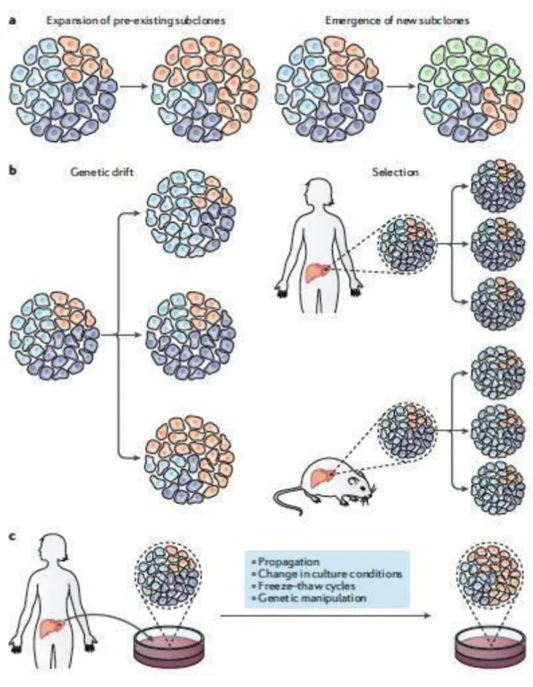
7. Tumor Heterogeneity : Overview 60
7.1 Implications of Heterogeneity 62
7.2 Heterogeneity of leukemic cells in CML 63
8. Transcriptional heterogeneity 64
8.1 Transcriptional heterogeneity in leukemia 64
9. Eukaryotic Elongation 2 Kinase 65
9.1 EEF2K in normal cells 65
9.21 EEF2K in cancer 66
10. Methods of single cell analyses 68
10.1 Single Cells and Computational analysis 72
10.2 Single cell isolation techniques 72
10.2.1 Data Analysis 74
10.3 C1 from Fluidigm 74
10.4 Monocle and Pseudotime 75
11. Single-cell applications in cancer 81
Objectives of the Thesis 82
Paper 1. Summary 83
Paper 1 Summary (in French) 86
Paper 1 – IN PRESS 89
Paper 2 Summary 104
Paper 2 Summary (in French) 107
Paper 2 IN PREPARATION 109
General Discussion 142
5-FU 5-Fluorouracil
5-LO 5-lipoxygenase
ABL Abelson murine leukemia viral oncogene
AHI 1 Abelson Helper Intergration Site 1
AHR aryl hydrocarbon receptor
AKT RAC-alpha serine/threonine-protein
ALL Acute Lymphoid Leukemia
Alox5 Arachidonate 5-lipoxygenase
AML Acute Myeloid Leukemia
Ang-1 angiopoietin-1
AP Acute Phase
ATP adenosine triphosphate
B-ALL Acute B Lymphoid Leukemia
BCL2 B-cell lymphoma 2
BCL-X1 B-cell lymphoma-extra large
BCR Breakpoint Cluster Region
BFU burst-forming unit
BM Bone Marrow
BMDW Bone Marrow Donors Worldwide
BMT Bone Marrow Transplantation
BP Blastic Phase
CAD coronary artery disease
CAFC cobblestones Area Forming Cell
CCgR complete cytogenetic remission
CD(number) Cluster Differentiation (number)
CFC Colony Forming Cells
CFU Colony Forming Unit
CFU-M Colony Forming Unit - Macrophage
CMP Col1a1 CP
common myeloid progenitor collagen a1 type 1
Chronic Phase
CRKL CRK Like Proto-Oncogene
CSC cancer stem cells
CVA cerebrovascular disease
CXCL CXC chemokine ligand
CXCR CXC chemokine receptor
DNA deoxyribonucleic acid
ECM extracellular matrix
EPO erythropoietin
ERK Mitogen-Activated Protein Kinase
ET essential thrombocythemia
FACS Fluorescence Activated Cell Sorting
FLT3-L Fms-Like Tyrosine kinase 3-ligand
G Granulocyte
GAB2 Growth Factor Receptor Bound Protein 2
GATA Globin Transcription Factor
G-CSF Granulocyte-Colony Stimulating Factor
GEF Guanine nucleotide exchange factors
GEMM granulocyte, erythrocyte, monocyte, megakaryocyte
GM granulo-macrophagic
GM-CSF Granulocyte and Macrophage-Colony Stimulating Factor
GTP Guanosine-5'-triphosphate
HCK Hemopoietic Cell Kinase
HOXB4 Homeobox B4
HSC Hematopoietic Stem Cells
HSPC Hematopoietic Stem and Progenitor Cell
IL-2 Interleukin-2
IL-3 Interleukin-3
IL-4 Interleukin-4
IL-6 Interleukin-6
IM Imatinib
IRIS International Randomized Study of Interferon
JAK Janus tyrosine kinases
KLF4 Kruppel-like factor 4
LIN Lineage
Lmo-2 LIM domain only 2
LSC Leukemic Stem Cell
LTB4 Leukotriene B4
LTC-IC long-term culture initiating cells
MCL-1 Induced Myeloid Leukemia Cell Differentiation Protein
M-CSF Macrophage-Colony Stimulating Factor
MDR-1 Multi Drug Resistance 1
MEK Mitogen-Activated Protein Kinase
MF primary myelofibrosis
Mip-1α Macrophage inhibitory protein-1α
MK megakaryocyte
MMP matrix metalloprotease
MMR Major Molecular Response
MPP immature multipotent progenitor
MR Molecular Response
mRNA Message Ribonucleic Acid
mTOR Mechanistic Target Of Rapamycin Kinase
NANOG Nanog Homeoboc
NK Natural Killer
OPN osteopontin
PAOD peripheral arterial occlusive disease
PDFG Platelet-Derived Growth Factor
PDX Patient Derived Xenograft
PF4 Platelet Factor-4
Ph Philadelphia Chromosome
PI3K Phosphatidyl-inositol-3-kinase
PML Promyelocytic Leukemia
PTEN Phosphatase and Tensin Homolog
PTH parathormone
PV Polycythemia Vera
SCF Stem Cell Factor
SDF-1 Stromal Derived Factor-1
SH Src Homology
Smo Smoothened
SOX2 SRY-Box Transcription Factor 2
SR1 Stem Regenin
SRC Sarcoma
STAT Signal Transducer and Activators of Transcription
STI selective tyrosine kinase inhibitors
STIM Stop IMatinib
TCDD 2,3,7,8-tetrachlorodibenzo-p-dioxin
TGFβ Transforming growth factor beta
TK Tyrosine Kinase
TKI Tyrosine Kinase Inhibitor
TNF-α Tumor Necrosis Factor-α
TPO thrombopoietin
WHO World Health Organization
CML is a myeloproliferative disorder that is currently classified in the context of myeloproliferative neoplasia along with polycythemia vera (PV), primary myelofibrosis (MF) essential thrombocythemia (ET) chronic neutrophil leukemia, chronic eosinophilic leukemia, and unclassifiable myeloproliferative neoplasias. 1
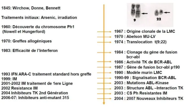
1.1 Historical background
The first descriptions of clinical cases corresponding to CML have been reported in 19th
century. In 1840s, two cases of patients presenting with fever, major splenomegaly, leukocytosis were published, almost simultaneously, by John Hughes Bennett2 in
Scotland; and Rudolph Virchow in Germany3. Both papers were preceded from a report
published in Paris, by Alfred Donne 4, that had described a possible case of leukemia,
although the lack of details did not allow conclusions to be drawn. Virchow described terms as ‘’white blood’’, that became ‘’Leukamie’’ in German, while Bennett created the term of ‘’leucocythaemia’’ or ‘’white cell blood’’5. About 30 years later, the origin of
leukemia was finally linked to the bone marrow by Ernest Neumann. CML have been afterwards treated with approaches such as administration of low doses of arsenic and starting from early 20th century by spleen irradiation.
The seminal discovery in the field of CML research occurred in 1960 where that the Philadelphia Chromosome was described by Nowell and Hungerford.6 The presence
of the abnormality was later confirmed by other studies and the aberrant chromosome was named as Philadelphia Chromosome (Ph). In 1973, due the evolution of the cytogenetics technology (banding), Janet Rowley was able to demonstrate that the Ph chromosome was formed by a translocation between Chromosome 22 and Chromosome 9 [t(9;22)].7 In the early 1980s, the Abelson gene (ABL) was identified on
the Ph chromosome (Chromosome 22) of CML patients, while normally it should be located on chromosome 9. 8In 1984, a group of scientists in Rotterdam unraveled the
BCR gene on Chromosome 22.,9 BCR-ABL fusion gene was clones identified by
Figure 1. Schematic representation of clinical and biological developments in the field of CML.
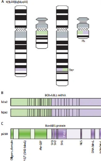
1.2 Philadelphia Chromosome
First described by Nowell and Hungerford, the Philadelphia Chromosome (Ph) is the small 9q- chromosome formed from the reciprocal t( 9;22) translocation13. The
presence of a translocation between the long arms of chromosomes 9 and 22 - t(9; 22) - was demonstrated in 1974 by Janet Rowley 14 and the BCR-ABL gene could be cloned
in 198415 This is the first neoplastic pathology characterized by the specific link between
a type of cancer and a biological marker.
The Philadelphia Chromosome is the pathognomic marker of CML, but can also, although it is rare, be detected in AML patients, and its presence is linked to a bad prognosis.16
The coexistence of normal HSC along with Ph1+ HSC has been demonstrated even at advanced phases of the disease 17During the process of translocation, the breakpoint
cluster region (BCR) gene, normally located on chromosome 22, where the breaks occurs in a small 5Kb region, fuses to the Abelson murine leukemia viral
ABL fusion gene18. The encoded BCR-ABL protein exhibits an exacerbated tyrosine
kinase activity, that plays a central role in the leukemogenesis process19. 1.3 The Bcr-Abl protein
1.3.1 The Bcr-Abl isoforms
While the ABL gene breakpoints highly variate upstream exon a2, there are only three known breakpoints regions in the M-BCR gene (between e13 and e14 or e14 and e15).
20 The BCR breakpoints are responsible for the three principal forms of BCR-ABL:
p190, p210 and p230. 21 Generally, the different BCR-ABL forms are linked to different
forms of leukemia. 18,22 The P190 form is found in 25% of cases of Ph-positive acute B
lymphoid leukemia (B-ALL) and less frequently in acute myeloid leukemia (AML) and very rarely in CML 23,24. The P230 is associated to neutrophilic CML.25 The most
common form, P210, is present in hematopoietic cells from CML stable phase and, also, in acute lymphoid (ALL) and myeloid leukemias (AML). 26–28
as a result of a break in the major region of the BCR gene (M-BCR) generating the fusion gene BCR-ABL which in turn generates Bcr-Abl mRNA and p210 Bcr-Abl protein.
The breakpoint occurring the e1 of BCR gene (m-BCR), gives rise to the truncated Bcr- Abl gene which in turn gives rise to p190 protein which is at the origin of Bcr-Abl- positive acute lymphoblastic leukemia (ALL). Finally, the e19a2 mRNA whose breakpoint is located in the micro region of the BCR gene (μ-BCR) gives the p230 protein found in the rare form of chronic neutrophilic leukemia characterized clinically by the proliferation of white blood mature cells (neutrophils) without presence of immature precursors fund in peripheral blood. 21
kinase domain, a Rho-GEF domain and a coiled-coiled oligomerization domain from Bcr. The coiled-coiled domain allows dimerization of the protein facilitating transphosphorylation of adjacent Bcr-Abl molecules. This part of Bcr is fused with SH domains, Abl DNA and actin binding domains. The SH1 domain has the Abl tyrosine kinase activity with the ATP binding domain and the catalytic phospho-transferase domain 18.
Although the fusion protein contains most of the coding sequences of Abl, the myristoylation site is lacking, contributing to the constitutive kinase activity of Bcr - Abl
29. Indeed, the Abl protein is capable of self-inhibition through this site and via molecular
interactions involving its SH2 and SH3 domains.
1.3.2 Consequences of BCR-ABL1 generation in hematopoietic cells
As early as the 1990s, the mouse models demonstrated the oncogenic role of Bcr-Abl. After transplantation of BCR-ABL-expressing stem cells into mice, a phenotype of myeloproliferative type-CML syndrome was developed in these animals but all the features of the disease could not be reproduced. 30,31 In fact, the transplantation of
retrovirally transduced 5-FU-enriched stem cells in irradiated mice generates a highly aggressive leukemia leading to rapid death of mice in 2-4 weeks. In parallel, in vitro experiments have demonstrated the oncogenic role of BCR-ABL which induces in hematopoietic cells, growth-factor-independence and resistance to apoptosis 32,33.
These experiments suggested therefore strongly that BCR-ABL fusion gene is responsible for the development of the disease.
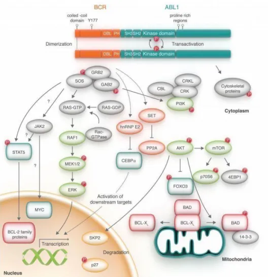
The Bcr-Abl protein causes abnormal activation of several signaling pathways in leukemic cells, giving them a proliferative and survival advantage. These activated pathways are related to growth factor dependence, apoptosis, proliferation and cell adhesion.
These pathways include Jak/STAT, PI3K/Akt and Ras/MEK, the major players in the pathogenic role.
The Bcr-Abl is an oncoprotein located mainly in the cytoplasm that has a constitutive tyrosine kinase activity34. The interaction of several cytoplasmic or membrane
components such as STATs and RAS respectively, will lead to activation of secondary signaling pathways controlling mitosis and cell survival 35.
1.3.2.1.1 JAK/STAT
Janus tyrosine kinases (JAK) are activated by cytokine receptors. The binding of the ligand on the receptors modifies their conformation allowing their dimerization and the activation of JAK by phosphorylation. These proteins are ubiquitous and play an important role in proliferation, differentiation, cell cycle and apoptosis of hematopoietic cells.36
Cellular models of CML have demonstrated that Bcr-Abl kinase activity increases activation of JAK2/STAT allowing proliferation and cell survival.36, activating JAK2 and
directly STAT5. JAK2 phosphorylates the tyrosine 177 residue of Bcr-Abl, allowing the activation of STAT5-independent cell signaling pathway 37
STAT proteins are transcription factors of which only STAT1, STAT3 and STAT5a and STAT5b are found in hematopoietic cells 38. STAT proteins participate in oncogenesis
by activating the transcription of target genes coding, for example, for the overexpression of anti-apoptotic proteins (Mcl-1, Bcl-x1, Bcl2) or cell cycle proteins (cyclin D1 or c-Myc). In cell lines or in primary CML cells, STAT5 is constitutively activated. 36,39. However, the use of STAT5 K/O mice model using conditional BCR-
ABL expression has shown that STAT5 was essential for the maintenance and development of leukemic cells. 40,41
It has been proposed that Bcr-abl can directly activate STAT5 and bypass the endogenous regulation of JAK2 to promote oncogenesis 42.
1.3.2.1.2 PI3K/AKT/CRK
The PI3K pathway (Phosphatidyl-inositol-3-kinase) modulates the activation of transcription factors promoting cell growth/survival and inhibition of cell death43.
a proto-oncogene, which has many downstream targets including mTOR. The mTOR pathway is responsible for controlling protein synthesis, cell size and autophagy. It has been observed that Bcr-Abl inhibits autophagy via mTOR and that TKI treatment restores this pathway 44
The Crk1 adapter protein is constitutively activated in the CML representing a marker whose phosphorylation demonstrates the presence of Bcr-Abl TK activity 45. The other
proteins related to Crk1 and Bcr-Abl are: STAT, Cbl, PI3K and Ras.46 Via these
interactions, Crkl appears indispensable for oncogenesis mediated by Bcr-Abl. In murine models, this interaction is necessary to obtain the phenotype of the myeloproliferative syndrome 47.
1.3.2.1.3 RAS / MEK
The Ras protein has a GTPase activity and is an initiator of the MEK-1, MEK-2 and ERK pathways.This pathway is activated in several cancers and is involved in proliferation and cell survival 48.
Bcr-Abl1 activates Ras via Grb2/Gab2 which allows the activation of MEK/ERK pathways and the transcription of target genes involved in cell proliferation and a decrease in the expression of genes involved in the cell cycle and apoptosis 49. The
Ras pathway is therefore activated constitutively by Bcr-Abl. In mouse models, inactivation of the Ras pathway attenuates the phenotype of CML 50.
Figure 5. : RAS Signaling pathway 50
1.3.2.1.4 SRC KINASES
The family of Src kinases is another group of Bcr-Abl target proteins, playing a major role in cell proliferation and differentiation. They are also involved in the transmission of extracellular signals 51.
In cellular models of CML, activation of some Src kinases is due to Bcr-Abl 52. Other studies have shown that Src kinases are essential for the transformation of BCR-ABL cell lines but also for the signal transduction of Bcr-Abl chimeric protein 51,53. In addition, Src kinases contribute to the activation of STAT5 and AKT by Bcr-Abl 54,55.
However, the importance of these kinases in the pathogenesis of the disease remains uncertain. In fact, mouse models show that Src kinases are not necessary for the initiation of CML but are rather involved in the pathophysiology of acute lymphoblastic leukemia 56.
1.3.2.1.5 Deregulation of apoptosis
Bcr-Abl has been shown to inhibit apoptosis in several ways, including stimulating anti-apoptotic pathways or modulating the expression of pro-apoptotic proteins. Bcr-
proteins Bcl2 and Bcl-x11. The PI3K/Akt and STAT5 pathways are important
mediators of apoptotic function. Activation of STAT5 by Bcr-Abl increases the expression of Bcl-X1 39 In addition, the PI3K pathway allows the retention of Bad
protein in the cytosol blocking mitochondrial depolarization preventing the activation of caspases required for apoptosis 57.
Mature blood cells have a limited lifespan and their production during the lifetime of an organism is ensured by the regulated activity of hematopoietic stem cells. In adult mammals, this process designed as hematopoiesis occurs primarily in the bone marrow, where billions of blood cells are produced every day. The different cell types are derived from very immature hematopoietic cells, or hematopoietic stem cells (HSCs), followed by a maturation process before gaining the bloodstream 59. HSCs are
multipotent cells that are able to regenerate and sustain complete hematopoiesis when transplanted into myeloablated recipients 60. This special ability is widely explored in
cancer treatment and other blood disorders via stem cell transplantation, the only curative approach in several types of leukemias, including CML until the advent of TKI therapies 61.
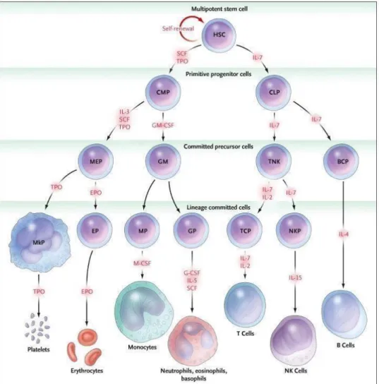
Hematopoiesis is a hierarchically organized process, departing from the most primitive hematopoietic stem cells, which give rise to lineage-progenitor cells, such as common myeloid and lymphoid progenitors (CMPs and CLPs, respectively) which in turn generate differentiated and cytologically identified blood cells 62,63. However,
experimental data giving rise to this classic organization has been obtained by transplantation of cell populations, rather that the analysis of steady state hematopoiesis. Recent technological advances allowed the follow-up up single clonal HSC without the stress induced by transplantation in mice 64 . These studies suggest
that in steady-state hematopoiesis, HSCs generate long-lived progenitors which can give rise themselves to differentiated hematopoietic cells 64. In these conditions, the
kinetics of this phenomenon has also been suggested to be different from the classical model, with early generation of MK progenitors from HSC then their rise to myeloid cells and lymphocytes 65 66 67 69.The organization of the branches has also been
questioned, such as the bifurcation of erythroid/megakaryocytic/myeloid versus lymphoid cell fates, as well as megakaryocytic and/or erythroid lineages 70–72. Furthermore, some groups have being demonstrating that the lineage-commitment may already be determined in primitive HSCs 73–75.
hematopoiesis process may be more complex and heterogenous than one thought. However, the elucidation of the clonal dynamics of HSC and their potential will require experimental data like what has been obtained in Wiskott-Adrich patients transplanted with genetically marked cells 68
2.1 Hematopoietic stem cells (HSC)
HSCs are characterized by 3 fundamental properties:
1 - self-renewal ability, ensuring their durability by giving both an identical daughter cell that keeps all the properties of the mother cell, but also able to differentiate itself. Self- renewal is possible due to the ability of asymmetric divisions probably under the influence of the niche, allowing the stem cells to generate daughter cells engaged in differentiation while remaining pluripotent.
2 – Ability to differentiate into all hematopoietic lineages
3 - Long-term quiescence capacity under the influence of factors related to the niche
or intrinsic factors 60
Hematopoietic stem cells (HSCs) are present in very small quantities in adult bone marrow (estimated at 1/100,000 in the bone marrow) and do not have specific morphological characteristics recognizable in conventional cytology.59
The concept of stem cells within hematopoiesis was initially raised by Till and McCulloch76. These authors have demonstrated that the hematopoietic reconstitution
of a lethally irradiated mouse receiving a bone marrow transplant passes through a stage of cell colony development in the spleen, each of these colonies coming from a cell called "CFU-spleen" (CFU- s). The presence of a stem cell at the origin of each of these colonies was deduced due to the fact that cytological analysis of CFU-S showed the presence of granulocytic, erythroid and megakaryocytic cells76. These dissected
and homogenized CFU-S could then reconstitute the hematopoiesis in other lethally irradiated recipient mice showing their hematopoietic potential which was found to be
of the hematopoiesis after serial grafting78. The formal demonstration of the existence
of myelo-lymphoid murine hematopoietic stem cells was subsequently carried out through defective retroviral-labeled stem cell transplantation experiments showing the presence of my same retroviral insertion at the level of myeloid and lymphoid cells. in the recipient mouse79. Since the development of characterization and isolation
techniques for stem cells with the discovery of the Sca1 antigen 80, it is currently
possible to purify murine stem cells by a combination of markers using Sca1 + cells, Kit + lin- or SLAM markers 7 for the long-term reconstitution of hematopoiesis in mice 81.
The development of in vitro tests made it possible to evaluate the intermediate compartment between the stem cells and the precursor and mature cells that can only be identified by morphological tests.
2.1.1 In vitro detection tests
To identify rare HSCs in the bulk of cells, one of the most frequently used technique is the flow cytometer. There are many surface markers that is currently used in both research and clinical for phenotyping and isolation of HSCs. Along with all the other criteria described above, these markers are also controversial among scientists. The cell surface protein CD34 is, the most important primitive human hematopoietic cell marker 1. It is estimated that only 1-5% of nucleated human bone marrow cells, around
1% of cord blood cells and less than 0.1% of normal peripheral blood cells express CD34 62,63,64. However, CD34 is not exclusively expressed in hematopoietic stem cells,
but also in progenitor cells Later the differentiation stage of the progenitor, lower is the expression of CD34. Therefore, despite CD34+ expression measured by flow cytometry is widely uses to isolate hematopoietic stem and immature progenitor cells for clinical engraftment, its expression alone does not guarantee an accurate measure of these populations, demanding a combination of HSC markers 86.
Another widely used strategy for this purpose is the elimination of lineage differentiation markers giving rose to a population designed as Lin-. Lineage antigens are not
to add CD38 88,89 and CD45RA, which are absent in primitive cells, together with CD90
(Thy-1), which is higher expressed in primitive as compared to differentiated cells
90,90,91. Obviously, more specific the combination of markers, rarer are the cells,
consequently, more difficult is to isolate them.
Functional tests have been developed to highlight and to quantify HSCs by long-term culture techniques.92–94. These cultures are based on the concept that the relatively quiescent most primitive cells do not generate clonogenic cells requiring an initial cycling period on a stroma and a suitable culture medium that will allow the differentiation of these cells into progenitors. The long-term culture technique (LTC-IC) remains the only one currently available for the in vitro identification of primitive cells in humans. However, even if this technique makes it possible to quantify and highlight a very immature population, the absence of differentiation markers continues to make HSCs difficult to detect. Another less used technology is the cobblestones generation cell detection technique (CAFC or Cobblestone area Forming cell). In this technique, foci of hematopoiesis are morphologically identifiable in the adherent layer in the form of "cobblestone areas" 95. In limiting dilution, cobblestone areas can be quantified
directly 96
2.1.2 In vivo detection tests
They make it possible to evaluate hematopoietic reconstitution in vivo after hematopoietic cell transplantation in mice in order to demonstrate the presence of stem cells. These are the only tests that formally identify the presence of genuine HSCs. Evaluation of the hematopoietic reconstitution potential of human stem cells has required the development of xenogeneic transplant systems in which immune responses are reduced to extreme. The most commonly used immunodeficient mice are NOD-SCID mice96, which show severe lymphopenia, macrophage deficiency and
directly into the NOD-SCID mouse, irradiated to destroy the hematopoiesis of the animal. Several months after transplantation, the presence of myeloid and lymphoid B human cells is sought in bone marrow and primary lymphoid organs using FACS analyses.97
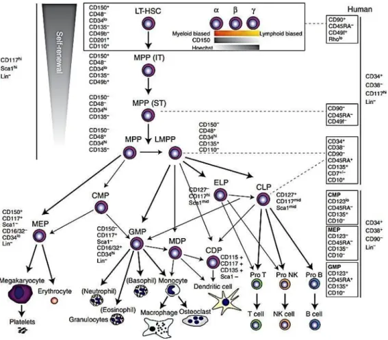
2.1.3 Progenitors and precursors and mature cells
The commitment of HSCs in a differentiation pathway leads first to the formation of highly clonogenic cells called hematopoietic progenitors. These progenitors are able to ensure hematopoiesis in the short and medium term.75
There are several types of progenitors: immature multipotent progenitors (MPPs) with increased proliferative capacity and more mature, lymphoid (LCP) or myeloid (MCP) progenitors with limited proliferation potential. LCP will differentiate B, T and NK progenitors. In the same way, the CMPs will give either erythroid progenitors (BFU-E, then CFU-E), or granulo-macrophagic progenitors (CFU-GM) themselves at the origin of the granular progenitors (CFU-G) and macrophage (CFU-M). These late progenitors are committed to a single cell type.55
Figure 7.Hematopoiesis and lineage differentiation98
Progenitors on a semi-solid medium were described in 1966 99. They are designed
under the term of colony forming cells (CFC). Several types of progenitors could currently be characterized: CFU-GEMM capable of generating colonies comprising granulocytic, macrophagic, megakaryocytic and erythroid cells, CFU-GM capable of forming granular and macrophagic colonies and BFU-E capable of forming erythroid colonies of different size depending on their proliferation potential.100
Figure 8. Examples of progenitors in a semi-solid medium
The differentiation of the progenitors results in the formation of precursors which are the last stage of maturation of blood cells. Their maturation is done by a morphological evolution (chromatin condensation, nucleoli disappearance and cytoplasm reduction).59
2.2 Regulation of the hematopoiesis
2.2.1 The microenvironment
In 1978, Schofield proposed the "niche" hypothesis to describe the physiological microenvironment that allows the survival of hematopoietic stem cells 101. The niche is
the microenvironment that protects stem cells from differentiation, apoptosis or other stimuli It is an anatomical structure comprising both cellular and non-cellular components integrating factors controlling the "fate" of stem cells 102. These niche
signals regulate the proliferation, survival and differentiation of stem cells, the adhesion of stem cells to stromal cells and/or the extracellular matrix (ECM) anchors stem cells in their niche near signals self-renewal. This specific environment linking stem cells with supporting cells can polarize stem cells into the niche to promote asymmetric cell division. Secreted factors may attract or, on the contrary, repel stem cells for their migration.
The reference model remains to date based on the anatomical location and distinguishes the osteoblastic niche and the vascular niche with the osteoblastic niche specialized in the maintenance and quiescence of long-term HSCs and, on the
of these 103
2.2.1.1 Osteoblastic niche
The first direct evidence that osteoblast cells are involved in hematopoiesis are studies in which human or murine osteoblast lines have been shown to secrete many cytokines that promote the proliferation of HSCs in culture. The direct role of osteoblasts in the regulation and maintenance of HSCs was shown in 2007 104. In this study, the
constitutive expression of PTH (parathormone) and its receptor, an important regulator of calcium homeostasis and bone formation / resorption, was achieved using the Col1a1 promoter (collagen a1 type 1). This activation resulted in a simultaneous increase in the number of osteoblasts and the number of HSCs. In addition, the in vitro maintenance of HSCs was more efficient with the stromal cells of these transgenic mice, presumably due to the increased number of osteoblasts in the stromal cell population compared to wild-type mice 105. The existence of this osteogenic niche has
been proven by a transgenic mouse model expressing the Herpes simplex virus thymidine kinase gene under the control of the osteoblast-specific type I collagen promoter, allowing depletion of these cells under the induction of ganciclovir 106.
Treatment with ganciclovir resulted in a decrease in spinal cellularity, in the number of HSCs and in progenitors of the different lineages. Discontinuation of the treatment restored the presence of osteoblasts in OM and restored bone marrow hematopoiesis (105)
2.2.1.2 Vascular niche
A close interaction between HSC and endothelial cells is not unexpected because they result from the same common embryonic precursor, the hemangioblast 107. Studies
have shown that endothelial cells are able to support HSCs in vitro in the absence of growth factors 108,109. The endothelial cells of the sinuses constitutively express
cytokines such as CXCL12 (CXC chemokine ligand 12) or SDF-1 (Stromal Derived Factor-1), adhesion molecules such as E-selectin and VCAM1 (vascular cell adhesion
"mobilization" towards the vascular niche involves the matrix metalloprotease (MMP) - 9 which induces the release of soluble SCF 112. An essential role of the vascular niche
is to assist HSC in migration phenomena during homing or peripheral mobilization. Vascular and / or perivascular cells can also create hematopoietic niches, since many HSCs are found around vessels and factors regulating the maintenance of HSCs 113.
The quiescent HSCs are even more numerous in the perivascular zones than in the endosteal zones 114. The Nagasawa group confirmed the importance of vessels in the
regulation of hematopoiesis by reporting that primitive HSCs have a very close interaction with reticular cells, strongly expressing SDF-1, found both near endothelial cells and in the body. endosteum 115. It has been proposed that the vascular niche
promotes the proliferation and differentiation of hematopoietic cells and progenitors while the osteogenic niche maintains the quiescence of HSCs 116.
2.2.1.3 Molecular mechanisms within the microenvironment
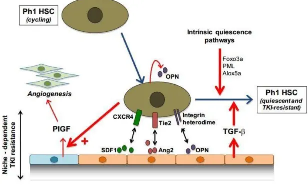
Factors secreted by osteoblasts also play a key role in maintaining HSCs in quiescence. Among these factors are angiopoietin-1 (Ang-1), osteopontin (OPN) and chemokine CXCL12. The binding of Ang-1 to its Tie2 receptor plays an important role in HSCs adhesion and their maintenance in quiescence 116. Ang-1 also increases the
expression of N-cadherins on the surface of osteoblasts, and promotes the anchoring of HSCs within the hematopoietic niche 117.
The OPN, recognized by the HSC via the CD44 receptor, acts as a negative regulator of their proliferation and ensures their localization, in a quiescent state 118 CXCL12 or
SDF-1 (Stromal Derived Factor-1) is produced by the stromal cells of the hematopoietic niche and interacts with the CXCR4 receptor, expressed on the surface of HSCs 103.
This chemokine is a chemo-attractant and its secretion regulates the migration and localization of HSCs within the niche 119. CXCL12 is also involved in regulating the
Growth factors occur at all levels of hematopoiesis regulating the reservoir of HSCs, but also cell expansion and cell differentiation. All these molecules, produced in vivo at low concentrations, interact with each other to modulate their effects. They are secreted either remotely, such as erythropoietin (EPO) or thrombopoietin (TPO) secreted respectively by the liver and kidney, or by the cells constituting the hematopoietic niche, such as G-CSF. The action of these factors necessarily requires their binding to receptors, present on the surface of hematopoietic cells, as well as the activation of signaling pathways. These factors may regulate hematopoiesis positively or negatively 121,122.
Among these factors, three play an essential role in the self-renewal of HSCs: TPO, SCF (Stem Cell Factor) and Flt3-L (Fms-Like Tyrosine kinase 3-ligand). TPO and its Mpl receptor are involved in maintenance and expansion of adult HSCs. Mpl is expressed on quiescent HSCs and this expression decreases with differentiation 124.
The SCF, coupled with its c-kit receptor, is also involved in the survival and / or expansion of HSCs. Finally, the Flt3-L receptor is expressed on primitive hematopoietic progenitors and plays an essential role in their survival, proliferation and differentiation
125. Other cytokines also occur during the early stages of hematopoiesis: IL-3
(Interleukin-3), IL-6 and GM-CSF (Granulocyte and Macrophage-Colony Stimulating Factor) Other cytokines have more restricted action spectra and intervene in late stages of hematopoiesis, or even during the terminal phases of differentiation: EPO, G-CSF (Granulocyte-Colony Stimulating Factor), M-CSF (Macrophage-Colony Stimulating Factor), IL-4, IL-7, IL-5 and IL-2.
2.2.1.4.2 Negative regulators of hematopoiesis
They play a determining role in hematopoietic homeostasis by inhibiting the self- renewal and / or differentiation of HSCs and thereby regulating the number of cells. They include TNF-α (Tumor Necrosis Factor-α), which induces the apoptosis of hematopoietic cells, PF4 (Platelet Factor-4) which inhibits megakaryopoiesis, but whose action also positively regulates viability, survival and adhesion of HSCs and progenitors 126 and Macrophage inhibitory protein-1α (Mip-1α), which restricts the
differentiation of primitive hematopoietic cells 127.
2.2.1.5 Other stimulating factors for HSCs and progenitors
Other pathways can be used to simulate the expansion of HSCs and progenitors. Rather than use cytokines, it is also possible to infect HSCs with recombinant retroviruses encoding transcription factors involved in their development, and whose
with the gene coding for HOXB4 homeoprotein. The constitutive overexpression of the HoxB4 gene in hematopoietic cells caused a strong and persistent expansion of the stem cell pool in vivo, without inducing their differentiation or malignant transformation, even in the long term 128. In humans, it has also been shown that the infection of HSCs
with a retroviral vector containing the coding sequence of the HoxB4 gene made it possible to obtain an expansion of these cells in vitro 129. More recently, Cooke’s team
has observed a massive expansion of HSCs and progenitors in all hematopoietic lineages with an AHR antagonist StemRegenin (SR1) 130.
2.2.1.6 Intrinsic regulation
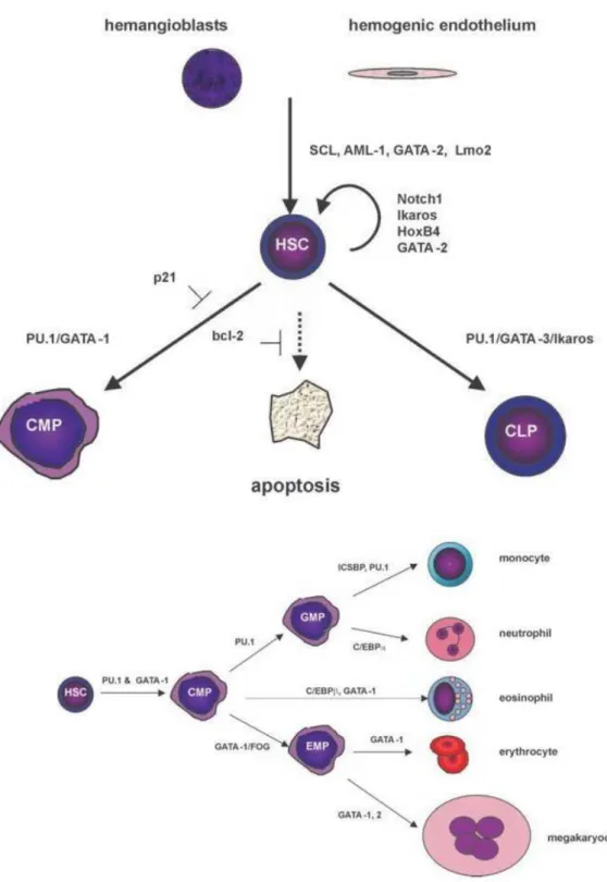
Many transcription factors occur during primitive or definitive hematopoiesis. Some have an important role in the renewal of the hematopoietic stem cell and others allow the differentiation of different lineages.
In a non-exhaustive way, the primitive hematopoiesis at the level of the hemangioblast is regulated by the Stem Cell Leukemia hematopoietic transcription factor (SCL), GATA-2, Lmo-2 and AML-1. The self-renewal of the stem cells is regulated by Ikaros, HOX B4, GATA-2 and Notch1 while the differentiation is done via a variety of factors such as the factors PU-1 and GATA1131,132
CML is known to affect the most immature hematopoietic cells, called leukemia stem cells (LSCs). During the last 30 years, one of the major challenges in the field of CML has been the possibility to identify LSC from normal HSC using specific markers. Indeed, both share the same surface markers, which makes FACS isolation of LSCs highly difficult and unreliable. In recent years, many studies have shown possible markers that could ultimately be unique to LSC, such as CD33 (Herrmann et al., 2012), CD36 135, CD44, CD47, CD52 136, IL1-RAP 136–139 and, last and most promising so far,
CD26 . Dipeptidyl peptidase-4 (DPP4; CD26) could contribute to the release of CML LSCs from bone marrow into the bloodstream 140,141. Studies have also shown that
CD26 can be detected in TKI resistant subclones 142. However, at a glance, all these
data remain controversial and inconclusive.
Hematopoietic cells maintain their pluripotent characteristics due to a special microenvironment, which is formed by a complex system that involves the presence of different cell types, such as mesenchymal stromal cells, osteoblasts, osteoclasts, endothelial cells, and neural cells combined with signalling pathways and specific metabolic conditions 143–145. It is believed that modifications in this microenvironment may be involved in the development of several hematological pathologies, such as CML. There is much evidence that this same niche that preserves the pluripotency of hematopoietic cells also contributes to the preservation of leukemic stem cells 146.
Many laboratories have been working to unravel the characteristics of the bone marrow niche and understand how they may contribute to the emergence of pathologies. Despite major advances in understanding this issue, there are still many challenges ahead. An important feature of CML is the presence of immature pluripotent leukemic cells in peripheral blood, whereas under normal conditions, HSC remain in the bone marrow 147.
3.1 Murine models
3.1.1 Retroviral transduction/transplantation model
The “gold standard” of murine CML model has been established as early as in 1990 by transplantation of 5-FU-enriched BCR-ABL-transduced bone marrow cells into an irradiated syngeneic recipient. This method leads to the development of CML-like a myeloproliferative disease, which however, is very aggressive as compared to CP CML
30. Subsequent studies have shown successful development of CML like syndrome in
100% of recipients in a very short time (2-4 weeks post transplantation) 148. In addition
to the rapid development of disease by recipients, another advantage is that influence of other genes and mutations can also be explored by this model 31. The aggressivity
of the leukemia, leading to death of mice in 3-4 weeks, did not allow therefore, the modelling of CP of human CML. This led to the development of inducible leukemia strategies allowing to model more closely the chronic phase.
3.1.2 SCLtTA/BCR-ABL transgenic model
In this model, BCR-ABL expression is modulated by a tetracycline-controlled transactivator (tTA) that specifically regulates BC ABL expression in the murine hematopoietic stem/progenitor cells. This model can develop a CML-like disease clinically similar do human CML (Li et al., 1999). Thus, the progress of the malignancy is done is a slower pace compared to the retroviral models, being more likelihood with human disease 149,150. This is also a better model for studying the endosteal and BM
microenvironment, as well as genomic instability 151.
Derivations of this model can be done to better adapt to each experimental profile, such as SCLtTA/BCR-ABL double transgenic model with a transposon-based insertional mutagenesis system, developed by Giotopoulos et al. (REF) In this version, it was possible to mimic the additional chromosomal instability and mutagenesis of blast crisis.
The xenotransplant model consist in engrafting human leukemic cells into immunocompromised mice and follow up the frequency of LSCs and Ph+ cells
152,153.This methodology which allows quite reproducibly AML in immunodeficient
mouse (Bonnet and Dick) has been very difficult to apply to CML as the growth of CML cells and progenitors from CP-CML patients are very difficult to obtain in vivo, requiring the administration of human growth factors to mice . Transplantation of cells from more advanced phases of the disease, such as blast crisis has been found to be more successful.
Methods of generating scaffolds using human mesenchymal cells to mimic the BM niche followed by injection of normal or leukemic cells have also been developed. 153–
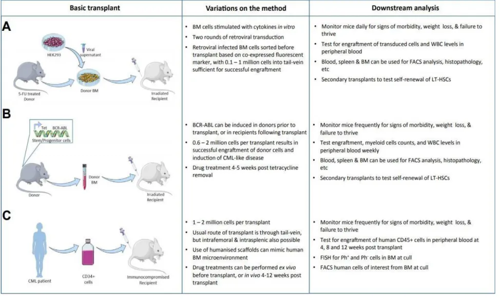
Figure 11. In vivo models of CML: A) Retroviral transduction/transplantation model B) SCLtTA/BCR-ABL transgenic model
and C) Xenograft model
156Tran sc ri p tio n al c o n tro l in d u ced b y Bc r-A b l an d it s ro le in leukem ic st em c el l h et ero genei ty 34
3.2 Cellular models
3.2.1 Cell Lines
In 1975, the K562 cell line carrying the Ph+ was established by Lozzio's team from a pleural effusion of a patient with blast crisis CML 157. This cell line has been used
extensively in the world, but it is not representative of a chronic phase of the disease and obviously cannot be compared to an equivalent without Ph1 chromosome. The extensive experience using this cell line which has been very useful in preclinical studies, showed also the spontaneous establishment of many different cell subclones, with different cytogenetic and molecular characteristics according to different laboratories. Over the years, several BC cells lines have been established from CML patients with different characteristics. The ability to evaluate directly the effects of BCR- ABL in a hematopoietic cell context has been subsequent established using BCR-ABL gene transfer strategies. In both murine and human cell lines, this has been accomplished by retrovirus-mediated BCR-ABL gene transfer. The murine lymphoid BaF/3 cell line the growth of which depends on IL-3, become transformed to IL-3 independence upon BCR-ABL expression. A similar outcome can be generate in humans using the UT7 line 158. Thus, starting from UT7 line, megakaryoblastic and
growth factor-dependent leukemic line, UT7-9 and UT7-11 clones were created by BCR-ABL gene transfer. These 2 clones express BCR-ABL RNA and protein and they are GM-CSF independent 158
3.2.2 Primary leukemic cells
The in vitro models of CML using primary leukemic cells include essentially the techniques of CFC and LTC-IC, which allow the quantification of CML progenitors and stem cells after long-term culture in the presence of permissive stroma such as MS-5
3.2.3 iPSC
IPSCs (induced pluripotent stem cells) result from the reprogramming of adult somatic cells into pluripotent stem cells, in many ways resembling embryonic stem cells. The first reprogrammed cells were derived from murine fibroblasts by the Yamanaka team in Japan 161. Four transcription factors were retained as reprogramming media OCT3
/ 4, SOX2, C-MYC and KLF4. In 2007, two other teams reported the derivation of iPSCs from human somatic cells 161,162. They were generated by reprogramming fibroblasts
either with retroviral vectors or with lentiviral vectors.
Because of their self-renewal capacity, iPS cell lines can be maintained in an undifferentiated state and, thanks to their pluripotency properties, it is then possible to differentiate them in all possible cell types.
Many iPSC patient-derived lines have been generated to model diseases. The first modelizations were carried out by the team of Park 163. These lines make it possible to
dispose of cellular material carrying abnormalities responsible for a pathology, to study them and to create an opportunity for the development of therapeutic strategies. The first CML iPSC lines were generated in 2011 164 and in subsequent years, the use of
4. Diagnosis.
The diagnosis is made most frequently in the chronic phase. The main symptoms and the usual signs of the disease are fatigue, anemia and splenomegaly. However, a significant proportion of patients are asymptomatic, the diagnosis being made fortuitously during a routine check-up. There are 600-800 new cases in France per year (Incidence 1/100000). The sex ratio is towards men (H / F = 1.2) and the average age is 53 years. 166
The BCR-ABL1 fusion gene is present in leukemic cells of all cases of CML but the karyotype and research of the Philadelphia chromosome is still needed during the initial assessment because no test can currently replace the prognostic value of cytogenetic remission complete at 6 months and the karyotype can detect clonal abnormalities associated undetectable by molecular biology. 167
4.1 Evolution of the disease and prognosis
The natural history of CML, which has been modified by the current TKI therapies, included typically 3 phases:
The chronic phase, lasting from 3 to 5 years, often identified following a systematic assessment showing hyperleukocytosis with significant myelemia. The cytological marrow examination through a bone marrow aspirate is mandatory, showing hyperplastic marrow involving myeloid cells. The diagnosis is confirmed by the karyotype that finds the Philadelphia chromosome and the BCR-ABL transcript.
According to the WHO 2016 classification the accelerated phase is characterized by: one or more of the following criteria:
• Persistence or increase of splenomegaly, not responding to treatment
• Persistent or increased number of leukocytes above 10 G / L, not responding to treatment
• Persistence of thrombocytopenia <100 G / L, unrelated to treatment • Presence of at least 20% of basophils in the blood
• Presence of 10-19% in the blood and / or bone marrow
• Presence of clonal cytogenetic abnormalities additional to Ph, including major abnormalities (doubling of Ph, trisomy 8, isochromosome 17q, trisomy 19), or a complex karyotype, or abnormalities in 3q26.
• Any new clonal abnormality in Ph + cells occurring during treatment
The blast phase or blast crisis marked by the presence of more than 20% medullary blasts. In most cases, the blasts are myeloid type and in 30% of cases the crisis is lymphoid type. This phase may be critical due the lack of a truly effective therapy. This evolution has now been dramatically changed due the use of tyrosine kinase inhibitors since 2001. 167–169170,171.
5. CML Therapy
5.1 Treatment History
Initial therapy of CML included, arsenical preparations from 1865 until the beginning of 20th century where splenic radiation has been introduced to therapy (1902). The
efficiencies of these therapies were transient and despite many efforts suing antileukasera (1932), benzene (1935), urethane (1950), leukapheresis (1960) and high dose combined chemotherapy ( COAP protocols) the prognosis remained the same with a median survival of 3-5 years, all patients succumbing to an acute leukemia 9. In
1953, David Galton introduced the alkylating agent, Busulfan, into clinics, with evidence of successful cytoreduction but no effect on survival 173. It was only in the 80’s, the
introduction of Interferon-α changed the therapeutic strategy as the first remissions with evidence of ph1-negative recovery were obtained with IFN albeit with modest increase in survival 174. The addition of cytarabine IFN-2b was the first therapy with a clear
evidence of cytogenetic remissions and survival advantage and this combination remained the standard therapy in patients not eligible for BMT until the introduction of TKI therapies and 175.
5.1.1 The introduce of TKIs, a turning point in CML history
The treatment of CML has undergone an important evolution, particularly in the 2000s with the arrival of targeted therapies: the tyrosine kinase inhibitors (TKIs). CML is the first cancer pathology caused by an acquired genetic abnormality that has benefited from targeted drugs.
After preclinical results showing the efficiency of tyrosine kinase inhibitors called “tyrphostin” in 1999176 CML treatment had its historical turnaround. A tyrosine kinase
inhibitor, initially developed to inhibit PDFG-receptor in solid tumors, has shown major efficiency in BCR-ABL-expressing cells. This drug, initially called STI-571 and later Imatinib, developed by Ciba-Geigy was developed into the clinical application by Novartis, has shown to be able to induce major cytogenetic responses. Not only did Imatinib significantly improved the lives of patients with CML, as it was also a pioneer
in target-specific treatment 177,178. Since then, second and third generation of TKIs have
been introducing into clinics.
5.1.2. Hematopoietic Stem Cell Transplantation (HSCT)
The first attempt to a human bone marrow transplant was in 1939 to a patient with aplastic anemia 179. Just after the World War II, researchers managed to recovery
normal hematopoiesis by marrow transplantation in a mouse model with marrow aplasia caused by radiation 180. In 1956, Barnes et al demonstrated an overcome of
acute leukemia in mouse models by bone marrow transplantation 181. In 1958, the first
BMT between unrelated donor was performed by Georges Mathé’s team in Paul Brousse hospital in Paris, for Yugoslavian researchers who were accidentally irradiated.182
Allogeneic transplants for leukemia between related donors and recipients was first performed in Seattle at Fred Hutchsion Center by E. Donnall Thomas’ team 183. Six
patients were pre-treated with radio and chemotherapy and then received an infusion of marrow from a normal donor. At the time, histocompatibility was not considered, and all patient died by 3 months post transplantation. Methods to identify human leukocyte antigens (HLA) in human were developed in the 60’s, allowing physicians to check compatibility between donor and patient. In 1979, despite a previous almost failed experiment reported with 100 transplantations, which only 13 were successful, Donnall Thomas was encouraged to continue and successfully engrafted 50% of 126 leukemic patients, being honoured with a Nobel Prize in 1990 for his discoveries 61,184,185. In
1988, the Bone Marrow Worldwide (BMDW) was funded, currently counting with more than 23 million registered donors in more than 70 countries 186,187.
BMT allows reconstitution of normal hematopoiesis through conditioning and in the allogeneic setting allows the elimination of leukemic cells via a graft versus leukemia (GVL) effect. The use of bone marrow transplants made it possible to understand very early that CML also represented a model of immunotherapy 188. Indeed, in all clinical
there has been a consistent increase in relapse rates189. These observations have
formed the basis of immunologic cellular therapies performed by donor lymphocyte (DLI) transfusion in patients who have relapsed after allogeneic transplant 190,191.
Currently the allogeneic transplant is no longer proposed in the first line, but only in case of resistance to TKI.
Until a few years ago, HSC allografting could be considered currently the only proven curative therapy for CML. However, recent TKI-discontinuation trials have shown that in some selected patients, long-term remissions without recurrence of CML can be observed. However, it is currently not known if these patients are cured, as late CML relapses have been reported as long as 20-25 years after allogeneic bone marrow transplantation. 192,193.
5.2 Conventional therapies
5.2.1 Chemotherapy
In the 1950s, chemotherapy was used to target rapidly dividing cells allowing the normalization of white blood cells in the blood. Chemotherapy does not only target cancer cells but also healthy cells and other dividing tissues with many side effects. The two most commonly used chemotherapeutic agents were busulfan and hydroxyurea. However, neither of these allowed to control the disease and the median survival of the patients did not exceed 5 years 194. These treatments often made it
possible to obtain hematologic remission but never cytogenetic with persistence of the Ph chromosome in 100% of the medullary mitoses.
Although, hydroxyurea, a cytoreductive agent, is still used to reduce tumor mass while waiting for cytogenetic or genetic confirmation at the time of diagnosis of CML.
5.2.2 Interferon alpha (IFNα)
IFN α has been used since the 1980s in the treatment of CML. It is a cytokine with antiproliferative properties. It was the first-line treatment until the arrival of TKIs and this despite a very variable tolerance according to the patients.
IFN α has an inhibitory action on the growth of leukemic progenitors. Its action seems to target progenitors already engaged in a certain differentiation but without sparing
the normal cells, explaining its hematological toxicity. Nevertheless, studies carried out in the 1980s have shown that IFNα induces, alone or in combination with chemotherapy (such as cytarabine), hematological remission in 60 to 80% of cases, complete cytogenetic remission in 15 to 35% of cases. patients and a good cytogenetic response (Ph + <35%) in 10 to 20% of additional patients 195,196 Modifications on INF- α formula
improved significantly its side effects – flu-like symptoms and severe fatigue – and INF-α and its combinations are still used today, in special cases 197,198
5.3 Targeted therapies - TKIs 5.3.1 1st generation TKI
5.3.1.1 Imatinib
Imatinib, or Glivec®, the first tyrosine kinase inhibitor (TKI) introduced therapeutically since 2001, drastically altered the natural course of the disease by revolutionizing management and prolonging survival. Indeed, in the era of targeted therapies by TKIs, responder patients (but only these) have a life expectancy identical to that of patients of the same age in the general population 199
Imatinib is a 2-phenylamine-pyrimidine derivative that inhibits the kinase activity of Bcr- Abl by competing for the ATP catalytic domain binding site of the chimeric protein 200.
In 1996, Druker reported the first in vitro effects of 2-phenylaminopyrimidine Abl1 tyrosine kinase inhibitor, then known as a 571 signal transduction inhibitor (STI) on cell lines derived from CML 177.
The interaction between imatinib and Bcr-Abl is possible in the inactive and dephosphorylated conformation of the oncogene 201. This makes it possible to stabilize
the oncogene in its inactive form and thus to block the autophosphorylation of the enzyme and then the transmission of the signal 202. Thus Imatinib is able to reduce the
formation of CML CFC by 90% with no inhibitory effect on normal cells 200,202.
Two years after the Phase I study, an international randomized study of interferon / cytarabine against STI (IRIS study) was conducted in 1100 newly diagnosed patients. The results of this study changed the management of patients in view of more than satisfactory results. The superiority of Imatinib was demonstrated in terms of
hematological, cytogenetic and molecular responses (97% versus 56% for the RHC, 74% versus 8% for the RCC and 85% versus 22% for the RMC) 203 . An 8 years follow-
up study report (IRIS) has shown that patients treated with Imatinib presented an overall survival rate of 85% and only an average of 0.9% (annual rate) progressed from AP to BC. Despite its satisfactory action, imatinib is less potent than the 2nd generation
TKI, which may not inhibit the kinase efficiently, allowing the development of secondary resistance and loss of response 107,108.
Figure 13. Mechanism of action of Imatinib.
5.3.2 2nd Generation TKIs
The identification of resistance to Imatinib led to focus research and development efforts on new TKIs effective against several types of acquired mutations.
5.3.2 1 Dasatinib
Similar to imatinib, dasatinib is also an ATP analogue derived from pyrido (2,3-d) pyrimidine. It is a broad-spectrum inhibitor since it targets in addition to Bcr-Abl (in its
active and inactive conformation). ), several proteins of the Src kinase family (such as Src, Lck, Yes, Fyn). 205,206 Dasatinib is active against the majority of kinase domain
mutations, except of T315I 207. It has proven 300-fold more potent than imatinib in
vitro208.
The phase 1 study of Dasatinib (140 mg / day) was conducted in 84 patients who did not respond to Imatinib. In 35% of patients with chronic or accelerated phase, it has been possible to obtain complete cytogenetic remission and in patients with blast phase a temporary improvement in hematological and cytogenetic responses 209.
Phase 2 included 387 chronic phase patients and showed complete 40 and 75% cytogenetic remission rates after 15 months of treatment in cases of imatinib resistance and intolerance, respectively (134). The overall survival of Imatinib- resistant and intolerant patients was 78% and 82% respectively, however only 30- 35% of patients remained for 5 years under Dasatinib 210.
Clinical trials comparing dasatinib versus imatinib have shown that CCgR rates were similar to imatinib (86% and 82%, respectively), while MMR and MR4.5 rates were better with dasatinib than imatinib (64% versus 46% and 17% versus 8%, respectively)
211–213.
5.3.2.2 Nilotinib
Nilotinib is also an aminopyrimidine and it was developed from Imatinib by crystallographic analysis. Like imatinib, it is a competitive inhibitor of ATP, with a higher affinity than imatinib for Bcr-Abl 214. It is able to bind to the inactive
conformation of the oncogene. In vitro and in vivo, this TKI prolongs the survival of mice injected with imatinib-resistant Bcr-Abl cells
The phase 1 study was conducted in patients resistant to Imatinib. Complete
cytogenetic remission was observed in 35% of these patients 215. The phase 2 study
included 321 chronic phase resistant or intolerant patients with Imatinib or both and the results. Complete cytogenetic remission rates were 45% and 78% overall survival at 4 years. As with Dasatinib, only 31% of patients remained on Nilotinib 216.
Nilotinib was approved in 2007 by the U.S FDA as an analogue of imatinib with much higher affinity for the ATP binding site. A clinical trial (ClinicalTrials.gov number,
NCT00471497.) comparing imatinib and nilotinib shows that patients treated with
nilotinib presented higher CCgR rates by 12 months (80% against 65% with imatinib) and also better MMR rates (44% compared to 22% with imatinib) 204,211,217. However,
nilotinib treatment is related to occurrence of vascular events, such as peripheral arterial occlusive disease (PAOD), coronary artery disease (CAD), cerebrovascular disease (CVA), and the risk to develop diabetes. It is also not indicated in case of F359V, E255K/V and Y253H mutations 218,219.
5.3.2.3 Bosutinib
Bosutinib is an oral tyrosine kinase inhibitor that targets Src/Abl activity. In Phases I and II studies including patients who previously had 2 or 3 other TKIs, 24% of patients achieved complete cytogenetic remission. After 2 years of treatment, the progression-free rate was 73% and 83% overall survival. With a median follow-up of 28 months, only 29% of patients remained on Bosutinib 220. Bosutinib was
subsequently tested in 286 patients resistant or intolerant to Imatinib, 47% of whom had complete cytogenetic remission. The phase III Bosutinib Efficacy and Safety in Newly Diagnosed CML (BELA) trial compared bosutinib to imatinib in CP-CML. Patients treated with bosutinib reached CCgR and MMR faster than with imatinib, but 12 months CCgR average rates were not different between the two drugs (70% versus 68%), while MMR rates were higher with bosutinib (41% versus 27%).221,222
5.3.3 3rd generation TKI (mutation T315I)
5.3.3.1 Ponatinib
Ponatinib is the first and, currently, the unique of 3rd generation TKI class. It is highly
potent due its action against a vast spectrum of kinase domains and the only drug able to overcome the T35I mutation problem. Its clinical use was approved by the U.S FDA in 2012 for patients with resistant CML or intolerant of prior TKI therapy. In 2013, due
its serious adverse vascular events, the U.S FDA withdraw ponatinib from the market. In 2014, after additional follow-up studies have showed satisfactory safety results, ponatinib marketing has been resumed, with initial lower doses. 223–226
5.4 Mechanisms of resistance
As early as 2003, it has been shown that resistance to TKI could occur during therapy (or in vitro models, using cell lines or patient cells (Review CML) These studies revealed different mechanisms of resistance including:
1- BCR-ABL independent mechanisms: efflux mechanisms and membrane transporters, the different mechanisms of persistence of leukemic cells (quiescent cells, gene instability and signaling pathways) (described in the next chapter
2- BCR-ABL-dependent mechanisms: BCR-ABL amplification and oncogene mutations
5.4.1 Efflux mechanisms and carrier hOCT1
Efflux transporters are frequently active transporters, belonging to the superfamily of ATP-binding cassette proteins (ABC). This family includes the P-glycoprotein (P-gp or ABCB1), responsible for the expulsion of Imatinib from the cell is encoded by the gene MDR-1 (Multi Drug Resistance 1). The Pgp pump allows the exit of endogenous or exogenous hydrophobic substrates such as therapeutic agents. This transporter is overexpressed in the cells of patients in the transformation phase and involved in resistance to blast phase chemotherapy 227, as well as resistance to Imatinib 228.
The hOCT transporter (human Organic Cation Transporter 1) has been shown to be a significant factor affecting the intracellular bioavailability of the active ingredient
229,230.Overexpression of OCT1 in patients promotes the efficacy of imatinib 231. Indeed,
a high rate of OCT1 seems to be predictive of a good response to Imatinib while patients with a low level of OCT1 require higher doses of Imatinib for an optimal response. Neither dasatinib 232 nor nilotinib 233 is affected by OCT1 activity.