HAL Id: hal-03076689
https://hal.archives-ouvertes.fr/hal-03076689
Submitted on 16 Dec 2020
HAL is a multi-disciplinary open access
archive for the deposit and dissemination of
sci-entific research documents, whether they are
pub-lished or not. The documents may come from
teaching and research institutions in France or
abroad, or from public or private research centers.
L’archive ouverte pluridisciplinaire HAL, est
destinée au dépôt et à la diffusion de documents
scientifiques de niveau recherche, publiés ou non,
émanant des établissements d’enseignement et de
recherche français ou étrangers, des laboratoires
publics ou privés.
Progression by Controlling Memory T Cell Response
Noymar Luque-Campos, Rafael Contreras-López, María Jose
Paredes-Martínez, Maria Jose Torres, Sarah Bahraoui, Mingxing Wei,
Francisco Espinoza, Farida Djouad, Roberto Elizondo-Vega, Patricia
Luz-Crawford
To cite this version:
Noymar Luque-Campos, Rafael Contreras-López, María Jose Paredes-Martínez, Maria Jose Torres,
Sarah Bahraoui, et al.. Mesenchymal Stem Cells Improve Rheumatoid Arthritis Progression by
Con-trolling Memory T Cell Response. Frontiers in Immunology, Frontiers, 2019, 10, pp.Front. Immunol.
10:798. �10.3389/fimmu.2019.00798�. �hal-03076689�
doi: 10.3389/fimmu.2019.00798 Edited by: Teun J. De Vries, VU University Amsterdam, Netherlands Reviewed by: Akio Morinobu, Kobe University, Japan Erik Lubberts, Erasmus University Rotterdam, Netherlands *Correspondence: Farida Djouad [email protected] Roberto Javier Elizondo-Vega [email protected] Patricia Luz-Crawford [email protected]
Specialty section: This article was submitted to Inflammation, a section of the journal Frontiers in Immunology Received: 21 November 2018 Accepted: 26 March 2019 Published: 16 April 2019 Citation: Luque-Campos N, Contreras-López RA, Paredes-Martínez MJ, Torres MJ, Bahraoui S, Wei M, Espinoza F, Djouad F, Elizondo-Vega RJ and Luz-Crawford P (2019) Mesenchymal Stem Cells Improve Rheumatoid Arthritis Progression by Controlling Memory T Cell Response. Front. Immunol. 10:798. doi: 10.3389/fimmu.2019.00798
Mesenchymal Stem Cells Improve
Rheumatoid Arthritis Progression by
Controlling Memory T Cell Response
Noymar Luque-Campos
1, Rafael A. Contreras-López
1, María Jose Paredes-Martínez
1,
Maria Jose Torres
2, Sarah Bahraoui
3, Mingxing Wei
4, Francisco Espinoza
5,
Farida Djouad
3*, Roberto Javier Elizondo-Vega
6* and Patricia Luz-Crawford
1*
1Laboratorio de Inmunología Celular y Molecular, Centro de Investigación Biomédica, Facultad de Medicina, Universidad de
los Andes, Santiago, Chile,2Escuela de Ingeniería Bioquímica, Pontificia Universidad Católica de Valparaíso, Valparaíso,
Chile,3IRMB, INSERM, Univ Montpellier, Montpellier, France,4Cellvax, SAS, Parc BIOCITECH, Romainville, France,5Cells
for Cells, Universidad de los Andes, Santiago, Chile,6Laboratorio de Biología Celular, Departamento de Biología Celular,
Facultad de Ciencias Biológicas, Universidad de Concepción, Concepción, Chile
In the last years, mesenchymal stem cell (MSC)-based therapies have become an
interesting therapeutic opportunity for the treatment of rheumatoid arthritis (RA) due to
their capacity to potently modulate the immune response. RA is a chronic autoimmune
inflammatory disorder with an incompletely understood etiology. However, it has been
well described that peripheral tolerance defects and the subsequent abnormal infiltration
and activation of diverse immune cells into the synovial membrane, are critical for RA
development and progression. Moreover, the imbalance between the immune response
of pro-inflammatory and anti-inflammatory cells, in particular between memory Th17 and
memory regulatory T cells (Treg), respectively, is well admitted to be associated to RA
immunopathogenesis. In this context, MSCs, which are able to alter the frequency and
function of memory lymphocytes including Th17, follicular helper T (Tfh) cells and gamma
delta (γδ) T cells while promoting Treg cell generation, have been proposed as a candidate
of choice for RA cell therapy. Indeed, given the plasticity of memory CD4
+T cells, it
is reasonable to think that MSCs will restore the balance between pro-inflammatory
and anti-inflammatory memory T cells populations deregulated in RA leading to prompt
their therapeutic function. In the present review, we will discuss the role of memory T
cells implicated in RA pathogenesis and the beneficial effects exerted by MSCs on the
phenotype and functions of these immune cells abnormally regulated in RA and how this
regulation could impact RA progression.
Keywords: mesenchymal stem cells, rheumatoid arthritis, T cell, plasticity, immunomodulatory
INTRODUCTION
Mesenchymal stem cells (MSCs) are multipotent stem cells able to exert immunosuppressive
functions on both the innate and the adaptive immune cells (
1
). They have been isolated from
almost all mesodermal tissues including bone marrow, adipose tissue, umbilical cord blood,
umbilical cord, placenta, menstrual fluid, and dental pulp (
2
–
5
). The International Society for
Cellular Therapy (ISCT) has defined minimal criteria for characterizing MSCs that include a
fibroblastic-like morphology, the expression of mesodermal markers such as CD90, CD105, and
CD73, the lack of hematopoietic marker expression such
as CD45, CD34, CD14, and the capacity to differentiate
into adipocytes, chondrocytes and osteoblasts (
6
). MSCs have
been reported as an interesting therapeutic cell candidate
for the treatment of autoimmune diseases such as RA, due
to their capacity to attenuate the exacerbated pathogenic
immune response observed in these patients (
7
). However,
given the complexity of RA disease as well as the mechanisms
involved in MSC immunosuppressive functions, it is mandatory
to decipher the mechanism by which MSC mediated their
immunosuppressive potential on the immune cell subsets
associated to RA to improve MSC-based therapy. In this context,
one of the main target for MSCs-based therapy are the pathogenic
memory T cells due to their critical role in autoimmune
disease progression including RA (
8
). Currently there is no
article focusing in discussing the importance of
targeting-memory T cells with MSCs-based therapy for autoimmune
disease treatment.
Therefore, in this review, we will focus on the effect of MSCs
on memory CD4
+T cells subsets and we will discuss about the
advantage that this knowledge could render to improve their
immunosuppressive properties in order to develop novel
MSCs-based therapy for RA treatment. During the development of this
review, we will discuss about the role of memory T cells in the
evolution of autoimmune disease focusing on RA and we will
infer studies between MSCs and their impact in memory T cells
and how the regulation of this populations could be a key player
on RA improvement.
MSC-BASED THERAPY FOR
AUTOIMMUNE DISEASE TREATMENT
MSCs have been largely propose as a therapeutic tool for
autoimmune disease treatment due to their potent suppressive
activity to inhibit proinflammatory cells from both the innate
and adaptive immune system. Indeed, it has been reported that
MSCs are able to modulate the differentiation and function
of myeloid cells toward immunosuppressive phenotypes. These
cells includes monocytes (
9
,
10
), dendritic cells (DCs) (
11
,
12
),
macrophages (
13
), myeloid-derived suppressor cells (MDSCs)
(
14
), and neutrophils (
15
). Furthermore, MSCs inhibits the
proliferation of T cells (
16
,
17
) and B cells (
18
), as well as their
functions. The mechanisms involved in this immunomodulation
include cell-cell contacts and the production of soluble factors
(
19
). Besides, MSCs are able to migrate to inflammatory sites
in order to interact and modulate proinflammatory immune
cells in the site of inflammation (
20
). For all this reasons,
we can currently count a totally of 707 MSC-related clinical
trials registered on the NIH Clinical Trial Database (https://
clinicaltrials.gov/). These clinical trials mainly tend to evaluate
the therapeutic efficacy and safety of MSCs from different
sources. Moreover, until December 2018 exists several clinical
trials targeting autoimmune disease treatment such as Multiple
Sclerosis (MS) (n = 29), Crohn’s Disease (n = 7), systemic lupus
erythematous (SLE) (n = 12), and RA (n = 14). In general,
the short-term and long term use of MSCs based therapy give
positive effects with no report of serious adverse events besides
some immediate type I hypersensitivity (pruritis, rash, fever) in
<15% of patients (
21
). For example, Riordan et al. evaluated the
safety and efficacy of the intravenous administration of umbilical
cord-derived MSCs (UC-MSCs) for the treatment 20 MS patients
(
22
). MS is an inflammatory disorder of the brain and spinal
cord in which focal lymphocytic infiltration leads to damage of
myelin and axon (
23
). The authors demonstrated that after 1
year, MRI scans of the brain and the cervical spinal cord showed
inactive lesions in 83.3% of the subjects followed (
22
). In another
study, an allogeneic adipose-derived stem cells (ASCs) was used
in a phase I/IIa clinical study for Crohn’s disease treatment (
24
).
Crohn’s disease is a systemic inflammatory chronic disorder that
affect the digestive tract (
25
). ASCs based treatment showed
that 69.2% of all the patients had a reduction of the number of
draining fistulas after 24 weeks post-injection compared to the
placebo group. Moreover, this study demonstrated that eASCs
infusion was safe and a beneficial therapy to treat perianal fistula
of Crohn’s disease patients (
24
). Finally optimistic results have
been obtained for SLE treatment using MSCs (
26
). SLE is a
multisystem autoimmune disease characterized by inflammation
of multiple organs owing to in part by loss of tolerance to
self-antigens and the production of autoantibodies (
27
). Wang
et al. demonstrated that after 12 months using two intravenous
infusions of UC-MSCs in 40 patients with refractory SLE a
well-tolerated safety profile with 32.5% (13/40) of patients achieving
a major clinical response and a significant decrease in
disease-activity (
26
).
However, despite these results there are still a lot of
controversy regarding the positive effects of MSCs based therapy
since their effect strongly depends on the etiology of the disease
and the degree of inflammation. Thus, it is very important
to understand the interaction between MSCs and pathogenic
immune cells such as memory T cells since they are main
players in the generation, pathogenesis, and progression of
autoimmune disease.
MEMORY T CELLS: KEY PLAYER IN THE
PATHOGENESIS OF
AUTOIMMUNE DISEASE
After infection or immunization, naive T cells undergo a clonal
expansion leading to a high frequency of antigen-specific T
cells with a rapid effector function. Naïve CD4
+T cells can
differentiate into multiple effector T helper (Th) cell subsets such
as Th1, Th2, Th17, and T follicular helper (Tfh) cells among
others, while naïve CD8
+T cells differentiate into cytotoxic
T lymphocytes (CTLs) (
28
). Once the initial response of the
adaptive immune system against an antigen ends, the organism
must return to the homeostasis through the contraction of
effector T cells. During this period the small amount of cells
that survive will eventually become part of the immunological
memory: immune cells that are able to respond rapidly to
a second round of a specific antigen previously encountered
(
29
). The generation and persistence of memory T cells is
an important feature of the adaptive immune system acquired
following antigen exposure that provides lifelong protection
against infections (
30
).
Memory T cells are an heterogeneous population of
cells classically distinguished by the expression of the
CD45RO isoform and by the absence of the CD45RA
(CD45RO
+CD45RA
−) (
31
,
32
). Lately, in human, specific
subsets of memory CD4
+and CD8
+T cells in peripheral
blood mononuclear cells (PBMCs) were identified through the
expression of CC-chemokine receptor 7 (CCR7), a chemokine
receptor that controls the homing to secondary lymphoid
organs (
33
). CCR7 negative memory T cells were found to
produce more effector cytokines, compared to the CCR7 positive
subset (
34
). Based on this finding, two subsets of memory T
cells were identified: CCR7
+central memory T cells (T
CM
)
and CCR7
−effector memory T cells (T
EM
) (
33
). Several
studies have been carried out to characterize the memory cells
present in PBMC using an extensive panel of markers. The
CD44
hi, CD45RO
hi, CD45RA
low, CD127
hi, CD62L
hiCCR7
hiT
CMcells are generated and reside in secondary lymphoid
tissues in the absence of antigen while CD44
hi, CD45RO
hi,
CD45RA
low, CD127
hi, L-selectin
lowCCR7
lowT
EMcells, are
generated in secondary lymphoid tissues and recirculate
between blood and non-lymphoid tissues in the absence of
antigen (
33
).
As mentioned before, the long-lived memory T cells in
the presence of secondary antigen exposure expand and
develop a more robust and stronger response. In the case
of autoimmune diseases memory T cells might become
harmful against self-antigens since these memory cells
exhibit a potent pathogenic response against self-tissues.
Moreover, due to their longevity, they are very difficult
to eliminate thus the development of novel therapies
directed against these cells are of main importance to
control autoimmunity.
In this context, the role of memory T cells in autoimmune
diseases has been studied. MS patients have an elevated numbers
of memory T cells (
35
–
37
), particularly of the T
EMsubsets (
38
,
39
). Recently it has been reported that memory CD4
+CCR9
+T cells are altered in MS patients and they could be mediate
the development of secondary progressive MS progression (
40
).
Also, it has been reported that memory T cells subpopulation
are increased in active Crohn’s disease patients (
41
,
42
). Indeed,
peripheral blood and intestinal mucosa memory T cells from
active Crohn’s disease patient have an increased intracellular
production of TNFα and correlate with the score of the disease
(CDAI). In addition, this peripheral blood memory T
cells-producing TNFα have an increased migratory profile to extra
nodal lymphoid tissues such as the intestinal mucosa (
43
).
Furthermore, there is evidence suggesting an augmentation of
CD4
+T
EM
cells population in SLE pathogenesis (
44
). Also,
the PD1
+ICOS
+T
CM
, and PD1
+ICOS
+T
EMsubpopulation are
increased in SLE patients and T
EMpositively cells correlated
with the severity of the disease (
45
). Likewise, it has been
observed an enrichment of CD4
+T
EM
-cell associated genes
within SLE loci, Crohn’s loci and RA loci (
46
). All this
evidence point memory T cell subsets as major contributors of
autoimmune pathogenicity.
Role of Memory T Cells in the Development
and Progression of RA
RA is an autoimmune disease characterized by the high
production of antibodies affecting a wide variety of
auto-antigens. Among them, the rheumatoid factor (RF) and
anti-citrullinated protein antibodies (ACPAs) have been the most
described (
47
). RA immunopathogenesis is characterized by
deficiencies in the immune response with predominance of
pro-inflammatory cells and an alteration of the peripheral immune
tolerance which involves in particular CD4
+T cells (
48
,
49
).
CD4
+T cells of RA patients undergo a premature transition
from a naïve to a memory phenotype. The resulting memory
CD4
+T cells are hyper-proliferative because of failures in
the cell cycle checkpoint which promote their differentiation
toward Th1 and Th17 pathogenic T cells (
50
). This was
confirmed in studies demonstrating that RA patients have large
numbers of memory CD4 T cells that infiltrate the inflamed
synovial membrane (
51
–
55
). Moreover, the increased frequency
of T
EMcell subset was observed in the synovial fluid from
RA patients (
55
). While T
EMcells have a short lifetime they
possess a potent effector function with a high capacity to secrete
pro-inflammatory cytokines allowing them to respond faster
to antigens present in the synovial fluid (
34
). All together,
these studies suggest the presence of highly activated and
differentiated memory CD4
+T cells with a high capacity
to produce pro-inflammatory cytokines in synovial fluid of
RA patients.
Conventional Therapy for RA Treatment
A large variety of drugs aiming at reducing the symptoms
and gradual progression of the disease are currently available.
Among them, synthetic disease-modifying anti-rheumatic drugs
(sDMARDs) including methotrexate (MTX), leflunomide,
sulfasalazine, and hydroxychloroquine, biologic response
modifiers referred as biologics (bDMARDs) and corticosteroids.
All these treatments target inflammation and are aimed at
improving both the quality of life and prognosis of RA patients
(
56
) through the prevention of structural damage (erosive
disease) and control of extra-articular symptoms. Since, RA
pathogenesis is associated to alterations of immune cell functions
and cytokine secretion produced in part by pro-inflammatory
CD4
+T memory responder cells, a wide variety of bDMARDs
have been proposed to target the latter cells. For instance, the
first bDMARD tested was aimed at reducing the production
of tumor necrosis factor alpha (TNF-α) (Infliximab), a
pro-inflammatory cytokine highly produced by memory T cells of
RA patients (
57
). Since then, other TNF-targeting agents such
as etanercept, adalimumab, certolizumab, and golimumab as
well as other biological agents such as anti-IL6 (tocilizumab),
anti-CTLA4 (abatacept), and anti-CD20 (Rituximab) were
developed (
56
). However, the treatment of some RA patients
with TNF inhibitors did not significantly reduce the frequency
of pathogenic Th17 cells revealing that a high range of patients
do not respond to this treatment (
57
). Later, an anti-interleukin
17 (IL-17) antibody (secukinumab) and anti-IL-17RA antibody
brodalumab (AMG827) were developed and evaluated in
clinical trials including RA patients with an inadequate response
to methotrexate. The phase II clinical study on RA patients
demonstrated that the administration of brodalumab did not
improve RA progression as revealed by the minimal response
criteria set designed by the American College of Rheumatology
(ACR) (
58
). Similar results were observed after secukinumab
administration in a phase Ib clinical study that included
moderate to severe RA patients (
59
). Indeed, the administration
of these drugs did not reduce the frequency of memory Th17
cells. Interestingly, patients with RA treated with TNF inhibitors,
possess pathogenic Th17 cells with a deleterious phenotype
because of the high production of granulocyte-macrophage
colony-stimulating factor (GM-CSF) (
57
). Indeed, GM-CSF is
indispensable for the differentiation of inflammatory dendritic
cells (infDCs) inducing the activation of memory CD4
+T cells
producing IL-17 (
60
,
61
). Thus, a monoclonal antibody against
GM-CSF has been developed and described to be effective
in clinical trial for RA treatment (
62
). However, despite this
promising result, the use of the anti-GM-CSF antibody has not
yet been approved (
62
).
Inhibitors of the Janus kinases (JAKs), such as Tofacitinib and
Baricitinib, have also been developed for RA treatment (
63
,
64
).
These inhibitors block the activation of signal transducer and
activator of transcription (STATs) signaling pathways, which
drive the signature of many cytokines including interleukin-7
(IL-7) and interleukin-15 (IL-15) that are important for memory
T cells proliferation and survival (
64
–
66
). Another approach
was the development of drugs that mimic mechanisms naturally
produced by our own immune system. For example, Abatacept
is a soluble recombinant human fusion protein comprising the
extracellular domain of human cytotoxic T-Lymphocyte Antigen
4 (CTLA-4). This protein binds to CD80 and CD86 receptors on
the antigen-presenting cells (APCs) and blocks the interaction
with T cells through the co-stimulatory molecule CD28 (
67
).
Clinical trials have shown promising results using Abatacept for
RA treatment (
68
). However, a subset of tissue-infiltrating CD4
+T cells from a group of RA patients have been shown to lose the
expression of CD28 while starting to express memory markers
(
54
,
69
). These latter cells exhibit a high capacity to produce
pro-inflammatory cytokines such as interferon-gamma (IFNγ)
and TNFα and cytotoxic activity (
69
–
73
). Remarkably, the effect
of bDMARD administration on memory T cell population has
never been addressed.
Although a significant progress has been made with the
current state of the art RA treatment for obtaining
long-term remission-induction, still between 20 and 30% of patients
with moderate-to-severe RA do not positively respond to
mono or combinations therapy (plus Methotrexate) with these
agents (
74
) thus the development of novel therapies targeting
pathogenic memory T cells seems to be ideal to improve
RA progression.
MSC-Based Therapy for RA Treatment
Despite the fact that MSCs based therapy for RA treatment is
one of the main autoimmune disease model use to study the
mechanism underlying the therapeutic effect of MSCs, nowadays,
RA MSCs-based clinical trials has been the least studied within
the autoimmune diseases. In this context, exist 14 MSC-based
therapy clinical trials for RA. Upon them, it has been reported
that the intravenous infusion of allogeneic bone marrow and
umbilical cord-derived MSC in a small group of refractory RA
patients resistant to the anti-TNF monoclonal antibody therapy,
led to a reduced erythrocyte sedimentation rate, improvement
on DAS28 clinical score and diminished on the serum
anti-cyclic citrullinated peptide (anti-CCP) antibody level, indicating
the efficacy of MSC treatment. However, the observed clinical
improvement was only partial and temporary because of the short
term follow-up (
75
). In another study, using allogeneic UC-MSCs
for RA treatment, the safety and effectiveness was demonstrated
in a larger number of patients (
76
). In this study, MSCs and
DMARDs were co-administrated intravenously in 172 patients
with active RA inducing a significant increase in the percentage
of regulatory CD4
+T cells (Treg) in the blood together with a
significant clinical improvement for up to 6 months. Moreover,
repeated infusion of MSCs after this period allowed an increased
therapeutic efficacy of the cells (
76
). More recently, in a phase
Ib/IIa clinical trial, the intravenous administration of allogeneic
expanded adipose-derived stem cells (ASCs) in a study that
included 53 patients with a placebo group was shown to be safe
and well tolerated in refractory RA patients (
77
).
Unfortunately at today there is no report that shows an
immune-monitoring of RA patients after MSCs infusion that
could allow us to compare the immune profile of RA patients
treated or not with MSCs with their clinical score before and
after MSCs infusion. Indeed, it is mandatory to deepen on how
MSCs affect the proinflammatory cells that are deregulated in
these patients in particular pathogenic memory T cells. This
information will surely help us to understand the mechanism by
which MSCs exert their therapeutic function that will allows us
to improve MSCs-based therapy.
IMMUNOMODULATORY ROLE OF MSCs
ON MEMORY T CELLS: FOCUS ON RA
Despite the significant advances that have been made in the
generation of novel therapies against RA, there are still a lot
of patients that do not respond to any treatments. Hence it is
reasonable to think that the resistance of pathogenic memory T
cells could be the main contributor to the absence of a beneficial
effect of these immunomodulatory therapies (
78
,
79
). Therefore,
it is mandatory for the successfully development of RA therapies
to target these specific T cells subsets. In this context, the effect
of MSCs on memory T cells have been investigated. For example,
Pianta et al. demonstrated that the conditioned medium derived
from the mesenchymal layer of the human amniotic membrane
(CM-hAMSC) strongly inhibits central memory (CD45RO
+CD62L
+) as well as effector memory (CD45RO
+CD62L
−) T cell
subsets, although the later ones to a lower extent (
80
). Also, using
Peripheral Blood Mononuclear Cells (PBMC) activated with
phytohemagglutinin (PHA), it has been shown that MSCs highly
inhibit the proliferation of T
CM, T
EM, and effector CD4
+T cells
(
81
). Moreover, Mareschi et al. observed that MSCs derived from
different tissues such as bone marrow and placenta were able to
decrease the proliferation of memory T cells (CD4
+CD45RO
+)
(
82
). In particular, PBMC stimulated with PHA were shown to
significantly decrease the frequency of CD4
+T
CM
and T
EMcells,
that produce TNF-α, IL-2, and IFNγ, when co-cultured with
BM-MSCs (
83
).
Thereby, all these studies aiming at the evaluation of the
inhibitory capacity of MSCs on human memory CD4
+T cells,
demonstrate a stronger immunomodulatory effect on the T
CMcell subset. However, the effect exerted by MSCs on memory
T cell subpopulations described to play a key role in RA
immunopathogenesis, such as memory Th17 cells, memory
Treg cells and memory Tfh cells among others still need to
be investigated. Then will be describe the effect of MSCs on
particular subpopulations memory T cells that could be related
to the RA immunopathogenesis.
Effects of MSCs on Effector Memory
Vγ9Vδ2 T Cells
A high frequency of effector memory Vγ9Vδ2 T cells has been
found in the peripheral blood and synovial fluid of RA patients.
These cells have a potent capacity to secrete inflammatory factors,
such as IFNγ and IL-17, and to present antigens (
84
). MSCs
display a potent capacity to suppress the proliferation of γδ T
cell, as well as their cytolytic responses and cytokine production
(
85
,
86
). This latter effect is mediated by the MSCs release of
the COX-2-dependent production of prostaglandin E2 (PGE2)
through their receptors, EP2 and EP4, expressed in Vγ9Vδ2 T
cells (
85
,
86
). These results suggest that MSCs exert a beneficial
effect in RA through their capacity to prevent the immune
response dysfunction mediated by γδ T cells via the inhibition of
inflammatory cytokine production and the improvement of the
anti-inflammatory response.
Interaction Between Pro-inflammatory
Memory Tfh Cells and MSCs
The production of auto-antibodies by B cells and thus the
production of autoantibodies in RA patients involves in part
the cooperation of Tfh cells (
87
). An association between an
increased percentage of ICOS
+blood memory Tfh cells,
auto-antibody titer of RA patient sera and the activity and/or severity
of RA (
88
,
89
). The differentiation of naïve CD4
+T cells isolated
from RA patients into Tfh cells was shown to be suppressed
by human UC-MSCs in part through the indoleamine
2,3-dioxygenase (IDO) activity of MSC induced by IFNγ produced
by Tfh cells (
87
). In the collagen-induced arthritis (CIA) model,
MSC injection prevented arthritis progression in mice by altering
both the number and function of Tfh cells (
87
). These results
indicate that MSCs might inhibit the differentiation of Tfh toward
the different memory subsets such as Tfh1, Tfh2, and Tfh17 and
consequently decrease the auto-reactive B cell number and the
production of auto-antibodies, such as anti-CCP.
Effects of MSC on Pro-inflammatory
Memory T Cells
Interactions between chemokines and their respective receptors
are key mediators of inflammation since they govern the
accumulation and homing of memory CD4
+T cells in the
synovial membrane of RA patients. Chemokine ligand 3 (CCL3),
CCL4, and CCL5 chemokines, which are highly produced by
different cell types present in the synovial tissue, bind to various
chemokine receptors such as CCR5 expressed at the surface of
memory T cells that are (
90
,
91
). CCR5 expression is increased
at the surface of synovial tissue and fluid T cells and correlated
with IFN-γ expression by synovial memory CD4
+T cells of RA
patients (
92
–
94
). Synovial memory CD4
+T cells also express
lymphotoxin-alpha (LT-α) that correlates with CCR6 expression
and the presence of lymphocytic aggregates in synovial tissue
(
95
). CCR6 was proposed to play a role in the development of
aggregates of CD4
+T cells that are characteristically found in
inflamed rheumatoid synovium (
94
).
As mentioned above, IL-17 plays a critical role in RA
inflammatory process. IL-17 enhances the production of
chemokines such as CCL20 and the stromal-derived factor
1 (SDF-1) by synoviocytes thus promoting the recruitment
of memory T cells to the synovium (
96
–
101
). One of the
mechanisms associated to the therapeutic effect of MSCs is
their capacity to migrate and home into inflamed tissues (
19
).
MSCs are well described to constitutively secrete a variety of
different chemokines such as CCL2 (MCP-1), CCL3
(MIP-1α), CCL4 (MIP-1β), CCL5 (RANTES), CCL7 (MCP-3), CCL20
(MIP-3α), CCL26 (eotaxin-3), CXCL1 (GROα), CXCL2 (GROβ),
CXCL5 (ENA-78), CXCL8 (IL-8), CXCL10 (IP-10), CXCL11
(i-TAC), CXCL12 (SDF-1), and CX3CL1 (fractalkine) (
102
–
104
).
Furthermore, BM-MSCs express several chemokine receptors
such as CXCR4, CCR1, CCR4, CCR7, CCR10, CCR9, CXCR5,
and CXCR6 involved in MSCs migration (
105
). Thus, such
MSCs could potentially migrate into the inflamed synovium and
interact with memory T cells, inhibit their proliferation rate
or/and alter their pro-inflammatory phenotype and finally reduce
inflammation in the synovial membrane.
CXCR4 plays a central role in the homing and retention
of CD4
+T cells (
96
,
106
). Interestingly, RA patients with
one or more susceptible HLA-DR haplotypes displayed a
significantly higher frequency of memory CXCR4
+CD4
+T
cells, suggesting that synovial migration and retention of
memory CXCR4
+CD4
+T cells is associated with sustained
auto-immunity and local inflammation. Moreover, the high frequency
of memory CXCR4
+CD4
+T cells correlated with the elevated
expression level of HLA-DR on B cells underlying that B cells
are important antigen-presenting cells in RA (
107
). Xie et al.
have reported that MSCs exhibit an increased CXCR4 expression
level when Notch signaling pathway was inhibited suggesting
that notch signaling regulates MSC migration and function
(
108
). Altogether these studies suggest that blocking of Notch
pathway might enhance MSC therapeutic effect by increasing
their capacity to migrate and home into the synovium where they
will interact with memory CXCR4
+CD4
+T cells and control
RA pathogenesis.
Effects of MSCs on Th17 and Treg Memory
T Cells
Th17 cells express the retinoic acid-related orphan nuclear
hormone receptor C (RORC) and secrete IL-17A along
with other cytokines, including IL-17F, IL-21, and IL-22.
Th17 cells are pro-inflammatory helper cells that protect the
organism against extracellular pathogens, including
Gram-negative bacteria, mycobacteria, and fungi (
109
). However,
their deregulation is associated with the generation of
auto-immune diseases including RA (
109
). On the other side, it
is well known that human Treg cells play a central role in
the maintenance of immune homeostasis and immunological
self-tolerance (
110
). Treg cells exert potent immunosuppressive
effects over effector T-cell proliferation and cytokine production
through cytokine-independent mechanism requiring
cell-to-cell contact. Treg cell-to-cells are characterized by high expression
level of CD25 (also referred as CD25 bright cells) and more
specifically, intracellular expression of the transcription factor
FoxP3 (
111
,
112
). Moreover, Treg are characterized by a low
expression of CD127 (IL-7 receptor alpha-chain) (
113
), and a
down-regulation of CD127 which is associated with regulatory
function acquisition (
114
). The imbalance between Th17 and
Treg cells has been largely associated with the RA pathogenesis
due to their close differentiation pathways but their completely
opposite function. (
115
,
116
). Indeed, Th17 cells are implicated
in RA development and progression and high levels of IL-17
have been reported in the synovial fluid of RA patients which is
positively correlated with the severity of the disease (
117
–
120
).
Furthermore, IL-17 is mainly produced by CD4
+CD45RO
+memory T cell (
121
,
122
). Another molecule, the chemokine
receptor CCR6, is expressed by memory Th17 cells and associated
with their capacity to migrate toward inflammatory joints in
response to CCL20 highly produced by T cells and synoviocytes
(
123
,
124
). On the other hand, CD4
+CD25
highTreg cells are
predominantly memory cells in the synovial fluid which is
enriched with CD4
+CD25
+CD127l
◦wFoxP3
+Treg cells in the
synovial fluid of RA patients (
111
,
125
,
126
). Furthermore,
while the percentage of memory Treg cells subsets significantly
increased in the synovial fluid of RA patients, it did not change in
their peripheral blood, and this increased frequency of memory
Treg correlated with the DAS28 (
127
). However, despite the
increased number of Treg in the synovial fluid, inflammation
is maintained suggesting an alteration of their functions in
RA patients. This was confirmed by a body of studies that
has demonstrated by the reduced regulatory functions of Treg
derived from the peripheral blood (
128
–
131
) and the synovial
fluid of RA patients (
132
). In line with these studies, Treg
cells isolated from patients with active RA did not inhibit the
secretion of pro-inflammatory cytokine such as IFN-γ and TNFα
released by T effector cells (
127
–
130
,
133
). Notoriously, TNFα
can inhibit the suppressive function of Treg (
129
) suggesting that
RA synovial fluid enriched in pro-inflammatory convert memory
Treg cells into cells producing pro-inflammatory cytokines such
as IL-17 unable to exert regulatory functions (
134
). An increased
percentage of memory CD45RA
−Foxp3
lownon-regulatory T
cells was reported in RA synovial fluid while it did not change
in the peripheral blood of patients (
55
). Memory non-Treg
cells produce IL-2, IFN-γ, and IL-17 and express high levels of
RORC (
135
,
136
).
MSCs are potent inhibitors of CD4
+T-bet
+CD183
+(Th1) and CD4
+RORγt
+CD161
+(Th17) cells proliferation
and significantly reduce their capacity to produce
pro-inflammatory cytokines such as IFN-γ, TNFα, and IL-1β
(Th1) and IL-17A and IL-22 (Th17) (
80
). Indeed, using memory
CD4
+CD45RO
+CCR6
+positive cells (Th17 cells), human
BM-MSCs have been shown to induce the generation of Th17
cells with regulatory features in an inflammatory environment
characterized by a decrease in RORC expression, an increase of
FoxP3 expression and the acquisition of immunosuppressive
functions (
137
).
Likewise, various studies have shown that MSCs have the
capacity to increase the percentage of Treg cells in vitro in
co-culture in mixed lymphocyte reactions (MLR) (
138
,
139
).
MSCs-derived PGE2 and transforming growth factor beta 1 (TGFβ1)
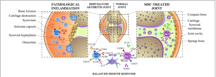
FIGURE 1 | MSCs dampen RA progression through the induction of the balance between memory Th17 and Treg cells. In RA, MSCs can diminish the frequency of pathogenic memory Th17 cells and the production of pro-inflammatory cytokines such as IL-17, IL-22, and GM-CSF and promote their differentiation toward an anti-inflammatory phenotype. In parallel, MSCs might also increase the capacity of memory Treg cells to produce anti-inflammatory cytokines such as IL-10 or TGFβ1 and prolong their immunosuppressive capacity maintaining their anti-inflammatory phenotype.
are not redundant players in this mechanism (
140
). This was
corroborated in a study with human adipose tissue-derived MSCs
that were able to reduce IL-17, TNF, and IFN-γ production and
to induce IL-10-producing T cells in vitro in collagen-specific
peripheral blood T cells of RA patients (
141
). It is well admitted
that MSCs co-cultured with purified CD4
+T cells induce the
expression of CD25
Highand FoxP3
+at the surface of these latter
T cells in a contact-dependent manner (
142
,
143
). The generation
of these CD4
+CD25
+FoxP3
+Treg has been shown to be, in part,
dependent on ICOSL expression by MSCs (
142
). Indeed, ICOS is
expressed on activated memory T cells, including Th17 cells, thus
through a contact cell-cell mechanism MSCs were proposed to
interact with memory Th17 cells and generate memory Treg cells.
In another study, it was reported that MSCs were able to recruit
both CD4
+CD25
+CD45RA
+and CD4
+CD25
+CD45RO
+Treg
cells, but the subpopulation of naïve Treg cells was recruited to
a higher extent. Additionally, MSC regulate and maintain the
suppressive function of memory Tregs cells over time (
144
).
Therefore, in the context of RA, the regulation of memory Treg
cell by MSCs is critical since they are more plastic than naive Treg
cell population (
136
).
Altogether, these studies provide evidence that MSCs do not
only increase the generation of Treg cells and the production
of IL-10 or TGFβ1 but also extend their immunosuppressive
capacity maintaining their phenotype (FoxP3
+CD127
low) and
functions (
140
,
144
). This is a critical function exerted by
MSC, considering that Treg from RA patients exhibit an
altered functionality. In addition, MSCs by suppressing the
secretion of IL17-A by effector-memory Th17 cells decrease
the acute or chronic activation of these cells in RA. Thus,
MSCs do not only inhibit the IL-17 production but also induce
the reprogramming of immunopathogenic memory Th17 cells
toward T cells with regulatory phenotype and functions (
137
)
(Summarized on Figure 1).
FUTURE PERSPECTIVE
MSCs are multipotent cells with broad immunomodulatory
properties, therefore, they have been proposed as the candidate
of choice for autoimmune diseases treatment including RA.
However, the clinical benefit for RA after 3 months of MSCs
administration have shown inconsistent positive effects. Thus,
it is necessary to increase the number of patients and studies
in order to draw robust conclusions regarding MSC therapeutic
effects in RA. Additionally, it is important to highlight that at
today, clinical trials using MSCs were injected in patients with
severe and refractory RA suggesting that MSCs treatment could
be more effective at early stages of the disease (
145
). Also, the
studies only evaluated the short-term efficacy of MSCs, from 3
to 8 months, and therefore the assessment of MSC long-term
efficacy still needs to be addressed.
Based on the topics exposed here we believe that further
studies needs to be address in order to evaluate the effect
of MSC treatment on pathogenic memory T cells derived
from RA patients. Since MSCs upon injection will migrate
to the site of inflammation were they will find an elevated
numbers of proinflammatory memory T cells it is essential
to evaluated the effect of MSCs on RA memory T cells that
has not been explored. Moreover, it is mandatory to achieve a
detailed immune-monitoring of RA patients that analyses the
dynamic of pathogenic and non-pathogenic memory T cells upon
MSCs infusion.
CONCLUSION
Memory T cells have been largely studied for their pivotal
role in the pathogenesis of auto-immune disease such as RA.
Although pro-inflammatory memory T cells-exhibit detrimental
effect in RA, their potential plasticity offers an approach yet
to be explored in order to better control RA progression.
In this context, MSCs, potent immunosuppressive cells that
are able to inhibit pro-inflammatory T cell proliferation and
functions while inducing the generation of regulatory T cells,
represent a strong candidate to choose for RA treatment.
Thus, deciphering the basis of the crosstalk between MSCs
and pathogenic memory T cells in RA will pave the way for
developing novel and potent strategies to successfully improve
MSC-based therapies.
AUTHOR CONTRIBUTIONS
NL-C, RC-L, FD, RE-V, and PL-C. wrote the manuscript with the
input of MP-M, MT, SB, MW, and FE.
FUNDING
This work was supported by Fondo Nacional de Desarrollo
Científico y Tecnológico 408 (FONDECYT) Iniciación 11160929,
Inserm, the University of Montpellier and the Société Française
de Rhumatologie (SFR).
REFERENCES
1. Le Blanc K, Mougiakakos D. Multipotent mesenchymal stromal cells and the innate immune system. Nat Rev Immunol. (2012) 12:383–96. doi: 10.1038/nri3209
2. Perry BC, Zhou D, Wu X, Yang F-C, Byers MA, Chu T-MG, et al. Collection, cryopreservation, and characterization of human dental pulp– derived mesenchymal stem cells for banking and clinical use. Tissue Eng Part C Methods. (2008) 14:149–56. doi: 10.1089/ten.tec.2008.0031
3. Trivanovi´c D, Mojsilovi´c S, Ili´c V, Krsti´c J, Jaukovi´c A, Oki´c Ðor ¯devi´c I, et al. Immunomodulatory capacity of human mesenchymal stem
cells isolated from adipose tissue, dental pulp, peripheral blood and umbilical cord Wharton’s jelly. Centr Eur J Immunol. (2013) 4:421–9. doi: 10.5114/ceji.2013.39756
4. González PL, Carvajal C, Cuenca J, Alcayaga-Miranda F, Figueroa FE, Bartolucci J, et al. Chorion mesenchymal stem cells show superior differentiation, immunosuppressive, and angiogenic potentials in comparison with haploidentical maternal placental cells: chorion MSCs outmatch other placental cells. Stem Cells Transl Med. (2015) 4:1109–21. doi: 10.5966/sctm.2015-0022
5. Luz-Crawford P, Torres MJ, Noël D, Fernandez A, Toupet K, Alcayaga-Miranda F, et al. The immunosuppressive signature of menstrual blood
mesenchymal stem cells entails opposite effects on experimental arthritis and graft versus host diseases: immunosuppresive signature of MenSC. Stem Cells. (2016) 34:456–69. doi: 10.1002/stem.2244
6. Dominici M, Le Blanc K, Mueller I, Slaper-Cortenbach I, Marini F, Krause DS, et al. Minimal criteria for defining multipotent mesenchymal stromal cells. The International Society for Cellular Therapy position statement. Cytotherapy. (2006) 8:315–7. doi: 10.1080/14653240600855905
7. Tanaka Y. Human mesenchymal stem cells as a tool for joint repair in rheumatoid arthritis. Clin Exp Rheumatol. (2015) 33(4 Suppl. 92):S58–62. Available online at: https://www.clinexprheumatol.org/abstract.asp?a=9879 8. Bhargava P, Calabresi PA. Novel therapies for memory cells in autoimmune
diseases: novel therapies for memory cells. Clin Exp Immunol. (2015) 180:353–60. doi: 10.1111/cei.12602
9. Cutler AJ, Limbani V, Girdlestone J, Navarrete CV. Umbilical Cord-derived mesenchymal stromal cells modulate monocyte function to suppress T cell proliferation. J Immunol. (2010) 185:6617–23. doi: 10.4049/jimmunol.1002239
10. Chen P-M, Liu K-J, Hsu P-J, Wei C-F, Bai C-H, Ho L-J, et al. Induction of immunomodulatory monocytes by human mesenchymal stem cell-derived hepatocyte growth factor through ERK1/2. J Leukoc Biol. (2014) 96:295–303. doi: 10.1189/jlb.3A0513-242R
11. Jiang X-X. Human mesenchymal stem cells inhibit differentiation and function of monocyte-derived dendritic cells. Blood. (2005) 105:4120–6. doi: 10.1182/blood-2004-02-0586
12. Chiesa S, Morbelli S, Morando S, Massollo M, Marini C, Bertoni A, et al. Mesenchymal stem cells impair in vivo T-cell priming by dendritic cells. Proc Natl Acad Sci USA. (2011) 108:17384–9. doi: 10.1073/pnas.1103650108 13. Németh K, Leelahavanichkul A, Yuen PST, Mayer B, Parmelee A, Doi K, et al.
Bone marrow stromal cells attenuate sepsis via prostaglandin E2–dependent reprogramming of host macrophages to increase their interleukin-10 production. Nat Med. (2009) 15:42–9. doi: 10.1038/nm.1905
14. Yen BL, Yen M-L, Hsu P-J, Liu K-J, Wang C-J, Bai C-H, et al. Multipotent human mesenchymal stromal cells mediate expansion of myeloid-derived suppressor cells via hepatocyte growth factor/c-Met and STAT3. Stem Cell Rep. (2013) 1:139–51. doi: 10.1016/j.stemcr.2013.06.006
15. Chen C-P, Chen Y-Y, Huang J-P, Wu Y-H. The effect of conditioned medium derived from human placental multipotent mesenchymal stromal cells on neutrophils: possible implications for placental infection. MHR Basic Sci Reprod Med. (2014) 20:1117–25. doi: 10.1093/molehr/gau062
16. Di Nicola M. Human bone marrow stromal cells suppress T-lymphocyte proliferation induced by cellular or nonspecific mitogenic stimuli. Blood. (2002) 99:3838–43. doi: 10.1182/blood.V99.10.3838
17. Krampera M. Bone marrow mesenchymal stem cells inhibit the response of naive and memory antigen-specific T cells to their cognate peptide. Blood. (2003) 101:3722–9. doi: 10.1182/blood-2002-07-2104
18. Corcione A. Human mesenchymal stem cells modulate B-cell functions. Blood. (2006) 107:367–72. doi: 10.1182/blood-2005-07-2657
19. Uccelli A, Moretta L, Pistoia V. Mesenchymal stem cells in health and disease. Nat Rev Immunol. (2008) 8:726–36. doi: 10.1038/nri2395
20. Griffin MD, Elliman SJ, Cahill E, English K, Ceredig R, Ritter T. Concise review: adult mesenchymal stromal cell therapy for inflammatory diseases: how well are we joining the dots?: MSC therapy for inflammatory diseases. Stem Cells. (2013) 31:2033–41. doi: 10.1002/stem.1452
21. Connick P, Kolappan M, Patani R, Scott MA, Crawley C, He X-L, et al. The mesenchymal stem cells in multiple sclerosis (MSCIMS) trial protocol and baseline cohort characteristics: an open-label pre-test: post-test study with blinded outcome assessments. Trials. (2011) 12:62. doi: 10.1186/1745-6215-12-62
22. Riordan NH, Morales I, Fernández G, Allen N, Fearnot NE, Leckrone ME, et al. Clinical feasibility of umbilical cord tissue-derived mesenchymal stem cells in the treatment of multiple sclerosis. J Transl Med. (2018) 16:57. doi: 10.1186/s12967-018-1433-7
23. Compston A, Coles A. Multiple sclerosis. Lancet. (2008) 372:1502–17. doi: 10.1016/S0140-6736(08)61620-7
24. Ibraheim H, Giacomini C, Kassam Z, Dazzi F, Powell N. Advances in mesenchymal stromal cell therapy in the management of Crohn’s disease. Exp Rev Gastroenterol Hepatol. (2018) 12:141–53. doi: 10.1080/17474124.2018.1393332
25. Lee SH, Kwon JE, Cho M-L. Immunological pathogenesis of inflammatory bowel disease. Intest Res. (2018) 16:26–42. doi: 10.5217/ir.2018.16.1.26 26. Wang D, Li J, Zhang Y, Zhang M, Chen J, Li X, et al. Umbilical cord
mesenchymal stem cell transplantation in active and refractory systemic lupus erythematosus: a multicenter clinical study. Arthritis Res Ther. (2014) 16:R79. doi: 10.1186/ar4520
27. Tsokos GC. Systemic lupus erythematosus. N Engl J Med. (2011) 365:2110– 21. doi: 10.1056/NEJMra1100359
28. Swain SL, Agrewala JN, Brown DM, Jelley-Gibbs DM, Golech S, Huston G, et al. CD4+ T-cell memory: generation and multi-faceted roles for CD4+ T cells in protective immunity to influenza. Immunol Rev. (2006) 211:8–22. doi: 10.1111/j.0105-2896.2006.00388.x
29. Farber DL, Yudanin NA, Restifo NP. Human memory T cells: generation, compartmentalization and homeostasis. Nat Rev Immunol. (2014) 14:24–35. doi: 10.1038/nri3567
30. Amsen D, Backer RA, Helbig C. Decisions on the road to memory. In: Katsikis PD, Schoenberger SP, Pulendran B, editors. Crossroads Between Innate and Adaptive Immunity IV. New York, NY: Springer New York (2013). p. 107–20. Available online at: http://link.springer.com/10.1007/978-1-4614-6217-0_12 (accessed June 15, 2018).
31. Smith SH, Brown MH, Rowe D, Callard RE, Beverley PC. Functional subsets of human helper-inducer cells defined by a new monoclonal antibody, UCHL1. Immunology. (1986) 58:63–70.
32. Sanders ME, Makgoba MW, Sharrow SO, Stephany D, Springer TA, Young HA, et al. Human memory T lymphocytes express increased levels of three cell adhesion molecules (LFA-3, CD2, and LFA-1) and three other molecules (UCHL1, CDw29, and Pgp-1) and have enhanced IFN-gamma production. J Immunol. (1988) 140:1401–7.
33. Sallusto F, Lenig D, Förster R, Lipp M, Lanzavecchia A. Two subsets of memory T lymphocytes with distinct homing potentials and effector functions. Nature. (1999) 401:708–12.
34. Sallusto F, Geginat J, Lanzavecchia A. Central memory and effector memory T cell subsets: function, generation, and maintenance. Annu Rev Immunol. (2004) 22:745–63. doi: 10.1146/annurev.immunol.22.012703.104702 35. Lovett-Racke AE, Trotter JL, Lauber J, Perrin PJ, June CH, Racke
MK. Decreased dependence of myelin basic protein-reactive T cells on CD28-mediated costimulation in multiple sclerosis patients. A marker of activated/memory T cells. J Clin Invest. (1998) 101:725–30. doi: 10.1172/JCI1528
36. Hedlund G, Sandberg-Wollheim M, Sjögren HO. Increased proportion of CD4+ CDw29+ CD45R– UCHL-1+ lymphocytes in the cerebrospinal fluid of both multiple sclerosis patients and healthy individuals. Cell Immunol. (1989) 118:406–12. doi: 10.1016/0008-8749(89)90388-2
37. Muraro PA, Pette M, Bielekova B, McFarland HF, Martin R. Human autoreactive CD4+ T cells from naive CD45RA+ and memory CD45RO+ subsets differ with respect to epitope specificity and functional antigen avidity. J Immunol. (2000) 164:5474–81. doi: 10.4049/jimmunol.164.10.5474 38. Mullen KM, Gocke AR, Allie R, Ntranos A, Grishkan IV, Pardo C, et al. Expression of CCR7 and CD45RA in CD4+ and CD8+ subsets in cerebrospinal fluid of 134 patients with inflammatory and non-inflammatory neurological diseases. J Neuroimmunol. (2012) 249:86–92. doi: 10.1016/j.jneuroim.2012.04.017
39. Zaffaroni M, Rossini S, Ghezzi A, Parma R, Cazzullo CL. Decrease of CD4+CD45+ T-cells in chronic-progressive multiple sclerosis. J Neurol. (1990) 237:1–4. doi: 10.1007/BF00319659
40. Kadowaki A, Saga R, Lin Y, Sato W, Yamamura T. Gut microbiota-dependent CCR9+CD4+ T cells are altered in secondary progressive multiple sclerosis. Brain. (2019) 142:916–31. doi: 10.1093/brain/awz012
41. Roman LI, Manzano L, De La Hera A, Abreu L, Rossi I, Alvarez-Mon M. Expanded CD4+CD45RO+ phenotype and defective proliferative response in T lymphocytes from patients with Crohn’s disease. Gastroenterology. (1996) 110:1008–19. doi: 10.1053/gast.1996.v110.pm8612987
42. De Tena JG, Manzano L, Leal JC, Antonio ES, Sualdea V, Álvarez-Mon M. Active Crohn’s disease patients show a distinctive expansion of circulating memory CD4+CD45RO+ CD28- T cells. J Clin Immunol. (2004) 24:185–96. doi: 10.1023/B:JOCI.0000019784.20191.7f
43. Tena JGD, Manzano L, Leal JC, Antonio ES, Sualdea V, Álvarez-Mon M. Distinctive pattern of cytokine production and adhesion molecule expression
in peripheral blood memory CD4+ T cells from patients with active Crohn’s disease. J Clin Immunol. (2006) 26:233–42. doi: 10.1007/s10875-006-9016-4 44. Fritsch RD, Shen X, Illei GG, Yarboro CH, Prussin C, Hathcock KS,
et al. Abnormal differentiation of memory T cells in systemic lupus erythematosus. Arthritis Rheum. (2006) 54:2184–97. doi: 10.1002/art.21943 45. Zhou H, Hu B, Huang N, Mo X, Li W, Zhang B, et al. Aberrant T cell
subsets and cytokines expression profile in systemic lupus erythematosus. Clin Rheumatol. (2018) 37:2405–13. doi: 10.1007/s10067-018-4124-0 46. Hu X, Kim H, Stahl E, Plenge R, Daly M, Raychaudhuri S. Integrating
autoimmune risk loci with gene-expression data identifies specific pathogenic immune cell subsets. Am J Hum Genet. (2011) 89:496–506. doi: 10.1016/j.ajhg.2011.09.002
47. Machold KP, Stamm TA, Nell VPK, Pflugbeil S, Aletaha D, Steiner G, et al. Very recent onset rheumatoid arthritis: clinical and serological patient characteristics associated with radiographic progression over the first years of disease. Rheumatology. (2006) Jul 28;46:342–9.
48. Firestein GS. Evolving concepts of rheumatoid arthritis. Nature. (2003) 423:356–61. doi: 10.1038/nature01661
49. Müller-Ladner U, Ospelt C, Gay S, Distler O, Pap T. Cells of the synovium in rheumatoid arthritis. Synovial fibroblasts. Arthritis Res Ther. (2007) 9:223. doi: 10.1186/ar2337
50. Yang Z, Shen Y, Oishi H, Matteson EL, Tian L, Goronzy JJ, et al. Restoring oxidant signaling suppresses proarthritogenic T cell effector functions in rheumatoid arthritis. Sci Transl Med. (2016) 8:331ra38. doi: 10.1126/scitranslmed.aad7151
51. Thomas R, McIlraith M, Davis LS, Lipsky PE. Rheumatoid synovium is enriched in CD45RBdim mature memory T cells that are potent helpers for B cell differentiation. Arthritis Rheum. (1992) 35:1455–65. doi: 10.1002/art.1780351209
52. Kohem CL, Brezinschek RI, Wisbey H, Tortorella C, Lipsky PE, Oppenheimer-Marks N. Enrichment of differentiated CD45RBdim,CD27-memory T cells in the peripheral blood, synovial fluid, and synovial tissue of patients with rheumatoid arthritis. Arthritis Rheum. (1996) 39:844–54. doi: 10.1002/art.1780390518
53. Morita Y, Yamamura M, Kawashima M, Harada S, Tsuji K, Shibuya K, et al. Flow cytometric single-cell analysis of cytokine production by CD4+ T cells in synovial tissue and peripheral blood from patients with rheumatoid arthritis. Arthritis Rheum. (1998) 41:1669–76. doi: 10.1002/1529-0131(199809)41:9<1669::AID-ART19>3.0.CO;2-G 54. Zhang X, Nakajima T, Goronzy JJ, Weyand CM. Tissue trafficking patterns
of effector memory CD4+ T cells in rheumatoid arthritis. Arthritis Rheum. (2005) 52:3839–49. doi: 10.1002/art.21482
55. Matsuki F, Saegusa J, Nishimura K, Miura Y, Kurosaka M, Kumagai S, et al. CD45RA–Foxp3low non-regulatory T cells in the CCR7–CD45RA– CD27+CD28+ effector memory subset are increased in synovial fluid from patients with rheumatoid arthritis. Cell Immunol. (2014) 290:96–101. doi: 10.1016/j.cellimm.2014.05.011
56. Koenders MI, van den Berg WB. Novel therapeutic targets in rheumatoid arthritis. Trends Pharmacol Sci. (2015) 36:189–95. doi: 10.1016/j.tips.2015.02.001
57. Andersson KME, Cavallini NF, Hu D, Brisslert M, Cialic R, Valadi H, et al. Pathogenic transdifferentiation of Th17 cells contribute to perpetuation of rheumatoid arthritis during anti-TNF treatment. Mol Med. (2015) 21:536– 43. doi: 10.2119/molmed.2015.00057
58. Genovese MC, Durez P, Richards HB, Supronik J, Dokoupilova E, Mazurov V, et al. Efficacy and safety of secukinumab in patients with rheumatoid arthritis: a phase II, dose-finding, double-blind, randomised, placebo controlled study. Ann Rheum Dis. (2013) 72:863–9. doi: 10.1136/annrheumdis-2012-201601
59. Martin DA, Churchill M, Flores-Suarez L, Cardiel MH, Wallace D, Martin R, et al. A phase Ib multiple ascending dose study evaluating safety, pharmacokinetics, and early clinical response of brodalumab, a human anti-IL-17R antibody, in methotrexate-resistant rheumatoid arthritis. Arthritis Res Ther. (2013) 15:R164. doi: 10.1186/ar4347
60. Greter M, Helft J, Chow A, Hashimoto D, Mortha A, Agudo-Cantero J, et al. GM-CSF controls nonlymphoid tissue dendritic cell homeostasis but is dispensable for the differentiation of inflammatory dendritic cells. Immunity. (2012) 36:1031–46. doi: 10.1016/j.immuni.2012.03.027
61. Reynolds G, Gibbon JR, Pratt AG, Wood MJ, Coady D, Raftery G, et al. Synovial CD4+ T-cell-derived GM-CSF supports the differentiation of an inflammatory dendritic cell population in rheumatoid arthritis. Ann Rheum Dis. (2016) 75:899–907. doi: 10.1136/annrheumdis-2014-206578
62. Behrens F, Tak PP, Østergaard M, Stoilov R, Wiland P, Huizinga TW, et al. MOR103, a human monoclonal antibody to granulocyte–macrophage colony-stimulating factor, in the treatment of patients with moderate rheumatoid arthritis: results of a phase Ib/IIa randomised, double-blind, placebo-controlled, dose-escalation trial. Ann Rheum Dis. (2015) 74:1058– 64. doi: 10.1136/annrheumdis-2013-204816
63. Fleischmann R, Kremer J, Cush J, Schulze-Koops H, Connell CA, Bradley JD, et al. Placebo-controlled trial of tofacitinib monotherapy in rheumatoid arthritis. N Engl J Med. (2012) 367:495–507. doi: 10.1056/NEJMoa1109071 64. Genovese MC, Kremer J, Zamani O, Ludivico C, Krogulec M, Xie L, et al.
Baricitinib in patients with refractory rheumatoid arthritis. N Engl J Med. (2016) 374:1243–52. doi: 10.1056/NEJMoa1507247
65. Geginat J, Sallusto F, Lanzavecchia A. Cytokine-driven proliferation and differentiation of human naive, central memory, and effector memory CD4(+) T cells. J Exp Med. (2001) 194:1711–9. doi: 10.1084/jem.194.12.1711 66. Boyle DL, Soma K, Hodge J, Kavanaugh A, Mandel D, Mease P, et al. The JAK inhibitor tofacitinib suppresses synovial JAK1-STAT signalling in rheumatoid arthritis. Ann Rheum Dis. (2015) 74:1311–6. doi: 10.1136/annrheumdis-2014-206028
67. Hünig T, Beyersdorf N, Kerkau T. CD28 co-stimulation in T-cell homeostasis: a recent perspective. ImmunoTargets Ther. (2015) 4:111–22. doi: 10.2147/ITT.S61647
68. Nam JL, Takase-Minegishi K, Ramiro S, Chatzidionysiou K, Smolen JS, van der Heijde D, et al. Efficacy of biological disease-modifying antirheumatic drugs: a systematic literature review informing the (2016) update of the EULAR recommendations for the management of rheumatoid arthritis. Ann Rheum Dis. (2017) 76:1113–36. doi: 10.1136/ annrheumdis-2016-210713
69. Warrington KJ, Takemura S, Goronzy JJ, Weyand CM. CD4+,CD28-T cells in rheumatoid arthritis patients combine features of the innate and adaptive immune systems. Arthritis Rheum. (2001) 44:13–20. doi: 10.1002/1529-0131(200101)44:1<13::AID-ANR3>3.0.CO;2-6 70. Park W, Weyand CM, Schmidt D, Goronzy JJ. Co-stimulatory pathways
controlling activation and peripheral tolerance of human CD4+CD28– T cells. Eur J Immunol. (1997) 27:1082–90. doi: 10.1002/eji.1830270507 71. Goronzy JJ, Weyand CM. Thymic function and peripheral T-cell
homeostasis in rheumatoid arthritis. Trends Immunol. (2001) 22:251–5. doi: 10.1016/S1471-4906(00)01841-X
72. Vallejo AN, Bryl E, Klarskov K, Naylor S, Weyand CM, Goronzy JJ. Molecular basis for the loss of CD28 expression in senescent T cells. J Biol Chem. (2002) 277:46940–9. doi: 10.1074/jbc.M207352200
73. Appay V, van Lier RAW, Sallusto F, Roederer M. Phenotype and function of human T lymphocyte subsets: consensus and issues: phenotype and function of human T lymphocyte subsets: consensus and issues. Cytometry Part A. (2008) 73A:975–83. doi: 10.1002/cyto.a.20643
74. Tayar JH, Suarez-Almazor ME. New understanding and approaches to treatment in rheumatoid arthritis. Br Med Bull. (2010) 94:201–14. doi: 10.1093/bmb/ldq007
75. Liang J, Li X, Zhang H, Wang D, Feng X, Wang H, et al. Allogeneic mesenchymal stem cells transplantation in patients with refractory RA. Clin Rheumatol. (2012) 31:157–61. doi: 10.1007/s10067-011-1816-0
76. Wang L, Wang L, Cong X, Liu G, Zhou J, Bai B, et al. Human umbilical cord mesenchymal stem cell therapy for patients with active rheumatoid arthritis: safety and efficacy. Stem Cells Dev. (2013) 22:3192–202. doi: 10.1089/scd.2013.0023
77. Álvaro-Gracia JM, Jover JA, García-Vicu-a R, Carre-o L, Alonso A, Marsal S, et al. Intravenous administration of expanded allogeneic adipose-derived mesenchymal stem cells in refractory rheumatoid arthritis (Cx611): results of a multicentre, dose escalation, randomised, single-blind, placebo-controlled phase Ib/IIa clinical trial. Ann Rheum Dis. (2017) 76:196–202. doi: 10.1136/annrheumdis-2015-208918
78. Yang J, Brook MO, Carvalho-Gaspar M, Zhang J, Ramon HE, Sayegh MH, et al. Allograft rejection mediated by memory T cells is resistant to regulation. Proc Natl Acad Sci USA. (2007) 104:19954–9. doi: 10.1073/pnas.0704397104