HAL Id: hal-01932007
https://hal.univ-lorraine.fr/hal-01932007
Submitted on 23 Nov 2018
HAL is a multi-disciplinary open access archive for the deposit and dissemination of sci-entific research documents, whether they are pub-lished or not. The documents may come from teaching and research institutions in France or abroad, or from public or private research centers.
L’archive ouverte pluridisciplinaire HAL, est destinée au dépôt et à la diffusion de documents scientifiques de niveau recherche, publiés ou non, émanant des établissements d’enseignement et de recherche français ou étrangers, des laboratoires publics ou privés.
Analyse quantitative de la bascule postérieure du
scaphoïde sur des arthroscanners du poignet chez les
patients ayant une rupture du ligament schapo-lunaire
Jacques De Verbizier
To cite this version:
Jacques De Verbizier. Analyse quantitative de la bascule postérieure du scaphoïde sur des arthroscan-ners du poignet chez les patients ayant une rupture du ligament schapo-lunaire. Sciences du Vivant [q-bio]. 2016. �hal-01932007�
AVERTISSEMENT
Ce document est le fruit d'un long travail approuvé par le jury de
soutenance et mis à disposition de l'ensemble de la
communauté universitaire élargie.
Il est soumis à la propriété intellectuelle de l'auteur. Ceci
implique une obligation de citation et de référencement lors de
l’utilisation de ce document.
D'autre part, toute contrefaçon, plagiat, reproduction illicite
encourt une poursuite pénale.
Contact : ddoc-thesesexercice-contact@univ-lorraine.fr
LIENS
Code de la Propriété Intellectuelle. articles L 122. 4
Code de la Propriété Intellectuelle. articles L 335.2- L 335.10
http://www.cfcopies.com/V2/leg/leg_droi.php
UNIVERSITÉ DE LORRAINE FACULTÉ DE MÉDECINE DE NANCY 2016
THÈSE
pour obtenir le grade de DOCTEUR EN MÉDECINE
Présentée et soutenue publiquement dans le cadre du troisième cycle de
Médecine Spécialisée
par
Jacques de VERBIZIER
le 30 septembre 2016
ANALYSE QUANTITATIVE DE LA BASCULE POSTERIEURE DU SCAPHOIDE SUR DES ARTHROSCANNERS DU POIGNET CHEZ LES PATIENTS AYANT UNE
RUPTURE DU LIGAMENT SCAPHO-LUNAIRE
Membres du jury :
Président : M. le Professeur BLUM Juges : M. le Professeur DAP
M. le Professeur DAUTEL
A mon Président du jury
Je tiens à exprimer mes plus vifs remerciements au Professeur Blum qui fut pour moi un directeur de thèse attentif et disponible malgré ses nombreuses charges.
Aux juges :
Pedro, j m m ’ m’ m T ’ e mes nombreuses questions, et tu ’ ’ m m que nous avons eues ainsi que tes conseils sont p Ton enthousiasme m’a m m . Pour tout cela merci.
Je voudrais m m P P ’ m m
m m m m ’ ’ sentation de m ’ pour des chirurgiens.
Famille
Amandine. Tu as changé ma vie. C’ ’ m ôtés une personne qui me comprend, qui me soutient m m’ m
’ ù ’ Mais en fait je te connais je pense mm ’ … ’ m …
A mes parents, le classique et efficace « je ne serais rien sans vous » est de mise. Merci beaucoup pour tout ce que vous avez fait, ce que vous faîtes et ce que v z m V m’ z pratiquement tout appris, notamment à faire du vélo avant que je tienne correctement sur mes 2 jambes (sans me doper, enfin je ne le crois pas). V m’ z s passions (peinture, sculpture, modélisme – m m ’ 2 m -, les randonnées – mêmes si ç m’ -, la radiologie). C’ âce à vous que je rédige ces lignes. Papa, forcément au boulot beaucoup de personnes me disent que je te ressemble énormément, e ’ m m ’ me faire.
Alice et Vincent. E œ -frère, vous avez un rôle capital à mon équilibre A ’ grandement à ce que je suis actu m m m’ lâché malgré nos quelques disputes inutiles. Je vous aime et je suis très fier de faire partie de votre famille parfaite, avec GuGu et MaMa.
Charlotte et Jomphop. Malheureusem C œ ’ ours tendance à vouloir te protéger. Alors que lorsque je deviens objectif et que je vois la femme que tu es devenue mm â m ’ ( toute ma famille). Votre bonne humeur et votre optimisme sont essentiels à notre famille. Merci pour tous ces souvenirs que vous nous apportez tous les jours depuis un peu plus de vingt ans.
Yaya. La plus fun des grand-mères C’ T ’ m ’ mple. Merci.
Grand-Papa. La pêche, les randonnées dans les Pyrénées dans des endroits sublimes, ’ age du ’U balades en bateau à Libourne. Tant de souvenirs gravés à tout jamais dans ma mémoire grâce à toi. Merci.
A mes beaux-parents. Merci pour votre acceuil. Je me suis tout de suite senti chez moi chez vous. ’ m m E ’ m z ’
Laurène et Pierre-Alain, Franck et Corinne. Qui aurait dit que le petit Piwit serait dans votre famille un jour E ’aime profiter des moments passés avec vous !
Amis
« Le bonheur n'est pas au sommet de la montagne mais dans la façon de la gravir ». J’ai de la chance de vous avoir tous à mes côtés car grâce à vous j’atteins le bonheur.
Les Luconi. Barbecue sur votre balcon, footbalcon®, mémoire flash, quelques soirées légendaires, le rugby, tennis, le cabare ’ m Q m m !
Les Hotton. S W EVG ( m … ’ mm un gant). Merci pour votre soutien et votre gentillesse ! Bisous à Clém et Pauline, vous êtes mes « 4 Fantastiques » !
V m’ m T F ’ m EVG . Notre amitié est comme la saga Star Wars®, immortelle.
Matteucci, Vous avez fait plus que de nous accueillir il y a un peu plus de 20 ans (ça ne nous rajeunit pas tout ç …). Vous avez mis en place la première pierre ’ m m de Metz.
Guillaume. The Superman. Merci pour le demi-tour du Mont-Blanc. Désolé d’ « … », mais ç ’ m m m m m ’ ’ ( û du Ping-P m …)
La Team. Ou plutôt THE TEAM !!!!!!!!! Vous êtes plus sympas et accueillants les uns que les autres. Même si m m m z ( m 18 ) ’ œ . Michael. Merci pour les partages de vidéos de voitures télécommandées.
Marvin et Fabien. Très beau couple, plein de bonheur. Heureux que vous vous rapprochiez de nous !
Amélie et Anthony. Mes petits Marseillais préférés. Marceau est top, bien que la distance nous éloigne beaucoup ’ !
Valentine m m ’ un peu éloigné, les soirées geek avec toi sont un pur bonheur. Je suis content que tu aies troqué ta vieille Xbox contre une PS4.
A mes co internes, tout particulièrement Anne-Sophie, Chloé, Marine, Rémi. Votre soutien et nos moments m m m V ’ contact en post internat.
A ’ côtoyer m m’ progresser tant professionnellement ’humainement. ’ z s pour m’ m m t leur humour tellement importants.
Mes élèves : Matthieu et Sophie. Sophie merci pour ta niaque. Un sacré bout de femme. Matthieu, toujours ’ ’ m m ?
SERMENT
«
A
u moment d'être admis à exercer la médecine, je promets et je jure d'être fidèle auxlois de l'honneur et de la probité. Mon premier souci sera de rétablir, de préserver ou de promouvoir la santé dans tous ses éléments, physiques et mentaux, individuels et sociaux. Je respecterai toutes les personnes, leur autonomie et leur volonté, sans aucune discrimination ’ vulnérables ou menacées dans leur intégrité ou leur dignité. Même sous la contrainte, je ne ferai pas usage de mes connaissances contre les lois de l'humanité. J'informerai les patients des décisions envisagées, de leurs raisons et de leurs conséquences. Je ne tromperai jamais leur confiance et n'exploiterai pas le pouvoir hérité des circonstances pour forcer les consciences. Je donnerai mes soins à l'indigent et à quiconque me les demandera. Je ne me laisserai pas influencer par la soif du gain ou la recherche de la gloire.
Admis dans l'intimité des personnes, je tairai les secrets qui me sont confiés. Reçu à l'intérieur des maisons, je respecterai les secrets des foyers et ma conduite ne servira pas à corrompre les mœ m agonies. Je ne provoquerai jamais la mort délibérément.
Je préserverai l'indépendance nécessaire à l'accomplissement de ma mission. Je n'entreprendrai rien qui dépasse mes compétences. Je les entretiendrai et les perfectionnerai pour assurer au mieux les services qui me seront demandés.
J'apporterai mon aide à mes confrères ainsi qu'à leurs familles dans l'adversité.
Que les hommes et mes confrères m'accordent leur estime si je suis fidèle à mes promesses ; que je sois déshonoré et méprisé si j'y manque ».
Table des matières
1. INTRODUCTION : ... 14
2. MATÉRIELS ET MÉTHODES : ... 15
2.1PATIENTS : ... 15
2.2ACQUISITION DES EXAMENS : ... 16
2.3ANALYSE DES EXAMENS : ... 17
2.4ANALYSE STATISTIQUE : ... 18
3. RÉSULTATS : ... 19
4. DISCUSSION : ... 25
1. INTRODUCTION ... 30
2. MATERIALS AND METHODS... 31
2.1PATIENTS ... 31 2.2IMAGING PROTOCOL ... 31 2.3IMAGE ANALYSIS ... 33 2.4STATISTICAL ANALYSIS... 35 3. RESULTS ... 35 4. DISCUSSION ... 42
ANALYSE QUANTITATIVE DE LA BASCULE POSTERIEURE DU SCAPHOIDE SUR DES ARTHROSCANNERS DU POIGNET CHEZ LES PATIENTS AYANT
UNE RUPTURE DU LIGAMENT SCAPHO-LUNAIRE
1. Introduction :
Les ruptures du ligament scapho-lunaire sont les plus fréquentes des lésions ligamentaires du carpe et sont principalement dues à un traumatisme direct du poignet en hyper extension [1-3].
La rupture du ligament scapho-lunaire entraî ’ m ï et du lunatum appelée dissociation scapho-lunaire.
La dissociation scapho-lunaire ’ [4] Watson et al. [5,6] sous le nom de scapholunate advanced collapse (SLAC).
Le traitement du SLAC dépend de son stade et peut aussi bien être conservateur que chirurgical. L m m ’ fonction du poignet [7].
La surface articulaire proximale du scaphoïde et la fossette scaphoïdienne radiale sont m m mm ’ ’ [8] E dissociation scapho-lunaire, la flexion palmaire et la bascule postérieure du scaphoïde sont ’ m m postérieure de la fossette articulaire radiale. Le déplacement postérieur du scaphoïde est m S AC [8 9] A m m ’ bascule postérieure du scaphoïde est importante pour déterminer la prise en charge thérapeutique [8].
La majorité des défauts d’ m est visible sur les radiographies [10 11] C ’ ï m ’ ’ tre part du fait des variations de position des patients pouvant influencer la ï ’ [8 11 12]
E m m ’IR ’arthroscanner sont fréquemment utilisés pour évaluer le ligament scapho-lunaire et le cartilage articulaire [13-15] ’ m m m m ’ m ï m -lunaire [16].
’angle formé sur une reformation sagittale ’ arthroscanner entre la surface articulaire radiale et le point le plus postérieur du scaphoïde (angle radio-scaphoïdien ) m ’ -scaphoï N ’ m ’ --scaphoïdien postérieur est corrélée au SLAC en cas de rupture du ligament scapho-lunaire. Cette information peut avoir une implication dans la prise en charge du patient.
2. Matériels et méthodes :
2.1 Patients :
De janvier 2009 à décembre 2013, 605 patients présentant des douleurs de poignet ont ’ P m patients, 150 ont été sélectionnés de manière randomisée.
’arthroscanner m ’ m -lunaire, a été m ’ -scaphoïdien postérieur. Les études rétrospectives basées sur ’ m ’ m ne nécessitent pas ’ m ’ dans notre hôpital.
Trente-huit patients ayant des antécédents chirurgicaux du poignet, des pathologies mm ’ La population finale de notre étude comporte donc cent douze patients.
E ’ m m scapho-lunaire sur ’arthroscanner, les patients ont été divisés en deux groupes : les patients ayant un ligament scapho-lunaire normal ou une rupture isolée du segment membraneux (groupe contrôle) ; les
patients ayant une rupture du segment postérieur et/ou antérieur du ligament scapho-lunaire (groupe témoin).
2.2 Acquisition des examens :
’ ’ A m I R200 (Siemens Healthcare) (55kVp et 7-10 mA) ; ou avec un Prestige (GE healthcare) (55kVp et 2-10 mA). Trois incidences ont été étudiées dans notre étude : face, profil et poing serré. Les radiographies de profil ont été acquises en position neutre. Les radiographies de face ont été m m ’ m m m ’ scapho-lunaire.
’ n de produit de contraste intra-articulaire a été réalisée sous guidage ’ radiologue spécialisé en ostéo-articulaire. Les articulations médio-carpienne et radio-carpienne ont été opacifiées chez tous les ’ m - m E ’ diffusion spontanée du produit de contraste dans ’ -carpienne, une inje ’ -carpienne était réalisée.
P ’ isition scanographique, les patients étaient en procubitus, la main au-dessus de la tête (position « superman ») ’ mises en place pour que le patient conserve son épaule en extension dans une position confortable.
’ ’ 2 : un scanner 64 barrettes (Aquilion 64, Toshiba Medical) et un scanner de 320 barrettes (Aquilion One, Toshiba Medical). Une acquisition hélicoïdale a été réalisée avec le scanner 64 barrettes avec les paramètres suivant : FOV de 170 mm, matrice de 520x520, 120kVp, 100 mAs, épaisseur de coupe de 0,5mm, pitch de 0,9 et la rotation du tube de 1 seconde.
Une acquisition volumique a été réalisée sur le scanner 320 barrettes avec les paramètres suivants : FOV de 160 mm, matrice de 520 x 520, 100kVp, 50 mAs, épaisseur de coupe de 0,5mm, rotation de tube de 0,5 seconde.
Les examens ont été reconstruits dans les 3 plans. Le plan coronal est défini comme 2 ï ’ t celui du radius. Le plan sagittal est perpendiculaire au précédant.
2.3 Analyse des examens :
Deux radiologues de 2 et 6 ’ m m U ’ î m 10 ’ a été réalisée ’ m m m ’ m toutes les mesures ont été réalisées sur les consoles PACS (V5 Impax, Agfa Healthcare). Un m 10 ’
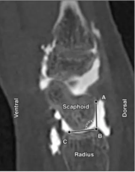
’ -scaphoïdien postérieur était mesuré sur les reformations sagittales en suivant la procédure suivante : nous faisions défiler les reformations sagittales afin ’ le point le plus postérieur du scaphoïde. m m ’ N une ligne passant par les points antérieur et postérieur de la surface articulaire radiale. Puis nous tracions une seconde ligne passant par le point le plus postérieur de la surface articulaire ï ’ mm ’ e radio-scaphoïdien postérieur (Fig 1).
Figure 1 : mesure de l’angle radio-scaphoïdien postérieur. La coupe ci-dessus est celle passant par le point le plus postérieur du scaphoide (A). Puis les points le plus postérieur (B) et antérieur (C) de la surface articulaire radiale sont identifiés. Deux lignes sont ensuite tracées : une partant du point le plus postérieur du scaphoïde (A) jusqu’au point le plus postérieur de la surface articulaire radiale (B) ; une seconde passant par les points B et C. L’angle formé par ces deux droites est l’angle radio-scaphoïdien postérieur (ligne pointillée).
Les angles scapho-lunaire, radio-lunaire et le diastasis scapho-lunaire ont aussi été m ’ scapho-lunaire était considéré comme normal 30° 70° [8 17 18] ’ radio-lunaire était considéré comme anormal au-dessus de 10° [8]. Le diastasis scapho-lunaire était défini comme étant une distance supérieure à 3 mm entre le scaphoïde et le lunatum sur les clichés de face [17].
’ (S AC) ’ W [3 19] I remaniements dégénératifs touchent la partie latérale de la surface articulaire radiale. Dans le II ’ m ’ -scaphoïdienne est touchée. Dans le stade III ’ -lunaire est atteinte en plus de la radio-scaphoïdienne.
2.4 Analyse statistique :
Les arthroscanners ont été utilisés comme référence diagnostique pour les lésions du ligament scapho- ’ m -lunaire était présente, les radiographies ont été utilisées pour grader le SLAC. Les angles scapho-lunaire, radio-lunaire,
radio- ï m ’ ’ t test pour des variables indépendantes. Une régression logistique multivariée a été utilisée pour générer des ROC et pour identifier le seuil permettant de déterminer la présence de SLAC. Les courbes ROC ont m ’ ctibilités inter et intra observateurs ont ’ (ICC) ’ANOVA
3. Résultats :
Dans notre étude ont été inclus 68 hommes et 44 femmes (sex ratio 1,5), la moyenne ’â (+/- SD) était de 41,3 +/- 12 ans.
Quarante-deux patients avaient un ligament scapho-lunaire intact (groupe contrôle) et 70 présentaient une rupture du ligament scapho-lunaire (groupe témoin).
Trente patients du groupe témoin étaient atteints de lésions dégénératives du carpe. Il y avait respectivement 4, 12 et 14 cas de SLAC I, II et III.
m ’ -scaphoïdien postérieur était réalisable chez tous les patients. La reproductibilité inter-observateurs était considérée comme bonne (ICC de 0,75). I ’ rence significative de la reproductibilité entre les patients sans et avec SLAC (ICC respectivement de 0,87 et 0,82). La reproductibilité intra-observateur était quasiment parfaite (ICC de 0,92).
’ -scaphoïdien postérieur était augmenté de manière significative chez les patients ayant une rupture du ligament scapho-lunaire (p < 0,0001).
m ’ -scaphoïdien postérieur était de 98,3° +/- 8° et de 99,6° +/- 7,2° dans le groupe contrôle contre 112,7°+/- 11,8° et de 111,3° +/- 12,7° dans le groupe témoin respectivement pour les lecteurs 1 et 2. Parmi les patients ayant une rupture du ligament scapho-lunaire ’ -scaphoïdien postérieur était plus élevé chez les patients ayant un SLAC. (p<0 0001 ) (F 2) I ’ ’ m ’ -scaphoïdien et le stade de SLAC. (p = 0,662 et p = 0,723 respectivement pour les premier et deuxième lecteur).
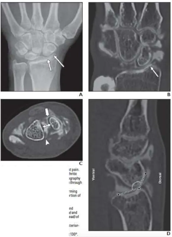
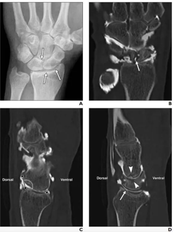
Figure 2 : femme de 49 ans ayant des douleurs chroniques du poignet.
A. Radiographie de face : remaniements dégénératifs de l’articulation radio-scaphoïdienne (flèches). B. Reconstruction frontale de l’arthroscanner : Erosions chondrales et irrégularité de la partie latérale
de la surface articulaire radiale (flèche).
C. Reformation oblique : diastasis scapho-lunaire (double flèche) et rupture complète du segment dorsal et partiel du segment ventral du ligament scapho-lunaire.
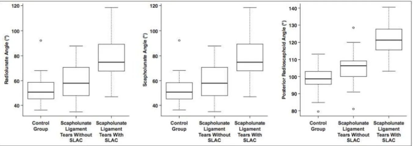
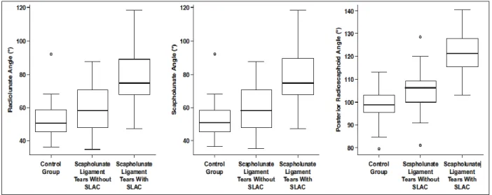
Les angles scapho-lunaire et radio-lunaire ont également été différents chez les patients sans et avec rupture de ligament scapho-lunaire (p<0,0001). Le tableau 1 compare les valeurs des angles radio-scaphoïdien postérieur, scapho-lunaire et radio-lunaire ’ radio-scaphoïdien postérieur permet une meilleure distinction des patients sans et avec SLAC. (Fig 3).
Figure 3 : distribution des angles radio-lunaire, scapho-lunaire et de l’angle radio-scaphoïdien postérieur. On note un chevauchement des angles dans les 3 groupes, mais l’angle radio-scaphoïdien postérieur permet une meilleure discrimination entre les patients sans et avec SLAC.
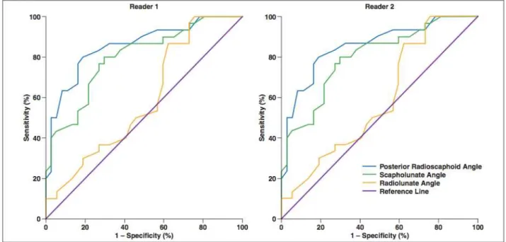
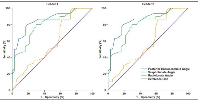
’ ROC m ’ -scaphoïdien postérieur était meilleur que les angles scapho-lunaire et radio-lunaire pour la différenciation des patients sans et avec SLAC (p<0,02) ( 4) E ’ ROC radio-scaphoïdien postérieur de 114° était considéré comme seuil permettant de différencier les patients sans et avec SLAC. Après relecture par le troisièm ’ -scaphoïdien postérieur avait une sensibilité de 80%, une spécificité de 89,7% une VPP de 85,7% et une VPN de 85,4%. Le tableau 2 montre les performances diagnostiques des ’ S AC z tients ayant une rupture du ligament scapho-lunaire.
Figure 4 : diagnostic de SLAC en utilisant les angles scaphoïdien postérieur, scapho-lunaire et lunaire pour les lecteurs 1 et 2. On note une meilleure puissance diagnostique du SLAC par l’angle radio-scaphoïdien postérieur pour les 2 lecteurs.
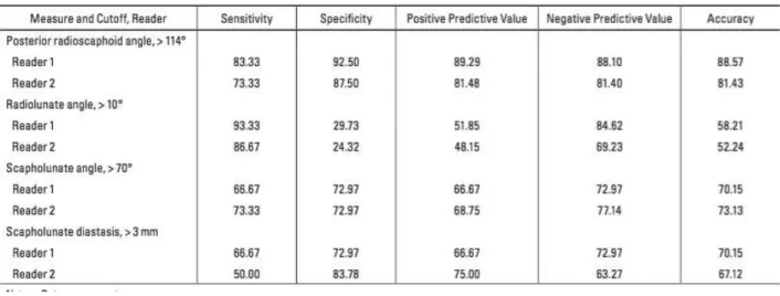
Tableau 2 : perfomance diagnostique des angles radio-scaphoïdien postérieur, radio-lunaire, scapho-lunaire et du diastasis scapho-lunaire pour l’identification de SLAC chez les patients ayant des lésions du ligament scapho-lunaire.
Avec un seuil de 114°, il y avait une discordance entre les deux lecteurs sur 14 patients (12,5% de la population). Les variations de mesure étaient principalement dues à des remaniements ostéophytiques de la partie post ’ -scaphoïdienne (12 cas). Une discordance a aussi été mise en évidence pour les angles scapho-lunaire, radio-lunaire et pour le diastasis scapho-radio-lunaire respectivement sur 8 (7,1%), 18 (16%) et 9 (8%) cas. Parmi les 40 patients ayant une rupture du ligament scapho-lunaire sans SLAC sur les radiographies standard, 4 avaient un angle radio-scaphoïdien postérieur de plus de 114°. Dans deux cas, même si les radiographies standard ne mettaient pas en évidence de SLAC, ’arthroscanner m ’ -scaphoïdienne. (fig. 5). Les deux autres patients étaient jeunes (18 et 22 ans) et avaient une instabilité clinique nécessitant une prise en charge chirurgicale.
Figure 5 : homme de 45 ans avec des douleurs persistantes du poignet deux ans après un traumatisme. A. Radiographie de face ne mettant pas en évidence de SLAC (flèches).
B. Arthroscanner en reformation frontale : rupture partielle du segment dorsal du ligament scapho-lunaire (flèche).
C. Réformation sagittale de l’arthroscanner passant par le point le plus postérieur du scaphoïde. L’angle radio-scaphoïdien postérieur est de 126°.
D. Reformation sagittale de l’arthroscanner : remaniements dégénératifs précoces de la surface articulaire radiale (flèche) et de l’interligne articulaire luno-capitale (têtes de flèche).
Parmi les 30 patients avec SLAC, 24 (80%) avaient un angle radio-scaphoïdien augmenté. Aucun des ô ’ -scaphoïdien postérieur au-dessus de 114°.
4. Discussion :
’ -scaphoïdien postérieur était significativement différent entre les patients sans et avec rupture du ligament scapho-lunaire (p<0,0001). Chez les patients ayant une rupture de ligament scapho-lunaire, un angle radio-scaphoïdien postérieur élevé était corrélé à la présence de lésions dégénératives (p<0,0001). Comparativement aux autres mesures de ’ ( -lunaire, radio-lunaire et diastasis scapho-lunaire), il y m m ’ -scaphoïdien postérieur, augmentant la performance de cet angle pour le diagnostic de SLAC. Dans la population étudiée, le seuil de 114° était op m ’ S AC E ô ’ -scaphoïdien postérieur anormal. Enfin, dans quatre cas chez les patients ayant une atteinte du ligament scapho- ’ radio-s ï m ’ S AC standard. Parmi ces quatre patients, deux avaient une chondropathie visible sur ’arthroscanner et deux avaient une instabilité scapho-lunaire clinique nécessitant une prise en C ’ -scaphoïdien postérieur et la présence de SLAC.
E ’ -lunaire, on visualise un déplacement postérieur et une ï ’ m es modifications de position du scaphoïde dans le développement de lésions dégénératives a été décrite dans la littérature [20, 21]. Après un m m ’ m ( -scaphoïdien postérieur supérieur à 114°) pourrait être utilisée comme marqueur de modifications biomécaniques potentiellement responsables du développement de lésions dégénératives du poignet. Le déplacement postérieur du scaphoïde est habituellement pris en considération par le chirurgien dans la décision du type de prise en charge chez le patient ayant une rupture du ligament scapho-lunaire.
’ m ’ m ï subjective, rendant difficile la décision du chirurgien [22-25].
Bien que l'angle radio-scaphoïdien postérieur ne soit pas mesurable sur les radiographies standard du fait de superpositions osseuses, il peut être mesuré sur une IR ’ ’ ’ -scaphoïdien postérieur est intéressante du fait de sa reproductibilité et de son caractère quantitatif.
’ ’ -scaphoïdien postérieur est m m ’ rticulation radio-scaphoïdienne. C’ pourquoi les ostéophytes postérieurs ne doivent pas être pris en compte.
N m I ’ ruptures du ligament scapho- ’ m diagnostic de rupture du ligament scapho-lunaire (95 % de sensibilité et 86 % de spécificité) [26 27] m ’ ’ -scaphoïdien ’ ’ rupture du ligament scapho-lunaire, ce qui m ’ m ’ de confrontation arthroscopique.
Il y avait un petit nombre de patients avec un angle radio-scaphoïdien augmenté sans S AC A ’ m ure connaissance du pronostic et ’ m ’ - ï ’ S AC prospective est nécessaire.
E ’ ’ -scaphoïdien postérieur offre un outil quantitatif et reproductible pour l'évaluation du déplacement postérieur du scaphoïde en cas de rupture du ligament scapho-lunaire. Les valeurs au-dessus du seuil de 114° sont fortement associées à la présence de SLAC sur les radiographies standard. Des valeurs anormales de ’ ngle radio-scaphoïdien postérieur sans SLAC sur les radiographies standard ont été z ’arthroscanner ou chez les patients ayant une instabilité scapho-lunaire clinique nécessitant une prise en charge U m ’ -scaphoïdien postérieur dans un contexte de rupture de ligament scapho-lunaire est donc un marqueur de modification biomécanique du poignet ayant des implications potentielles sur la prise en charge thérapeutique du patient.
Références
1. Viegas SF, Yamaguchi S, Boyd NL, Patterson RM. The dorsal ligaments of the wrist: anatomy, mechanical properties, and function. J Hand Surg Am 1999; 24:456–468
2. Viegas SF, Patterson RM, Peterson PD, et al. Ulnarsided perilunate instability: an anatomic and biomechanic study. J Hand Surg Am 1990; 15:268–278
3. Kitay A, Wolfe SW. Scapholunate instability: current concepts in diagnosis and management. J Hand Surg Am 2012; 37:2175–2196
4. Vender MI, Watson HK, Wiener BD, Black DM. Degenerative change in symptomatic scaphoid nonunion. J Hand Surg Am 1987; 12:514–519
5. Watson HK, Weinzweig J, Zeppieri J. The natural progression of scaphoid instability. Hand Clin 1997; 13:39–49
6. Watson HK, Ballet FL. The SLAC wrist: scapholunate advanced collapse pattern of degenerative arthritis. J Hand Surg Am 1984; 9:358–365
7. Rohman EM, Agel J, Putnam MD, Adams JE. Scapholunate interosseous ligament injuries: a retrospective review of treatment and outcomes in 82 wrists. J Hand Surg Am 2014; 39:2020–2026
8. Linscheid RL, Dobyns JH, Beabout JW, Bryan RS. Traumatic instability of the wrist: diagnosis, classification, and pathomechanics. J Bone Joint Surg Am 1972; 54:1612–1632
9. Pappou IP, Basel J, Deal DN. Scapholunate ligament injuries: a review of current concepts. Hand (N Y) 2013; 8:146–156
10. Short WH, Werner FW, Green JK, Masaoka S. Biomechanical evaluation of the ligamentous stabilizers of the scaphoid and lunate. Part II. J Hand Surg Am 2005; 30:24–34
11. Wolfe SW, Neu C, Crisco JJ. In vivo scaphoid, lunate, and capitate kinematics in flexion and in extension. J Hand Surg Am 2000; 25:860–869
12. Garcia-Elias M, Lluch AL, Stanley JK. Three-ligament tenodesis for the treatment of scapholunate dissociation: indications and surgical technique. J Hand Surg Am 2006; 31:125–134
13. Meade TD, Schneider LH, Cherry K. Radiographic analysis of selective ligament sectioning at the carpal scaphoid: a cadaver study. J Hand Surg Am 1990; 15:855– 862
14. Penteado FT, dos Santos JB, Caporrino FA, de Moraes VY, Belloti JC, Faloppa F. Scaphoid non-union advanced collapse classifications: a reliability study. J Hand Microsurg 2012; 4:12–15
15. Cerezal L, de Dios Berná-Mestre J, Canga A, et al. MR and CT arthrography of the wrist. Semin Musculoskelet Radiol 2012; 16:27–41
16. Magee T. Comparison of 3-T MRI and arthroscopy of intrinsic wrist ligament and TFCC tears. AJR 2009; 192:80–85
17. Strauch RJ. Scapholunate advanced collapse and scaphoid nonunion advanced collapse arthritis: update on evaluation and treatment. J Hand Surg Am 2011; 36:729–735
18. Schimmerl-Metz SM, Metz VM, Totterman SM, Mann FA, Gilula LA. Radiologic measurement of the scapholunate joint: implications of biologic variation in scapholunate joint morphology. J Hand Surg Am 1999; 24:1237–1244
19. Blum A, Bresler F, Voche P, Merle M, Regent D. CT arthrography of the wrist. In: Gilula LA, Yin Y, eds. Imaging of the wrist and hand. Philadelphia, PA: Saunders, 1996:367–400
20. Gilula LA, Mann FA, Dobyns JH, Yin Y; IWI Terminology Committee. Wrist terminology as defined by the International Wrist Investigators' Workshop (IWIW). J Bone Joint Surg Am 2002; 84(suppl 1):S1–S66
21. Szabo RM. Scapholunate ligament repair with capsulodesis reinforcement. J Hand Surg Am 2008; 33:1645–1654
22. Leventhal EL, Moore DC, Akelman E, Wolfe SW, Crisco JJ. Conformational changes in the carpus during finger trap distraction. J Hand Surg Am 2010; 35:237–244
23. Crema MD, Zentner J, Guermazi A, Jomaah N, Marra MD, Roemer FW. Scapholunate advanced collapse and scaphoid nonunion advanced collapse: MDCT arthrography features. AJR 2012; 199:[web]W202–W207
24. Garcia-Elias M. The treatment of wrist instability. J Bone Joint Surg Br 1997; 79:684–690
25. Klausmeyer MA, Fernandez DL, Caloia M. Scaphocapitolunate arthrodesis and radial styloidectomy for posttraumatic degenerative wrist disease. J Wrist Surg 2012; 1:47–54
26. Vishwanathan K, Hearnden A, Talwalkar S, et al. Reproducibility of radiographic classification of scapholunate advanced collapse (SLAC) and scaphoid nonunion advanced collapse (SNAC) wrist. J Hand Surg Eur Vol 2013; 38:780–787
27. Bille B, Harley B, Cohen H. A comparison of CT arthrography of the wrist to findings during wrist arthroscopy. J Hand Surg Am 2007; 32:834–841
POSTERIOR RADIOSCAPHOID ANGLE AS A PREDICTOR OF WRIST DEGENERATIVE JOINT DISEASE IN PATIENTS WITH SCAPHOLUNATE
LIGAMENT TEARS
1. Introduction
Scapholunate ligament tears are the most common type of wrist ligament injury and are typically caused by hyperextension of the pronated hand during falls and collisions [1–3]. Scapholunate ligament ruptures lead to a loss of synchronous motion and alignment between the scaphoid and the lunate, known as scapholunate dissociation. Scapholunate dissociation is associated with wrist osteoarthrosis [4], which is described by Watson et al. [5, 6] as scapholunate advanced collapse (SLAC), which not only can cause pain and disability but also has prognostic implications. Treatment of SLAC wrist depends on the disease stage and can be either conservative or surgical (e.g., arthrodesis, first row carpectomy, partial or complete scaphoid resection, or denervation). Treatment is mainly focused on pain resolution, and some surgical procedures can be associated with a loss of wrist function [7].
The scaphoid proximal articular surface and the scaphoid fossa of the distal radius are elliptical in shape and are normally congruent, like two spoons fitting together [8]. In cases of scapholunate dissociation, palmar flexion and dorsal displacement of the scaphoid are responsible for increased compressive and shear stress on the lateral aspect of the radioscaphoid fossa and the dorsal rim of the scaphoid fossa of the radius. Dorsal scaphoid displacement has been associated with the development of SLAC wrist [8, 9]. After trauma, dorsal displacement and flexion of the scaphoid are important factors in determining whether surgical therapy is needed [8].
Most alterations of carpal alignment are well seen on standard anteroposterior and lateral views [10, 11]. However, the dorsal displacement of the scaphoid is difficult to assess on radiographs. Bone superimposition and patient positioning issues, such as small degrees of pronation or supination of the wrist, may influence the relative projection of the scaphoid and the radius on a lateral view [8, 11, 12]. In addition to radiographs, MRI and CT arthrography are frequently used to evaluate the scapholunate ligament and associated cartilage lesions [13–15]. Regardless of the evaluation method, objective diagnostic criteria for the evaluation of scaphoid dorsal displacement in cases of scapholunate dissociation are lacking [16].
The angle formed between the radial articular surface plane and the posterior-most point of the scaphoid on sagittal CT images (posterior radioscaphoid angle) could be an indicator of radioscaphoid incongruence. We hypothesize that the posterior radioscaphoid angle is a predictor of SLAC wrist in patients with scapholunate ligament tear. This information may have prognostic and therapeutic implications in patients with scapholunate dissociation.
2. Materials and Methods
2.1 Patients
Between January 2009 and December 2013, 605 patients with wrist pain underwent radiography and CT arthrography in our institution. Among these patients, 150 were randomly selected and their images were retrospectively evaluated. CT arthrography was used for the measurement of the posterior radioscaphoid angle because it is the most frequent study performed for patients suspected of having scapholunate ligament injuries at our institution. Retrospective studies based on the anonymous analysis of images acquired with conventional imaging methods do not require ethics committee approval at our institution. Thirty-eight patients with previous wrist surgery, known inflammatory joint disease of the wrist, and other types of carpal instability were excluded, giving a final study population of 112 patients.
On the basis of the integrity of the components of the scapholunate ligament on CT arthrography, subjects were divided in two groups: patients with normal scapholunate ligament or isolated tear of the membranous component (control group) and patients with tears of the dorsal or volar components of the scapholunate ligament, herein designated as scapholunate ligament rupture (study group).
2.2 Imaging Protocol
Radiography was performed in a radiography and fluoroscopy room with an Axiom Iconos R200 (Siemens Healthcare) system using a small focal spot tube (0.6 mm), 55 kVp, and 7–10 mA; or with a Prestige (GE Healthcare) system using a small focal spot tube (0.6 mm), 55 kVp, and 2–10 mA. Digitalization was performed with an AGFA Diagnostic Center
evaluation involved at least three views: posteroanterior, lateral, and posteroanterior with a clenched fist. The lateral view was acquired in neutral rotation with the arm abducted 90° from the torso and the forearm flexed 90° to the arm. The posteroanterior view was acquired with the arm abducted 90° from the torso and the elbow extended with the palm of the hand flat on the table. The posteroanterior view with clenched fist was acquired with the same arm or elbow position as the latter but with a slight wrist pronation to allow optimal visualization of scapholunate diastasis. Intraarticular injection of contrast agent was performed under strict asepsis by a musculoskeletal radiologist using fluoroscopic guidance. The midcarpal and radiocarpal compartments were injected in all patients. The midcarpal compartment was injected first. Then, if spontaneous diffusion of the contrast medium to the radiocarpal compartment was not seen, radiocarpal injection was performed. A 25-gauge needle and 3 mL of a nondiluted arthrography-dedicated iodinated contrast medium (iodixanol, 270 mg I/mL; Visipaque, GE Healthcare) were used.
For CT acquisition, the patients were prone with the wrist overhead (superman position). Arm and elbow supports were used to keep the arm in a comfortable height with the elbow in extension. The wrist was centered in the gantry by positioning the patients slightly lateral on the scanner table to avoid elbow flexion. CT was performed with two different scanners, a 64-MDCT scanner (Aquilion 64, Toshiba Medical) and a 320-MDCT scanner (Aquilion One, Toshiba Medical). Helical acquisition
was performed with the 64-MDCT scanner with the following parameters: FOV of 170 mm, 520 X 520 matrix, 120 kVp, 100 mAs, slice thickness of 0.5 mm, pitch of 0.9, and tube rotation speed of 1 second. Sequential (volumic) acquisition was performed in the 320-MDCT scanner with the following parameters: FOV of 160 mm, 520 X 520 matrix, 100 kVp, 50 mAs, slice thickness of 0.5 mm, tube rotation time of 0.5 second, and 1.5-mm slices with a 0.3-mm gap. Iterative reconstruction was used on the 320-MDCT scanner. Images were reconstructed in all orthogonal planes using bone and soft-tissue kernels. The coronal plane was defined in the native axial images by the bi-styloid axis and the long axis of the radius. The sagittal plane was perpendicular to the bi-styloid axis and showed the third metacarpal, the capitate bone, and the radius in a single image.
2.3 Image Analysis
Two radiologists with 2 and 6 years of clinical experience analyzed all images independently. A training session was conducted with 10 cases not included in the study for the homogenization of measurements. Radiographs and CT arthrography images were evaluated in two different readout sessions with a 2-week interval between them. Images were evaluated and all measurements were performed on a PACS workstation (V5 Impax, Agfa HealthCare). A third reader with 10 years of experience in clinical radiology served as adjudicator and reviewed all the discordant cases.
All CT arthrograms were evaluated with a bone window setting (window, 2500 HU; level, 300 HU). The three components of the scapholunate ligament—dorsal, volar, and membranous—were analyzed on axial, sagittal, and coronal reformatted images. Scapholunate ligament ruptures were characterized by a clear loss in continuity of the dorsal or the ventral portion of the ligament.
The posterior radioscaphoid angle was measured on sagittal reformatted images using the following procedure: Sagittal images were browsed to identify the image depicting the posterior-most point of the scaphoid. Osteophytic formations and periosseous calcifications were not considered in the analysis. If this point was similar in two or more images, the one located closest to the center of the scaphoid fossa of the radius was selected for analysis. A line passing through the dorsal and volar rims of the scaphoid fossa of the radius was drawn on this image. Then, a second line was drawn, passing through the dorsal rim of the radius and the posterior-most point of the scaphoid. The angle between these lines was designated the posterior radioscaphoid angle (Fig. 1).
Fig. 1—Posterior radioscaphoid angle measurement. Image that shows posterior-most point of scaphoid bone (A) is selected. Then posterior (B) and anterior (C) rims of scaphoid fossa of radius are identified. Two lines are then drawn, one from dorsal rim of scaphoid fossa to posterior-most point of scaphoid bone (A to B) and another between dorsal and ventral rims of radial scaphoid fossa (B to C). Angle formed by these two lines is posterior radioscaphoid angle (dotted line).
The scapholunate angle, radiolunate angle, and scapholunate diastasis were also measured on conventional radiographs as follows: The scapholunate angle was measured on the lateral view. A line passing though the volar and dorsal horns of the lunate bone was drawn. A line perpendicular to that was considered to represent the main axis of the lunate. A line passing through the most proximal point of the proximal scaphoid pole and the most distal point of the distal pole was considered to represent the main axis of the scaphoid. The angle between the axis of the lunate and the scaphoid was measured [17, 18] and was considered normal if it was between 30° and 70° [8].
Also in the lateral view, the radiolunate angle was measured between a line parallel to the main axis of the distal radius and that of the main axis of the lunate bone (as described previously). A radiolunate angle over 10° was considered abnormal [8]. The scapholunate diastasis was measured in the posteroanterior and the posteroanterior with clenched fist views. The shortest distance between the scaphoid and the lunate bones was measured. The scapholunate diastasis was considered abnormal when it was over 3 mm in at least one of these views [17].
The severity of degenerative carpal disease (SLAC wrist) was evaluated on conventional radiographs with the Watson classification system [3, 19]. In stage I, osteoarthritic changes are seen in the radial portion of the radioscaphoid (e.g., osteophyte
formation or loss of the normal curvature of the radial styloid). In stage II SLAC, the rest of the radioscaphoid joint is affected with progression to diffuse narrowing of the joint space. Stage III SLAC is characterized by narrowing of the capitolunate joint in addition to radioscaphoid degenerative changes.
2.4 Statistical Analysis
CT arthrography was used as the diagnostic reference standard for scapholunate ligament tears. When a scapholunate ligament rupture was present, radiography was used to evaluate the presence of SLAC wrist. Continuous measures (scapholunate angle, radiolunate angle, and posterior radioscaphoid angle) were compared using the t test for independent samples. Binormal and multivariate logistic regression models were computed to generate ROC and diagnostic cutoff values for SLAC wrist. ROC analyses of the different measurements were compared with the Delong test. Interobserver agreement between readers and the intraobserver agreement were assessed by calculating the intraclass correlation coefficient (ICC) value and ANOVA. ICC values of 0–0.20 were considered slight agreement, 0.21–0.40 indicated fair agreement, 0.41–0.60 indicated moderate agreement, 0.61–0.80 indicated substantial agreement, and values of 0.81–1 were accepted as almost perfect agreement.
3. Results
There were 68 male and 44 female patients in the study population (male-to-female ratio, 1.5:1), and the mean (+/-SD) age was 41.3 +/-12 years (range, 15–68 years). On CT arthrography, 42 patients had an intact scapholunate ligament (control group) and 70 had a scapholunate ligament rupture (study group). Thirty of the study group patients had associated degenerative joint disease (SLAC wrist) and 40 did not. There were four, 12, and 14 cases of SLAC wrist types I, II, and III, respectively.
The calculation of posterior radioscaphoid angle was possible in all patients. Interobserver agreement of the measurements was considered substantial (ICC, 0.75). There was no meaningful difference in the interobserver agreement between patients with and
without SLAC wrist (ICC, 0.87 and 0.82, respectively). The intraobserver agreement was almost perfect (ICC, 0.924).
The posterior radioscaphoid angle was statistically significantly larger in patients with scapholunate ligament rupture (p < 0.0001). The mean posterior radioscaphoid angle values were 98.3° 8° and 99.6° 7.2° in the control group and 112.7° 11.8° and 111.3° +/-12.7° in the study group for readers 1 and 2, respectively. Among patients with a ruptured scapholunate ligament, posterior radioscaphoid angle values were higher in patients with SLAC wrist (122° 9.3° and 105.6° 7.9° for reader 1 and 120° 11.4° and 104.7° +/-9.2° for reader 2 in patients with and without SLAC wrist, respectively; p < 0.0001) (Fig. 2). There was no statistically significant correlation between posterior radioscaphoid angle and SLAC wrist severity (p = 0.662 and p = 0.723 for readers 1 and 2, respectively).
Fig. 2—49-year-old woman with chronic wrist pain.
A, Anteroposterior radiograph shows osteoarthritic changes in radioscaphoid joint (arrows). Arthrography showed abnormal passage of contrast medium through ruptured scapholunate ligament (not shown).
B, Coronal post arthrography CT image confirming chondral erosion and irregularity in lateral portion of scaphoid fossa of radius (arrow).
C, Oblique axial CT image demonstrating scapholunate diastasis (two-headed arrow) and complete rupture of dorsal component (arrow) and partial rupture of ventral component (arrowhead) of scapholunate ligament. D, Sagittal CT arthrography image shows posteriormost point of scaphoid. Lines show posterior
Scapholunate and radiolunate angles were also different in patients with and without scapholunate ligament ruptures (p < 0.0001). Table 1 shows a comparison between the values of posterior radioscaphoid, scapholunate, and radiolunate angles in the study population. There was some overlap between the values of these three angles among control and study group patients. Posterior radioscaphoid angle values allowed the best distinction between patients with and without SLAC (Fig. 3).
Fig. 3—Box-plot graphs showing distribution of radiolunate, scapholunate, and posterior radioscaphoid angles in studied patients. Although some degree of overlap occurs between values in these groups in all types of measurement, posterior radioscaphoid angle values allow better discrimination between patients with and without scapholunate advanced collapse (SLAC) wrist. Horizontal lines in boxes denote medians, dashed lines and whiskers denote 95% CIs, and circles denote outliers.
TABLE 1: Posterior Radioscaphoid, Scapholunate, and Radiolunate Angle Values in the Study Population, by Reader
ROC analysis showed that the posterior radioscaphoid angle was better than the scapholunate and radiolunate angles for the differentiation of patients with and without SLAC wrist (p < 0.02) (Fig. 4). On the basis of the ROC analysis of posterior radioscaphoid angle values, 114° was selected as the cutoff for optimal performance for the differentiation of patients with and without SLAC wrist. After adjudication by the third reader, posterior radioscaphoid angle evaluation yielded a sensitivity of 80.0%, specificity of 89.7%, positive predictive value of 85.7%, negative predictive value of 85.4%, and accuracy of 85.5%. Table 2 shows the diagnostic performance of all the measurements for the identification of SLAC wrist in patients with a scapholunate ligament rupture.
Fig. 4—ROC graphs for diagnosis of scapholunate advanced collapse wrist using posterior radioscaphoid, scapholunate, and radiolunate angles for readers 1 and 2. Note that for both readers, posterior radioscaphoid angle offers best diagnostic performance, underscoring association of this angle with degenerative joint disease in patients with scapholunate ligament ruptures.
With 114° as cutoff, there was discordance between readers 1 and 2 in the classification of 14 patients (12.5%). Variations in the measurements were mainly due to the presence of osteophytes in the posterior surface of the scaphoid or in the dorsal rim of the radial fossa (12 cases). In two cases, discordance was caused by a large scapholunate diastasis, which accounted for a change in the angulation of the scaphoid fossa of the radius in the slice depicting the posterior-most point of the scaphoid. Discordance was also seen for the scapholunate angle, radiolunate angle, and scapholunate diastasis in eight (7.1%), 18 (16.0%),
After a consensus reading, of the 40 patients with a scapholunate ligament rupture and without radiographic SLAC wrist, four had an abnormal posterior radioscaphoid angle. In two of these cases, although radiography showed no signs of degenerative joint disease, chondral damage (fissuring, thinning, and irregularity) of the radioscaphoid joint was seen on CT arthrograms (Fig. 5). The other two patients were young (18 and 22 years old), had significant clinical disability, and underwent surgery. Of the 30 patients with SLAC wrist, 24 (80%) had abnormal posterior radioscaphoid angle. None of the patients in the control group had abnormal posterior radioscaphoid angle values.
Fig. 5—45-year-old man with persistent wrist pain 2 years after wrist trauma.
A, Posteroanterior radiography shows no signs of scapholunate advanced collapse wrist, with normalappearing radioscaphoid and midcarpal joint lines (arrows). Arthrography showed abnormal passage of contrast medium through ruptured scapholunate ligament (not shown).
B, Coronal post-arthrography CT image shows irregular partially ruptured dorsal portion of scapholunate ligament (arrow).
C, Sagittal CT arthrography image shows posterior-most point of scaphoid. Lines show posterior radioscaphoid angle, which was measured at 126°.
D, Sagittal post-arthrography CT image shows early degenerative changes at radial articular surface (arrow) and at lunocapitate joint line, characterized by chondral wearing and subchondral bone irregularity
4. Discussion
Posterior radioscaphoid angle values were statistically significantly different between patients with and without scapholunate ligament ruptures (p < 0.0001). In patients with scapholunate ligament ruptures, high posterior radioscaphoid angle values were correlated with the presence of wrist degenerative joint disease (i.e., SLAC wrist; p < 0.0001). Compared with other classic measures of carpal instability evaluated (scapholunate angle, radiolunate angle, and scapholunate diastasis), posterior radioscaphoid angle values in patients with and without SLAC wrist showed less overlap, yielding a better performance for the diagnosis of SLAC wrist. In the studied population, the cutoff value of 114° was deemed optimal for the identification of SLAC wrist. Using this cutoff, none of the patients without scapholunate ligament ruptures had abnormal posterior radioscaphoid angle values. Finally, in four cases, the posterior radioscaphoid angle was abnormal in the absence of radiographic SLAC wrist. Two of these patients had unequivocal chondral damage visible on CT arthrography, and the other two had significant anomalies of carpal function warranting early surgical treatment. These findings indicate that high posterior radioscaphoid angle is strongly associated with SLAC wrist.
Posterior displacement and flexion of the scaphoid bone in the context of scapholunate ligament tears is related to loss of wrist homoeostasis. The importance of these positional abnormalities of the scaphoid in the development of degenerative wrist changes is well reported in the literature [20, 21]. After trauma, the presence of posterior scaphoid displacement (posterior radioscaphoid angle > 114°) could be used as a marker of the biomechanical importance of the injury and the potential for the development of degenerative wrist disease. Posterior scaphoid displacement is usually taken in consideration when deciding whether to operate on patients with scapholunate ligament tears. In addition, capsulodesis, ligament reconstruction, and intercarpal arthrodesis are indicated according to the presence or absence of scaphoid positional changes [22–25]. Up to now, the evaluation of posterior scaphoid displacement has been subjective, hampering its use for therapeutic decision making. The posterior radioscaphoid angle had better inter- and intraobserver agreement than did the Watson radiographic classification of SLAC wrist (ICCs of 0.75 and 0.92 vs 0.59 and 0.65, respectively), with a sensitivity and specificity over 80% for the detection of SLAC wrist [26]. Therefore, the posterior radioscaphoid angle is an objective measurement that may be better than the Watson radiographic classification of SLAC wrist. Although the posterior radioscaphoid angle is not measurable on conventional radiographs
because of superimposition issues, it can be measured on sagittal MR images if patients are positioned adequately. Posterior radioscaphoid angle evaluation is thus interesting because it is quantitative and reproducible.
The main difficulties in measuring posterior radioscaphoid angle were related to the presence of osteophytosis of the bony landmarks used and to large degrees of scapholunate dissociation. Posterior osteophytes should not be considered in the determination of the posterior-most point of the scaphoid, but sometimes it is hard to define the boundary between osteophytes and normal cortical bone. The expected outline of the posterior scaphoid cortex should be used as the reference for calculation when osteophytes are present. In cases of large scapholunate diastasis, the posterior- most point of the scaphoid is displaced radially, and in this position (closer to the radial styloid), a dorsal angulation of the radial articular surface plane is seen, leading to underestimation of the posterior radioscaphoid angle.
Various limitations of this study need to be acknowledged. There was no arthroscopic confirmation of the scapholunate ligament ruptures. CT arthrography has a high performance (95% sensitivity and 86% specificity) for the diagnosis of scapholunate ligament tears [26, 27]. The aim of this study was to show the association between the posterior radioscaphoid angle and degenerative wrist disease, as opposed to testing the diagnostic performance of this angle for the diagnosis of scapholunate ligament ruptures, which limits the effect of the lack of arthroscopic correlation on the presented results. The degree of scaphoid flexion could influence the posterior radioscaphoid angle values, especially in the presence of posterior osteophytes. To limit the influence of scaphoid flexion on the posterior radioscaphoid angle, osteophytes and other bone contour irregularities were not considered in the calculations. There was a small number of patients with abnormal posterior radioscaphoid angle and no radiographic SLAC wrist. Had they been more numerous, a better understanding of the prognostic implications of abnormal posterior radioscaphoid angle would have been possible. A prospective study is necessary to fully appreciate the prognostic value of posterior radioscaphoid angle values.
In conclusion, posterior radioscaphoid angle analysis offers a quantitative tool for the evaluation of posterior displacement of the scaphoid bone in cases of scapholunate ligament tear. Posterior radioscaphoid angle was invariably measurable and substantially reproducible. Values over 114° were strongly associated with radiographic SLAC wrist. Abnormal posterior radioscaphoid angle values without radiographic SLAC wrist were found in patients
treated surgically. Abnormal posterior radioscaphoid angle values in the context of acute scapholunate ligament tears are indicative of significant biomechanical changes in the wrist with potential implications for patient management.
Bibliography
1. Viegas SF, Yamaguchi S, Boyd NL, Patterson RM. The dorsal ligaments of the wrist: anatomy, mechanical properties, and function. J Hand Surg Am 1999; 24:456–468
2. Viegas SF, Patterson RM, Peterson PD, et al. Ulnarsided perilunate instability: an anatomic and biomechanic study. J Hand Surg Am 1990; 15:268–278
3. Kitay A, Wolfe SW. Scapholunate instability: current concepts in diagnosis and management. J Hand Surg Am 2012; 37:2175–2196
4. Vender MI, Watson HK, Wiener BD, Black DM. Degenerative change in symptomatic scaphoid nonunion. J Hand Surg Am 1987; 12:514–519
5. Watson HK, Weinzweig J, Zeppieri J. The natural progression of scaphoid instability. Hand Clin 1997; 13:39–49
6. Watson HK, Ballet FL. The SLAC wrist: scapholunate advanced collapse pattern of degenerative arthritis. J Hand Surg Am 1984; 9:358–365
7. Rohman EM, Agel J, Putnam MD, Adams JE. Scapholunate interosseous ligament injuries: a retrospective review of treatment and outcomes in 82 wrists. J Hand Surg Am 2014; 39:2020–2026
8. Linscheid RL, Dobyns JH, Beabout JW, Bryan RS. Traumatic instability of the wrist: diagnosis, classification, and pathomechanics. J Bone Joint Surg Am 1972; 54:1612–1632
9. Pappou IP, Basel J, Deal DN. Scapholunate ligament injuries: a review of current concepts. Hand (N Y) 2013; 8:146–156
10. Short WH, Werner FW, Green JK, Masaoka S. Biomechanical evaluation of the ligamentous stabilizers of the scaphoid and lunate. Part II. J Hand Surg Am 2005; 30:24–34
11. Wolfe SW, Neu C, Crisco JJ. In vivo scaphoid, lunate, and capitate kinematics in flexion and in extension. J Hand Surg Am 2000; 25:860–869
12. Garcia-Elias M, Lluch AL, Stanley JK. Three-ligament tenodesis for the treatment of scapholunate dissociation: indications and surgical technique. J Hand Surg Am 2006; 31:125–134
13. Meade TD, Schneider LH, Cherry K. Radiographic analysis of selective ligament sectioning at the carpal scaphoid: a cadaver study. J Hand Surg Am 1990; 15:855– 862
14. Penteado FT, dos Santos JB, Caporrino FA, de Moraes VY, Belloti JC, Faloppa F. Scaphoid non-union advanced collapse classifications: a reliability study. J Hand Microsurg 2012; 4:12–15
15. Cerezal L, de Dios Berná-Mestre J, Canga A, et al. MR and CT arthrography of the wrist. Semin Musculoskelet Radiol 2012; 16:27–41
16. Magee T. Comparison of 3-T MRI and arthroscopy of intrinsic wrist ligament and TFCC tears. AJR 2009; 192:80–85
17. Strauch RJ. Scapholunate advanced collapse and scaphoid nonunion advanced collapse arthritis: update on evaluation and treatment. J Hand Surg Am 2011; 36:729–735
18. Schimmerl-Metz SM, Metz VM, Totterman SM, Mann FA, Gilula LA. Radiologic measurement of the scapholunate joint: implications of biologic variation in scapholunate joint morphology. J Hand Surg Am 1999; 24:1237–1244
19. Blum A, Bresler F, Voche P, Merle M, Regent D. CT arthrography of the wrist. In: Gilula LA, Yin Y, eds. Imaging of the wrist and hand. Philadelphia, PA: Saunders, 1996:367–400
20. Gilula LA, Mann FA, Dobyns JH, Yin Y; IWI Terminology Committee. Wrist terminology as defined by the International Wrist Investigators' Workshop (IWIW). J Bone Joint Surg Am 2002; 84(suppl 1):S1–S66
21. Szabo RM. Scapholunate ligament repair with capsulodesis reinforcement. J Hand Surg Am 2008; 33:1645–1654
22. Leventhal EL, Moore DC, Akelman E, Wolfe SW, Crisco JJ. Conformational changes in the carpus during finger trap distraction. J Hand Surg Am 2010; 35:237–244
23. Crema MD, Zentner J, Guermazi A, Jomaah N, Marra MD, Roemer FW. Scapholunate advanced collapse and scaphoid nonunion advanced collapse: MDCT arthrography features. AJR 2012; 199:[web]W202–W207
24. Garcia-Elias M. The treatment of wrist instability. J Bone Joint Surg Br 1997; 79:684–690
25. Klausmeyer MA, Fernandez DL, Caloia M. Scaphocapitolunate arthrodesis and radial styloidectomy for posttraumatic degenerative wrist disease. J Wrist Surg 2012; 1:47–54
26. Vishwanathan K, Hearnden A, Talwalkar S, et al. Reproducibility of radiographic classification of scapholunate advanced collapse (SLAC) and scaphoid nonunion
advanced collapse (SNAC) wrist. J Hand Surg Eur Vol 2013; 38:780–787 27. Bille B, Harley B, Cohen roscopy. J Hand Surg Am 2007; 32:834–841
VU
NANCY, le 15 juillet 2016
Le Président de Thèse
NANCY, le 31 août 2016
Pour le Doyen de la Faculté de
Médecine
Le Vice-Doyen,
Professeur Alain BLUM
Professeur Marc DEBOUVERIE
AUTORISE À SOUTENIR ET À IMPRIMER LA THÈSE/ 9185
NANCY, le 2 septembre 2016
LE PRÉSIDENT DE L’UNIVERSITÉ DE LORRAINE,
RÉSUMÉ
Objectifs
m ’ ’ -scaphoïdien postérieur (PRSA) évaluant la bascule postérieure du ï ’ ligament scapho-lunaire (SLAC wrist). Matériels et méthodes
Nous avons réalisé une étude rétrospective sur 150 patients ayant réalisé des radiographies standard et un arthroscanner de poignet dans le cadre de douleurs du poignet. Deux groupes ont été définis : un groupe témoin et un groupe cas comportant les patients présentant une rupture du ligament scapho-lunaire (LSL). Ce dernier a été divisé en deux suivant la présence de SLAC ou non. La présence de SLAC, les angles scapho-lunaire et radio-lunaire ont été évalués sur les radiographies standard. Les examens ont été analysés par 2 radiologues ; un troisième radiologue a relu les cas discordants.
Nous avons étudié le lien entre cet angle et la présence ou non de SLAC, et comparé le PRSA aux angles mesurés sur les radiographies standard.
Résultats
Une différence significative a été mise en évidence entre le PRSA et les angles mesurés sur les radiographies pour distinguer les groupes rupture du LSL sans et avec SLAC.
La concordance inter-observateurs est excellente (ICC : 0,75). De plus, une valeur du PRSA supérieure à 114° permet de détecter les SLAC avec des valeurs de sensibilité et de spécficité élevées (Se : 80% ; Sp : 89,7%).
Conclusion
’ -scaphoïdien postérieur, étudiant la bascule postérieure du scaphoïde, est fortement associé à la présence de SLAC.
TITRE EN ANGLAIS
Posterior Radioscaphoid Angle as a Predictor of Wrist Degenerative Joint Disease in Patients With Scapholunate Ligament Tears.
THÈSE : MÉDECINE SPÉCIALISÉE – ANNÉE 2016 MOTS CLES
Instabilité scapho-lunaire. SLAC. Ligament scapho-lunaire. Recul du scaphoide.
INTITULÉ ET ADRESSE : UNIVERSITÉ DE LORRAINE Faculté de Médecine de Nancy 9, avenue de la Forêt de Haye