HAL Id: hal-02929367
https://hal.archives-ouvertes.fr/hal-02929367
Submitted on 26 Nov 2020
HAL is a multi-disciplinary open access
archive for the deposit and dissemination of
sci-entific research documents, whether they are
pub-lished or not. The documents may come from
teaching and research institutions in France or
abroad, or from public or private research centers.
L’archive ouverte pluridisciplinaire HAL, est
destinée au dépôt et à la diffusion de documents
scientifiques de niveau recherche, publiés ou non,
émanant des établissements d’enseignement et de
recherche français ou étrangers, des laboratoires
publics ou privés.
The Redox Chemistry of Mono- and Bis(acyl)phosphane
Oxides
Michal Zalibera, Pierre-Nicolas Stebe, Kurt Dietliker, Hansjorg Grützmacher,
Martin Spichty, Georg Gescheidt
To cite this version:
Michal Zalibera, Pierre-Nicolas Stebe, Kurt Dietliker, Hansjorg Grützmacher, Martin Spichty, et al..
The Redox Chemistry of Mono- and Bis(acyl)phosphane Oxides. European Journal of Organic
Chem-istry, Wiley-VCH Verlag, 2014, 2014 (2), pp.331-337. �10.1002/ejoc.201301214�. �hal-02929367�
FULL PAPER
DOI: 10.1002/ejoc.200((will be filled in by the editorial staff))
The Redox Chemistry of (Bis)Acylphosphaneoxides
Michal Zalibera,
[a]Pierre-Nicolas Stébé,
[b]Kurt Dietliker,
[c]Hansjörg Grützmacher,
[c]Martin
Spichty,
[b]and Georg Gescheidt
*[a]Dedicated to … on the occasion of … ((optional))
Keywords: Photoinitiator / Radical anion / Cyclovoltammetry / EPR Mono- and bisacylphosphaneoxides are usually utilized as photoinitiators for radical polymerizations. This manuscript reveals the electrochemically reversible reductions of these molecules, leading to the corresponding radical anions.
The radical anions are characterized by EPR and ENDOR spectroscopies. They display remarkable delocalization and ion-pairing phenomena, which can be rationalized by theoretical calculations.
____________
[a] Dr. Michal Zalibera, Prof. Dr. G. Gescheidt Institute of Physical and Theoretical Chemistry Graz University of Technology
Stremayrgasse 9, A-8010 Graz (Austria) Fax: (+43 316 873 32202)
E-mail: g.gescheidt-demner@tugraz.at Homepage: URL of homepage [b] P.-N. Stébé, Dr. M. Spichty
Laboratory of Molecular and Cellular Biology, Differentiation and the Cell Cycle
Ecole Normale Supérieure de Lyon
46, allée d’Italie, 69364 Lyon, cedex 07, (France) [c] Dr. K. Dietliker, Prof. Dr. H. Grützmacher
Laboratory of Inorganic Chemistry ETH Zürich
Supporting information for this article is available on the WWW under http://dx.doi.org/10.1002/ejoc.xxxxxxxxx.
Introduction
Mono(acyl) and bis(acyl)phosphaneoxides, MAPOs and BAPOs, are among the most successful photoinitiators utilized to start radical polymerization reactions with a very high efficiency[1].
They are widely used for a variety of coatings processes and are especially useful for the curing of thick layers and highly pigmented formulations. The unique bleaching of the long-wavelengths absorption makes them especially suitable for use in white coatings.[2] Apart from their industrial relevance, the parent
molecules and their follow-up products have prominent optical absorption bands[1b, 1c] and are well suited for spectroscopic
investigations[1f, 3] providing detailed understanding of the
elemental chemical steps involved in these applications.
Despite their widespread use, the fundamental chemistry of MAPOs and BAPOs is little explored. A yet underestimated property of phosphaneoxides is their ability to accept electrons. It was shown that a variety of phosphaneoxides can be reduced by chemical and electrolytic reduction, yielding EPR spectra. [4] We
have performed cyclovoltammetric measurements and recorded
EPR/ENDOR spectra of one-electron reduced photoinitiators MAPOs and BAPOs 1–4. Their electronic properties have been investigated with density functional theory (DFT) calculations.
Scheme 1. Bis- and Mono-Acylphosphaneoxides 1-4
Results and Discussion
Cyclic voltammograms (CV) of 1–4 (Figure 1) reveal quasi-reversible reduction peaks with E1/2 between –1.8 and –2.04 V vs.
Fc+/Fc (Table 1). The reduction potentials of
bisacylphosphaneoxides 1 and 4 are approximately 200 mV less negative than those of monoacyl compounds 2 and 3. Notably, the reduction potentials of 1–4 are markedly less negative than those of acetophenone (AC) and benzophenone (BP) [5], indicating that the
R3P=O moiety contributes to the electron-accepting ability in 1–4
and allows electron delocalization in spite of its pyramidal geometry. [6]
Figure 1. Cyclic voltammograms of 1–4 (0.5 mM, solvent, CH3CN; supporting electrolyte, 0.1 M tetrabutylammonium perchlorate) using a Pt working electrode, Pt counter electrode and Ag pseudo-reference electrode (potentials are given vs. Fc+/Fc) and 100 mVs-1 scan rate. The direction of
the potential sweep is indicated by the arrow.
Table 1. Reduction potentials and CV peak separations for 1–4 (all values
vs. Fc+/Fc)
1 2 3 4 BP[c] AP[c]
E1/2 / V[a] –1.86 –2.03 –2.04 –1.81 –2.23 –2.60
Ep / mV[b] 89 98 99 97 – –
[a] E1/2 = (Epc+Epa)/2, Epc, Epa are cathodic and anodic peak potentials,
respectively [b] Ep = (Epa - Epc) [c] Taken from Ref [5] and recalculated to
Fc+/Fc as the reference
The quasi-reversible character of the voltammetric peak indicates that radical anions of 1–4 should be rather persistent. Indeed electrochemical (Pt-mesh working electrode) and chemical (K metal in THF, DME, and DME/HMPA) reduction (Scheme 1) led to well-distinguishable EPR spectra, attributed to 1•––4•–. In chemically reduced samples, signal intensities remain constant at temperatures below −40 °C. Warming of the solutions to room temperature leads to a substantial decrease of the EPR signal, revealing the decomposition of radical anions into EPR silent products.
Scheme 2. Reduction of 1–4 leading to the corresponding persistent radical
anions 1−–4−
The EPR spectra are only fairly resolved and dominated by 31P
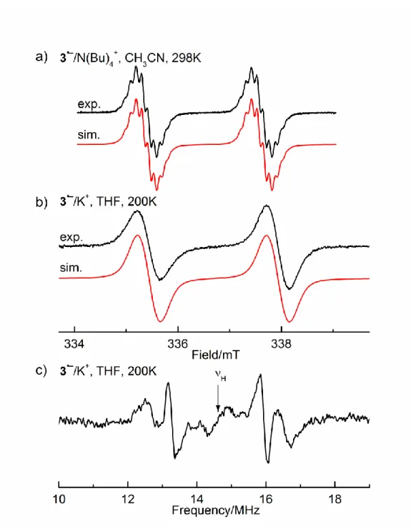
isotropic hyperfine coupling constants (hfcs) of 2–2.5 mT. Noteworthy, the EPR spectra are better resolved, when the radical anions are generated by electrolysis (Figure 2 and SI).
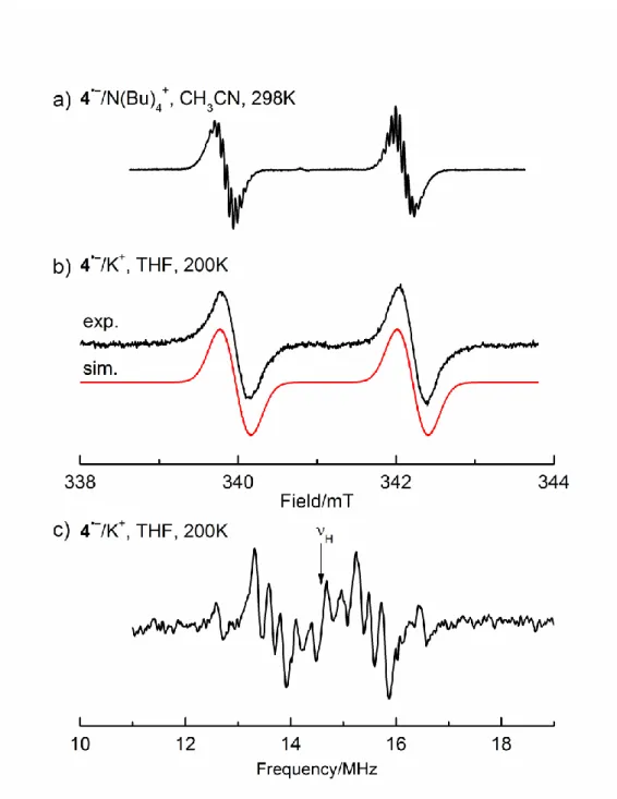
Figure 2. a) EPR spectra obtained by electrochemical reduction (Pt cathode,
g = 2.0042) of 1 and 2 (solvent CH3CN, supporting electrolyte,
N(Bu)4ClO4), with the simulation, T =298K; b) EPR spectrum of 1 after
chemical reduction with K in THF, T = 200 K, with the simulation; c) corresponding ENDOR spectrum (THF, T = 200 K).
We were able to obtain ENDOR spectra for all radical anions generated by the K-metal reduction. They provided, the absolute values of the 1H hfcs, not discernible in the EPR spectra, as well as
the 31P hfcs. This does not only lead to a satisfying simulation of
the EPR spectra, but also allows estimation of the amount of spin transferred into the aryl and alkyl substituents (Table 2).
As an example, Figure 2 shows the experimental EPR and ENDOR spectra of 1•– together with the corresponding EPR simulations. The 1H hfcs of 0.060 mT (6 H), 0.039 mT (12 H), and
0.012 mT (4 H) can be easily discerned in the ENDOR spectrum, whereas they are not resolved in the EPR signals. The EPR data of 1•––4•– are summarized in Table 2.
The 1H hfcs multiplicities obtained for BAPO 1•– indicate, that
the spin and the charge are evenly distributed within the two mesityl substituents, whereas the P-phenyl group does not carry detectable spin. The two mesityl groups are not equivalent in BAPO 4•–, presumably due to the decrease of the overall symmetry by the 3,6-dioxoheptyl substituent at P(O). MAPO 2•– reveals three
1H hfcs of 0.142, 0.165, and 0.100 mT, due to 6, 3, and 2 protons,
respectively, attributable to the ortho and para methyl groups and the two meta protons of the mesitoyl moiety. Analogous multiplicities hold for MAPO 3•–. Accordingly, it can be deduced, that delocalization is exclusively confined to the electron-withdrawing substituents. The (not markedly) differing absolute values of the sets of hfcs indicate, that the alkyl and phenyl substituents produce electronic effects, however, without participating in electron delocalization.
The development of DFT calculations in recent years, has provided relatively accurate predictions of hfc values in organic radicals [7]. It is thus rather remarkable that for 1•––4•– the 1H and 31P hfcs calculated by DFT (Table 2) differ significantly from the
experimental values. In addition to the basis sets given in Table 3, we tested for the most populated conformer of 1•– a large variety of different basis sets (including EPR-III, cc-pVTZ), their impact was marginal (data not shown). The calculation systematically overestimates the 1H hfcs. This may be traced back to ion pairing,
where ion pairs between the two (formally negative) oxygen atoms and the counterion could lead to conformations, which markedly alter the spin distribution and thereby change the hyperfine coupling in the mesitoyl substituent. Indeed, with ion-paired states included into the calculations, the agreement between experimental and calculated 1H hfcs improves substantially.
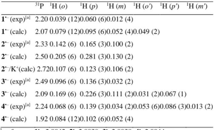
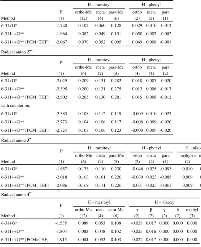
Table 2. 31P and 1H hfcs (/mT, values in parentheses indicate the number of
equivalent nuclei) together with their calculated counterparts (UB3LYP[PCM=THF]/6-311++G**) and g factors for 1•––4•– (K+/THF, T =
200 K). Calculated hfcs < 0.03 mT are not listed (see instead Table S2, Supporting Information). 31P 1H (o) 1H (p) 1H (m) 1H (o') 1H (p') 1H (m') 1•– (exp)[a] 2.20 0.039 (12) 0.060 (6) 0.012 (4) 1•– (calc) 2.07 0.079 (12) 0.095 (6) 0.052 (4) 0.049 (2) 2•– (exp)[a] 2.33 0.142 (6) 0.165 (3) 0.100 (2) 2•– (calc) 2.50 0.205 (6) 0.281 (3) 0.130 (2) 2•–/K+(calc) 2.72 0.107 (6) 0.123 (3) 0.106 (2) 3•– (exp)[a] 2.49 0.096 (6) 0.136 (3) 0.032 (2) 3•– (calc) 2.09 0.169 (6) 0.226 (3) 0.111 (2) 0.031 (2) 0.067 (1) 4•– (exp)[a] 2.24 0.068 (6) 0.139 (3) 0.034 (2) 0.053 (6) 0.086 (3) 0.013 (2) 4•– (calc) 1.92 0.084 (12) 0.102 (6) 0.052 (4) [a]. g factors: 1•–, 2.0042; 2•– 2.0039; 3•– 2.0038; 4•– 2.0044
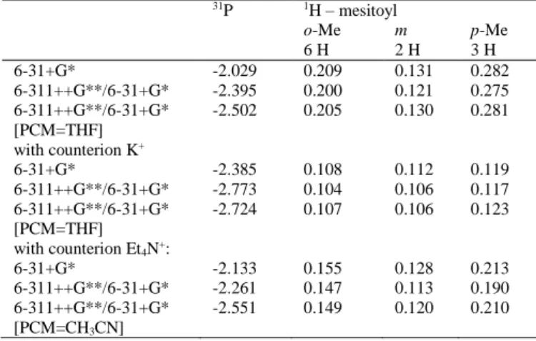
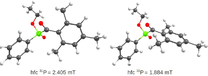
A more detailed insight into these ion-pairing phenomena, is presented with MAPO radical anion 2•– as a paradigm. The results are summarized in Table 3. Changes of the basis set have some influence on the 31P hfc, while the 1H hfcs hardly alter. However,
introduction of either K+ or Et
4N+, as the counterion, leads to a
marked change in the 1H hfcs. The hfcs for the six equivalent
methyl protons in the ortho position and the three para (methyl) protons are almost halved in the presence of K+, and the effect is
well distinguishable but less pronounced for Et4N+. Clearly, this is
not a consequence of the use of different basis-sets, but a result of the specific interactions between the radical anion and its environment. The ion pairing leads also to a shift in the population of conformational states. In the absence of the counterion, there exists only a single conformational minimum for 2a•– with the two oxygen bonds in trans configuration (Figure 3). In the presence of K+/Et
4N+ additional conformers can be identified. The
conformational equilibrium shifts from trans to cis upon ion pairing. The cis conformer 2b•– has significantly lower 1H hfcs than the
trans conformers 2a’ or 2a’’.
Figure 3. The calculated conformers of 2•–.in the absence (top) and presence (bottom) of the counterion K+. Dominant conformers are indicated
by a frame. The 1H hfcs of the mesitoyl ring are shown (in mT) as well as
the relative energy (UB3LYP[PCM=THF]/6-311++G**, in kcal/mol) for the
ion pairs.
For the 31P hfcs of 1•––4•– the deviation between calculation
The calculation of the 31P hfcs is extremely sensitive to the
geometry of these radical anions. For example, a rotation of the mesitoyl ring between two rotamers of 3•– can alter the 31P hfc by
0.6 mT (see also Figue S3). A more accurate calculation of the 31P
hfc for MAPO and BAPO radical anions may therefore require an approach that integrates accurate calculation of conformer populations (conformational free energies) and explicit treatment of solvent and counterion effects.
Table 3. 31P and 1H hfcs (/mT) for 2•– with different basis sets, solvent
models, and counterions. The hfcs of the phenyl substituents are not listed since they are substantially smaller than those of the mesitoyl substituent. The complete data are presented in the Supporting Information Table S2).
31P 1H – mesitoyl o-Me m p-Me 6 H 2 H 3 H 6-31+G* -2.029 0.209 0.131 0.282 6-311++G**/6-31+G* -2.395 0.200 0.121 0.275 6-311++G**/6-31+G* [PCM=THF] -2.502 0.205 0.130 0.281 with counterion K+ 6-31+G* -2.385 0.108 0.112 0.119 6-311++G**/6-31+G* -2.773 0.104 0.106 0.117 6-311++G**/6-31+G* [PCM=THF] -2.724 0.107 0.106 0.123 with counterion Et4N+: 6-31+G* -2.133 0.155 0.128 0.213 6-311++G**/6-31+G* -2.261 0.147 0.113 0.190 6-311++G**/6-31+G* [PCM=CH3CN] -2.551 0.149 0.120 0.210
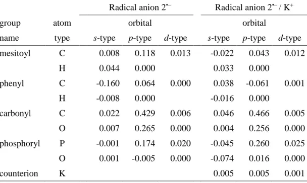
DFT calculations indicate, that the spin and the charge in 1•–– 4•–, are delocalized within the π-electron system of the molecule. This includes the two mesitolyl substituents and the PO unit in 1•– , and one mesitolyl unit and the PO group in 2•–, as shown in Figure 4. The largest contributions to this π-type spin system are from the
p-orbitals of the carbonyl group (ca. 70% for 2•–), the phosphorus atom (15%) and the mesitoyl carbons (12%). The singly-occupied molecular orbital of 2•– (Figure 4) reflects this distribution. Although the values of the 31P hfcs are relatively high, they reflect
a small amount of the spin population at the P atom (since even a low spin population at phosphorus leads to large 31P hfc values[8]).
This is nicely illustrated by comparing 2•– with the corresponding phosphorus-centred diphenyl-phosphanoyl radical Ph2(P•)=O, P2
(obtained by the photo-induced homolytic cleavage of the P-CO bond in 2). P2 is well described, since its reactivity is the basis for the success of 2, as photoinitiator of radical polymerization.[1a, 1b, 9]
In P2, the 31P hfc amounts to 36.0 mT[3g, 3i, 10], more than one order
of magnitude larger than in 2•–. The reason for this immense difference is two-fold: i) the overall phosphorus spin population in P2 is exceedingly higher, and ii) the s-orbital character in P2 is more pronounced.
Thus, experiment and calculation both suggest that 1•––4•– are π-type delocalized radical anions.
Figure 4. The lowest-energy conformers of 1•– and2•– and their singly occupied molecular orbitals.
The reason for the diminished symmetry in 4•– is very likely connected with the alkoxy chain, which, together with a single carbonyl group, is involved in a strong complexation of the counterion. Accordingly, an asymmetric spin distribution among the two mesitoyl units is induced. Here, binding and unbinding of the counterion, are slow at the hyperfine EPR time scale diminishing the overall symmetry.
Conclusions
We have shown that phosphane oxides display, in addition to their photochemical reactivity, a well-defined redox chemistry and form remarkably persistent radical anions. The electron distribution in these radical anions reflects the π*-orbital. This property relates to the photochemistry of BAPOs where upon n-π* excitation, one P-C bond breaks and the acyl group is cleaved off selectively in a Norrish type I reaction.[10a] A related follow-up reaction, the
cleavage [R1R2P(=O)-COMes]− R1R2P(=O)− + MesCO
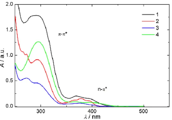
contributes to the thermal instability of MAPO and BAPO radical anions. However, MAPOs and BAPOs may possess valuable properties, since π-π* transition-based electron transfer reactions could be coupled to the n-π* type chemistry, as such transitions can be excited selectively, since they are well separated (Figure 4).
Figure 5. UV-VIS spectra of 1–4 displaying the regions for π-π* and n-π*
transitions.
These distinct photochemical characteristics together with the electron distribution in the one-electron reduced stages of acylphosphane oxides, which is markedly different from the parent molecules, opens new opportunities for their chemical reactivity. This is the subject of on-going investigations.
Experimental Section
Materials. (Photoinitiators (PIs)
2,4,6-bis(trimethylbenzoyl)-phenylphosphane oxide 1 (Irgacure® 819) 2,4,6-trimethylbenzoyl)-diphenylphosphane oxide 2 (Lucirin® TPO) and Ethyl-2,4,6-trimethylbenzoylphenylphosphinate 3 (Lucirin® TPO-L) were purchased from BASF SE and were used as received.
The BAPO derivative 2,4,6- bis(trimethylbenzoyl)-(2-(2-methoxyethoxy)ethyl)phosphane oxide 4 was prepared in 92 % yield as yellow oil as reported in ref.[1h] Acetonitrile
(ROTIPURAN®, ≥99,5 %, p.a.; Roth) and Tetrabutylammonium perchlorate (N(Bu)4ClO4, puriss. electrochemical grade, Fluka,
dried under reduced pressure at 340 K for 24 h prior to use) were used in the electrochemical experiments. Tetrahydrofuran (THF, Sigma-Aldrich) was heated to reflux over a Na/K alloy and stored over a Na/K alloy under high vacuum. Its deep blue color in combination with benzophenone was used as an indicator for rigorously water-free conditions.
Cyclic voltammograms (CV) were obtained with a PG580
potentiostat (Uniscan, UK) using the standard three electrode electrochemical cell with platinum disk working (WE) and counter (CE) electrodes and a silver wire pseudoreference electrode (RE). Approximately 1 mM sample solutions were prepared in CH3CN
with 0.1 M N(Bu)4ClO4 supporting electrolyte and purged with
argon for 10 min before each experiment. CVs were recorded at the 100 mV s–1 scan rate. All potentials are given vs. Fc/Fc+ used as
internal reference and are uncorrected from ohmic drop.
EPR/ENDOR Spectroscopy Radical anions of 1–4 were prepared
by controlled potential electrolysis in the 0.2 M
N(Bu)4ClO4/CH3CN solutions and/or by the chemical reduction in
THF. The electrolysis was performed at ambient temperature, in situ in the cavity of the EPR spectrometer using EPR
electrochemical flat cell (Pt mesh WE, Pt wire CE and Ag wire RE) and applying potential 100 mV more negative than the half-wave potential (E1/2) of the corresponding reduction process.
Samples for the chemical reduction were prepared in a special three compartment EPR sample tube connected to the vacuum line. K-metal mirror was sublimated to the wall of the tube and about 0.4 mL of THF was freshly condensed to dissolve the investigated
compound. The sample was successively degassed by three freeze-pump-thaw cycles and sealed under high vacuum. Reductions were performed by contact of the THF solution of the parent molecule with the K-metal mirror in the evacuated sample tube.
Two Bruker X-band spectrometers, EMX (100 kHz field modulation) and ESP 300 (equipped with an ENDOR unit, 12.5 kHz field modulation), both with an EUROTHERM temperature control unit, were used to record the cw-EPR and ENDOR spectra. Typical experimental conditions for the EPR spectra were 2 mW microwave power and 0.03 mT field modulation. ENDOR spectra were recorded with 20 mW microwave power, 0.1 mT field modulation, 10 MHz pump frequency, 100 kHz modulation depth and field locked at the maximum of the high field line of the phosphorus doublet. Spectra were analyzed with WinEPR and SimFonia software provided by the spectrometers manufacturer as well as with WinSim[11], a public domain program.
Calculations
Software: Molecular dynamic simulations were performed with the
program CHARMM [12], version c36b1; clustering was done with
the trajectory analysis tool Wordom0.22[13], version 0.22; and
density functional theory (DFT) calculations were carried out with the program Gaussian09[14], RevA02.
Conformational sampling: A systematic conformational analysis
was performed for radical anion 2•– on the level UB3LYP/6-31+G* by a constrained scanning around the P-C(=O) bond and one of the P-C(Ph) bonds. Subsequent unconstrained geometry minimization and subsequent removal of conformational duplicates yielded a single conformer. In addition, we optimized the geometry of 2•– in the presence of counterions K+ and Et
4N+ which yielded three
conformers.
For the more flexible systems 1, 3 and 4 we initially performed for their neutral state a 1-s molecular dynamics simulation at 350K using the Merck force-field MMFF (parameters were obtained from SwissParam[15]). Conformations were extracted every 100 ps
and minimized for 2000 steps of adopted basis Newton-Raphson prior to clustering[16] using the root-mean-square deviation of
non-hydrogen atoms as clustering criterion; the leader algorithm was applied with a cutoff criterion of 0.5 Å. Cluster headers were minimized as radical anions on the DFT level UB3LYP/6-31+G* and conformational duplicates were removed. This yielded six, ten and 44 confomers for radical anions 1•–, 3•– and 4•–, respectively.
Calculation of electronic properties: Based on the
UB3LYP/6-31+G*-minimized structures, the potential energy and spin densities were determined by single-point calculations on the level UB3LYP/6-311++G**. In addition, we tested the effect of the solvent (THF) using the continuum solvation model PCM.[17]
Finally, the isotropic hyperfine coupling constants were calculated through a Boltzmann-weighted average over all confomers; the weight of a given conformer i was
𝑝(𝑖) =𝑒
−𝐸(𝑖) 𝑘B𝑇
𝑍
with kB the Boltzmann constant, T the temperature, and E(i) the
single-point energy of conformer i; Z is the sum of all weights. For chemically identical nuclei the mean value is reported in Table S2 (assuming fast exchange).
Supporting Information Analytical details on 4,EPR/ENDOR spectra of 3•– and 4•–, the complete set of calculated EPR data of 1•––4•–, calculated
orbital spin populations of 2•–, and a figure displaying populated conformations in 3•–.
Financial support from Graz University of Technology and the ProBono Stiftung (MZ) is gratefully acknowledged. PNS is supported by a fellowship from the Rhône-Alpes Region. PNS and MS are grateful for a CIBLE grant (Rhône-Alpes Region) and a grant for access to French supercomputing resources from the Grand Equipement National de Calcul Intensif (DARI x2013076381. We are also grateful to the Pôle Scientifique de Modélisation Numérique (ENS de Lyon) for computer time
[1] aK. Dietliker, T. Jung, J. Benkhoff, H. Kura, A. Matsumoto, H. Oka, D. Hristova, G. Gescheidt, G. Rist, Macromol. Symp. 2004, 217, 77-97; bS. Jockusch, N. J. Turro, J. Am. Chem. Soc. 1998, 120, 11773-11777; cI. V. Khudyakov, N. J. Turro, Photochem. UV Curing 2006, 241-251; dM. Spichty, N. J. Turro, G. Rist, J.-L. Birbaum, K. Dietliker, J.-P. Wolf, G. Gescheidt, J. Photochem. Photobiol. A 2001, 142, 209-213; eN. J. Turro, I. V. Khudyakov, Res. Chem. Intermed. 1999, 25, 505-529; fM. Weber, N. J. Turro, D. Beckert, Phys. Chem. Chem. Phys. 2002, 4, 168-172; gY. Yagci, S. Jockusch, N. J. Turro, Macromolecules 2010, 43, 6245-6260; hA. Huber, A. Kuschel, T. Ott, G. Santiso-Quinones, D. Stein, J. Braeuer, R. Kissner, F. Krumeich, H. Schoenberg, J. Levalois-Gruetzmacher, H. Gruetzmacher, Angew. Chem., Int. Ed. 2012, 51, 4648-4652. [2] N. S. Kenning, B. A. Ficek, C. C. Hoppe, A. B. Scranton, Polym. Int.
2008, 57, 1134-1140.
[3] aA. Borer, R. Kirchmayr, G. Rist, Helv. Chim. Acta 1978, 61, 305-324; bK. Dietliker, S. Broillet, B. Hellrung, P. Rzadek, G. Rist, J. Wirz, D. Neshchadin, G. Gescheidt, Helv. Chim. Acta 2006, 89, 2211-2225; cU. Kolczak, G. Rist, K. Dietliker, J. Wirz, J. Am. Chem. Soc. 1996, 118, 6477-6489; dM. Griesser, D. Neshchadin, K. Dietliker, N. Moszner, R. Liska, G. Gescheidt, Angew. Chem., Int. Ed. 2009, 48, 9359-9361; eG. S. Ananchenko, D. I. Potapenko, P. A. Purtov, E. G. Bagryanskaya, R. Z. Sagdeev, Appl. Magn. Reson.
2004, 26, 65-82; fH. Bock, K. Ruppert, C. Nather, Z. Havlas, H. F.
Herrmann, C. Arad, I. Gobel, A. John, J. Meuret, S. Nick, A. Rauschenbach, W. Seitz, T. Vaupel, B. Solouki, Angewandte Chemie-International Edition in English 1992, 31, 550-581; gI. Gatlik, P. Rzadek, G. Gescheidt, G. Rist, B. Hellrung, J. Wirz, K. Dietliker, G. Hug, M. Kunz, J.-P. Wolf, J. Am. Chem. Soc. 1999, 121, 8332-8336; hD. Hristova, I. Gatlik, G. Rist, K. Dietliker, J.-P. Wolf, J.-L. Birbaum, A. Savitsky, K. Moebius, G. Gescheidt,
Macromolecules 2005, 38, 7714-7720; iA. N. Savitsky, M. Galander, K. Mobius, Chem. Phys. Lett. 2001, 340, 458-466; jT. N. Makarov, A. N. Savitsky, K. Moebius, D. Beckert, H. Paul, J. Phys. Chem. A
2005, 109, 2254-2263; kG. W. Sluggett, P. F. McGarry, I. V.
Koptyug, N. J. Turro, J. Am. Chem. Soc. 1996, 118, 7367-7372. [4] aV. Cerri, P. Furderer, F. Gerson, P. Tordo, Nouveau Journal De
Chimie-New Journal of Chemistry 1980, 4, 109-112; bV. Cerri, M. Boyer, P. Tordo, Tetrahedron Lett. 1979, 1437-1440.
[5] P. J. Wagner, R. J. Truman, A. E. Puchalski, R. Wake, J. Am. Chem. Soc. 1986, 108, 7727-7738.
[6] H. Bock, U. Lechnerknoblauch, P. Hanel, Chem. Ber. Recl. 1986, 119, 3749-3765.
[7] aV. Barone, M. Biczysko, J. Bloino, F. Egidi, C. Puzzarini, J. Chem. Phys. 2013, 138, 234303/234301-234303/234314; bM. Renz, M. Kaupp, J. Phys. Chem. A 2012, 116, 10629-10637; cA. Polimeno, V. Barone, J. H. Freed, Computational Strategies for Spectroscopy 2012, 549-582; dL. Hermosilla, C. Sieiro, P. Calle, M. Zerbetto, A. Polimeno, J. Phys. Chem. B 2008, 112, 11202-11208; eL. Hermosilla, P. Calle, J. M. G. de la Vega, C. Sieiro, J. Phys. Chem. A 2005, 109, 7626-7635.
[8] J. R. Morton, K. F. Preston, Journal of Magnetic Resonance 1978, 30, 577-582.
[9] aC. S. Colley, D. C. Grills, N. A. Besley, S. Jockusch, P. Matousek, A. W. Parker, M. Towrie, N. J. Turro, P. M. W. Gill, M. W. George, J. Am. Chem. Soc. 2002, 124, 14952-14958; bI. Gatlik, P. Rzadek, G. Gescheidt, G. Rist, B. Hellrung, J. Wirz, K. Dietliker, G. Hug, M. Kunz, J.-P. Wolf, J. Am. Chem. Soc. 1999, 121, 8332-8336; cM. Weber, N. J. Turro, D. Beckert, Phys. Chem. Chem. Phys. 2002, 4, 168-172.
[10] aP. Laurino, H. F. Hernandez, J. Braeuer, K. Krueger, H. Gruetzmacher, K. Tauer, P. H. Seeberger, Macromol. Rapid Commun. 2012, 33, 1770-1774; bL. Cataldo, C. Dutan, S. K. Misra, S. Loss, H. Grutzmacher, M. Geoffroy, Chemistry-a European Journal 2005, 11, 3463-3468; cW. B. Gara, B. P. Roberts, Journal of the Chemical Society-Perkin Transactions 2 1978, 150-154; dN. J. Turro, I. V. Khudyakov, Chem. Phys. Lett. 1992, 193, 546-552. [11] D. R. Duling, Journal of Magnetic Resonance, Series B 1994, 104,
105-110.
[12] B. R. Brooks, C. L. Brooks, III, A. D. Mackerell, Jr., L. Nilsson, R. J. Petrella, B. Roux, Y. Won, G. Archontis, C. Bartels, S. Boresch, A. Caflisch, L. Caves, Q. Cui, A. R. Dinner, M. Feig, S. Fischer, J. Gao, M. Hodoscek, W. Im, K. Kuczera, T. Lazaridis, J. Ma, V.
Ovchinnikov, E. Paci, R. W. Pastor, C. B. Post, J. Z. Pu, M. Schaefer, B. Tidor, R. M. Venable, H. L. Woodcock, X. Wu, W. Yang, D. M. York, M. Karplus, J. Comput. Chem. 2009, 30, 1545-1614. [13] M. Seeber, M. Cecchini, F. Rao, G. Settanni, A. Caflisch,
Bioinformatics 2007, 23, 2625-2627.
[14] M. J. Frisch, G. W. Trucks, H. B. Schlegel, G. E. Scuseria, M. A. Robb, J. R. Cheeseman, G. Scalmani, V. Barone, B. Mennucci, G. A. Petersson, H. Nakatsuji, M. Caricato, X. Li, H. P. Hratchian, A. F. Izmaylov, J. Bloino, G. Zheng, J. L. Sonnenberg, M. Hada, M. Ehara, K. Toyota, R. Fukuda, J. Hasegawa, M. Ishida, T. Nakajima, Y. Honda, O. Kitao, H. Nakai, T. Vreven, J. A. Montgomery, J. E. Peralta, F. Ogliaro, M. Bearpark, J. J. Heyd, E. Brothers, K. N. Kudin, V. N. Staroverov, R. Kobayashi, J. Normand, K.
Raghavachari, A. Rendell, J. C. Burant, S. S. Iyengar, J. Tomasi, M. Cossi, N. Rega, J. M. Millam, M. Klene, J. E. Knox, J. B. Cross, V. Bakken, C. Adamo, J. Jaramillo, R. Gomperts, R. E. Stratmann, O. Yazyev, A. J. Austin, R. Cammi, C. Pomelli, J. W. Ochterski, R. L. Martin, K. Morokuma, V. G. Zakrzewski, G. A. Voth, P. Salvador, J. J. Dannenberg, S. Dapprich, A. D. Daniels, Farkas, J. B. Foresman, J. V. Ortiz, J. Cioslowski, D. J. Fox, Wallingford CT, 2009.
[15] V. Zoete, M. A. Cuendet, A. Grosdidier, O. Michielin, J. Comput. Chem. 2011, 32, 2359-2368.
[16] F. Rao, M. Karplus, Proceedings of the National Academy of Sciences of the United States of America 2010, 107, 9152-9157. [17] M. Cossi, V. Barone, R. Cammi, J. Tomasi, Chem. Phys. Lett. 1996,
255, 327-335. ____________
Received: ((will be filled in by the editorial staff)) Published online: ((will be filled in by the editorial staff))
Entry for the Table of Contents
((Please choose one layout.))
Layout 1:
((Key Topic))
(Bis)Acylphosphaneoxides, well established photoinitiators for radical polymerization, display a remarkable redox chemistry. They act as efficient electron acceptors. The corresponding radical anions are rather persistent and open novel opportunities for the use of this class of molecules.
Michal Zalibera, Pierre-Nicolas Stébé, Kurt Dietliker, Hansjörg Grützmacher, Martin Spichty, and Georg Gescheidt*) …….. Page No. –
Page No. ((Title))
Keywords: Photoinitiator / Radical anion / Cyclovoltammetry / EPR)
Supporting Information
Supporting Information
The Redox Chemistry of (Bis)Acylphosphaneoxides
Michal Zalibera,a Pierre-Nicolas Stébé,c Kurt Dietliker, b Hansjörg Grützmacher, b Martin Spichty,c and Georg Gescheidt*,a
a Institute of Physical and Theoretical Chemistry, Graz University of Technology, Stremayrgasse 9, A-8010 Graz
bDepartment of Chemistry and Applied Biosciences, HCI H 131, CH-8093 Zürich
c Laboratory of Molecular and Cellular Biology, Differentiation and the Cell Cycle, Ecole Normale Supérieure de Lyon, 46, allée d’Italie,
69364 Lyon, cedex 07, France
1 Experimental
1.1 Characterization of 4
1
H NMR (300 MHz, C
6
D
6, 25 °C, TMS): δ = 6.68 (s, 4H, Mes-CH), 3.79 (trd, 2H,P-CH
2-CH
2-O), 3.34
(m, 2H, O-CH
2-), 3.18 (m, 2H, O-CH
2-), 3.11 (s, 3H, O-CH
3), 2.58 (dtr, 2H, P-CH
2-), 2.54 (s, 12H,
o-Mes-CH
3), 2.08 (s, 6H, p-Mes-CH
3) ppm;
13
C NMR (63 MHz, C
6
D
6): δ = 215.96 (d, J
PC= 56.2 Hz, C=O), 140.68 (s, Mes-C
4), 136.60 (s, Mes-C
1),
136.41 (s, Mes-C
3,5), 129.25 (s, Mes-C
2,6), 71.53 (s, O-CH
2), 70.12 (s, CH
2-O), 63.62 (d,
2JCP
= 4.65 Hz,
P-CH
2-CH
2), 58.25 (s, O-CH
3), 28.20 (d,
2JCP
= 54.78 Hz, P-CH
2-CH
2), 20.83 (s, Mes p-CH
3), 20.09 (s,
Mes o-CH
3) ppm;
31
P NMR (121.5 MHz, C
6
D
6, 25 °C, H
3PO
4) δ = 25.1 (m) ppm;
FT-IR (ATR): ν = 2915 (vw), 1754 (vw), 1674 (w, υ[C=O]), 1650 (vw), 1607 (w), 1444 (w), 1422 (w),
1381 (w), 1354 (w), 1297 (vw), 1278 (vw), 1250 (vw), 1200 (m), 1180 (w), 1136 (w), 1112 (m), 1102
(m), 1034 (w), 1010 (w), 959 (w), 887 (w), 859 (m), 850 (m), 774 (w), 743 (w), 704 (w), 676 (w), 643
(vw), 618 (w) cm
-1;
HRMS (ESI) calc. for C
25H
33NaO
5P 467.1958, found 467.1956.
2 Results
Figure S1. a) EPR spectrum obtained by electrochemical reduction (Pt cathode, g = 2.0038) of 3 (solvent
CH
3CN, supporting electrolyte, N(Bu)
4ClO
4), with the simulation ; T =298K; b) EPR spectrum of 3 after
chemical reduction with K in THF, T = 200 K, with the simulation; c) corresponding ENDOR spectrum
(THF, T = 200 K).
Figure S2. a) EPR spectrum obtained by electrochemical reduction (Pt cathode, g = 2.0044) of 4 (solvent
CH
3CN, supporting electrolyte, N(Bu)
4ClO
4); T =298K; b) EPR spectrum of 4 after chemical reduction
with K in THF, T = 200 K, with the simulation; c) corresponding ENDOR spectrum (THF, T = 200 K).
Table S1. EPR data of 1
•––3
•–(hfcs are given in mT) in CH
3CN/N(Bu)
4+298K
31
P
1H (o)
1H (p)
1H (m)
1H (o')
1H (p')
1H (m') g factor
1
•–(exp)
a2.35 0.053 (12) 0.060 (6)
2.0042
2
•–(exp)
a2.43 0.119 (6)
0.165 (3) 0.104 (2)
2.0039
3
•–(exp)
a2.23 0.094 (6)
0.134 (3) 0.026 (2)
2.0038
aSpectra were recorded in CH
2.2 Calculations
Table S2. Calculated isotropic hyperfine coupling constants (in mT) for radical anions 1
•–-4
•–. Values in
parentheses indicate the number of chemically equivalent nuclei.
Radical anion 1●–
P ortho-Me meta para-Me ortho meta para
Method (1) (12) (4) (6) (2) (2) (1) 6-31+G* -1.728 0.102 0.060 0.128 0.029 0.010 -0.012 6-311++G** -1.986 0.082 0.049 0.101 0.050 0.007 -0.002 6-311++G** [PCM=THF] -2.067 0.079 0.052 0.095 0.049 0.008 -0.001 H – mesitoyl H - phenyl Radical anion 2●–
ortho-Me meta para-Me ortho meta para
Method (1) (6) (2) (3) (4) (4) (2) 6-31+G* -2.029 0.209 0.131 0.282 0.010 0.007 -0.020 6-311++G** -2.395 0.200 0.121 0.275 0.012 0.006 -0.017 6-311++G** [PCM=THF] -2.502 0.205 0.130 0.281 0.015 0.008 -0.012 with counterion: 6-31+G* -2.385 0.108 0.112 0.119 -0.009 0.010 -0.023 6-311++G** -2.773 0.104 0.106 0.117 -0.008 0.009 -0.020 6-311++G** [PCM=THF] -2.724 0.107 0.106 0.123 -0.008 0.009 -0.020 P H – mesitoyl H - phenyl Radical anion 3●–
ortho-Me meta para-Me ortho meta para methylen methyl
Method (1) (6) (2) (3) (2) (2) (1) (2) (3) 6-31+G* -1.657 0.173 0.110 0.230 -0.046 0.025 -0.093 0.010 0.016 6-311++G** -2.018 0.163 0.101 0.220 -0.039 0.023 -0.085 0.009 0.015 6-311++G** [PCM=THF] -2.086 0.169 0.111 0.226 -0.031 0.023 -0.067 0.009 0.016 H – alkoxy P H – mesitoyl H - phenyl Radical anion 4●–
ortho-Me meta para-Me α β γ δ methyl
Method (1) (12) (4) (6) (2) (2) (2) (2) (3)
6-31+G* -1.535 0.089 0.053 0.108 -0.028 0.017 0.000 0.000 0.000
6-311++G** -1.804 0.083 0.048 0.102 -0.023 0.016 0.000 0.000 0.000
6-311++G** [PCM=THF] -1.915 0.084 0.052 0.103 -0.022 0.017 0.000 0.000 0.000