RESEARCH OUTPUTS / RÉSULTATS DE RECHERCHE
Author(s) - Auteur(s) :
Publication date - Date de publication :
Permanent link - Permalien :
Rights / License - Licence de droit d’auteur :
Institutional Repository - Research Portal
Dépôt Institutionnel - Portail de la Recherche
researchportal.unamur.be
University of Namur
Alginate@TiO2 hybrid microcapsules as a reservoir of beta INS-1E cells with controlled insulin delivery
Leroux, Grégory; Neumann, Myriam; Meunier, Christophe F.; Michiels, Carine; Wang, Li; Su, Bao Lian
Published in:
Journal of Materials Science
DOI:
10.1007/s10853-020-04576-9 Publication date:
2020
Document Version
Publisher's PDF, also known as Version of record
Link to publication
Citation for pulished version (HARVARD):
Leroux, G, Neumann, M, Meunier, CF, Michiels, C, Wang, L & Su, BL 2020, 'Alginate@TiO2 hybrid
microcapsules as a reservoir of beta INS-1E cells with controlled insulin delivery', Journal of Materials Science, vol. 55, no. 18, pp. 7857-7869. https://doi.org/10.1007/s10853-020-04576-9
General rights
Copyright and moral rights for the publications made accessible in the public portal are retained by the authors and/or other copyright owners and it is a condition of accessing publications that users recognise and abide by the legal requirements associated with these rights. • Users may download and print one copy of any publication from the public portal for the purpose of private study or research. • You may not further distribute the material or use it for any profit-making activity or commercial gain
• You may freely distribute the URL identifying the publication in the public portal ?
Take down policy
If you believe that this document breaches copyright please contact us providing details, and we will remove access to the work immediately and investigate your claim.
Alginate@TiO
2Hybrid Microcapsules as a
Reservoir of beta INS-1E Cells with Controlled
Insulin Delivery
Grégory Leroux,§ Myriam Neumann,§ Christophe F. Meunier,§ Carine Michiels,‡ Li Wang§,* and
Bao-Lian Su§,#,*
§Laboratory of Inorganic Materials Chemistry (CMI), University of Namur, 61 Rue de
Bruxelles, B-5000 Namur, Belgium.
‡Laboratory of Biochemistry and Cell Biology (URBC), Namur Research Institute for Life
Sciences (NARILIS), University of Namur, 61 Rue de Bruxelles, B-5000 Namur, Belgium.
#State Key Laboratory of Advanced Technology for Materials Synthesis and Processing,
Wuhan University of technology, Luoshi Road 122, Wuhan 430070, China.
ABSTRACT: Designing biocompatible materials to encapsulate xenogeneic insulin-releasing β-cells for transplantation has been considered as a promising alternative to avoid the immunosuppression and drawbacks of the treatment of Type 1 diabetes mellitus (T1D) by direct islet transplantation. The current work for the first time studied a hybrid alginate@TiO2
microcapsule as a reservoir for rat insulinoma-derived INS-1E cells, as a β-cell surrogate, towards the treatment of T1D. The hybrid microcapsule is composed of an alginate core as a biocompatible matrix for cell encapsulation and a crack-free TiO2 shell as a semi-permeable
membrane to prevent cell leakage, protect encapsulated cells from immune attacks, as well as allow the diffusion of nutrients and the secretion of insulin. Compared to most-commonly used pure alginate microcapsules, the insulin-secreting INS-1E cells encapsulated in our alginate@TiO2 microcapsules revealed higher metabolic activity and maintained the insulin
secretion over more than 6 weeks. This study highlights that our designed alginate@TiO2
hybrid microcapsules can serve as an ideal reservoir for cell encapsulation towards the treatment of T1D, thus further promoting the development of artificial organs for cell therapy.
KEYWORDS: hybrid hydrogels, cell encapsulation, biocompatibility, insulin secretion, type 1 diabetes
INTRODUCTION
Type 1 diabetes mellitus (T1D), as a kind of human disorders involving hormone or proteins deficiency, has increasingly become a big human health threat nowadays1. It consists in the
autoimmune destruction of insulin secreting β-cells and hampers thus the glucose homeostasis. Currently the most common treatment is the discontinuous insulin injection which remains however a major burden in everyday life and cannot eliminate all complications of the disease2.
Transplantation of islets has evolved into a prospective treatment as it allows the recovery of an endogenous insulin secretion and the control of glycaemia in insulin-dependent patients3-4.
However, this treatment still presents various restrictions such as very limited donor supply, in general one donor for 300 patients. Furthermore, a patient requires generally 2-3 donors5. About
90 % of insulin-dependence recurrence 5-years post-transplantation has been observed6. A
strong and life-long immunosuppressive medication is also required for patients, which leads to various side-effects7-8.
An alternative to avoid immunosuppression and its drawbacks is to separate the islets (or β-cells) from the host immune system by introducing a semi-permeable membrane that would prevent the entry of immune defenders while allowing the diffusion of essential nutrients, oxygen, metabolites and insulin9-13. This kind of cell therapy methodologies has been largely
used since first introduced by Bisceglie 80 years ago14-17. Alginate-based microcapsules are the
most-used hydrogels for this medical treatment, because of their high biocompatibility, low cytotoxicity, low cost, and mild gelation conditions18-21. For instance, alginate microcapsules
have been used for the encapsulation of insulin-secreting islet cells in order to restore long-term euglycemic maintenance without immunosuppression22-23. However, they usually present
poor mechanical resistance and thus low stability in vitro and in vivo.
Hybridization of hydrogels with inorganic compounds has been developed as a promising method to improve the properties of the hydrogels or equip the hydrogels with new functions
to widen their application range or improve their efficiency in applications24-25. For example, the
incorporation of carbon materials within hydrogels can enhance their electronical conductivity26, thus realizing the applications in cell therapy towards cardiac or neural diseases
or bone injury27-30; hybridizing microcapsules with gold nanoparticles enables to reinforce the
mechanical strength of hydrogels and facilitate cell adhesion, spreading, and proliferation, which makes possible the precise cell differentiation control31; coordinating minerals with
hydrogels can introduce the hydrogels with controllable stiffness to control the behaviours of encapsulated cells precisely32-33. Recently, new structured hybrid microcapsules which are
composed of inorganic shells and an organic core have been reported and attracted a lot of attention34-35. The inorganic shell enhances the stability of the microcapsule and provides
controlled permeability, while the organic core offers a highly biocompatible environment for the encapsulated cells, which makes this structured microcapsule a promising cell reservoir with long-term viability and activity for cell therapy36-38. For example, alginate@TiO
2 hybrid
microcapsules have been demonstrated to be a suitable reservoir of HepG2 cells. They showed biocompatible and possessed controlled porosity to confirm the long-term cell viability, the cell activity, and the release of the cell metabolic39. Importantly, these microcapsules also
presented high in vivo biocompatibility and in vivo stability when implanted into rats, thus contributing to their potential in cell therapy. However, the previous studies were implemented based on the model HepG2 cells which do not present any therapeutic use, so that further investigation is still highly needed towards practical application of hybrid microcapsules in cell therapy.
Herein, we first time investigate a hybrid alginate@TiO2 microcapsule as a reservoir for
insulin-secreting β-cells to maintain glucose homeostasis towards the cell therapy of T1D. Compared with the most-commonly used pure alginate microcapsules, hybrid alginate@TiO2
stability without compromising the nutrient diffusion and insulin secretion, thus preventing the leakage of encapsulated cells and providing them with a long-term stable environment for growth, proliferation and metabolism. Biocompatible hybrid alginate@TiO2 microcapsules are
prepared through a facile one-step process under mild conditions. Alginate serves as a matrix for cell encapsulation and a template for the formation of TiO2 shells by the hydrolysis and
condensation of the precursor. Rat insulinoma-derived INS-1E cells, which are able to secrete insulin in response to glucose stimulation in the physiological range40-41, are encapsulated in our
hybrid alginate@TiO2 microcapsules. The morphology and structure of the hybrid
microcapsules are investigated in detail. The viability of the encapsulated INS-1E cells are monitored, as well as their glucose-stimulated insulin secretion, in order to advance the development of cell encapsulation techniques for the treatment of T1D.
MATERIALS AND METHOD
Chemicals. Titanium(IV) bis(ammonium lactato)dihydroxide solution (TiBALDH, 50 wt.%
in H2O), poly(diallyldimethylammonium) chloride solution (PDDAC, average Mw
100,000-200,000, 20 wt. % in H2O), alginic acid sodium salt, from brown algae (for immobilization of
microorganisms), calcium chloride (dihydrate, 99 %), ethylene glycol-bis(β-aminoethyl ether)-N,N,N’,N’-tetraacetic acid (EGTA, tetrasodium salt, 97 %), hydrochloric acid (Molecular Biology Grade, 36.5-38.0 %), sodium hydroxide (reagent grade, 98 %, anhydrous), chloroform (anhydrous, 99 %), glutaraldehyde (50 wt. % in H2O), fluorescein diacetate (FDA, used as cell
viability stain), glucose solution (BioUltra, for molecular biology, ~20 % in H2O), sodium
chloride (ACS reagent, 99,0 %), potassium chloride (for molecular biology, 99.0 %), potassium phosphate monobasic (plant cell culture tested, 99 %), magnesium sulfate heptahydrate (Plant Cell Culture Tested), sodium bicarbonate (Plant Cell Culture Tested), albumin, from bovine serum (BSA, 98 % (agarose gel electrophoresis, lyophilized powder, essentially fatty acid free,
essentially globulin free)) and dimethyl sulfoxide (BioReagent, for molecular biology, suitable for plant cell culture, 99.9 %) were purchased from Sigma-Aldrich. Ethanol (absolute) was purchased from Fisher. PBS (Phosphate Buffered Saline, 0.0067 M (PO4), For Cell Culture)
was purchased from Lonza. Forskolin (Coleus forskohlii in DMSO) was purchased from Merck Millipore. Roswell Park Memorial Institute medium (RPMI, 1640), foetal bovine serum, HEPES buffer solution (1 M), sodium pyruvate (100 mM, 100X), L-Glutamine (200 mM, 100X) and 2-mercaptoethanol (50 mM) were purchased from Invitrogen (Casbald, USA).
Sodium alginate powder was purified according to the method proposed by de Vos et al42.
Briefly, the commercial powder was dissolved in an aqueous solution of EGTA 1 mM to remove Mg2+ and Ca2+ cations. Subsequently, the solution was filtered and washed by HCl,
chloroform and butanol to eliminate remaining ions and proteins. Finally, the purified alginate powder was obtained by adjusting pH to 7.0, adding absolute ethanol for precipitation and drying using a vane pump.
Cell culture. Rat insulin-secreting cell line INS-1E sensitive to glucose stimulation was
provided by Prof. Pierre Maechler (University of Geneva, Department of Cell Physiology and Metabolism) and Prof. Patrick Gilon (Université Catholique de Louvain, EDIN, Endocrinology, Diabetes and Nutrition division). The Roswell Park Memorial Institute medium (RPMI, 1640) used for the culture of INS-1E cells was supplemented with HEPES (10 mM), sodium pyruvate (1 mM), glutamine (2 mM), 2-mercaptoethanol (0.05 mM) and 10 % of foetal bovine serum. Cells were incubated in 75-cm2 flasks (Costar, Lowell, USA) at 37°C
in a humid atmosphere of 5 % CO2, and the medium was replaced by fresh one every 2 days.
Once the cells were 100 % confluent (4 or 5 days), they were washed with PBS (Phosphate Buffer Saline) before treatment with a trypsin/EDTA mixture. Trypsinized cells were suspended in fresh medium for encapsulation. For further culture, cells were diluted and placed in new 75-cm2 flasks containing 15 mL of fresh culture medium with serum.
Cell encapsulation. The pH of the solutions used for cell encapsulation were previously
adjusted to 7.4.
Cell encapsulation in pure alginate microcapsules. An INS-1E cell suspension was mixed
with 3 wt.% sodium alginate. The final cell density was 1.1*106 cells mL-1. This mixture was
dropped in a CaCl2 solution (100 mM) using sterilized 3.0 mL plastic Pasteur pipettes (VWR).
The droplets were incubated for 5 min to obtain microcapsules with solid calcium alginate external crusts and inner cell-containing sodium alginate core. The microcapsules were finally recovered, placed in test tubes filled with fresh culture medium with serum and maintained in cell culture conditions (37 °C, 5 % CO2).
Cell encapsulation in alginate@TiO2 microcapsules. The encapsulation procedure of
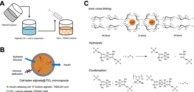
INS-1E cells in alginate@TiO2 microcapsules is showed in Figure 1 A. In detail, an INS-1E cell
suspension was first mixed with 3 wt.% sodium alginate supplemented with titanium precursor (TiBALDH, 250 mM). The final cell density was 1.1*106 cells mL-1. This mixture was extruded
into a buffered aqueous solution containing CaCl2 (100 mM) and PDDAC (100 mM). The
droplets were incubated for 5 min to obtain microcapsules with a solid white crust. The microcapsules were finally recovered, placed in test tubes filled with fresh culture medium supplemented with serum and maintained in cell culture conditions (37 °C, 5 % CO2).
Material characterization. Microcapsules were dried through supercritical drying with
liquid carbon dioxide, after being incubated in 3 % glutaraldehyde and dehydrated with ethanol. Morphological properties were analyzed using field emission scanning electron microscopy (FE-SEM, JEOL JSM-7500F), after being sputter-coated with gold. The elemental composition of the internal core and external shell of alginate@TiO2 microcapsules was
determined using an energy-dispersive X-rays (EDX) analysis system with an acceleration voltage of 15.00 kV and a working distance of 8 mm.
Cell viability. The viability of encapsulated INS-1E cells was monitored by their oxygen
consumption using a Clark’s cell vessel (HansaTech). Briefly, 3 microcapsules were put in the device, containing 1.0 mL of fresh culture medium. The relative respiration of INS-1E cells was determined by oxygen consumption. The oxygen consumption of the INS-1E cells just after the encapsulation was set as the reference value (100 %). Cell survival was confirmed using fluorescein diacetate (FDA), a vital dye stain. INS-1E cells were incubated in FDA solution (5 µM) for 5 minutes at room temperature. Pictures were then taken using a fluorescence microscope (Multizoom AZ100, Nikon) either in brightfield mode or in fluorescent mode at 536/40 nm with a colour camera (DSRi1, Nikon).
Glucose-stimulated insulin secretion. An evaluation of insulin secretion of the
microcapsules with entrapped INS-1E cells was performed as a function of time. Briefly, culture medium was removed from test tubes containing hybrid microcapsules. Microcapsules were washed twice with Hank’s balanced sodium salt solution (HBSS) (114 mmol L-1 NaCl,
4.7 mmol L-1 KCl, 1.2 mmol L-1 KH
2PO4, 1.16 mmol L-1 MgSO4, 20 mmol L-1 HEPES, 2.5 mmol
L-1 CaCl
2, 25.5 mmol L-1 NaHCO3 and 0.2 % BSA, pH 7.2). The first wash was just a quick rinse,
while for the second wash, the microcapsules were incubated in HBSS for 2 hours at 37°C (5% CO2). Then, the secretagogues diluted in HBSS were added another 2-hour incubation. A
glucose solution of 3 mM was used for the determination of basal insulin secretion level, while 15 mM glucose and 15 mM glucose supplemented with 1 µM forskolin were used for stimulated secretion, using independent samples for each assay. The supernatants were finally recovered to measure insulin concentration using a Rat/Mouse Insulin ELISA Kit provided by Merck Millipore.
Statistical analysis. The concentration of insulin secreted by encapsulated INS-1E cells
when stimulated by 15 mM glucose (or 15 mM glucose supplemented with 1 µM forskolin) was compared to basal secretion (stimulation by 3 mM glucose). Results were analyzed by
unpaired Student’s t-test, using XLSTAT 2013 software (Addinsoft) and with p < 0.05 being considered as significant.
RESULTS AND DISCUSSION
Microcapsule formation and cell encapsulation. The cell-laden alginate@TiO2
microcapsules were formed by dropping the mixture of alginate, TiBALDH and INS-1E cells into CaCl2 / PDDAC solution, as depicted in Figure 1A. When the drops were immersed into
the solution, a series reactions took place in the outmost layer of the drops, resulting in the formation of microcapsules. The reactions mainly include three ones as listed in Figure 1C. The sodium alginate in the interface of the drops and the solution reacted with calcium ions by cross-linking to form calcium alginate shells around the drops. Simultaneously, PDDAC adsorbed on the formed calcium alginate shells by electrostatic force. The adsorbed PDDAC further induced the hydrolysis and condensation of the TiBALDH in the outmost layer of the drops to form a TiO2 layer. With the prolongation of incubation, three above-mentioned
reactions continuously occurred to thicken the calcium alginate and TiO2 shells, and finally
contributed to the formation of alginate@TiO2 microcapsules (Figure 1B). The INS-1E cells
were mainly encapsulated in the core of alginate@TiO2 microcapsules which was most
composed of biocompatible alginate. The calcium alginate and TiO2 shells could protect the
encapsulated cells from immune defenders and maintain the nutrient diffusion and insulin secretion.
Figure 1. Formation of cell-laden alginate@TiO2 microcapsules. (A) Encapsulation
procedure of INS-1E cells in alginate@TiO2 microcapsules. (B) Schematic diagram of an
INE-1E cells-laden alginate@TiO2 microcapsules. (C) Ionic cross-linking of alginate and
hydrolysis and condensation processes of TiBALDH in the formation of alginate@TiO2
microcapsules.
Material characterization. The morphology, structure and elemental composition of
alginate@TiO2 microcapsules with encapsulated INS-1E cells were characterized. Pure
alginate microcapsules with encapsulated INS-1E cells were set as a control. Both cell-laden microcapsules were supercritically dried with CO2, in order to preserve their original structure.
The dried microcapsules were characterized using scanning electron microscopy (SEM) equipped with an energy-dispersive X-rays (EDX) analysis system.
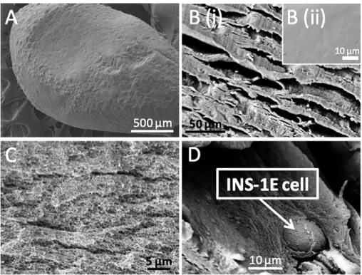
Figure 2. Morphology and structure of a cell-laden alginate microcapsule. SEM
micrographs of a cell-laden alginate microcapsule (A), its surface (B) and core (C), as well as encapsulated INS-1E cells (D). The microcapsule was incubated in culture medium for 6 weeks, except for the microcapsule shown in B(ii) which was observed directly after preparation and set as a control. The microcapsules were supercritically dried with CO2 prior
to analyses.
Figure 2A depicts the SEM micrograph of a pure alginate microcapsule with encapsulated INS-IE cells after 6-week incubation in culture medium. The microcapsule shows spherical morphology with a diameter of ~2000 μm. Figure 2B (i) gives the enlarged SEM micrograph of the external surface of the microcapsules in which some cracks can be observed. On the contrary, a very smooth surface can be seen in pure alginate microcapsules without any further incubation (Figure 2B (ii)). The difference between the two surfaces indicates that the surface evolution of pure alginate microcapsules from smooth to cracked by long-term incubation, demonstrating the lack of long-term stability of pure alginate microcapsules. The cracks of the
alginate cores. The core of the microcapsule presents a sponge-like structure with highly porosity (Figure 2C), which was formed by the loss of a large content of aqueous solution during the supercritically drying. This sponge-like structured core could not only provide enough space to be filled with nutrient for cell growth, proliferation and metabolism, but also maintain the mass transport and the cell-to-cell communication inside microcapsules. Figure 2D shows encapsulated INS-1E cells in the microcapsule, confirming the presence of the cells in this cell-laden microcapsule.
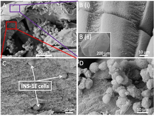
Figure 3. Morphology and structure of a cell-laden alginate@TiO2 microcapsule. SEM
micrographs of a cell-laden alginate@TiO2 microcapsule (A), its surface (B) and core (C), as
well as encapsulated INS-1E cells (D). The microcapsule was kept in culture medium for 6 weeks and supercritically dried with CO2 prior to analyses.
Figure 3A presents a cross-section of an alginate@TiO2 microcapsule, highlighting the
presence of well dispersed cells in the core and a condensed shell. Figure 3B shows that the surface of the alginate@TiO2 microcapsules is still very dense without cracks even after 6-week
of TiO2. PDDAC is a positive charged linear polymer. When it was adsorbed on the surface of
the microcapsules, it would contribute to the condensation of alginate on the outmost layer owing to its interaction with the negative charged alginate, thus consolidating the calcium alginate shell. Meanwhile, PDDAC induced the hydrolysis and condensation of TiBALDH to form a uniform and robust TiO2 layer which could further enhance the mechanical stability of
shells. As a comparison, on the surface of pure alginate microcapsules some cracks of about 10 µm are observed after 6-week incubation which is shown in Figure 2B(i). Since the molecular size of immunoglobulin G (IgG)43, the smallest defender of our immune system, is
around 17-20 nm on the basis of the equation of Stokes44-46, these immunoglobulin molecules
can easily penetrate through the cracks of 10 µm and kill encapsulated INS-1E cells encapsulated. In addition, these large cracks would lead to the leakage of encapsulated cells, thus decreasing functional activity of cell-laden microcapsules in cell therapy. Therefore, pure alginate microcapsules present serious defects in terms of stability to be applied in cell encapsulation towards cell therapy. Whilst alginate@TiO2 hybrid microcapsules remain intact
after long-term incubation in culture medium, evidencing their enhanced mechanical stability and suggesting their more effective protection against immune defenders. The core of the alginate@TiO2 microcapsule (Figure 3C) presents the same structural properties as those of
pure alginate microcapsules (Figure 2C), which indicates that the formation of TiO2 mainly
occurred at the external surface of alginate microcapsules and would not influence the inner structure of alginate, thus confirming their biocompatible structure towards the encapsulated cells. It is worth to note that the encapsulated INS-1E cells were grouped into small clusters (Figure 3D), which is in the same manner as free cells during culture, in addition to the presence of extracellular matrix at their surface, thus suggesting a good health state of the encapsulated cells and proving biocompatibility of the alginate@TiO2 matrix.
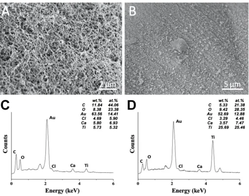
Figure 4. Elemental composition of an alginate@TiO2 microcapsule. SEM micrographs of
the core (A) and the shell (B) of an alginate@TiO2 microcapsule. EDX spectra and elemental
percentage of the core (C) and the shell (D) of the alginate@TiO2 microcapsule. The
microcapsule was supercritically dried with CO2 immediately after synthesis prior to analyses.
EDX analyses were performed on alginate@TiO2 microcapsules to identify the elemental
composition of the inner and the outer parts of the microcapsules. Figure 4 presents the morphology and EDX spectra of the inner core (A, C) and the external shell (B, D) of alginate@TiO2 microcapsule. The presence of the CKα, OKα and CaKα peaks in either the
sponge-structured core (Figure 4A) or the dense shell (Figure 4B) indicates that the microcapsules are mainly made of alginate and calcium alginate. Ti element can be detected at the external surface of the alginate@TiO2 microcapsule (25.46 at.%) (Figure 4D), indicating
that a TiO2 layer was formed around the microcapsule as a shell which could enhance the
stability of microcapsules. A small portion of Ti element (5.32 at.%) can be detected in the core of the microcapsule (Figure 4C), which suggests that the formation of TiO2 also took part
in the inner core but with a slow reaction rate due to the absence of polycations. The different percentage of Ti element between the core (5.32 at.%) and the shell (25.46 at.%) of the microcapsules confirms their core@shell structure which not only maintains the biocompatibility of the alginate core towards the encapsulated cells but also enhances the stability and mechanical strength of the microcapsules through the introduction of an inorganic shell for long-term applications.
Cell viability. The viability of INS-1E cells encapsulated in either pure alginate or
alginate@TiO2 microcapsules was monitored over time by the evaluation of oxygen
consumption and FDA staining, in order to test the biocompatibility of the microcapsules towards encapsulated cells for their potential application in cell therapy.
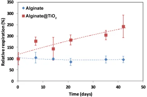
The oxygen consumption of encapsulated cells was measured using a Clark’s cell device. This parameter is directly proportional to the number of living cells in the microcapsules. Figure 5 presents the relative respiration as a function of time with 100 % corresponding to the oxygen consumption of INS-1E cells encapsulated in one microcapsule just after encapsulation. The viability of INS-1E cells was maintained for 6 weeks as the relative respiration slightly increased over the whole period. It is worth to note that the respiration activity of the cells encapsulated in alginate@TiO2 microcapsules increased up to ~250 %,
whereas that of the cells encapsulated in pure alginate microcapsules remained ~100 %. The rapid increase in respiration activity of INS-1E cells encapsulated in alginate@TiO2
microcapsules can be attributed to cell proliferation, suggesting very good biocompatibility of our designed hybrid microcaspules towards INS-1E cells. Please note that the relative respiration of alginate@TiO2 varied more largely at 14 days and 42 days, which could result
from the shell crack of few alginate@TiO2 microcapsules after long-term incubation. Owing to
the cell proliferation and the degradation of alginate core, the shell of few microcapsules appeared cracked after a long-time incubation, while the most others were still crack-free. Pure
alginate microcapsules are highly compatible towards encapsulated cells and support their growth and proliferation, however, the oxygen comsuption which can be linked to the number of living cells has not increased. One possible explanation of this phenomenon is the cell leakage from the pure alginate microcapsules due to the surface crack appearance of the most of the microcapsules upon incubation.
Figure 5. Oxygen consumption of encapsulated INS-1E cells. Oxygen consumption of the
INS-1E cells encapsulated in alginate and alginate@TiO2 microcapsules. 100 % corresponds
to the oxygen consumption of INS-1E cells encapsulated within one microcapsule (1.3*102
μmol h-1 microcapsule-1). Results are presented as means ± 1 S.D. (n = 3).
The viability of the cells encapsulated in alginate@TiO2 microcapsules was further
confirmed by the characterization of FDA-stained cells using fluorescent microscope. Living cells with intact plasma membrane can produce green fluorescein after stained by FDA due to the presence of active esterase in cytoplasm.
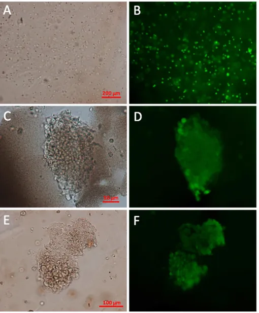
Figure 6. Viability of encapsulated INS-1E cells. Optical micrograph of the FDA-stained
INS-1E cells encapsulated in alginate@TiO2 microcapsules 1-h (A), 35-day (C) and 42-day (E)
post-encapsulation. Fluorescent micrograph of the FDA-stained INS-1E cells encapsulated within alginate@TiO2 microcapsules 1-h (B), 35-day (D) and 42-day (F) post-encapsulation.
Optical and fluorescent micrographs of FDA-stained INS-1E cells encapsulated in alginate@TiO2 microcapsules are depicted in Figure 6. 1-h post-encapsulation, cells were
homogeneously dispersed in microcapsules (Figure 6A) and more than 90 % of them were alive (Figure 6B). After 35 days, the encapsulated INS-1E cells were present as clusters in the microcapsules (Figure 6C and 6E), which was in accordance with the observations made by
SEM (Figure 3A and 3D) and the growth state of free cell in culture plate. In addition, the cell clusters exhibited green fluorescence (Figure 6D and 6F), demonstrating the high viability of the encapsulated cells. Combined with the results of oxygen consumption, it can be concluded that our designed alginate@TiO2 hybrid microcapsules, in comparison with pure alginate
microcapsules, could be a better candidate as a cell resevoir to encapsulate INS-1E cells with long-term viability for the treatment of T1D.
Glucose-stimulated insulin secretion. In order to evaluate the potential of our designed
cell-laden alginate@TiO2 microcapsules used for the treatment of T1D, the insulin secretion of
encapsulated INS-1E cells stimulated by glucose were studied by the measurement of insulin concentration in the supernatant of the secretagogue media 2- and 6-week post-encapsulation. Cell-laden pure alginate microcapsules were set as a control. The results were indicated as a stimulation index which is defined by the increase in insulin secretion when stimulated by glucose or glucose supplemented with forskolin minus the basal secretion. Forskolin is an activator of adenylate cyclase that stimulates the release of insulin.
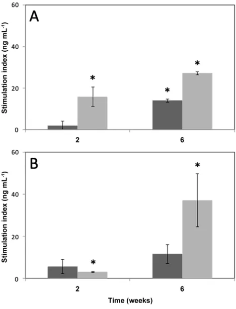
Figure 7. Insulin secretion of encapsulated INS-1E cells. Insulin secretion of the INS-1E
cells encapsulated in alginate (A) and alginate@TiO2 (B) microcapsules under the stimulation
of glucose (15 mM) or glucose (15 mM) supplemented with forskolin (1 µM). Stimulation index is defined by the increase in insulin secretion (ng mL-1) when stimulated by glucose or
glucose supplemented with forskolin minus the basal secretion (3 mM). Values are means ± 1 S.D. (n = 3). * p < 0.05 compared to the basal insulin secretion.
glucose, the insulin secretion of the cells encapsulated in pure alginate microcapsules or in alginate@TiO2 microcapsules and incubated for 2 weeks shows no difference to that of basal
levels, however, an increased insulin secretion could be detected for the cells encapsulated in the pure alginate microcapsules or in alginate@TiO2 microcapsules and incubated for 6 weeks
because of cell proliferation. In order to confirm the responses of encapsulated cells to glucose more precisely, forskolin was used as an additive stimulator to amplify insulin secretion. After 2-week or 6-week incubation for the cells encapsulated in pure alginate microcapsule and in alginate@TiO2 hybrid microcapsules, the insulin secretion stimulated with glucose
supplemented with forskolin shows a highly significant difference (p < 0.05) in comparison with basal levels. Both experiments indicate that encapsulated INS-1E cells can significantly respond to glucose stimulation and maintain their metabolic activity. It is worth to note that the cells encapsulated in alginate@TiO2 microcapsules 6-week post-encapsulation show
significantly increased insulin secretion levels compared to that 2-week post-encapsulation. The increase of insulin secretion over time was attributed to the enhanced metabolic activity of encapsulated cells and cell proliferation, supporting the results presented in Figures 4 and 5. Please note the cells encapsulated in pure microcapsules showed a less increase of insulin secretion levels between 2-week and 6-week post-encapsulation. This could result rather from the metabolic activity enhancement of encapsulated cells than cell proliferation, as the amount of encapsulated cells was not increased based on the result presented in Figure 5. In addition, this result evidences that alginate@TiO2 microcapsules displayed well adapted porosity for the
entry of nutrients and the secretion of insulin. Although pure alginate microcapsules also allow the diffusion of nutrients and insulin as shown by the results presented in Figures 5 and 7, these microcapsules suffer from crack formation during incubation. These cracks could result in the cell leakage and the loss of their immune-isolation.
On the basis of the previous study39, alginate@TiO
2 hybrid microcapsules, in comparison with
the most-used pure alginate microcapsules, not only present long-term mechanical stability and high biocompatibility towards HepG2 cells, but also possess well-controlled porosity to maintain the nutrient diffusion and metabolic releasing of the encapsulated HepG2 cells. However, HepG2 cells are only a kind of model cells for the fundamental investigation and show no function in cell therapy. The practical application of alginate@TiO2 hybrid
microcapsules in cell therapy still remains a lot of problems. In this work, the functional insulin-secreting INS-1E cells were first time encapsulated in alginate@TiO2 hybrid
microcapsules, thus being further developped from the last research and much closer to the practical treatment of T1D. In the case of INS-1E cells, the core of the alginate@TiO2 hybrid
microcapsules provided the encapsulated cells with a biocompatible aqueous environment for their growth, proliferation and metabolism. In addition, their robust TiO2 layer not only offered
a long-term stability to prevent cell leakage, but also presented well controlled porosity for the diffusion of glucose as well as insulin secreted by the encapsulated cells (Figure 1B). This makes them an ideal reservoir of INS-1E cells for the controlled delivery of insulin in the treatment of T1D.
CONCLUSIONS
The highlights of this work can be summarized into the points listed below:
• Alginate@TiO2 hybrid microcapsules were synthesized by the ionic cross-linking of
alginate and the hydrolysis and condensation of TiBALDH, thereafter first time being applied to encapsulate rat insulinoma-derived INS-1E cells, aiming at the treatment of T1D.
• Alginate@TiO2 hybrid microcapsules, compared to the most-used pure alginate
microcapsules, demonstrated improved long-term stability and less cell leakage, making them a better reservoir for INS-1E cells to protect them from immune attacks for a long time.
• The rat insulinoma-derived INS-1E cells encapsulated in alginate@TiO2 maintained
insulin secretion as a response to glucose stimulation 6-week post-encapsulation, evidencing the potential application of the cell-laden alginate@TiO2 microcapsules in the treatment of T1D.
Consequently, alginate@TiO2 hybrid microcapsules could be an ideal reservoir for the
encapsulation of INS-1E cells towards the treatment of the increasingly widespread T1D, as well as offer a draft to design artificial organs for cell therapy in future.
AUTHOR INFORMATION
Corresponding Authors
*E-mail: li.wang@unamur.be; Tel: 0032 485 262580; Fax: 0032 81 725414 (L. Wang) *E-mail: bao-lian.su@unamur.be; Tel: 0032 81 724531; Fax: 0032 81 725414 (B. L. Su)
Notes
The authors declare no competing financial interest.
ACKNOWLEDGMENT
This work was supported by Program for Changjiang Scholars and Innovative Research Team in University (IRT_15R52) and “Algae Factory” (1610187) European H2020 program financed by FEDER and Wallonia Region of Belgium. G. Leroux thanks University of Namur for his assistant position to realize his PhD research. We thank Prof. Pierre Maechler of the University of Geneva and Prof. Patrick Gilon of Université Catholique de Louvain for their supply of the rat insulin-secreting cell line INS-1E.
1. Van Belle, T. L.; Coppieters, K. T.; Von Herrath, M. G., Type 1 diabetes: etiology, immunology, and therapeutic strategies. Physiological Reviews 2011, 91 (1), 79-118.
2. Tauschmann, M.; Hovorka, R., Technology in the management of type 1 diabetes mellitus - current status and future prospects. Nature Reviews Endocrinology 2018, 14 (8), 464-475.
3. Shapiro, A. M. J.; Pokrywczynska, M.; Ricordi, C., Clinical pancreatic islet transplantation. Nature Reviews Endocrinology 2016, 13, 268.
4. Ellis, C.; Ramzy, A.; Kieffer, T. J., Regenerative medicine and cell-based approaches to restore pancreatic function. Nature Reviews Gastroenterology and Hepatology 2017, 14 (10), 612.
5. Shapiro, A. J.; Ricordi, C.; Hering, B. J.; Auchincloss, H.; Lindblad, R.; Robertson, R. P.; Secchi, A.; Brendel, M. D.; Berney, T.; Brennan, D. C., International trial of the Edmonton protocol for islet transplantation. New England Journal of Medicine 2006, 355 (13), 1318-1330.
6. Ryan, E. A.; Paty, B. W.; Senior, P. A.; Bigam, D.; Alfadhli, E.; Kneteman, N. M.; Lakey, J. R.; Shapiro, A. J., Five-year follow-up after clinical islet transplantation. Diabetes
2005, 54 (7), 2060-2069.
7. Chang, R.; Faleo, G.; Russ, H. A.; Parent, A. V.; Elledge, S. K.; Bernards, D. A.; Allen, J. L.; Villanueva, K.; Hebrok, M.; Tang, Q., Nanoporous Immunoprotective Device for Stem-Cell-Derived β-Cell Replacement Therapy. ACS Nano 2017, 11 (8), 7747-7757.
8. Jenssen, T.; Hartmann, A., Emerging treatments for post-transplantation diabetes mellitus. Nature Reviews Nephrology 2015, 11, 465.
9. Desai, T.; Shea, L. D., Advances in islet encapsulation technologies. Nature Reviews
Drug Discovery 2017, 16 (5), 338.
10. Foster, G. A.; García, A. J., Bio-synthetic materials for immunomodulation of islet transplants. Advanced Drug Delivery Reviews 2017, 114, 266-271.
11. Calafiore, R.; Basta, G., Clinical application of microencapsulated islets: actual prospectives on progress and challenges. Advanced Drug Delivery Reviews 2014, 67, 84-92. 12. Ma, M.; Chiu, A.; Sahay, G.; Doloff, J. C.; Dholakia, N.; Thakrar, R.; Cohen, J.; Vegas, A.; Chen, D.; Bratlie, K. M.; Dang, T.; York, R. L.; Hollister-Lock, J.; Weir, G. C.; Anderson, D. G., Core-shell hydrogel microcapsules for improved islets encapsulation. Advanced
Healthcare Materials 2013, 2 (5), 667-672.
13. Dolgin, E., Diabetes: Encapsulating the problem. Nature 2016, 540, S60.
14. Bisceglie, V., Über die antineoplastische immunität. Zeitschrift für Krebsforschung
1934, 40 (1), 122-140.
15. Mount, N. M.; Ward, S. J.; Kefalas, P.; Hyllner, J., Cell-based therapy technology classifications and translational challenges. Philosophical Transactions of the Royal Society B:
Biological Sciences 2015, 370 (1680), 20150017.
16. Trounson, A.; McDonald, C., Stem cell therapies in clinical trials: progress and challenges. Cell Stem Cell 2015, 17 (1), 11-22.
17. Leonard, A.; Dandoy, P.; Danloy, E.; Leroux, G.; Meunier, C. F.; Rooke, J. C.; Su, B.-L., Whole-cell based hybrid materials for green energy production, environmental remediation and smart cell-therapy. Chemical Society Reviews 2011, 40 (2), 860-885.
18. Sun, J.; Tan, H., Alginate-based biomaterials for regenerative medicine applications.
Materials 2013, 6 (4), 1285.
19. Lee, K. Y.; Mooney, D. J., Alginate: properties and biomedical applications. Progress
in Polymer Science 2012, 37 (1), 106-126.
20. Seliktar, D., Designing cell-compatible hydrogels for biomedical applications. Science
21. Strand, B. L.; Coron, A. E.; Skjak-Braek, G., Current and future perspectives on alginate encapsulated pancreatic islet. Stem Cells Translational Medicine 2017, 6 (4), 1053-1058.
22. Vegas, A. J.; Veiseh, O.; Gürtler, M.; Millman, J. R.; Pagliuca, F. W.; Bader, A. R.; Doloff, J. C.; Li, J.; Chen, M.; Olejnik, K., Long-term glycemic control using polymer-encapsulated human stem cell-derived beta cells in immune-competent mice. Nature Medicine
2016, 22 (3), 306.
23. Bochenek, M. A.; Veiseh, O.; Vegas, A. J.; McGarrigle, J. J.; Qi, M.; Marchese, E.; Omami, M.; Doloff, J. C.; Mendoza-Elias, J.; Nourmohammadzadeh, M.; Khan, A.; Yeh, C.-C.; Xing, Y.; Isa, D.; Ghani, S.; Li, J.; Landry, C.-C.; Bader, A. R.; Olejnik, K.; Chen, M.; Hollister-Lock, J.; Wang, Y.; Greiner, D. L.; Weir, G. C.; Strand, B. L.; Rokstad, A. M. A.; Lacik, I.; Langer, R.; Anderson, D. G.; Oberholzer, J., Alginate encapsulation as long-term immune protection of allogeneic pancreatic islet cells transplanted into the omental bursa of macaques. Nature Biomedical Engineering 2018, 2 (11), 810-821.
24. Wang, L.; Neumann, M.; Fu, T.; Li, W.; Cheng, X.; Su, B.-L., Porous and responsive hydrogels for cell therapy. Current Opinion in Colloid & Interface Science 2018, 38, 135-157. 25. Mehrali, M.; Thakur, A.; Pennisi, C. P.; Talebian, S.; Arpanaei, A.; Nikkhah, M.; Dolatshahi-Pirouz, A., Nanoreinforced hydrogels for tissue engineering: biomaterials that are compatible with load-bearing and electroactive tissues. Advanced Materials 2017, 29 (8). 26. Ahadian, S.; Yamada, S.; Ramón-Azcón, J.; Estili, M.; Liang, X.; Nakajima, K.; Shiku, H.; Khademhosseini, A.; Matsue, T., Hybrid hydrogel-aligned carbon nanotube scaffolds to enhance cardiac differentiation of embryoid bodies. Acta Biomaterialia 2016, 31, 134-143. 27. Shin, J.; Choi, E. J.; Cho, J. H.; Cho, A.-N.; Jin, Y.; Yang, K.; Song, C.; Cho, S.-W., Three-dimensional electroconductive hyaluronic acid hydrogels incorporated with carbon nanotubes and polypyrrole by catechol-mediated dispersion enhance neurogenesis of human neural stem cells. Biomacromolecules 2017, 18 (10), 3060-3072.
28. Shin, S. R.; Jung, S. M.; Zalabany, M.; Kim, K.; Zorlutuna, P.; Kim, S. b.; Nikkhah, M.; Khabiry, M.; Azize, M.; Kong, J.; Wan, K.-T.; Palacios, T.; Dokmeci, M. R.; Bae, H.; Tang, X.; Khademhosseini, A., Carbon-nanotube-embedded hydrogel sheets for engineering cardiac constructs and bioactuators. ACS Nano 2013, 7 (3), 2369-2380.
29. Hao, T.; Li, J.; Yao, F.; Dong, D.; Wang, Y.; Yang, B.; Wang, C., Injectable fullerenol/alginate hydrogel for suppression of oxidative stress damage in brown adipose-derived stem cells and cardiac repair. ACS Nano 2017, 11 (6), 5474-5488.
30. Pacelli, S.; Maloney, R.; Chakravarti, A. R.; Whitlow, J.; Basu, S.; Modaresi, S.; Gehrke, S.; Paul, A., Controlling adult stem cell behavior using nanodiamond-reinforced hydrogel: implication in bone regeneration therapy. Scientific Reports 2017, 7 (1), 6577. 31. Heo, D. N.; Castro, N. J.; Lee, S.-J.; Noh, H.; Zhu, W.; Zhang, L. G., Enhanced bone tissue regeneration using a 3D printed microstructure incorporated with a hybrid nano hydrogel. Nanoscale 2017, 9 (16), 5055-5062.
32. Zhang, K.; Feng, Q.; Xu, J.; Xu, X.; Tian, F.; Yeung, K. W.; Bian, L., Self-assembled injectable nanocomposite hydrogels stabilized by bisphosphonate-magnesium (Mg2+)
coordination regulates the differentiation of encapsulated stem cells via dual crosslinking.
Advanced Functional Materials 2017, 27 (34).
33. Heinemann, S.; Heinemann, C.; Wenisch, S.; Alt, V.; Worch, H.; Hanke, T., Calcium phosphate phases integrated in silica/collagen nanocomposite xerogels enhance the bioactivity and ultimately manipulate the osteoblast/osteoclast ratio in a human co-culture model. Acta
Biomaterialia 2013, 9 (1), 4878-4888.
34. Dandoy, P.; Meunier, C. F.; Michiels, C.; Su, B.-L., Hybrid shell engineering of animal cells for immune protections and regulation of drug delivery: towards the design of “artificial
35. Desmet, J.; Meunier, C.; Danloy, E.; Duprez, M.-E.; Lox, F.; Thomas, D.; Hantson, A.-L.; Crine, M.; Toye, D.; Rooke, J., Highly efficient, long life, reusable and robust photosynthetic hybrid core-shell beads for the sustainable production of high value compounds.
Journal of Colloid and Interface Science 2015, 448, 79-87.
36. Dandoy, P.; Meunier, C. F.; Leroux, G.; Voisin, V.; Giordano, L.; Caron, N.; Michiels, C.; Su, B.-L., A hybrid assembly by encapsulation of human cells within mineralised beads for cell therapy. PLoS One 2013, 8 (1), e54683.
37. Desmet, J.; Meunier, C. F.; Danloy, E. P.; Duprez, M.-E.; Hantson, A.-L.; Thomas, D.; Cambier, P.; Rooke, J. C.; Su, B.-L., Green and sustainable production of high value compounds via a microalgae encapsulation technology that relies on CO2 as a principle reactant.
Journal of Materials Chemistry A 2014, 2 (48), 20560-20569.
38. Zhang, B.-B.; Wang, L.; Charles, V. R.; Rooke, J. C.; Su, B.-L., Robust and biocompatible hybrid matrix with controllable permeability for microalgae encapsulation. ACS
Applied Materials & Interfaces 2016, 8 (14), 8939-8946.
39. Leroux, G.; Neumann, M.; Meunier, C. F.; Fattaccioli, A.; Michiels, C.; Arnould, T.; Wang, L.; Su, B.-L., Hybrid alginate@TiO2 porous microcapsules as a reservoir of animal cells
for cell therapy. ACS Applied Materials & Interfaces 2018, 10 (44), 37865-37877.
40. Merglen, A.; Theander, S.; Rubi, B.; Chaffard, G.; Wollheim, C. B.; Maechler, P., Glucose sensitivity and metabolism-secretion coupling studied during two-year continuous culture in INS-1E insulinoma cells. Endocrinology 2004, 145 (2), 667-678.
41. Hohmeier, H. E.; Mulder, H.; Chen, G.; Henkel-Rieger, R.; Prentki, M.; Newgard, C. B., Isolation of INS-1-derived cell lines with robust ATP-sensitive K+ channel-dependent and-independent glucose-stimulated insulin secretion. Diabetes 2000, 49 (3), 424-430.
42. de Vos, P.; Spasojevic, M.; de Haan, B. J.; Faas, M. M., The association between in vivo physicochemical changes and inflammatory responses against alginate based microcapsules. Biomaterials 2012, 33 (22), 5552-5559.
43. Morris, P. J., Immunoprotection of therapeutic cell transplants by encapsulation. Trends
in Biotechnology 1996, 14 (5), 163-167.
44. Nagy, J. A.; Herzberg, K. T.; Masse, E. M.; Zientara, G. P.; Dvorak, H. F., Exchange of macromolecules between plasma and peritoneal cavity in ascites tumor-bearing, normal, and serotonin-injected mice. Cancer Research 1989, 49 (19), 5448-5458.
45. Desai, T. A.; Hansford, D.; Ferrari, M., Characterization of micromachined silicon membranes for immunoisolation and bioseparation applications. Journal of Membrane Science
1999, 159 (1), 221-231.
46. Carturan, G.; Dal Toso, R.; Boninsegna, S.; Dal Monte, R., Encapsulation of functional cells by sol-gel silica: actual progress and perspectives for cell therapy. Journal of Materials