HAL Id: tel-02280080
https://tel.archives-ouvertes.fr/tel-02280080
Submitted on 6 Sep 2019
HAL is a multi-disciplinary open access
archive for the deposit and dissemination of sci-entific research documents, whether they are pub-lished or not. The documents may come from teaching and research institutions in France or abroad, or from public or private research centers.
L’archive ouverte pluridisciplinaire HAL, est destinée au dépôt et à la diffusion de documents scientifiques de niveau recherche, publiés ou non, émanant des établissements d’enseignement et de recherche français ou étrangers, des laboratoires publics ou privés.
nucleus in muscle cells
Petra Gimpel
To cite this version:
Petra Gimpel. Mechanisms of non-centrosomal MTOC formation at the nucleus in muscle cells. Cellular Biology. Université Pierre et Marie Curie - Paris VI; Freie Universität (Berlin), 2017. English. �NNT : 2017PA066442�. �tel-02280080�
MTOC formation at the nucleus
in muscle cells
Dissertation
In fulfillment of the requirements for the Joint Degree (Cotutelle)
”Doctor rerum naturalium” (Dr. rer. nat.)
integrated in the International Graduate School for Myology
”MyoGrad”
in the Department of Biology, Chemistry and Pharmacology
at the Freie Universität Berlin
and in Cotutelle Agreement
with the Ecole Doctorale ”Complexité du Vivant”
at the Université Pierre et Marie Curie Paris 6
Submitted by
Petra Gimpel
from Berlin
Portugal and Center for Research in Myology, UMRS 974, Paris, France) and Prof. Dr. Simone Spuler (Experimental and Clinical Research Center, Berlin, Germany). The experiments were conducted at the Center for Research in Myology, UMRS 974, Paris, France and at the Charité Berlin, Campus Mitte, CharitéCrossOver, Berlin, Germany.
Thesis jury:
Prof. Dr. Simone Spuler, thesis supervisor in Berlin (1st reviewer for FU Berlin) Prof. Dr. Sigmar Stricker (2nd reviewer for FU Berlin)
Dr. Christian Kähler, postdoctoral research fellow at FU Berlin
Dr. Edgar R. Gomes, thesis supervisor in Paris Dr. Michel Bornens, examiner from UPMC
Dr. Mónica Bettencourt-Dias, external examiner (1st reviewer for UPMC)
Dr. Antoine Guichet, external examiner (2nd reviewer for UPMC)
no other aid and sources than noted.
This work was done in collaboration with the laboratory of Prof. Dr. Brian Burke (In-stitute of Medical Biology, Agency for Science, Technology and Research (A*STAR), Singapore) and the laboratory of Dr. Jan Schmoranzer (Charité Universitätsmedizin Berlin, Campus Mitte, CharitéCrossOver, Berlin, Germany). All experiments that were performed by our collaborators are clearly indicated within each figure legend.
"Are microtubules growing from nuclear pore complexes in muscle cells?" With this seemingly simple question my supervisor Dr. Edgar R. Gomes had fired the starting gun for an exciting and fascinating PhD project, which turned out to be much more complex than initially thought. I would like to thank Dr. Edgar R.
Gomes for the opportunity and all his support to explore this starting question in
every possible direction that I aimed at.
I am deeply grateful for the excellent supervision that I received from Dr. Bruno
Cadot, for all of the efforts he put into this project, for his never-ending enthusiasm
and motivation and for his support inside and outside the lab. This project would not have been possible without him.
I would like to thank Prof. Dr. Simone Spuler, Prof. Dr. Sigmar Stricker,
Dr. Mónica Bettencourt-Dias and Dr. Antoine Guichet for reading and
evaluating my PhD thesis and being part of my thesis jury. In addition, I thank Dr.
Michel Bornens and Dr. Christian Kähler for accepting to be members of my
thesis jury.
Dr. Michel Bornens and his group laid the foundation for my PhD project back
in 1985 with their discoveries that microtubules and microtubule-organizing centers are reorganized to the nucleus in skeletal muscle cells. I am thus especially hon-ored that he takes part in my thesis defense to discuss the recent progress in this field.
I want to thank Prof. Dr. Simone Spuler for giving me the opportunity to be part of the MyoGrad PhD program through which I was able to perform my PhD in Paris and Berlin and to attend international conferences. The MyoGrad summer schools helped me a lot to gain substantial knowledge within the muscle field outside my personal project and allowed me to get in contact with international scientists. I want to thank Susanne Wissler for all her support and advice regarding adminis-trative work at both universities, for arranging flights between Berlin and Paris and
I would like to thank la Fondation pour la Recherche Médicale (FRM) for supporting my work with a fourth-year scholarship (FDT20160435051) and the
Université Franco-Allemande (UFA) for providing me a mobility aid stipend
within the cotutelle de thèse program (CT-46-14-II).
I would like to thank Dr. Renata Basto, Dr. Vanessa Ribes, Dr. Fanny
Pilot-Storck, Prof. Dr. Simone Spuler and Dr. Jan Schmoranzer for being
part of my thesis committee and their guidance in finding the right direction for my project.
I am grateful for the excellent training that I received from Dr. Jan Schmoranzer in order to perform 3D-SIM and SD-dSTORM microscopy. I thank him for accepting me to be part of his laboratory and for his support during my lab relocation. I would like to thank Dr. Robyn Brackin for showing me the fascinating possibilities and advantages of Nikon spinning disk confocal microscopy.
I want to acknowledge Prof. Dr. Brian Burke and Dr. Yin Loon Lee for the fruitful collaboration which was ultimately important for the success of this project. I especially want to thank Dr. Yin Loon Lee for all the scientific discussions and for teaching me how to perform BioID experiments. Moreover, I want to thank him for making my stay in Singapore such a memorable experience.
I thank Prof. Dr. Markus Schülke for hosting and supporting me in his lab-oratory during my stay in Berlin. Thanks to all members of the AG Schülke/
AG Seelow, especially to Ioanna Polydorou, Franziska Seifert and Esther Gill, for welcoming me in the laboratory and their technical and organizational
support. I especially want to thank Ioanna for her motivation, the discussions and for continuing to improve my Greek.
Thanks to all other members of the Gomes/Cadot group and all other teams of the 105 building for creating such an enjoyable work atmosphere during my three years in Paris and for the very inspiring discussions during work seminars and in between: Dr. William Roman, Dr. David Ollitrault, Dr. Rosamaria
I want to thank Dr. Judite Costa, Sara Ferreira and Telma Carrilho for their organizational support and for sending packages with lab material back and forth between Lisbon, Paris and Berlin.
I would like to thank Dr. Sestina Falcone, Aurore Besse, Dr. Sonia
Alonso-Martin, Dr. Maria Grazia Biferi, and Mathilde Cohen-Tannoudji for
intro-ducing me to the world of Gym Suédoise, for all the sportive activities and funny moments.
I would like to thank Dr. Valérie Vilmont, Dr. Despoina Mademtzoglou,
Dr. Audrey Der Vartanian and Jean-François Darrigrand for being both
- great colleagues and wonderful friends -, for all the shared moments inside and outside the lab, for all the favors and help, the intercultural exchange and for making me feel home in Paris.
I thank Dr. Xenia Naj for her immense friendship, for our shared adventure ex-ploring Paris and San Francisco, her contagious happiness and her talent to discover the best restaurants.
Finally, I want to express my gratitude to my wonderful family and friends outside the scientific world for their optimism and understanding, no matter how time consuming my work turned out to be. Most importantly I wish to thank my
parents and my husband Tobias for their never-ending support every single day,
Acknowledgements III
List of Figures XI
List of Tables XIII
List of Abbreviations XV Abstract XIX Zusammenfassung XXI Résumé XXIII 1 Introduction 1 1.1 Skeletal muscle . . . 1
1.1.1 Structure of skeletal muscle . . . 1
1.1.2 Myogenesis . . . 3
1.2 Nuclear positioning and the nuclear envelope . . . 4
1.2.1 SUN domain proteins . . . 5
1.2.2 KASH domain proteins and their connections to the cytoskele-ton . . . 7
1.2.3 Tissue-specific KASH splice variants . . . 10
1.2.4 The LINC complex in muscle disorders . . . 11
1.2.5 Nesprin-1/2 and Sun1/2 knockout mouse models . . . 12
1.2.6 Nuclear positioning in skeletal muscle cells . . . 13
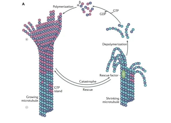
1.3 Microtubules . . . 18
1.3.1 Microtubule structure and dynamics . . . 18
1.3.2 Regulation of MT dynamics . . . 20
1.3.3 Microtubule nucleation . . . 22
1.4.2 Non-centrosomal MTOCs . . . 34
1.4.3 Non-centrosomal MTOCs in skeletal muscle cells . . . 37
1.5 Aim of the project . . . 41
2 Materials and Methods 43 2.1 Materials . . . 43
2.1.1 Chemicals and disposables . . . 43
2.1.2 Solutions and media . . . 43
2.1.3 Molecular weight standards . . . 48
2.1.4 Plasmids . . . 48
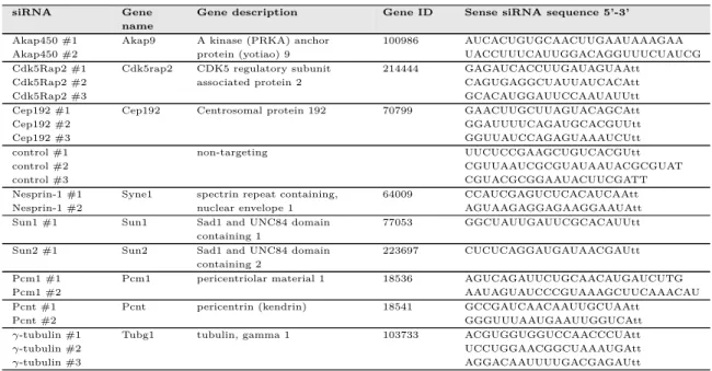
2.1.5 Small interfering RNAs (siRNAs) . . . 48
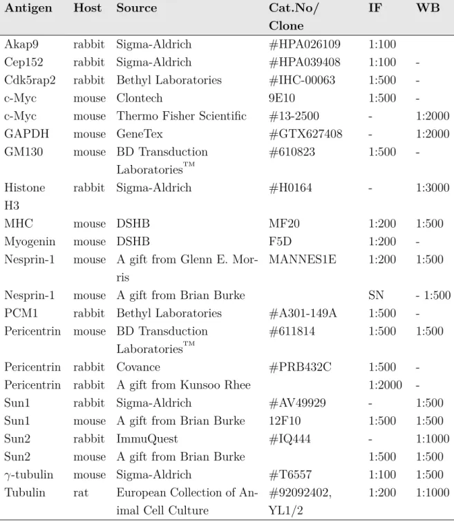
2.1.6 Antibodies . . . 61
2.2 Molecular biological methods . . . 63
2.2.1 Transformation of chemically competent E. coli . . . 63
2.2.2 Plasmid DNA isolation from E. coli cultures . . . 63
2.3 Cell biological methods . . . 63
2.3.1 Mammalian cell lines . . . 63
2.3.2 Cell Cultivation . . . 64
2.3.3 Transfection of DNA . . . 64
2.3.4 Transfection of siRNA . . . 65
2.3.5 Genereation of Nesprin-1-depleted C2C12 cells using CRISPR/ Cas9 . . . 66
2.3.6 Generation of pTripZ-mycBirA*-Nesprin-1α stable C2C12 cell lines . . . 66
2.3.7 Mouse strains and primary myoblasts . . . 67
2.3.8 Immunofluorescence (IF) . . . 67
2.3.9 Microtubule regrowth assay . . . 67
2.4 Biochemical methods . . . 68
2.4.1 Preparation of cell lysates . . . 68
2.4.2 SDS-PAGE and Western Blot . . . 68
2.4.3 Immunoprecipitation (IP) using GFP-Trap R beads . . . . 70
2.4.4 Proximity-dependent biotin identification method (BioID) 70 2.4.5 Mass spectrometry and data processing . . . 71
2.5 Computer simulations . . . 72
2.6.3 Structured-illumination microscopy (SIM) . . . 74
2.6.4 SD-dSTORM . . . 75
2.7 Image Analysis . . . 75
2.7.1 Manual quantification of Pericentrin/Akap450 at the NE . 75 2.7.2 CellProfiler analysis . . . 75
2.7.3 Spreading factor analysis . . . 76
2.7.4 Western Blot quantification . . . 77
2.8 Statistical analysis . . . 77
3 Results 79 3.1 Recruitment of centrosomal proteins to the NE . . . 79
3.2 Identifying the nuclear receptor for NE localization of centrosomal proteins . . . 81
3.2.1 Nesprin-1 - a potential receptor for Pericentrin? . . . 81
3.2.2 Validating the LINC complex as nuclear receptor . . . 83
3.2.3 Pericentrin, Akap450, and PCM1 are mislocalized in SYNE1 (23560 G>T) patient myotubes . . . 88
3.3 Interdependent recruitment of centrosomal proteins . . . 93
3.4 Involvement of Nesprin-1α in NE localization of centrosomal proteins 97 3.4.1 Nesprin-1α expression increases during myogenic differentia-tion . . . 98
3.4.2 BioID identifies Akap450, Pcnt, and Pcm1 to be associated with Nesprin-1α in myotubes . . . 98
3.4.3 Nesprin-1α is involved in binding of the PACT domain . . 102
3.4.4 Nesprin-1α rescues Pericentrin and Akap450 mislocalization in Nesprin-1-depleted cells . . . 104
3.5 Microtubule nucleation from the nucleus in differentiated muscle cells 104 3.5.1 The LINC complex is involved in MT nucleation from the NE 104 3.5.2 Akap450 is required for MT nucleation from the NE . . . . 108
3.6 Nuclear positioning . . . 111
3.6.1 Nesprin-1 is important for nuclear positioning . . . 111
3.6.2 Cytosim as computational model for nuclear movement . . 114
3.6.3 Akap450 is required for nuclear positioning . . . 118
4.2 Involvement of Sun1 and Sun2 in MTOC relocalization to the nucleus 124 4.3 Centrosomal proteins involved in NE-mediated MT nucleation . . 126 4.4 The PACT domain localizes to the NE during myogenic differentiation 128 4.5 The Golgi complex as non-centrosomal MTOC in skeletal muscle cells 130 4.6 Nuclear positioning . . . 132 4.7 Outlook . . . 135
5 Publications 137
6 Appendix 139
1.1 Structure of skeletal muscle. . . 2
1.2 Simplified scheme of embryonic myogenesis. . . 4
1.3 Schematic structure of SUN domain proteins. . . 6
1.4 Nesprins connect the NE to different cytoskeletal components. . . 9
1.5 Different nuclear movements occur during skeletal muscle formation. 18 1.6 Microtubule assembly and disassembly. . . 19
1.7 γ-tubulin complexes and their proteins. . . . 23
1.8 Current model of γ-TuRC arrangement. . . . 26
1.9 Kinesin and dynein motor proteins. . . 28
1.10 Models of the centrosome architecture. . . 31
1.11 Scheme of Pericentrin isoforms . . . 32
1.12 Non-centrosomal MT arrays in polarized epithelial cells and neurons. 36 1.13 MTOC rearrangements during myogenic differentiation. . . 39
2.1 Workflow of the CellProfiler analysis. . . 76
2.2 Model of the spreading factor analysis. . . 77
3.1 Time-lapse video microscopy of dsRed-PACT during myogenic differ-entiation. . . 80
3.2 Workflow of the siRNA screen. . . 82
3.3 Hits of the siRNA screen showing decreased Pericentrin levels at the NE. . . 84
3.4 SD-dSTORM reveals Pericentrin and Nesprin-1 colocalization at the NE. . . 85
3.5 The LINC complex is required for recruiting centrosomal proteins during myogenic differentiation. . . 86
3.6 Pericentrin protein levels remain unchanged upon Nesprin-1 depletion. 87 3.7 Sun1/2 are required for Pericentrin recruitment to the nucleus. . . 87
3.10 Akap450, PCM1, and Pericentrin are mislocalized together with Golgi fragments in SYNE1 (23560 G>T) patient myotubes. . . 92 3.11 Centrioles are enclosed by Golgi fragments in human myotubes. . 93 3.12 Akap450 is recruited to the NE independently of Pcm1, Pcnt or
Cdk5Rap2. . . 94 3.13 Pcm1 is recruited to the NE independently of Akap450, Pcnt or
Cdk5Rap2. . . 95 3.14 Pcnt recruitment to the NE requires Pcm1 but is independent of
Akap450 or Cdk5Rap2. . . 96 3.15 Cdk5Rap2 recruitment to the NE is dependent on Pcnt but
indepen-dent of Akap450 or Pcm1. . . 97 3.16 Nesprin-1α is upregulated during myogenic differentiation. . . . . 98 3.17 Model for BioID applied to BirA*-Nesprin-1α. . . . 99 3.18 BioID identifies Akap450, Pcnt and Pcm1 as Nesprin-1α-vicinal
pro-teins in myotubes. . . 101 3.19 Nesprin-1α is sufficient to recruit the PACT domain to the nucleus. 103 3.20 Reintroduction of Nesprin-1α in Nesprin-1-depleted cells rescues NE
localization of Pericentrin and Akap450. . . 105 3.21 MTs emanate from foci containing Pericentrin and
Nesprin-1α/Nesprin-1. . . 106 3.22 MT nucleation from the NE requires Nesprin-1 and Sun1/2. . . . 108 3.23 MT organization and nucleation in human wild type and SYNE1
(23560 G>T) patient myotubes. . . 109 3.24 MT nucleation from the NE does not require Pcnt, Cep192, or
Cdk5Rap2. . . 112 3.25 MT nucleation from the NE is mediated by Akap450. . . 113 3.26 Nesprin-1 is required for nuclear positioning in muscle cells. . . 116 3.27 MT nucleation from the NE is required for proper nuclear positioning. 118 3.28 Akap450 is required for nuclear positioning. . . 119 4.1 Model for MTOC formation at the nucleus in skeletal muscle cells. 122
2.1 Buffers and Media for Molecular Biological Methods . . . 43
2.2 Buffers and Media for Cell Culture . . . 44
2.3 Buffers for Cell Biological Methods. . . 45
2.4 Buffers for Biochemical Methods. . . 46
2.5 Molecular weight standards. . . 48
2.6 Plasmids used for overexpression. . . 48
2.7 siRNAs . . . 49
2.8 siRNAs used for the siRNA screen . . . 49
2.9 Primary antibodies. . . 62
6.1 Results of the siRNA screen. . . 139
6.2 Table S1 Cytosim simulation parameters . . . 144
Ab antibody
AchRs acetylcholine receptors
AKAP450 A-kinase anchoring protein 450
AMP adenosine monophosphate
ATP adenosine triphosphate
A.U. arbitrary units
Cdk5Rap2 CDK5 Regulatory Subunit Associated Protein 2
Cep152 centrosomal protein of 152 kDa
Cep192 centrosomal protein of 192 kDa
CG-NAP centrosome and Golgi localized PKN-associated protein
CH domain calponin homology domain
CMD congenital muscular dystrophy
CNM centronuclear myopathy
DNA deoxyribonucleic acid
DOX doxycycline
D-PLP Drosophila Pericentrin-like protein
DTT dithiothreitol
EB end-binding protein
EDMD Emery-Dreifuss muscular dystrophy
EM electron microscopy
ER endoplasmic reticulum
GAPDH glyceraldehyde-3-phosphate dehydrogenase
GCP γ-tubulin complex protein
GFP green fluorescent protein
HCl hydrochloric acid
hrs hours
IF Immunofluorescence
KLC kinesin light chain
LINC linker of nucleoskeleton and cytoskeleton
LIS1 lissencephaly 1
NaOH sodium hydroxide
NUDE nuclear distribution E
MAPs microtubule-associated proteins
MHC myosin heavy chain
min minutes
ms milliseconds
MOZART mitotic-spindle organizing protein associated with a ring of γ-tubulin
MT microtubule
MTOC microtubule-organizing center
MuSK muscle-specific tyrosine kinase
N1α Nesprin-1α
NE nuclear envelope
NMJ neuromuscular junction
NPC nuclear pore complex
ONM outer nuclear membrane
PACT Pericentrin-Akap450 centrosomal targeting
PBS phosphate buffered saline
PCM pericentriolar material
Pcm1 pericentriolar material 1
PCR polymerase chain reaction
PFA paraformaldehyde
Plk1 polo-like kinase 1
Plp Pericentrin-like protein
PNS perinuclear space
RNA ribonucleic acid
SUN Sad1/UNC-84
rpm revolutions per minute
s seconds
SF spreading factor
SR sarcoplasmic reticulum
+TIPs microtubule plus-end-tracking proteins
TBS Tris buffered saline
γ-TuNA γ-tubulin ring complex-mediated nucleation activa-tor
γ-TuSC γ-tubulin small complex γ-TuRC γ-tubulin ring complex
The accurate position of the nucleus within a cell is important for many biological processes such as cell migration or differentiation and relies on the connections between the nuclear envelope (NE) and the cytoskeleton. Nuclear positioning is par-ticularly controlled during skeletal muscle formation, when post-mitotic myoblasts fuse to form multinucleated myotubes and when myotubes further differentiate into mature myofibers. Defects in nuclear positioning were shown to affect muscle function and are linked to many muscle diseases, such as Centronuclear Myopathies (CNM) and Emery-Dreifuss Muscular Dystrophy (EDMD). Nuclear movement
dur-ing myogenic differentiation is highly dependent on microtubules (MTs) which adopt a unique organization during the muscle differentiation process. In comparison to proliferating myoblasts, where MTs are radially nucleated from the centrosome, MTs are nucleated from the NE in dense bundles parallel to the cell axis in differentiated myotubes. This dramatic reorganization of the MT network during muscle develop-ment is accompanied by a redistribution of the Golgi complex and proteins from the centrosome (e.g. Pericentrin, Pcm1, Akap450) to the NE. Thus, the nucleus takes over the function as a non-centrosomal microtubule-organizing center (MTOC) during myogenic differentiation. However, the molecular mechanisms underlying centrosomal protein recruitment and anchoring to the NE as well as their capacity to organize MTs are still unknown.
Here, we performed a siRNA screen against 299 genes encoding NE-transmembrane or NE-associated proteins and identified Nesprin-1, a KASH (Klarsicht/ANC-1/SYNE homology) domain protein located at the outer nuclear membrane (ONM), involved in the recruitment of Pericentrin to the NE during myogenic differentiation. In C2C12 myotubes depleted for Nesprin-1 or in myotubes from a congenital muscular dystrophy patient carrying a nonsense mutation within the Nesprin-1/SYNE1 (23560 G>T) gene, several centrosomal proteins, including Pericentrin, Akap450, Pcm1, and Cdk5Rap2, are mislocalized in the cytoplasm in close proximity to mislocalized Golgi fragments. During skeletal muscle formation, a muscle-specific isoform of Nesprin-1,
with Akap450, Pericentrin and Pcm1 in C2C12 myotubes. Moreover, re-expression of Nesprin-1α in Nesprin-1-depleted cells is sufficient to rescue the observed recruitment defects, thus confirming Nesprin-1α as the major receptor at the NE to anchor centrosomal proteins. However, Nesprin-1α together with Sun1/Sun2 are not only important for recruiting centrosomal proteins but also for MTOC activity. MTs are nucleated from the NE in differentiated muscle cells, whereas MTs grow from cytoplasmic foci containing mislocalized Golgi fragments and centrosomal proteins in Nesprin-1- or Sun1/Sun2-depleted cells. Among the recruited centrosomal proteins, solely Akap450 seems to be required for MT nucleation from the NE. Using computer simulations and knockdown experiments, we demonstrate that Akap450-Nesprin-1-mediated MT nucleation from the nucleus has a biological relevance for nuclear positioning during myotube formation.
Taken together, our data suggest a model where Nesprin-1α, when upregulated dur-ing myogenic differentiation, forms a complex with Sun1/Sun2 and thereby anchors centrosomal proteins at the nucleus. In turn, Akap450 plays a key role in inducing MT nucleation, thereby assuring proper nuclear positioning during skeletal muscle formation. These findings strengthen our understanding on how non-centrosomal MTOCs form in muscle and how defects within the underlying mechanism can potentially contribute to nuclear positioning defects in muscular dystrophies.
Die korrekte Position des Zellkerns ist für viele biologische Prozesse, z.B. während der Zellmigration oder der Differenzierung, wichtig und erfordert Verbindungen zwischen der Kernmembran und dem Zytoskelett. Die Positionierung des Zellkerns ist vor allem während der Bildung des Skelettmuskels streng kontrolliert, wenn postmitotische Myoblasten miteinander fusionieren, um multinukleäre Myotuben zu bilden, und wenn Myotuben weiter in reife Muskelfasern differenzieren. Fehlpo-sitionierte Zellkerne stehen im Verdacht, die Muskelfunktion zu beeinträchtigen und hängen somit oft mit verschiedenen Muskelerkrankungen, wie z.B. der zen-tronukleären Myopathie und der Emery-Dreifuss-Muskeldystrophie, zusammen. Die Bewegung von Zellkernen ist während der myogenen Differenzierung von Mikrotubuli abhängig, die während des Differenzierungsprozesses umfangreich umorganisiert wer-den. Während Mikrotubuli in proliferierenden Myoblasten radial vom Zentrosom nukleiert werden, werden sie in differenzierten Myotuben von der Kernmembran in dichten, parallel zur Zellachse verlaufenden, Bündeln nukleiert. Diese drastische Umstrukturierung des Mikrotubuli-Netzwerkes geht während der Muskelentwicklung mit einer Umordnung des Golgi-Komplexes und von zentrosomalen Proteinen (z.B. Pericentrin, Pcm1, Akap450) zur Kernmembran einher. Der Zellkern übernimmt somit während der myogenen Differenzierung die Funktion eines nicht-zentrosomalen Mikrotubulus-Organisationszentrums. Der molekulare Mechanismus, wie zentro-somale Proteine rekrutiert und an der Kernmembran verankert werden, und wie Zellkerne die Fähigkeit erlangen Mikrotubuli zu organisieren, ist jedoch bisher unge-klärt.
In der vorliegenden Arbeit haben wir mit Hilfe einer funktionalen siRNA-Analyse 299 verschiedene Gene untersucht, die transmembranäre Kernmembranproteine oder Kernmembran-assoziierte Proteine kodieren. Dabei haben wir Nesprin-1, ein KASH-(Klarsicht/ANC-1/SYNE Homologie) Domänen-Protein der äußeren Kernmembran, als potenziellen Rezeptor für die Rekrutierung von Pericentrin zur Kernmembran während der myogenen Differenzierung identifiziert. In Nesprin-1-defizienten C2C12
einige zentrosomale Proteine, Pericentrin, Akap450, Pcm1 und Cdk5Rap2, im Zyto-plasma in der Nähe von mislokalisierten Golgi-Fragmenten fehllokalisiert. Während der Bildung des Skelettmuskels, wird Nesprin-1α, eine muskelspezifische Isoform von Nesprin-1, verstärkt exprimiert. Mit Hilfe der BioID-Methode konnte gezeigt werden, dass Nesprin-1α stark mit Akap450, Pericentrin und Pcm1 in C2C12 Myotu-ben assoziiert ist. Weiterhin konnte gezeigt werden, dass die Reexprimierung von Nesprin-1α in Nesprin-1-defizienten Zellen ausreichend ist, um die beobachteten Rekrutierungsdefekte zentrosomaler Proteine zu beheben. Nesprin-1α konnte somit als wesentlicher Rezeptor für die Verankerung zentrosomaler Proteine an der Kern-membran bestätigt werden. Nesprin-1α wurde jedoch im Zusammenhang mit Sun1/Sun2 nicht nur für die Rekrutierung zentrosomaler Proteine als wichtig er-wiesen, sondern auch für die Aktivität des Mikrotubulus-Organisationszentrums. Während Mikrotubuli in differenzierten Zellen von der Kernmembran nukleiert wurden, wurden sie in Nesprin-1- oder Sun1/Sun2-defizienten Zellen von zytoplas-matischen Stellen, die mislokalisierte Golgi-Fragmente und zentrosomale Proteine enthielten, gebildet. Von den zentrosomalen Proteinen, die bekannt sind, während der myogenen Differenzierung zur Kernmembran zu lokalisieren, war lediglich Akap450 benötigt, um die Mikrotubuli-Nukleation an der Kernmembran zu induzieren. Mit Hilfe von Computersimulationen und Knockdown-Experimenten konnten wir zeigen, dass die durch Akap450 und Nesprin-1 vermittelte Mikrotubuli-Nukleation von der Kernmembran eine wichtige Rolle bei der Positionierung der Zellkerne innerhalb von Myotuben spielt.
Zusammengefasst können wir anhand unserer Daten ein Modell vorschlagen, in dem Nesprin-1α, wenn es während der myogenen Differenzierung hochreguliert wird, einen Komplex mit Sun1/Sun2 bildet und dabei zentrosomale Proteine am Zellkern verankert. Anschließend induziert Akap450 die Nukleation von Mikro-tubuli an der Kernmembran und gewährleistet somit die korrekte Positionierung von Zellkernen während der Bildung des Skelettmuskels. Insgesamt verstärken unsere Ergebnisse unser allgemeines Verständnis, wie nicht-zentrosomale Mikrotubulus-Organisationszentren im Muskel gebildet werden und wie Defekte in den zugrun-deliegenden Mechanismen zu potenziellen Fehlern bei der Zellkernpositionierung in Muskeldystrophien beitragen können.
Le juste positionnement du noyau au sein des cellules est important pour de nom-breux processus cellulaires tel que la migration ou la différenciation cellulaire. Ce positionnement s’opère grâce à la connexion entre l’enveloppe nucléaire (NE) et le cytosquelette. La régulation du positionnement du noyau est particulièrement importante durant la formation musculaire lorsque les myoblastes post-mitotique fusionnent afin de former des myotubes multinucléés et lorsque ces myotubes se différencient en myofibres matures. Il a été démontré qu’un défaut du positionnement nucléaire affectait la fonction musculaire et ce défaut du positionnement nucléaire est une caractéristique de certaines myopathies telles que les myopathies centronucléaires (CNM) et la dystrophie d’Emery-Dreifuss (EDMD). Le mouvement nucléaire lors de la différenciation myogénique dépend des microtubules (MTs), qui adoptent une organisation unique lors de la maturation musculaire. Contrairement aux myoblastes où les MTs irradient du centrosome, dans les myotubes différenciés la nucléation des MTs commence au niveau de l’enveloppe nucléaire pour former des faisceaux parallèles à l’axe de la cellule. Cette réorganisation du réseau microtubulaire lors du développement musculaire s’accompagne de la redistribution de l’appareil de Golgi et des protéines centrosomales (ex. Pericentrin, Pcm1 et Akap450) vers l’enveloppe nucléaire. Ainsi, le noyau adopte le rôle de centre organisateur des microtubules (MTOC) aux dépens du centrosome. Néanmoins, les mécanismes moléculaires
régis-sant le recrutement et l’ancrage de protéines centrosomales à l’enveloppe nucléaire ainsi que leur capacité à réorganiser le réseau microtubulaire demeurent inconnus.
Dans cette étude, nous avons réalisé un criblage par ARN interférence ciblant 299 gènes codant des protéines contenant un domaine transmembranaire inséré au niveau de l’enveloppe nucléaire ou associé à cette dernière. Nous avons identifié la protéine Nesprin-1 qui contient un domaine KASH (Klarsicht/ANC-1/SYNE homology) et qui est localisée à la membrane nucléaire externe. Nesprin-1 recrute la protéine Pericentrin à l’enveloppe nucléaire lors de la différenciation myogénique. Dans des myotubes C2C12 appauvris en Nesprin-1 ainsi que dans des myotubes
centrosomales incluant Pericentrin, Akap450, Pcm1 et Cdk5Rap2, sont mal loca-lisées dans le cytoplasme à proximité de fragments de l’appareil de Golgi, qui sont également mal localisés. Nous avons remarqué que lors de la formation musculaire, l’expression d’une isoforme spécifique de Nesprin-1, Nesprin-1α, augmentait. Grace à l’utilisation de la méthode d’identification BioID, nous démontrons que Nesprin-1α est associé à Akap450, Pericentrin et Pcm1 dans les myotubes C2C12. De plus, la réexpression de Nesprin-1α dans des cellules déplétées en Nesprin-1 est suffisante pour corriger les défauts de recrutement de ces protéines à l’enveloppe nucléaire. Ceci confirme l’importance de Nesprin-1α comme récepteur clé au niveau de l’enveloppe nucléaire pour l’ancrage des protéines centrosomales. En parallèle à leur activité de recrutement des protéines centrosomales à l’enveloppe nucléaire, Nesprin-1α et Sun1/Sun2 sont également nécessaires à l’activité du MTOC. En effet, la diminution de Nesprin-1 ou de Sun1/Sun2 dans les myotubes engendre des altérations dans la localisation des fragments de l’appareil de Golgi ainsi que des protéines centrosomales. Parmi les protéines centrosomales recrutées par Nesprin-1, seul Akap450 semble nécessaire à la nucléation des MTs à l’enveloppe nucléaire. Grâce à la modélisation informatique et des expériences d’ARN interférence, nous avons démontré que la nucléation des microtubules par Akap450 et Nesprin-1 a une fonction biologique pour positionner de manière précise le noyau lors du développement des myotubes.
Ainsi, nos données permettent d’établir un modèle où l’accroissement de Nesprin-1α durant la différenciation myogénique permettrait son association avec Sun1/Sun2, engendrant l’ancrage de protéines centrosomales à l’enveloppe nucléaire. Ceci permet-trait à Akap450 d’induire la nucléation des MTs assurant ainsi le juste positionnement du noyau lors de la formation musculaire. Cette étude renforce notre compréhension du fonctionnement du centre organisateur des microtubules non-centrosomal et nous éclaire sur les conséquences d’une défaillance de ce système dans les dystrophies musculaires.
1.1 Skeletal muscle
Coordinated movement is one of the major characteristics of all animals that is afforded by muscles. Generally, muscles can be divided into three main types, cardiac muscle that is found in the heart, smooth muscles that are present in the walls of some internal organs (e.g. blood vessels, stomach) and skeletal muscle. Skeletal muscle is one of the most specialized and fascinating tissue of the human body that accounts for about 40% of the total body mass. Skeletal muscles are connected to bones through tendons and are controlled by the somatic nervous system to initiate voluntary movements. In contrast to most of the skeletal muscles, movements accomplished by cardiac and smooth muscles are not under conscious control and are thus involuntary. The architecture of skeletal muscle is unique and specially constructed to allow for quick and repetitive contractions.
1.1.1 Structure of skeletal muscle
Skeletal muscles contain multinucleated muscle fibers, also termed myofibers or muscle cells, that are well-arranged into bundles (fascicules) and encircled by a layer of connective tissue, termed the perimysium (Figure 1.1). A large number of fascicules is further grouped and surrounded by another connective tissue layer, known as the epimysium, to finally form a distinct muscle (Frontera and Ochala, 2015).
Individual muscle fibers are cylindrical cells that are surrounded by a cell membrane, known as the sarcolemma. The cytoplasm of each muscle fiber, termed sarcoplasma, contains parallel arranged myofibrils, which in turn are composed of small regularly repeating units, termed sarcomeres. In resting muscle, sarcomeres are approximately 2 µm long but they can reduce their length by approximately 70% when the muscle contracts (Au, 2004). From an ultrastructural perspective, sarcomeres are made up of two different filament types - thick filaments, containing myosin II molecules, and thin filaments, comprising predominantly actin and tropomyosin-troponin complexes.
Thin filaments are attached by dimeric α-actinin molecules at one site to the Z-disc, which forms the boundary between adjacent sarcomeres and appears in electron micrographs of longitudinal sections as a densely stained band (Luther, 2009). On both sites of the Z-disk are the I-bands, which appear as lightly stained regions in electron micrographs and solely consist of actin thin filaments. Thin filaments extend from the Z-disc and interdigitate with thick filaments, the latter forming the so-called A-band. Thick filaments are crosslinked within the M-band at the middle of the sarcomere (Agarkova and Perriard, 2005). During muscle contraction, thick and thin filaments basically slide past each other, resulting in a shortening of the sarcomere (Herzog et al., 2015; Holmes and Geeves, 2000).
Figure 1.1: Structure of skeletal muscle. Skeletal muscles are connected to the skeleton by
tendons. Tendons are contiguous with the epimysium, a layer of connective tissue that surrounds the entire muscle. The perimysium surrounds individual muscle fibers and groups them into bundles (fascicules). Muscle fibers are connected to the central nervous system through motor neurons. Individual muscle fibers contain multiple myofibrils which themselves are composed of repeating contractile units, the sarcomeres. Nuclei are located at the periphery, under the sarcolemma of the muscle fiber. Modified from (Tajbakhsh, 2009). Reprinted by permission (License #4120890420701) from Wiley Company: J Intern Med. 266(4):372-89, copyright (2009).
Common to all eukaryotic cells, muscle fibers contain several organelles, including nuclei for gene regulation, the Golgi complex for protein sorting, a three-dimensional network of mitochondria for energy production (Dahl et al., 2015), and a specialized form of the endoplasmic reticulum, termed the sarcoplasmic reticulum (SR), which stores and releases calcium (Mazzarello et al., 2003; Rossi and Dirksen, 2006). However, in contrast to other eukaryotic cells, nuclei do not localize within the middle of the muscle fiber but at the periphery under the sarcolemma.
1.1.2 Myogenesis
Multinucleated muscle fibers are formed during embryonic development in a tightly regulated multi-step process, known as myogenesis. Embryonic myogenesis begins with a process known as determination, when embryonic precursor cells commit to the myogenic lineage. Subsequently, the committed myoblasts proliferate, withdraw from the cell cycle and differentiate into postmitotic myocytes which fuse to form multinucleated myotubes (Figure 1.2). Maturation of myotubes finally leads to the formation of muscle fibers (Bentzinger et al., 2012; Buckingham et al., 2003; Tajbakhsh, 2009). These processes are spatiotemporally controlled by the combined action of multiple transcription factors, growth factor-induced signaling pathways [Wnt, Bone Morphogenetic Proteins (BMPs), Notch, Sonic hedgehog (Shh), p38] and epigenetic regulatory factors (Bentzinger et al., 2012; Cisternas et al., 2014; Jin et al., 2016; Knight and Kothary, 2011). During embryonic skeletal muscle formation, determination and differentiation of muscle cells is strongly dependent on four members of the myogenic regulatory factor (MRF) family: Myf5 (myogenic factor 5), MRF4 (muscle-specific regulatory factor 4), MyoD (myoblast determination protein) and Myogenin (Sabourin and Rudnicki, 2000). MRFs are basic helix-loop-helix (bHLH) transcription factors that activate the expression of muscle-specific genes during myogenesis. Myf5 and MyoD are important for the commitment of precursor cells to the myogenic lineage, whereas Myogenin and MRF4 regulate the differentiation of committed myoblasts into elongated myocytes and their subsequent fusion into myotubes (Bentzinger et al., 2012; Berkes and Tapscott, 2005). Terminal differentiation of muscle cells triggers the expression of genes encoding for proteins that are important for muscle fiber architecture and function, including myosin heavy (MHC) and light chains (MLC), and muscle creatine kinase (Berkes and Tapscott, 2005). Other important transcription factors during myogenesis include the paired-box transcriptional factors Pax3 and Pax7, which act upstream of MRFs (Buckingham and Relaix, 2015), the myocyte enhancer factor 2 (MEF2) protein
family (Pon and Marra, 2015), and the six family of homeo-box proteins (Relaix and Buckingham, 1999; Wu et al., 2014).
It is important to note that some muscle precursor cells escape the differentiation program and reside as quiescent muscle stem cells, so-called satellite cells, under the basal lamina. Quiescent satellite cells can be activated upon muscle injury and transit into the proliferating myoblast state. Activated satellite cells give thus rise to a population of myoblasts which can differentiate and fuse with the damaged muscle fiber (Le Grand and Rudnicki, 2007; Relaix and Zammit, 2012; Yin et al., 2013).
Figure 1.2: Simplified scheme of embryonic myogenesis. During embryonic development,
muscle progenitor cells that arise from the somites express Pax3/Pax7. Certain embryonic progenitors commit to the myogenic lineage and the committed myoblasts differentiate into myocytes, which further fuse with each other to form myotubes. These processes are tightly regulated by a plethora of transcription factors and signaling cascades. Taken from (Jin et al., 2016), https://doi.org/10.1016/j.bbrep.2016.04.009, reprinted under the terms of the Creative Commons user license, https://creativecommons.org/licenses/by-nc-nd/4.0/ [02.06.2017].
1.2 Nuclear positioning and the nuclear envelope
In skeletal muscle, nuclei are positioned at the periphery under the sarcolemma of the muscle fiber. However, not only during skeletal muscle formation but in a diversity of cellular processes, such as cell migration, cell division and zygote formation, is the positioning of the nucleus strictly controlled (Gundersen and Worman, 2013). The nuclear envelope (NE) plays a major role in the regulation of nuclear positioning processes.
The NE is a double membrane that forms a boundary between the nucleus and the cytoplasm and thereby functions to shelter the genome but also to couple the
nucleus to the cytoskeleton. It comprises two lipid bilayers, the inner nuclear mem-brane (INM) and the outer nuclear memmem-brane (ONM) which is continuous with the endoplasmic reticulum (ER) (Franke et al., 1981). The INM and ONM are separated by a 40-50 nm luminal gap, the perinuclear space (PNS). Together, INM and ONM are believed to contain more than 100 potential transmembrane proteins, yet their composition may vary depending on the cell type or differentiation status (Chen et al., 2006; Datta et al., 2009; Korfali et al., 2010; Liu et al., 2009; Malik et al., 2010; Schirmer et al., 2003; Wilkie et al., 2011). Underlying the INM is a 10-20 nm thin protein network, the nuclear lamina that is mainly composed of A- and B-type lamins (Burke and Stewart, 2013). The nuclear lamina provides structural support for the NE but moreover regulates chromatin organization and gene expression (Guelen et al., 2008; Kind and van Steensel, 2010; Shimi et al., 2008; Towbin et al., 2013).
Two key structures of the NE that span INM and ONM include the nuclear pore com-plexes (NPCs) and the linker of nucleoskeleton and cytoskeleton (LINC) comcom-plexes. NPCs are multi-protein channels that comprise more than 30 different nucleoporins (Nups) and regulate nucleocytoplasmic transport (Beck and Hurt, 2017). LINC complex are built of transmembrane SUN (Sad1/UNC-84) domain proteins at the INM that bind within the PNS to KASH (Klarsicht/ANC-1/SYNE homology) do-main proteins of the ONM (Starr and Fridolfsson, 2010). On the nucleoplasmic site, SUN domain proteins interact with the nuclear lamina, chromatin and other INM proteins. On the cytoplasmic site, KASH domain proteins interact with cytoskeletal components, including actin filaments, microtubules, and intermediate filaments. SUN and KASH domain proteins span together as the LINC complex the entire NE and can thus transmit forces from the cytoskeleton to the nuclear lamina (Figure 1.4 B).
Several components of the NE have been linked with a diversity of diseases, including laminopathies, bone disorders, and accelerated aging syndromes (Stewart et al., 2007; Wilkie et al., 2011; Worman, 2012). However, most diseases resulting from mutations in nuclear transmembrane or NE-associated proteins affect muscle.
1.2.1 SUN domain proteins
Sun proteins were firstly discovered by genetic characterization of C. elegans mutants showing nuclear positioning defects (Horvitz and Sulston, 1980; Malone et al., 1999). These studies firstly identified Unc-84 with a characteristic C-terminal domain that displayed high homology to the C-terminus of Sad1 in Schizosaccharomyces pombe
and to two human proteins, named Sun1 and Sun2 (Figure 1.3). This conserved C-terminal domain (∼ 175 amino acids) was thus termed the SUN (Sad1/UNC-84) domain (Malone et al., 1999).
Figure 1.3: Schematic structure of SUN domain proteins. Different domains of SUN
domain proteins from different species are drawn to scale. Ce, Caenorhabditis elegans; Dm, Drosophila melanogaster; Hs, human; Sp, Schizosaccharomyces pombe; SPB, spindle pole body. Modified from (Tzur et al., 2006). Reprinted by permission (Licence #4121560829884) from Macmillan Publishers Ltd: Nat Rev Mol Cell Biol. 7(10):782-8., copyright (2006).
Sun proteins are evolutionarily conserved and can be found in various species ranging from Drosphila melanogaster (Klaroid) and Saccharomyces cerevisiae (Mps3) to plants (Graumann et al., 2010; Jaspersen et al., 2006; Kracklauer et al., 2007). In mammalian cells, Sun proteins are encoded by at least five genes, Sun1-5. However, only Sun1 and Sun2 are ubiquitously expressed, whereas the expression of Sun3, Sun4 (also termed SPAG4) and Sun5 (also termed SPAG4L) is largely restricted to testis (Crisp et al., 2006; Frohnert et al., 2011; Göb et al., 2010; Pasch et al., 2015; Shao et al., 1999). Besides their prominent SUN domain, Sun proteins contain at least one transmembrane and a more variable N-terminal region that is orientated towards the nucleoplasm and mediates binding to various nuclear components, including lamins and chromatin (Figure 1.3) (Chi et al., 2007; Crisp et al., 2006; Haque et al., 2006; Hodzic et al., 2004; Oza et al., 2009; Wang et al., 2006). Whereas Sun1 localizes to the INM independently of A- or B-type lamins, Sun2 seems to require A-type lamins for its retention at the NE, as Sun2 mislocalizes to cytoplasmic membranes (e.g. ER) in laminA/C-deficient fibroblasts (Crisp et al., 2006; Haque et al., 2006; Hasan et al., 2006). The C-terminal domain of Sun proteins extends into the PNS and
comprises the SUN domain as well as a preceding coiled-coil region that likely forms a triple coiled-coil to induce trimerization of SUN domains (Nie et al., 2016; Sosa et al., 2013; Wang et al., 2012; Zhou et al., 2012). These trimeric SUN domains form a platform for the interaction with three KASH domain proteins which together form higher-order LINC complexes (Chang et al., 2015).
1.2.2 KASH domain proteins and their connections to the
cytoskeleton
The family of KASH domain proteins were described based on a conserved C-terminal region in Drosophila Klarsicht, C. elegans ANC-1 and in mammalian Syne-1/Nesprin-1, which was thus termed the Klarsicht/ANC-1/SYNE homology (KASH) domain (Starr and Han, 2003). The KASH domain comprises the transmembrane domain and a region of ∼ 40 amino acids that reaches into the PNS and interacts with SUN domains (Starr and Fridolfsson, 2010). The KASH domain is sufficient to localize these proteins to the ONM (Fischer et al., 2004; McGee et al., 2006; Starr and Han, 2002). Upon targeting to the ONM, the large but more variable N-terminus of KASH domain proteins can bind to different cytoskeletal components and thus functions, for example, to position the nucleus within the cell or to tether the centrosome to the nucleus.
In mammalian cells, KASH domain proteins are broadly known as Nuclear Envelope SPectRIN repeat proteins (Nesprins or Syne according to the gene name), of which there are at least five known up to now: Nesprin-1, -2, -3, -4 and KASH5, the latter being specific to germ cells (Morimoto et al., 2012; Roux et al., 2009; Wilhelmsen et al., 2005; Zhang et al., 2001).
Binding to actin filaments
The giant isoforms of Nesprin-1 (∼ 1 MDa) and Nesprin-2 (∼ 800 kDa) contain besides their C-terminal KASH domains, multiple spectrin repeats (74 in the case of Nesprin-1 and 56 in the case of Nesprin-2) and N-terminal paired calponin homology (CH) domains that bind directly to actin (Figure 1.4 A, B) (Zhang et al., 2002; Zhen et al., 2002). Connections between Nesprin-2 and actin were shown to regulate nuclear shape of fibroblasts and keratinocytes, as these cells displayed severe nuclear morphological changes (e.g. NE blebbing, giant nuclei) in Nesprin-2 knockout mice lacking the CH domains (Lüke et al., 2008). Interactions of Nesprins with actin are moreover important to mediate nuclear anchorage and positioning in various
cell types (Luxton et al., 2010; Starr and Han, 2002). In polarizing fibroblasts, the nucleus moves to the rear of the cell prior to migration to reposition the centrosome towards the leading edge (Gomes et al., 2005). This nuclear movement depends on a retrograde flow of actin cables that are linked to the nucleus by Nesprin-2 giant and Sun2. Nesprin-2 giant and Sun2 congregate actin into linear arrays on the dorsal site of the nucleus and form so-called transmembrane actin-associated nuclear (TAN) lines (Luxton et al., 2010, 2011). Additional interaction of Nesprin-2 giant with the formin FHOD1 is required for TAN line formation and rearward nuclear movement (Kutscheidt et al., 2014).
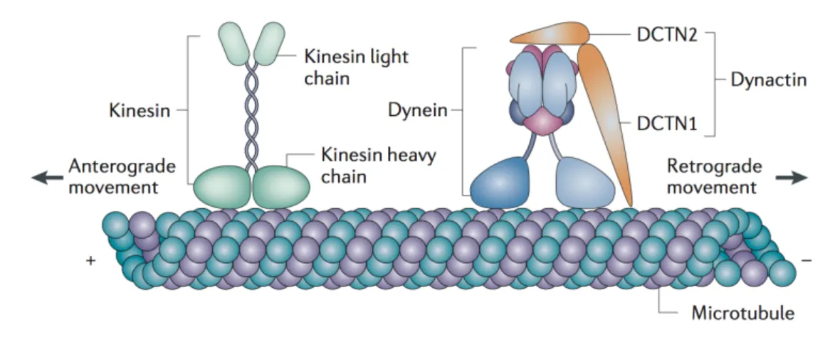
Binding to microtubules (MTs)
Nesprin-1, Nesprin-2, Nesprin-4 in secretory epithelial cells, and KASH5 in sper-matocytes bind to MTs through direct or indirect association with motor proteins, including kinesins and dynein/dynactin (see section 1.3.4 for a description of kinesin and dynein motor proteins) (Roux et al., 2009; Schneider et al., 2011; Wilson and Holzbaur, 2015; Yu et al., 2011; Zhang et al., 2009). However, only the KASH-less isoform p50Nesp1, which is composed of the spectrin repeats 48-51 of Nesprin-1 and
localized to processing bodies (P-bodies), was shown by cosedimentation assays to di-rectly interact with MTs so far (Rajgor et al., 2014). The interaction between KASH domain proteins and MT-dependent motor proteins is often direct, although not all binding sites have been mapped so far. Nesprin-2 associates with dynein/dynactin and kinesin-1 motors at the NE to regulate centrosome-coupling to the nucleus during neuronal migration of the developing mouse brain and during nuclear migration in the mouse retina (Yu et al., 2011; Zhang et al., 2009). In contrast, Nesprin-4 binds directly to the light chain of kinesin-1 and, when ectopiclly expressed, disengages the centrosome and the Golgi complex from the nucleus (Roux et al., 2009). KASH5 interacts with dynein/dynactin and recruits these motor proteins together with Sun1 to telomere ends. Through the recruitment of dynein/dynactin to the NE, KASH5 and Sun1 mediate chromosome movement during meiosis (Horn et al., 2013; Morimoto et al., 2012).
Binding to intermediate filaments
Two different isoforms for Nesprin-3 have been described: Nesprin-3α and Nesprin-3β. Nesprin-3α comprises an unique N-terminal region that can bind to the actin-binding domain of plectin, which in turn interacts with intermediate filaments (Wilhelmsen et al., 2005). Additonally, Nesprin-3α can interact with the CH domains of Nesprin-1
and Nesprin-2, thus forming diverse Nesprin networks at the NE that are able to connect to different cytoskeletal components at the same time (Lu et al., 2012).
Figure 1.4: Nesprins connect the NE to different cytoskeletal components. (A)
Schematic domain structure of the Nesprin family. Nesprins contain a C-terminal Klarsicht/ANC-1/SYNE homology (KASH) domain, several spectrin repeats and variable N-terminal regions. The giant isoforms of Nesprin-1 and Nesprin-2 possess actin-binding calponin homology (CH) domains at their N-termini. KASH5 is a meiosis-specific KASH protein and is not depicted here. (B) Nesprins localize to the outer nuclear membrane by binding to SUN domain proteins of the inner nuclear membrane (INM). Nesprin-1 and Nesprin-2 interact directly with actin filaments through their N-terminal CH domains and indirectly with microtubules (MTs) through kinesin-1 and dynein motor proteins. Nesprin-3α binds to intermediate filaments through plectin. Nesprin-4 binds to MTs through kinesin-1. Some smaller Nesprin isoforms may also localize to the INM. Within the nucleoplasm, Sun proteins interact with the nuclear lamina (lamins), chromatin and other proteins of the INM. Reprinted from (Cartwright and Karakesisoglou, 2014), 29:169-79, Nesprins in Health and Disease, copyright (2014), with permission from Elsevier (Licence #4118400155065).
1.2.3 Tissue-specific KASH splice variants
The giant isoforms of Nesprin-1 and Nesprin-2 are in general ubiquitously expressed and predominant in the majority of tissues but they additionally produce tissue-specific isoforms through alternative initiation of transcription and mRNA splicing (Duong et al., 2014; Rajgor et al., 2012; Zhang et al., 2001). Tissue-specific expression of certain Nesprin isoforms is believed to contribute to specific nucleus-cytoskeleton connections which account for the individual biomechanical properties of highly specialized cell types, e.g. muscle cells (Mellad et al., 2011). Many of these alternative-spliced isoforms contain the C-terminal KASH domain and subsequently localize to the ONM or in a few cases to the INM (smaller isoforms), respectively. However, also KASH-less isoforms that are unable to contribute to LINC complex formation were described to localize throughout the cell but their exact function in vivo remains elusive so far (Duong et al., 2014; Razafsky and Hodzic, 2015). The expression of smaller Nesprin-1 and Nesprin-2 isoforms is in some tissues dominant (Duong et al., 2014; Rajgor et al., 2012; Zhang et al., 2001). In spleen, for example, expression of Nesprin-1β-1 is highly increased and in cardiac and skeletal muscle two smaller isoforms, Nesprin-1α-2 (hereafter referred to as Nesprin-1α) and Nesprin-2α-1, are exclusively expressed (Duong et al., 2014). During skeletal muscle differentiation, expression of Nesprin-1α was shown to be highly upregulated, both on mRNA and protein levels (Holt et al., 2016; Randles et al., 2010; Zhang et al., 2001). Nesprin-1α is structurally an N-terminally truncated version of Nesprin-1 giant: It contains the C-terminal KASH domain, seven spectrin repeats but lacks the N-terminal CH domains (Apel et al., 2000; Mislow et al., 2002b). Nesprin-1α contains additionally a short and unique N-terminal segment but apart from that is identical to the C-terminal region of Nesprin-1 giant (Simpson and Roberts, 2008). From a functional perspective, Nesprin-1α was suggested to homodimerize through its N-terminal spectrin repeat domains and to recruit the scaffolding protein muscle A-kinase anchoring protein (mAKAP) to the NE in cardiac myocytes (Mislow et al., 2002a; Pare et al., 2005). Additionally, Nesprin-1α was demonstrated to bind lamin A and emerin (Mislow et al., 2002a,b). Although Nesprin-1α was proposed to localize to the INM in these studies, this seems due to its size of ∼ 112 kDa rather unlikely. Together these three studies concluded that Nesprin-1α might function as a scaffold protein and molecular link for signaling molecules at the NE. However, a clear biological function for Nesprin-1α in skeletal muscle remains to be determined.
1.2.4 The LINC complex in muscle disorders
Various components of the LINC complex, including Nesprin-1/-2 and Sun1/2, as well as lamin A/C, are mutated in muscular dystrophies as exemplified by Emery-Dreifuss muscular dystrophy (EDMD) (Bonne et al., 1999; Mamchaoui et al., 2011; Meinke et al., 2014; Zhang et al., 2007b). EDMD is a neuromuscular disorder characterized by joint contractures that appear early in childhood as well as progressive skeletal muscle weakness and wasting and cardiomyopathy (Pillers and Von Bergen, 2016). Mutations within the LMNA or EMD gene, encoding lamin A/C or the INM protein emerin, respectively, were initially identified to cause autosomal-dominant and X-linked forms of EDMD (Bione et al., 1994; Bonne et al., 1999). Heterozygous mutations within the SYNE1 and SYNE2 gene, encoding for Nesprin-1 and Nesprin-2, respectively, were additionally associated with EDMD (Zhang et al., 2007b). Fibroblasts and muscle cells from these patients displayed marked alterations of nuclear shape and mislocalization of Nesprins, emerin and Sun2. Moreover, the interactions between Nesprins-1/-2, emerin and lamin A/C were either decreased or completely abolished, suggesting that any changes in LINC complex composition, stability or functionality can cause severe muscle disorders (Wheeler et al., 2007; Zhang et al., 2005). Mutations within the SUN1 and SUN2 gene have been further identified in patients with EDMD-like phenotypes (Meinke et al., 2014). Fibroblasts overexpressing one of the five disease-causing Sun1/2 variants (SUN1 G68D, SUN1 G338S, SUN1 W377C, SUN2 A56P, SUN2 R620C) failed to move their nuclei towards the rear of the cell and to reposition their centrosomes prior to migration. Primary myoblasts from a patient with compound heterozygous SUN1 p.G68D/p.G338S variants showed increased levels of SUN1 and Nesprin-2 at the NE but a decreased capability to interact with emerin. Interestingly, in vitro differentiated myotubes from this patient showed disorganized myonuclei which failed to recruit the centrosomal protein Pericentrin to the NE and to nucleate MTs (Meinke et al., 2014).
A homozygous nonsense mutation within the SYNE1 (23560 G>T) gene (Protein: E7854X) was furthermore identified in patients suffering from congenital muscular dystrophy (CMD), a heterogenous group of inherited myopathies characterized by early onset of progressive muscle weakness (Mamchaoui et al., 2011; Voit et al., 2007, 2002). Quantitative PCR studies of these SYNE1 (23560 G>T) CMD patient cells showed that the mRNA levels of both Nesprin-1 giant and Nesprin-1α were highly reduced by 78% and 85%, respectively (Holt et al., 2016).
muscle diseases, is still a puzzling question. It is likely that changes within LINC complex components affect the nucleoskeleton-cytoskeleton coupling and thus render particularly muscle cells incapable to withstand mechanical forces during muscle contraction. However, muscle cells also undergo an extensive isoform switch from giant to smaller muscle-specific isoforms during myogenic differentiation (Randles et al., 2010). This suggests that also defects in gene expression of these tissue-specific isoforms could contribute to cause muscle diseases. Finally, the LINC complex is crucial for correct nuclear positioning during skeletal muscle formation and any defects in the underlying mechanisms were suggested to affect muscle function (see also section 1.2.5 and 1.2.6) (Elhanany-Tamir et al., 2012; Metzger et al., 2012).
1.2.5 Nesprin-1/2 and Sun1/2 knockout mouse models
Several knockout mouse models for components of the LINC complex were created to understand cellular functions of Nesprins and Sun proteins in vivo and in human muscle disorders. However, existing Nesprin-1 and Nesprin-2 knockout strains display a high variability in the severeness of the observed phenotypes.
Mice overexpressing either the KASH-domain of Nesprin-1 or Nesprin-2 under a muscle-specific promoter, displace endogenous Nesprin-1 and Nesprin-2 from the nucleus in muscle fibers due to the dominant-negative effect of the artificial KASH-construct. These mice display nuclear positioning defects with decreased numbers of extrasynaptic nuclei and increased numbers of non-synaptic nuclei (Grady et al., 2005; Zhang et al., 2007c). Studies with single Nesprin-1 or Nesprin-2 KASH-domain knockout mice, respectively, further clarified that Nesprin-1 but not Nesprin-2 is important for the anchorage of synaptic and non-synaptic nuclei in myofibers (Zhang et al., 2007c). Although single KASH-domain knockout mice are viable, double-homozygous KASH-domain knockout mice of Nesprin-1 and Nesprin-2 die within 20 min after birth from respiratory failure, suggesting that Nesprin-1 and Nesprin-2 fulfill partially redundant but essential functions (Zhang et al., 2007c). Another study, however, showed that homozygous mice lacking the KASH domain die around birth, while surviving mice develop typical features of EDMD, including muscle weakness, kyphoscoliosis, cardiac conduction defects, as well as centralized and misaligned nuclei (Puckelwartz et al., 2009). Both Nesprin-1 KASH-knockout mouse models show an important role for Nesprin-1 in nuclear positioning but they differ greatly regarding the viability of the mice and the observed EDMD-like phenotypes (Puckelwartz et al., 2009; Zhang et al., 2007c). These discrepancies might result from the different genetic mouse backgrounds used in the two studies
for generating the knockout lines as well as from the different strategies used to disrupt the KASH domain, resulting in slightly different C-terminal regions of the truncated Nesprin-1 protein. To make the picture even more complicated, two more recent knockout mouse models were generated by Ju Chen’s laboratory. They first created knockout mice lacking all Nesprin-1 isoforms harboring the C-terminal spectrin repeats (KASH-containing and KASH-less Nesprin-1 isoforms are depleted) (Zhang et al., 2010). These knockout mice die postnatally between day 2 and 11 (60%), whereas surviving mice show decreased body weights, a decreased exercise capability, a large proportion of centralized nuclei and a lack of nuclei clusters under the NMJ. Secondly, Nesprin-1 knockout mice specifically lacking the N-terminal actin-binding CH domains were viable and displayed no obvious effects regarding nuclear shape, nuclear positioning or localization of other LINC complex components (Stroud et al., 2017). Moreover, in the same study they described Nesprin-1α-specific homozygous knockout mice which mostly die within 5 min after birth. Surviving mice showed growth retardation, kyphosis and nuclear positioning defects. Interestingly, in Nesprin-1α-knockout myofibers, Kif5b was absent at the NE, thus supporting a role for Nesprin-1α/Nesprin-1 in recruiting Kif5b/Kinesin-1 to the nucleus to support Kinesin-mediated nuclear movement in skeletal muscle cells (Stroud et al., 2017; Wilson and Holzbaur, 2015).
As Nesprin-1 is anchored to the ONM by Sun proteins, it is not surprising that also Sun1 and Sun2 double-knockout mice show displacement of Nesprin-1 from the NE and thus severe synaptic and non-synaptic nuclear positioning defects (Lei et al., 2009). However, Sun1 seems to have a more pronounced role in nuclear anchorage as the number of synaptic nuclei is partially decreased in Sun1 but not Sun2 knockout mice (Lei et al., 2009).
1.2.6 Nuclear positioning in skeletal muscle cells
Skeletal muscle fibers are multinucleated cells that arise from the fusion of hundreds of differentiated myoblasts (myocytes) (Abmayr and Pavlath, 2012). As a result, mature myofibers can contain more than 100 nuclei which are predominantly posi-tioned at the periphery of the cell, while some nuclei are found in clusters under the neuromuscular junction (NMJ) (Bruusgaard et al., 2003; Grady et al., 2005; Kummer et al., 2004). Peripheral nuclei are equally spaced, resulting in a maximum distance between adjacent nuclei (Bruusgaard et al., 2003). This positioning is believed to create ’myodomains’, where each nucleus is responsible for the transcrip-tional activity within its surrounding area, thus leading to decreased requirements
to transport mRNAs over long distances throughout the myofiber (Pavlath et al., 1989).
Mispositioning of myonuclei is often a hallmark of muscle diseases, such as Emery-Dreifuss muscular dystrophy (EDMD) or Centronuclear myopathy (CNM), suggest-ing that correct nuclear positionsuggest-ing is important for muscle function (Cohn and Campbell, 2000; Jungbluth et al., 2008; Metzger et al., 2012). However, a direct relation between nuclear positioning and muscle function still needs confirmation. At least four distinct nuclear movements or positioning processes - (1) nuclear cen-tration, (2) nuclear spreading, (3) nuclear dispersion and (4) nuclear clustering - can be observed during skeletal muscle formation (Cadot et al., 2015). The underlying mechanisms that drive and regulate these nuclear movements are only recently beginning to emerge.
Nuclear centration
After fusion of a differentiated myoblast with a pre-existing myotube, the myoblast nucleus moves rapidly towards the myotube center with an approximate speed of 0.88 µm/min (Cadot et al., 2012; Englander and Rubin, 1987) (Figure 1.5 A, centering). This nuclear movement, termed centration, requires an intact and dynamic MT net-work, the small GTPase Cdc42 and the dynein/dynactin complex. In fact, altering MT dynamics with the help of the MT-stabilizing drug taxol or low concentrations of nocodazole prevents nuclear centration movements after myoblast fusion (Cadot et al., 2012). The dynein/dynactin complex is in the presence of Par6 and Par3 proteins recruited to the nucleus in differentiated myoblasts and myotubes. There, it acts to transport the myoblast nucleus towards the minus-end of MTs emanating from myotube nuclei (Figure 1.5 B). Reciprocally, dynein/dynactin molecules located at myotube nuclei can move towards the minus-end of MTs emanating from the myoblast nucleus, thereby pulling the myoblast nucleus towards the center of the myotube (Cadot et al., 2012).
Nuclear spreading
Once nuclei are aligned in the middle of the myotube, they start to spread in parallel to the long axis of the myotube with an average speed of 0.2 µm/min (Cadot et al., 2015) (Figure 1.5 A, spreading). During these nuclear spreading movements, nuclei were observed to rotate and to undergo pausing events before they continue to translocate (Capers, 1960; Cooper and Konigsberg, 1961; Wilson and Holzbaur, 2012). Similar to nuclear centration movements, nuclear spreading depends on
an intact MT network (Englander and Rubin, 1987). Up to know three different models were proposed to explain these nuclear spreading movements. First, nuclear spreading was suggested to require an anti-parallel network of MTs, with each MT minus-end being anchored at one nucleus, as well as cytoplasmic complexes of Kif5b/kinesin-1 and Map7. Through the MT plus-end-directed motor activity of Kif5b, these Kif5b/Map7 complexes would act in concert to push nuclei apart from each other (Metzger et al., 2012) (Figure 1.5 C [1]). Secondly, nuclear spreading was proposed to depend on NE-localized Kif5b and to a lesser extent on NE-localized dynein molecules that transport myotube nuclei as cargo towards the plus-end or minus-end of MTs, respectively (Wilson and Holzbaur, 2012) (Figure 1.5 C [2]). In this model, Kif5b/kinesin-1 is recruited to the NE by Nesprin-1 or Nesprin-2 which share a common kinesin light chain-binding sequence (’LEWD’ motif); interfering with the expression of either Nesprin1/2 or Kif5b/kinesin-1 affects nuclear spreading and nuclear rotation (Wilson and Holzbaur, 2012, 2015). How dynein molecules are linked to the NE is less understood but might involve Nesprins or unknown binding partners at the nucleus (Zhang et al., 2009). The third model was suggested based on muscle morphogenesis studies in Drosophila melanogaster (Figure 1.5 C [3]). Dynein is transported to the cell cortex of the myofiber pole by kinesin-mediated movement towards the MT plus-end (Folker et al., 2014). This transport requires Sunday Driver (Syd), a family member of the JNK-interacting proteins (JIP), which mediates complex formation of kinesin and dynein (Schulman et al., 2014). At the cell cortex, dynein is anchored by the protein Pins/Raps (partner of inscuteable; also termed rapsynoid) and is thus able to pull on MTs to move nuclei in the direc-tion of the myofiber pole. Thereby, interacdirec-tions between MTs and the cell cortex are stabilized by CLIP-190 and are important for correct myonuclear positioning (Folker et al., 2012). However, if this model is also important for nuclear spreading
movements in mammalian muscle cells remains to be determined.
Nuclear dispersion
After nuclei are equally spread throughout the myotube, they move towards the periphery of the cell and finally get anchored below the sarcolemma (Figure 1.5 A, dispersion). In contrast to nuclear centration and spreading movements, nuclear dispersion seems to be dependent on the actin cytoskeleton but independent of the MT network (Falcone et al., 2014). Additionally, nuclear movement to the periphery involves Nesprins that couple myonuclei to actin, as well as the actin polymeriza-tion factor N-WASP, which activates the actin-nucleapolymeriza-tion capacity of the Arp2/3
complex (Falcone et al., 2014; Takenawa and Suetsugu, 2007). N-WASP itself is activated through interaction with Amphyphysin-2, a protein that is encoded by the AMPH2/BIN1 gene and often found mutated in centronuclear myophathy (Falcone et al., 2014; Nicot et al., 2007). Other factors that contribute to nuclear positioning at the periphery include desmin intermediate filaments and blood vessels (Chapman et al., 2014; Ralston et al., 2006; Shah et al., 2004; Tokuyasu et al., 1983). Myonuclei were demonstrated to localize in close proximity to blood vessels, suggesting that blood vessels could locally stimulate nuclear movement to the periphery (Ralston et al., 2006). In contrast, desmin was shown to regulate nuclear spacing by acting as a repellent between nuclei (Ralston et al., 2006). Finally, nuclear anchorage at the periphery requires LINC complexes, including Nesprin-1 and Sun1/2, and at least in part desmin intermediate filaments (Chapman et al., 2014; Elhanany-Tamir et al., 2012; Lei et al., 2009; Zhang et al., 2010, 2007c).
Nuclear clustering
Typically 3-8 nuclei cluster and get anchored under the neuromuscular junction (NMJ) (Bruusgaard et al., 2003; Sanes and Lichtman, 2001) (Figure 1.5 A, clustering). These so-called synaptic nuclei are highly specialized as they express NMJ-specific genes (Nazarian et al., 2005). The mechanisms underlying nuclear movements to the NMJ as well as nuclear clustering at synaptic contact sites is still poorly understood. However, studies with chick muscle cultures suggested that nuclear clustering might be linked to the clustering of acetylcholine receptors (AchRs) at post-synaptic membranes (Englander and Rubin, 1987). Recruitment of AchRs to the post-synaptic membrane requires a complex signaling pathway involving the neuron-derived ligand Agrin, the co-receptor low-density lipoprotein receptor-related protein 4 (LRP4) and the muscle-specific tyrosine kinase (MuSK) (Kim et al., 2008; Zong and Jin, 2013). MuSK was demonstrated to interact with Nesprin-1, which is increased at synaptic nuclei in comparison to non-synaptic nuclei, suggesting that connections between Nesprin-1 and MuSK might be important for synaptic nuclei anchorage beneath the NMJ (Apel et al., 2000). In line with this hypothesis, transgenic mice overexpressing the dominant-negative KASH domain of Nesprin-1 displayed anchorage defects of synaptic nuclei due to displacement of endogenous Nesprin-1 from the NE (Grady et al., 2005).